Review

Gut Microbiota and Immune System in Necrotizing Enterocolitis and Related Sepsis

1
Interdisciplinary Department of Medicine, Section of Microbiology and Virology, School of Medicine, University of Bari, 70124 Bari, Italy
2
Department of Clinical Disciplines, School of Technical Medical Sciences, University of Elbasan “A. Xhuvani”, 3001 Elbasan, Albania
3
Istituti Clinici Scientifici Maugeri IRCCS, Pneumology and Respiratory Rehabilitation Unit, Institute of Bari, 70124 Bari, Italy
4
Doctoral School, eCampus University, 22060 Novedrate, Italy
*
Author to whom correspondence should be addressed.
Gastrointest. Disord. 2024, 6(2), 431-445; https://doi.org/10.3390/gidisord6020029
Submission received: 19 April 2024 / Revised: 3 May 2024 / Accepted: 6 May 2024 / Published: 10 May 2024

Abstract

:
A severe condition of sepsis can be a complication of necrotizing enterocolitis (NEC), which can occur in premature infants and becomes a medical challenge in the neonatal intensive care unit (NICU). It is a multifactorial intestinal disease (can affect both the small and large intestine) that can lead to ischemia of the intestinal tissues that evolves into acute organ necrosis. One of these factors is that different types of nutrition can influence the onset or the progression of the disease. Cow-milk-based infant formulas have been shown to cause it in premature infants more frequently than human milk. Recently, nutrition has been shown to be beneficial after surgery. Several issues still under study, such as the pathogenesis and the insufficient and often difficult therapeutic approach, as well as the lack of a common and effective prevention strategy, make this disease an enigma in daily clinical practice. Recent studies outlined the emerging role of the host immune system and resident gut microbiota, showing their close connection in NEC pathophysiology. In its initial stages, broad-spectrum antibiotics, bowel rest, and breastfeeding are currently used, as well as probiotics to help the development of the intestinal microbiota and its eubiosis. This paper aims to present the current knowledge and potential fields of research in NEC pathophysiology and therapeutic assessment.

1. Introduction

Necrotizing enterocolitis (NEC) is an intestinal inflammatory disease that occurs mainly in premature neonates with a wide range of effects, from minor lesions that are confined to the intestinal mucosa up to extensive necrosis of the intestinal wall that can cause perforation. Intestinal necrosis is the most common cause of death that occurs in premature neonates and especially in those with an exceptionally low birth weight [1]. Although the early recognition and treatment of this disease have improved over time based on clinical findings, it represents a serious complication in neonatal survivors in the neonatal intensive care unit. In 1888, Paltauf first described newborn clinical cases with this disease, while the term NEC to describe the disease was first used by Schmid and Quaiserk in 1953 [2]. In 1965, Mizrahi and collaborators described this disease as “intestinal syndrome characterized by emesis, abdominal distension, gastrointestinal bleeding and shock in premature new-borns” [3]. However, decades of systematic research have not fully elucidated the pathogenetic mechanism of NEC, while its timely diagnosis remains difficult [4,5]. It has been noted that NEC usually occurs in infants in the first three weeks of life having an extremely low birth weight with a gestational time <28 weeks [6,7,8]. About 10% of NEC cases occur in full-term infants, while 90% occur in premature newborns. NEC is a major clinical challenge, as its impact on infants admitted to NICUs are 1–5%, mostly involving those (7–14% incidence) with a birth weight <1500 g. Its incidence in the population is between 0.5–5 cases per 1000 newborns [9]. Its mortality is inversely correlated with birth weight and gestational age, which can be up to 50%, especially if it is being treated surgically [10,11]. Thus, it occurs in 1 to 3 cases in every 1000 births and is the most common gastrointestinal emergency in premature infants. Globally, the incidence of the disease reaches up to 13% of newborns at ≤33 weeks of gestation or babies with birth weights ≤2500 g and mortality rates between 30–50%. Finally, NEC occurs less often after ≥35 weeks of gestation [12,13,14]. Symptoms in premature infants and the time of onset, for the most part, are inversely proportional to gestational time (i.e., the earlier the newborn, the more obvious the signs). The initial symptoms include intolerance to enteral feeding and failure to thrive, abdominal distention, and bloody stools. In the advanced stage of the disease, erythema appears on the skin of the anterior abdominal wall, accompanied by cardiovascular and neurosensorial disturbances (such as bradycardia, hypotension, lethargy, etc.), hyperpyrexia, oliguria, etc. According to Bell staging, we have stage I (lethargy, apnea attacks, decreased nutrition, mild abdominal distension/ileus, and blood in stool), stage II (severe abdominal distension, bowel pneumothorax, and/or portal air), and stage III (full thickness intestinal necrosis, perforation, peritonitis, sepsis, and death) [15,16]. Note that clinical manifestations begin after the onset of bowel feeding. Symptoms can rapidly progress to abdominal discoloration with intestinal perforation, peritonitis, and sepsis requiring intensive care and medical support [17,18]. Symptoms in some children do not manifest themselves clinically, and they may develop a septic condition without the disease being diagnosed. The condition can rapidly progress from mild to severe within 72 h. Laboratory hematological test results are usually neutropenia, thrombocytopenia, unbalanced hemostasis (prolongation of partial thromboplastin time), metabolic acidosis, anemia, and altered sepsis biomarkers [18,19,20,21]. Non-severe cases of NEC can be effectively treated with intestinal food retention, stomach decompression with a nasogastric tract, and the initiation of broad-spectrum antibiotics. Some advanced cases can involve extensive intestinal necrosis, and the complications after surgery can be fatal. Most treatment strategies for the disease are achieved by the pharmacological and clinical support of vital signs and/or surgery [22,23,24]. The choice of treatment depends on the stage of NEC. When newborns show symptoms, such as apnea, bradycardia, or gastric and abdominal distension, the disease is treated with pharmaceuticals because it is in its initial phase. Surgery is performed when the intestine has extensive necrotic areas or is necrotic. Moreover, the surgical procedure is performed when the pharmacological therapy has failed. The type of surgery chosen is based on the stage of the disease [25]. First, fluids can be drained with the introduction of special catheters (Penrose drainages), and if this method fails, then a partial or total removal of the intestine is performed. As we discuss further, NEC is a multifactorial disease whose main risk factors are prematurity, which disrupts the acquisition of gut microbiota in preterm infants that correlates with the altered development of the immune system and the immaturity of gastrointestinal system, and low weight at birth [26,27].

2. Predisposing Factors for NEC

Data available about risk factors for NEC are relatively few. The disease is multifactorial, and elucidation of the predisposing factors is difficult. Table 1 shows the best-known predisposing risk factors associated with NEC [25,28,29,30].
As mentioned above, a premature baby is one born at a gestational age of less than 37 weeks. Risk factors usually include diabetes mellitus, hypertension, multiple pregnancies, obesity, pregnancy complications (such as placental abruption and others), abuse of substances (such as cocaine, methamphetamine, and others), tabagism, drugs (such as antibiotics, corticosteroids, FANS, and H-2 receptor antagonist), and others [31,32,33,34].
It is estimated that at least 75% of premature neonates will survive with appropriate treatment. Premature birth is the most common cause of infant death in the world [35,36]. Often, survival has been associated with complications in the health and development of the child. As we mentioned, prematurity is one of the main causes of low-birth-weight babies, which is an aggravating factor for the child’s health. Low birth weight is not a direct cause of mortality but is associated with adversities such as respiratory distress syndrome, bronchopulmonary dysplasia, cardiovascular disturbances, inadequate functions of the immune system (limited capacity to moderate inflammation and infection), neurological disturbances, and others [25,28]. Thus, we can divide this factor into low birth weight (1500–2499 g), exceptionally low birth weight (1000–1499 g), and extremely low birth weight (<1000 g), which are associated with many morbidities, such as chronic pulmonary diseases, retinopathy, and NEC [37]. Newborns with elevated health clinical risk conditions or severe comorbidities have a greater probability to develop NEC. The inhomogeneity of the lungs, a problem that affects neonates born <32 weeks of gestation, results in altered O2 and CO2 exchange and insufficient tissue oxygenation. Cardiac complications due to prematurity limit the availability of oxygen and nutrients to other tissues and organs [38]. Even the intestinal function is a concern. Certain drugs, such as H2 agonists in premature infants, are associated with an increase in incidence because maintaining the acidic pH at normal values reduces the possibility of colonization by bacteria [7]. Vasoconstrictive drugs, such as nonsteroidal anti-inflammatory drugs (NSAIDs) (such as cyclooxygenase inhibitors, indomethacin, etc.), used in the treatment of cardiac complications in premature babies, can also hinder the spread to the intestine [39,40]. A low Apgar score at birth, cardiac defects, intestinal blockage, the use of inotropes, and reduced respiratory function are some factors. The clinical events that can reduce the diffusion in the intestine or even the saturation of O2 in the blood are associated with NEC [41]. It has been observed that septicemia is associated with a higher incidence of the occurrence of NEC. In addition, inflammation and infectious agents can cause ischemia in the intestine [7]. The importance of infectious agents is well known in the manifestation of NEC. Sepsis is due to microorganisms that colonize premature newborns during childbirth (i.e., Escherichia coli, Listeria, Klebsiella, etc.), or to microorganisms that colonize neonatal intensive care units (i.e., Staphylococcus aureus, Staphylococcus epidermidis, E. coli, Pseudomonas, and Klebsiella) [18,42,43,44].
There have been two known conditions regarding the relationship between enteral feeding and NEC, which predominate in enterally fed preterm infants. A range of protective ingredients contained in breast milk can be a preventative test for the onset of the condition or for treatment [45]. The enteral feeding of the premature newborn with maternal or artificial milk is an aggravating factor in the onset of NEC. However, it has been found that maternal milk reduces the risk of developing the disease, but does not exclude its manifestation, since it is a multifactorial condition. Maternal milk contains immunity and anti-inflammatory factors that reduce the severity of the disease or prevent its onset [46,47]. The beginning of the feeding, after surgery for the disease, is conducted with an elementary diet, with a solution comprising amino acids or hydrolyzed casein, with maltodextrin and lipids across the chain. In general, the use of lactose should be avoided during recovery [47]. Recent research has suggested the epigenetic effects of human milk on the intestinal genomic structure and thus, an important risk factor in the pathogenesis of the disease involves the interaction between the type of enteral nutrition and the intestinal microbiota. Indeed, the indications for parenteral nutrition (PN) in premature and non-premature infants are very low birth weight (<1500 g) or extremely low birth weight (<1000 g). Due to the immaturity of the intestine, full bowel feeding may not be tolerated; however, delaying feeding can lead to negative outcomes throughout the newborn [48,49].

3. Biomolecular Mechanisms of NEC

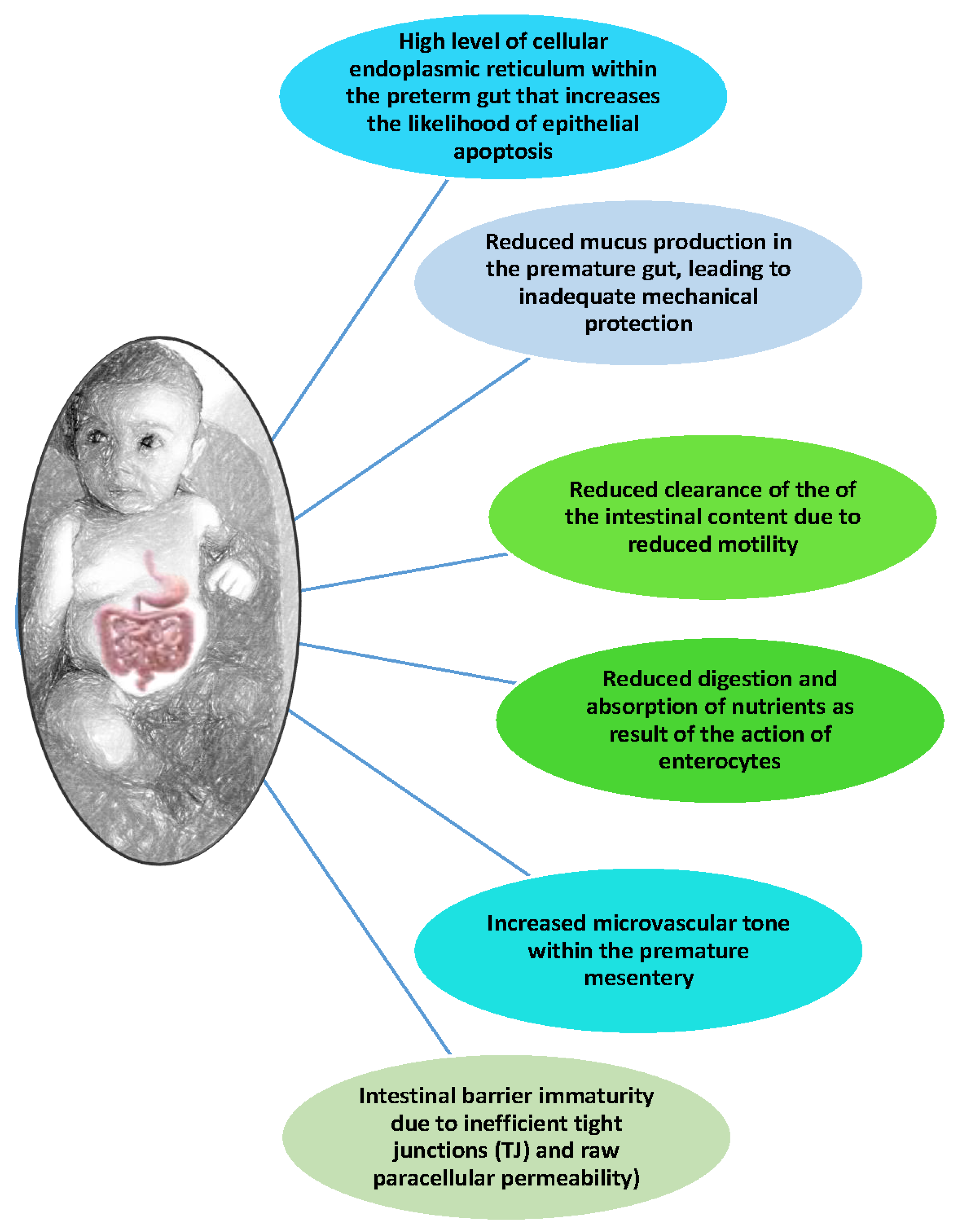
3.1. Hyperactive Premature Intestine and Inflammatory Response

There are differences between the mature intestine and the intestine of a premature baby. Several studies showed significant differences in the bacterial colonization, microcirculatory diffusion, and immune maturation of the intestinal system. Thus, these differences may be those that cause the pathogenesis of the disease and demonstrate its multifactorial nature. The increased expression of toll-like receptor 4 (TLR4) in the preterm intestinal tract reflects the function of its manifestations in regulating physiological intestinal development [50]. Toll-like receptors, TLRs, are found on the cell surface and act as sensors of infections, and simultaneously play a role in its initial inflammation and immune response. Microorganisms express molecules that function as receptors for TLRs, such as lipopolysaccharides and lipoteichoic acid. The activation of these receptors by normal intestinal microbial flora contributes to the integrity of the intestinal epithelium, while at the same time protecting it from injury [51,52]. During preterm labor, intestinal levels of TLR4 and the activation of TLR4 remain elevated in the lining of the preterm intestine by Gram negative bacteria that colonize it, leading to various harmful effects, including enterocyte apoptosis, the attenuation of the mucosa, and enhanced pro-inflammatory cytokines, which overall lead to the development of NEC [53]. Furthermore, the translocation of Gram negative bacteria through the mucosa to the intestine leads to the activation of TLR4 in the endothelial lining of the preterm intestine, resulting in reduced blood flow and intestinal growth, with subsequent necrosis and ischemia [54,55]. Thus, TLRs are essential for intestinal epithelial homeostasis after damage or injury. Furthermore, the overexpression of some TLRs is responsible for inducing a pro-inflammatory response in the intestinal epithelium of preterm infants with the absence of a eubiotic intestinal microbiota. This explanation for the pathogenesis of the disease partly explains the reasons why the premature newborn is at risk of developing NEC and why the disease occurs after intestinal bacterial colonization. The premature intestine is characterized by reduced peristalsis, a thin mucous layer, a reduced number of tight junctions, increased apoptosis of enterocytes, and reduced regeneration of enterocytes [56]. These deficiencies can lead to a breakdown of the “permeability” of the intestine, thus facilitating the penetration of certain pathogenetic or opportunist microorganisms (bacterial, viruses, and others) from the lumen. On the other hand, the reduced structural integrity and functionality of the intestine causes insufficient digestion and absorption of energy, proteins, and other nutrients necessary for growth and immune protection. Patient response to infection and clinical outcome implies a balance between pro-inflammatory and anti-inflammatory cytokines. Not only NEC but several adverse effects in neonates (i.e., brain damage and bronchopulmonary dysplasia) appear to be associated with cytokine production in response to maternal, fetal, or neonatal infections. Mediators investigated in infants include tumor necrosis factor-α (TNFα), interleukins 1 (IL-1), 4 (IL-4), 6 (IL-6), 8 (IL-8), 10 (IL -10), and 12 (IL-12), platelet-activating factor (PAF), and leukotrienes. The release of various inflammatory mediators in response to infection offers the possibility of early laboratory diagnosis of the infection. The potential markers for the laboratory diagnosis, not only of NEC or bacterial sepsis, but also pneumonia, etc., are TNFα, IL-6, and IL-8. [57,58]. Interferons (INF) α and β are normal, but the synthesis of INF-γ is low. INF-α levels are higher in infants with sepsis, but the response may be less clear than in adults. Interleukin-2 activity in the umbilical cord blood of full-term and preterm infants has been reported to be higher than that of adults. Interleukin-6 levels are also elevated in the serum of neonates with sepsis and/or NEC [57,58,59]. A premature immune system is not able to easily detect pathogenic agents or protect the organism from infections, due to some related factors such as (a) changes in the expression of toll-like receptors (TLR), in particular TLR4 and TLR9, which are involved in the recognition of pathogenic microorganisms and the activation of the immune system, (b) the reduced production of IgA, IgM, IgG, and defensin, and (c) the regulation of pro-inflammatory TLRs and/or pro-inflammatory cytokines, such as TNF-α, IL-6, IL-8, and IL-1β [60]. As we mentioned, for premature newborns, hypoxia–ischemia and respiratory complications limit the supply of O2 and component nutrients to the intestine. The hypoxia conditions lead to activated adenosine monophosphate, which reduces the energy consumption of anabolic mechanisms, such as Na+/K+-ATPase activity and catalytic processes, in the attempt to conserve energy [61,62]. These intracellular responses may be the springboard for pathology initiation. Over time, this oxidative catabolic system may fail to maintain the digestive and absorptive functionality and cellular integrity experience increased intestinal sensitivity to uncontrollable inflammation and necrosis [63]. The lack of an adequate supply of O2 causes tissue hypoxia that manifests itself as an imbalance of the acid/base ratio, leading to a severe state of metabolic acidosis in the tissues supplied by the mesenteric vessels and subsequently creates vasospasm, causing a further reduction in the supply of O2 from the blood to the tissues. This prolonged ischemia also leads to malabsorption syndrome. In a hypoxic intestinal environment, the intestinal introduction of nutrients can lead to the burden of using O2 for maintaining the natural barrier of the intestinal wall rather than for digestion [64]. At the tissue level, hypoxic conditions or vasoconstrictive drugs can lead to an insufficient supply of the nutrients and oxygen that are necessary to produce energy, produce immune cells, create membrane proteins to protect intestinal integrity, and conduct processes of digestion and absorption. Therefore, the inability to maintain the structure and function of the intestinal wall due to hypo-polarization can be a cause of the disease (Figure 1) [65].
The role of the inflammatory activation response in disrupting the integrity of the intestinal barrier has been an important research area in the pathogenesis of NEC. Intestinal epithelial cells produce cytokines involved in infection and damage the intestinal barrier. Important among these is the role of interleukin 6 in the promotion of T lymphocytes and in the production of antibodies by B lymphocytes. The production of TNFα is induced by cytokines and some microbial pathogenic factors, which in turn promotes the cytotoxic response and programmed cell apoptosis and death [62,65]. PAF is one of the most important and most studied inflammatory mediators involved in the pathogenesis of NEC. This is because it is a phospholipid factor produced by inflammatory cells (basophils and white blood cells), endothelial cells, platelets, and bacteria. It has an inflammatory and vasoactive effect on some organs, also causing a severe lowering of blood pressure. It is synthesized after activation of the phospholipase A2II and it is formed through a precursor compound, which is Liso-PAF. PAF receptors (PTAFRs) exist in many cells and their action is conducted through the activation of the G protein receptor, a pair of PAFs found in high concentrations, in intestinal epithelial cells and in the ileum, which is also the most frequent site of NEC [66]. The activation of PAF receptors promotes the production of other cytokines and secondary mediators, such as TNFα, IL-6, IL-8, IL-1, leukotrienes, and NO. Furthermore, their action is through the apoptosis of intestinal epithelial cells. Based on this hypothesis, the apoptosis of intestinal epithelial cells involves the loss of epithelial integrity, increased permeability and mucosal complexity, and the entry of the microbes into the intestinal wall. In experimental models, PAF has been found to cause capillary damage, myocardial and renal dysfunction, neutropenia, thrombocytopenia, and hypotension [67]. The enzyme PAF–acetyl hydrolase, which cleaves and inactivates PAF, is at low concentrations in preterm infants. Finally, increased levels of PAF in plasma and feces were found in infants exhibiting NEC, findings that indirectly reinforce its key role in the pathogenesis of the disease [68].

3.2. The Role of Intestinal Microbiota

The intestinal microbiota is a complex and fragile balanced system of microorganisms that has only been fully studied in recent years. Knowledge of its interactions and precise functions is still under study. In the international literature, the terms gut microbiota and gut microbiome are often confused, but there is a significant difference between them [69,70]. More specifically, the term gut microbiota refers to the community of microorganisms living in a person’s gastrointestinal tract consisting of members of bacteria, their viruses (mainly phage), archaea, fungi, as well as eukaryotic microorganisms, and thus can be used to characterize the intestinal tract. The term gut microbiome refers to all the genes of an individual’s gut microbes. Several microorganisms can colonize the gastrointestinal tract, ultimately shaping the intestinal microbiota [69]. The intestinal microbiota could be described as a “microbial organ” due to the homeostasis of the organism and its participation in several functions such as digestion, absorption of component nutrients, elimination of waste substances, natural immunity, and others [69,70]. Those microbial communities (consisting of several types of cells) can communicate with each other and with the host. They can consume, store, and convert energy, mediate chemical pathways, and can regenerate themselves. Their genome contains over one hundred times more genes than that of humans [71,72]. The surface it occupies is estimated to be around 200 m2, thus providing the necessary space for all the processes of digestion, growth, and the production of microorganisms to take place [73]. The epithelial surface occupied by the gut microbiota can be up to 400 m2 and is the second largest surface of the human body after the surface of the respiratory apparatus [74]. It is also reported that around 1014 living bacteria can be found in the gastrointestinal tract of an adult human, ten times the number of eukaryotic cells in all the tissues of the human body [69,73] Thus, gut microbiota eubiosis together with the intestinal mucosa are a natural barrier for the immune system against, for example, pathogenic microorganisms, harmful compounds in the intestine, autoimmune and metabolic diseases, precancerous conditions, and others [75,76,77,78]. The microbial composition of the intestinal flora mainly consists of four genera of bacteria, Bacteroidota (23%), Bacillota (64%), Actinomycetota (3%), and Pseudomonadota (8%), as well as some archaea, eukaryotes, and viruses [79,80]. Of the 70 genera present in each organism, the dominant are those of Bacteroidetes and Bacillota, which represent over 90% of the total bacterial load of the population, and 95% of all bacteria are classified as phylum of Bacillota belong to the Clostridia spp., while Bacteroidota phyla are made up of four main species: Bacteroides thetaiotaomicron, Bacteroides vulgaris, Bacteroides distasonis, and Bacteroides fragilis [71,79,80]. The other genera of bacteria are present in small proportions. Due to the prevailing anaerobic conditions in the large intestine, exclusively anaerobic genera such as Bacteroides, Eubacterium, Bifidobacterium, Fusobacterium, Peptostreptococcus, Clostridium, and Ruminococcus are dominant, while aerobic genera such as Escherichia, Enterobacter, Enterococcus, and Lactobacillus are present in minor populations [69,80,81].
From the moment of birth, the formation of the gut microbiota begins. The first stage at which the newborn meets microorganisms is its method of birth, whether via normal delivery or not. Infants’ direct contact with germs from the mother’s genital tract during normal birth affects the development of the gut microbiota, as can be seen from the similarity of the infants’ bacteria to those in their mother’s vagina [69,82]. The newborns born by caesarean delivery, on the other hand, have a different composition of intestinal microbiota. Furthermore, it was observed that the microbiota of monozygotic and dizygotic adult twins were equally like those of the rest of their siblings, thus underlining the fact that the colonization of microbiota by the common mother was a more determining factor for them than their genetic background [69,83]. There is a debate regarding certain recent studies showing the presence of bacteria in placental tissue, umbilical cord blood, amniotic fluid, and fetal membranes from healthy infants with no signs of infection or inflammation, with the main phyla being Bacillota, predominantly Staphylococcus spp., while Pseudomonadota phyla (such as E. coli, Klebsiella pneumoniae, and Serratia marcescens) are found. Bacterial genera such as Escherichia and Streptococcus initially predominate. After the first year of life, the intestinal microbiota begins resembling that of a young adult, followed by the period during which the intestinal microbiota exerts the greatest effect on the baby’s immune system. The complete composition of the main bacterial populations, however, does not stabilize at least until the first two and a half years of life [48,70,84]. The second phase involving continuous training is nutrition with breast milk, which naturally has a key role both in shaping the intestinal microbiota and in protecting the newborn from pathogenic bacteria [83,84,85,86]. It is also known that the mother’s milk, in addition to antibodies, also contains other factors such as glycomacropeptides (GMPs) and lactoferrin (the major iron-transferring protein of humans) produced from the breakdown of casein, which protect the underdeveloped intestine and immature gut microbiota from infectious agents. It was found that infants fed exclusively with breast milk showed a sharp increase in Bifidobacterium spp. and a simultaneous decrease in E. coli and Streptococcus spp., while Clostridium spp. was absent [48,69,87,88].
How bacterial pathogens participate in the pathophysiology of NEC is not yet clear. One important clue regarding the participation of bacteria is the appearance of the disease in different cases resulting from the growth of separate microorganisms, so it cannot be considered that the development of the disease is due to a separate bacterial agent [89]. In addition, the researchers found that endotoxemia and an H2 content of 30% demonstrated an association of bacteria with disease pathogenesis. The bacterial diversity in the intestinal microbiota disappeared shortly before disease appearance with the subsequent predominance of pathogenic bacteria. This state of the microbial invasion of the intestinal mucosa can be a secondary factor in its pathogenesis. To date, several microorganisms have been implicated, including the first among them being Gram negative bacteria, mainly Enterobacteriaceae, followed by Gram positive cocci, parasites, and viruses. Various bacteria have been isolated from fecal cultures of neonates with NEC, such as E. coli, Klebsiella, Enterobacter, Pseudomonas, Salmonella, Clostridioides difficile, coagulase-negative Staphylococci, etc. [90,91]. In the last decade, the important role of probiotics and prebiotics in reducing the incidence of NEC in experimental models and in clinical trials has been established. Probiotics are a heterogeneous class of bacteria in the production of lactic acid starting from the final fermentation product. Important among them are Bifidobacterium spp. and Lactobacillus spp., which are present in the eubiotic gut microbiota of healthy newborns. Also, prebiotics are oligosaccharides that are not digested, and favor the development of a normal eubiotic intestinal microbiota [92]. However, many studies report that the colonization of neonatal intensive care units with bacteria is responsible for the occurrence of micro-epidemics and may be another factor causing NEC development [93]. This also has to do with the intestinal microbiota, which collaborates for the absorption and production of metabolic substances such as SCAFs, which at extremely high levels are not protective of the immune system’s homeostasis [4,69,94]. Interestingly, NEC occurs only when infants have been enterally fed. This may be related to intestinal microbiota load. Premature infants have a lower diversity of microbes and a higher percentage of potentially harmful species (such as Proteus mirabilis). The inefficient digestion and absorption of food in the intestinal lumen allows the gut microbiota to use the nutrients for their development and proliferation [95].
The overgrowth of bacteria combined with an underdeveloped immune system and intestinal structure can facilitate bacterial adhesion to the intestinal wall and increase mucosal permeability. The displacement of bacteria can initiate the inflammatory processes involved in necrotic enterocolitis. Proton pump inhibitors or H2 blockers have also been linked to NEC due to changes in the gut microbiota [96,97].
The mechanism of action is unclear, but it could be that H2 antagonists increase intestinal pH, thus promoting protein growth and the overgrowth of intestinal microbiota that can lead to dysbiosis (major opportunist and pathogen presence). The interaction between a dysbiotic microbiota and the intestinal epithelium has been associated with an increase in leukocytes, indicating inflammation of the mucosa [98]. This inflammatory state can cause NEC, but can also lead to a severe form of it. As we mentioned, the early initiation of enteral feeding (first 48 h), with both breast and modified cow’s milk, is considered an aggravating factor for the onset of NEC. In general, the delayed initiation of milk administration is associated with a reduced incidence of NEC. However, it has been observed that it is difficult to determine the time interval of the early initiation of milk administration accurately and validly. Furthermore, feeding with a large amount of milk, as well as a sudden increase in the amount of milk given, appears to be associated with a higher incidence of NEC [99]. As reported above, breast milk plays a protective role in the case of NEC, although it is not entirely clear how it works in a protective manner. Breastfeeding strengthens the immune system of premature babies, reduces intestinal inflammation, protects the intestinal mucosa, and is better tolerated by newborns. All this evidence also indicates their importance in the development of the intestinal microbiota and its dysbiosis, which may have a key role in aggravating or improving the predisposition or the clinical course of NEC [100,101]. Indeed, several studies have shown that the administration of probiotics is associated with a reduction in the incidence of NEC, especially in the first days of life of premature infants. Its significant contribution to the optimal intestinal function of infants, can probably avoid dysbiosis that can lead to the downregulation of the crosstalk gut axes (such as gut/lung, gut/brain, and others), thereby reducing the the incidence of NEC. Despite this, the most appropriate probiotic has not been fully clarified (Figure 2) [43,44,70,95,102]. Noteworthily, during the recent COVID-19 pandemic, a rising number of NEC cases in several countries was reported [103,104,105,106,107]. How SARS-CoV-2 can promote NEC in neonates is still unclear, but we can hypothesize that the high affinity of the virus for the gut mucosa may have a role in such a process [108,109,110,111]. According to the most recent studies, the virus may induce a strong paracrine activation involving the release of certain enteric hormones able to promote intestinal functions (i.e., vasoactive intestinal peptide (VIP)) after binding with ACE2 receptors of the intestinal mucosa, as well as an intense local immune response [112,113,114,115,116,117,118]. Noteworthily, during the last decade, some clinical trials have been started to evaluate the therapeutic potential of colostrum to prevent and treat the risk of NEC in preterm and very low-birth-weight infants [119,120,121,122,123]. The preliminary results from this study show that the oropharyngeal administration of colostrum, maternal or bovine, is very effective at significantly decreasing NEC severity [124,125,126,127]. Additionally, a study was aimed at evaluating the effect of colostrum in immune regulation and its effect on NEC and NEC-related sepsis, showing that increased levels of FOXP3 T regulatory cells after bovine colostrum administration could be the most influencing factor [128].

4. Conclusions

As noted, NEC has various predisposing and etiological factors and a prevalence in most cases in very low-weight premature newborns. The main factors believed to be involved in the pathogenesis of NEC are the hyperactive premature intestine, intestinal microbiota, and enteral nutrition. The clinical diagnosis must be timely because there can be severe and even fatal complications. Several studies have been dedicated to understanding the biomechanisms between epidemiology and etiology. This seems to indicate that the state of the health of the newborn is linked to the mode of nutrition. However, it has not been clarified precisely which eating habits are protective or aggravating in the incidence of NEC, and for this reason further investigation is needed. The quantity and quality of nutrition is also important for the development of the young microbiota. The way forward would be to investigate the role of metabolites and the microbiota in the course of the disease, not only as a cause but also as a co-factor of severe symptoms, and whether the use of probiotics or other nutritional supplements (i.e., commercially available bovine colostrum) can prevent either the disease or the onset of symptoms.

Author Contributions

Concept and design: I.A.C. and L.S. Acquisition, analysis and interpretation of data: M.C. and I.A.C. Drafting of the manuscript: I.A.C. and E.G. Critical revision of the manuscript for important intellectual content: all authors. Project administration: L.S. Supervision: S.T. and L.S. All authors have read and agreed to the published version of the manuscript.

Funding

This research received no external funding.

Institutional Review Board Statement

Not applicable.

Informed Consent Statement

Not applicable.

Data Availability Statement

No new data were created or analyzed in this study. Data sharing is not applicable to this article.

Conflicts of Interest

The authors declare no conflict of interest.

References

  1. Sylvester, K.G.; Liu, G.Y.; Albanese, C.T. Necrotizing Enterocolitis. In Pediatric Surgery (Ch. 94), 7th ed.; Elsevier Inc.: Cham, Switzerland, 2012; pp. 1187–1207. [Google Scholar] [CrossRef]
  2. Garbi-Goutel, A.; Brévaut-Malaty, V.; Panuel, M.; Michel, F.; Merrot, T.; Gire, C. Prognostic value of abdominal sonography in necrotizing enterocolitis of premature infants born before 33 weeks gestational age. J. Pediatr. Surg. 2014, 49, 508–513. [Google Scholar] [CrossRef]
  3. Kenton, A.B.; O’Donovan, D.; Cass, D.L.; Helmrath, M.A.; Smith, E.O.; Fernandes, C.J.; Washburn, K.; Weihe, E.K.; Brandt, M.L. Severe thrombocytopenia predicts outcome in neonates with necrotizing enterocolitis. J. Perinatol. 2005, 25, 14–20. [Google Scholar] [CrossRef]
  4. Bizzarro, M.J.; Ehrenkranz, R.A.; Gallagher, P.G. Concurrent bloodstream infections in infants with necrotizing enterocolitis. J. Pediatr. 2014, 164, 61–66. [Google Scholar] [CrossRef]
  5. Savarino, G.; Carta, M.; Cimador, M.; Corsello, A.; Giuffrè, M.; Schierz, I.A.M.; Serra, G.; Corsello, G. Necrotizing enterocolitis in the preterm: Newborns medical and nutritional Management in a Single-Center Study. Ital. J. Pediatr. 2021, 47, 226. [Google Scholar] [CrossRef]
  6. Bell, M.J. Emerging trends in neonatal intestinal disease. J. Perinatol. 2008, 28, 383–384. [Google Scholar] [CrossRef] [PubMed]
  7. Gephart, S.M.; McGrath, J.M.; Effken, J.A.; Halpern, M.D. Necrotizing enterocolitis risk: State of the science. Adv. Neonatal Care 2012, 12, 77–87; quiz 88–89. [Google Scholar] [CrossRef] [PubMed]
  8. Bradshaw, W.T. Necrotizing enterocolitis: Etiology, presentation, management, and outcomes. J. Perinat. Neonatal Nurs. 2009, 23, 87–94. [Google Scholar] [CrossRef]
  9. Cordova, J.; Sriram, S.; Patton, T.; Jericho, H.; Gokhale, R.; Weinstein, D.; Sentongo, T. Manifestations of Cow’s-Milk Protein Intolerance in Preterm Infants. J. Pediatr. Gastroenterol. Nutr. 2016, 62, 140–144. [Google Scholar] [CrossRef]
  10. Robinson, J.R.; Rellinger, E.J.; Hatch, L.D.; Weitkamp, J.-H.; Speck, K.E.; Danko, M.; Blakely, M.L. Surgical necrotizing enterocolitis. Semin. Perinatol. 2017, 41, 70–79. [Google Scholar] [CrossRef] [PubMed]
  11. Lee, J.S.; Polin, R.A. Treatment and prevention of necrotizing enterocolitis. Semin. Neonatol. 2003, 8, 449–459. [Google Scholar] [CrossRef]
  12. Alsaied, A.; Islam, N.; Thalib, L. Global incidence of Necrotizing Enterocolitis: A systematic review and Meta-analysis. BMC Pediatr. 2020, 20, 344. [Google Scholar] [CrossRef] [PubMed]
  13. Zani, A.; Pierro, A. Necrotizing enterocolitis: Controversies and challenges. F1000Research 2015, 4, 1373. [Google Scholar] [CrossRef] [PubMed]
  14. Tan, X.; Zhou, Y.; Xu, L.; Zhang, L.; Wang, J.; Yang, W. The predictors of necrotizing enterocolitis in newborns with low birth weight: A retrospective analysis. Medicine 2022, 101, e28789. [Google Scholar] [CrossRef]
  15. Han, J.; Liu, G.; Yu, M.; Li, G.; Cao, J.; Duan, L.; Huang, L. Analysis of Factors Influencing Outcomes in Preterm Infants With Necrotizing Enterocolitis. Front. Pediatr. 2022, 10, 768107. [Google Scholar] [CrossRef] [PubMed]
  16. Ergenekon, E.; Tayman, C.; Özkan, H. Turkish Neonatal Society Necrotizing Enterocolitis Diagnosis, Treatment and Prevention Guidelines. Turk. Arch. Pediatr. 2021, 56, 513–524. [Google Scholar] [CrossRef] [PubMed]
  17. Yee, W.H.; Soraisham, A.S.; Shah, V.S.; Aziz, K.; Yoon, W.; Lee, S.K.; Canadian Neonatal Network. Incidence and timing of presentation of necrotizing enterocolitis in preterm infants. Pediatrics 2012, 129, e298–e304. [Google Scholar] [CrossRef] [PubMed]
  18. Charitos, I.A.; Topi, S.; Castellaneta, F.; D’agostino, D. Current Issues and Perspectives in Patients with Possible Sepsis at Emergency Departments. Antibiotics 2019, 8, 56. [Google Scholar] [CrossRef] [PubMed]
  19. Song, R.; Subbarao, G.C.; Maheshwari, A. Haematological abnormalities in neonatal necrotizing enterocolitis. J. Matern.-Fetal Neonatal Med. 2012, 25 (Suppl. S4), 14–17. [Google Scholar] [CrossRef] [PubMed]
  20. Di Serio, F.; Lovero, R.; D’Agostino, D.; Nisi, L.; Miragliotta, G.; Contino, R.; Man, A.; Ciccone, M.M.; Santacroce, L. Evaluation of procalcitonin, Vitamin D and C-reactive protein levels in septic patients with positive emocoltures. Our preliminary experience. Acta Med. Mediterr. 2016, 32, 1911–1914. [Google Scholar] [CrossRef]
  21. Santacroce, L.; Colella, M.; Charitos, I.A.; Di Domenico, M.; Palmirotta, R.; Jirillo, E. Microbial and Host Metabolites at the Backstage of Fever: Current Knowledge about the Co-Ordinate Action of Receptors and Molecules Underlying Pathophysiology and Clinical Implications. Metabolites 2023, 13, 461. [Google Scholar] [CrossRef]
  22. Johnston, B.C.; Sadeghirad, B.; Florez, I.D.; Chang, Y.; Forutan, F.; Zeraatkar, D.; Morgan, R.L.; Shahid, S.; Bala, M.M.; Beyene, J.; et al. Comparative Effectiveness of Prophylactic Therapies for Necrotizing Enterocolitis in Preterm Infants: Protocol for a Network Meta-Analysis of Randomized Trials. Int. J. Prev. Med. 2018, 9, 83. [Google Scholar] [CrossRef] [PubMed]
  23. Bottalico, L.; Charitos, I.A.; Potenza, M.A.; Montagnani, M.; Santacroce, L. The war against bacteria, from the past to present and beyond. Expert Rev. Anti-Infect. Ther. 2022, 20, 681–706. [Google Scholar] [CrossRef] [PubMed]
  24. Santacroce, L.; Di Domenico, M.; Montagnani, M.; Jirillo, E. Antibiotic Resistance and Microbiota Response. Curr. Pharm. Des. 2023, 29, 356–364. [Google Scholar] [CrossRef] [PubMed]
  25. Ginglen, J.G.; Butki, N. Necrotizing Enterocolitis. In StatPearls; StatPearls Publishing: Treasure Island, FL, USA, 2022. Available online: https://www.ncbi.nlm.nih.gov/books/NBK513357/ (accessed on 10 March 2024).
  26. Chauhan, K.; Kashif, S.; Awadalla, S. Role of the Penrose drain in the management of VLBW infants with perforated necrotising enterocolitis. Ir. Med. J. 2007, 100, 533–534. [Google Scholar] [PubMed]
  27. Geng, Q.; Wang, Y.; Li, L.; Guo, C. Early postoperative outcomes of surgery for intestinal perforation in NEC based on intestinal location of disease. Medicine 2018, 97, e12234. [Google Scholar] [CrossRef] [PubMed]
  28. Kaplina, A.; Kononova, S.; Zaikova, E.; Pervunina, T.; Petrova, N.; Sitkin, S. Necrotizing Enterocolitis: The Role of Hypoxia, Gut Microbiome, and Microbial Metabolites. Int. J. Mol. Sci. 2023, 24, 2471. [Google Scholar] [CrossRef] [PubMed]
  29. Wang, Z.-L.; An, Y.; He, Y.; Hu, X.-Y.; Guo, L.; Li, Q.-Y.; Liu, L.; Li, L.-Q. Risk factors of necrotizing enterocolitis in neonates with sepsis: A retrospective case-control study. Int. J. Immunopathol. Pharmacol. 2020, 34, 2058738420963818. [Google Scholar] [CrossRef] [PubMed]
  30. Hassani, S.e.M.e.; Niemarkt, H.J.; Derikx, J.P.M.; Berkhout, D.J.C.; Ballón, A.E.; de Graaf, M.; de Boode, W.P.; Cossey, V.; Hulzebos, C.V.; van Kaam, A.H.; et al. Predictive factors for surgical treatment in preterm neonates with necrotizing enterocolitis: A multicenter case-control study. Eur. J. Pediatr. 2021, 180, 617–625. [Google Scholar] [CrossRef]
  31. Stewart, A.; Graham, E. Preterm birth: An overview of risk factors and obstetrical management. Dev. Disabil. Res. Rev. 2010, 16, 285–288. [Google Scholar] [CrossRef] [PubMed]
  32. Cionci, N.B.; Lucaccioni, L.; Pietrella, E.; Ficara, M.; Spada, C.; Torelli, P.; Bedetti, L.; Lugli, L.; Di Gioia, D.; Berardi, A. Antibiotic Exposure, Common Morbidities and Main Intestinal Microbial Groups in Very Preterm Neonates: A Pilot Study. Antibiotics 2022, 11, 237. [Google Scholar] [CrossRef]
  33. Baer, R.J.; Chambers, C.D.; Ryckman, K.K.; Oltman, S.P.; Rand, L.; Jelliffe-Pawlowski, L.L. Risk of preterm and early term birth by maternal drug use. J. Perinatol. 2019, 39, 286–294. [Google Scholar] [CrossRef] [PubMed]
  34. U.S. Department of Health & Human Services. National Institute on Drug Abuse. Available online: https://nida.nih.gov/publications/research-reports/substance-use-in-women/substance-use-while-pregnant-breastfeeding (accessed on 10 June 2023).
  35. Stafford, I.A.; Rodrigue, E.; Berra, A.; Adams, W.; Heard, A.J.; Hagan, J.L.; Stafford, S.J. The strong correlation between neonatal early-onset Group B Streptococcal disease and necrotizing enterocolitis. Eur. J. Obstet. Gynecol. Reprod. Biol. 2018, 223, 93–97. [Google Scholar] [CrossRef] [PubMed]
  36. American College of Obstetricians and Gynecologists; Society for Maternal-Fetal Medicine. Obstetric Care consensus No. 6: Periviable Birth. Obstet. Gynecol. 2017, 130, e187–e199. [Google Scholar] [CrossRef] [PubMed]
  37. Vilanova, C.S.; Hirakata, V.N.; Buriol, V.C.d.S.; Nunes, M.; Goldani, M.Z.; da Silva, C.H. The relationship between the different low birth weight strata of newborns with infant mortality and the influence of the main health determinants in the extreme south of Brazil. Popul. Health Metr. 2019, 17, 15. [Google Scholar] [CrossRef] [PubMed]
  38. Capozzi, G.; Santoro, G. Patent ductus arteriosus: Patho-physiology, hemodynamic effects and clinical complications. J. Matern. Neonatal Med. 2011, 24 (Suppl. S1), 15–16. [Google Scholar] [CrossRef] [PubMed]
  39. Slaughter, J.L.; Reagan, P.B.; Bapat, R.V.; Newman, T.B.; Klebanoff, M.A. Nonsteroidal anti-inflammatory administration and patent ductus arteriosus ligation, a survey of practice preferences at US children’s hospitals. Eur. J. Pediatr. 2016, 175, 775–783. [Google Scholar] [CrossRef] [PubMed]
  40. Hammers, A.L.; Sanchez-Ramos, L.; Kaunitz, A.M. Antenatal exposure to indomethacin increases the risk of severe intraventricular hemorrhage, necrotizing enterocolitis, and periventricular leukomalacia: A systematic review with metaanalysis. Am. J. Obstet. Gynecol. 2015, 212, 505.e1–505.e13. [Google Scholar] [CrossRef] [PubMed]
  41. Bohn, D.; Dargaville, P.A.; Davis, P.G.; Hutchison, A.A.; Owen, L.S. Acute Neonatal Respiratory Failure. Pediatric and Neonatal Mechanical Ventilation; Springer: Berlin/Heidelberg, Germany, 2013; pp. 1185–1265. [Google Scholar] [CrossRef]
  42. Sharma, C.M.; Agrawal, R.P.; Sharan, H.; Kumar, B.; Sharma, D.; Bhatia, S.S. “Neonatal Sepsis”: Bacteria & their Susceptibility Pattern towards Antibiotics in Neonatal Intensive Care Unit. J. Clin. Diagn. Res. 2013, 7, 2511–2513. [Google Scholar] [CrossRef] [PubMed]
  43. Trentadue, R.; Fiore, F.; Massaro, F.; Papa, F.; Iuso, A.; Scacco, S.; Santacroce, L.; Brienza, N. Induction of mitochondrial dysfunction and oxidative stress in human fibroblast cultures exposed to serum from septic patients. Life Sci. 2012, 91, 237–243. [Google Scholar] [CrossRef] [PubMed]
  44. Ejiofor, O.S.; Ajunwa, O.M.; Ezeudu, C.E.; Emechebe, G.O.; Okeke, K.N.; Ifezulike, C.C.; Ekejindu, I.M.; Okoyeh, J.N.; Osuala, E.O.; Oli, A.N. The Bacteriology and Its Virulence Factors in Neonatal Infections: Threats to Child Survival Strategies. J. Pathog. 2018, 2018, 4801247. [Google Scholar] [CrossRef]
  45. Eaton, S.; Rees, C.M.; Hall, N.J. Current Research on the Epidemiology, Pathogenesis, and Management of Necrotizing Enterocolitis. Neonatology 2017, 111, 423–430. [Google Scholar] [CrossRef] [PubMed]
  46. Verduci, E.; Banderali, G.; Barberi, S.; Radaelli, G.; Lops, A.; Betti, F.; Riva, E.; Giovannini, M. Epigenetic effects of human breast milk. Nutrients 2014, 6, 1711–1724. [Google Scholar] [CrossRef] [PubMed]
  47. Altobelli, E.; Angeletti, P.M.; Verrotti, A.; Petrocelli, R. The Impact of Human Milk on Necrotizing Enterocolitis: A Systematic Review and Meta-Analysis. Nutrients 2020, 12, 1322. [Google Scholar] [CrossRef] [PubMed]
  48. Bresesti, I.; Salvatore, S.; Valetti, G.; Baj, A.; Giaroni, C.; Agosti, M. The Microbiota-Gut Axis in Premature Infants: Physio-Pathological Implications. Cells 2022, 11, 379. [Google Scholar] [CrossRef] [PubMed]
  49. Embleton, N.D.; Simmer, K. Practice of parenteral nutrition in VLBW and ELBW infants. World Rev. Nutr. Diet. 2014, 110, 177–189. [Google Scholar] [CrossRef] [PubMed]
  50. Andrews, R.E.; Coe, K.L. Clinical Presentation and Multifactorial Pathogenesis of Necrotizing Enterocolitis in the Preterm Infant. Adv. Neonatal Care 2021, 21, 349–355. [Google Scholar] [CrossRef] [PubMed]
  51. Sameer, A.S.; Nissar, S. Toll-Like Receptors (TLRs): Structure, Functions, Signaling, and Role of Their Polymorphisms in Colorectal Cancer Susceptibility. BioMed Res. Int. 2021, 2021, 1157023. [Google Scholar] [CrossRef] [PubMed]
  52. Rakoff-Nahoum, S.; Paglino, J.; Eslami-Varzaneh, F.; Edberg, S.; Medzhitov, R. Recognition of commensal microflora by toll-like receptors is required for intestinal homeostasis. Cell 2004, 118, 229–241. [Google Scholar] [CrossRef] [PubMed]
  53. Lu, P.; Sodhi, C.P.; Hackam, D.J. Toll-like receptor regulation of intestinal development and inflammation in the pathogenesis of necrotizing enterocolitis. Pathophysiology 2014, 21, 81–93. [Google Scholar] [CrossRef] [PubMed]
  54. Gomart, A.; Vallée, A.; Lecarpentier, Y. Necrotizing Enterocolitis: LPS/TLR4-Induced Crosstalk Between Canonical TGF-β/Wnt/β-Catenin Pathways and PPARγ. Front. Pediatr. 2021, 9, 713344. [Google Scholar] [CrossRef]
  55. Yazji, I.; Sodhi, C.P.; Lee, E.K.; Good, M.; Egan, C.E.; Afrazi, A.; Neal, M.D.; Jia, H.; Lin, J.; Ma, C.; et al. Endothelial TLR4 activation impairs intestinal microcirculatory perfusion in necrotizing enterocolitis via eNOS–NO–nitrite signaling. Proc. Natl. Acad. Sci. USA 2013, 110, 9451–9456. [Google Scholar] [CrossRef] [PubMed]
  56. Siggers, R.H.; Siggers, J.; Thymann, T.; Boye, M.; Sangild, P.T. Nutritional modulation of the gut microbiota and immune system in preterm neonates susceptible to necrotizing enterocolitis. J. Nutr. Biochem. 2011, 22, 511–521. [Google Scholar] [CrossRef]
  57. McAdams, R.M.; Juul, S.E. The role of cytokines and inflammatory cells in perinatal brain injury. Neurol. Res. Int. 2012, 2012, 561494. [Google Scholar] [CrossRef]
  58. Machado, J.R.; Soave, D.F.; da Silva, M.V.; de Menezes, L.B.; Etchebehere, R.M.; Monteiro, M.L.G.d.R.; dos Reis, M.A.; Corrêa, R.R.M.; Celes, M.R.N. Neonatal sepsis and inflammatory mediators. Mediat. Inflamm. 2014, 2014, 269681. [Google Scholar] [CrossRef]
  59. Chiesa, C.; Pellegrini, G.; Panero, A.; De Luca, T.; Assumma, M.; Signore, F.; Pacifico, L. Umbilical cord interleukin-6 levels are elevated in term neonates with perinatal asphyxia. Eur. J. Clin. Investig. 2003, 33, 352–358. [Google Scholar] [CrossRef]
  60. Melville, J.M.; Moss, T.J.M. The immune consequences of preterm birth. Front. Neurosci. 2013, 7, 79. [Google Scholar] [CrossRef] [PubMed]
  61. Caplan, M.S.; Sun, X.; Hsueh, W. Hypoxia, PAF, and necrotizing enterocolitis. Lipids 1991, 26, 1340–1343. [Google Scholar] [CrossRef]
  62. Ward, J.B.J.; Keely, S.J.; Keely, S.J. Oxygen in the regulation of intestinal epithelial transport. J. Physiol. 2014, 592, 2473–2489. [Google Scholar] [CrossRef] [PubMed]
  63. Shulhan, J.; Dicken, B.; Hartling, L.; Larsen, B.M. Current Knowledge of Necrotizing Enterocolitis in Preterm Infants and the Impact of Different Types of Enteral Nutrition Products. Adv. Nutr. Int. Rev. J. 2017, 8, 80–91. [Google Scholar] [CrossRef]
  64. Tappenden, K.A. Provision of phosphorylatable substrate during hypoxia decreases jejunal barrier function. Nutrition 2002, 18, 168–172. [Google Scholar] [CrossRef]
  65. Gieryńska, M.; Szulc-Dąbrowska, L.; Struzik, J.; Mielcarska, M.B.; Gregorczyk-Zboroch, K.P. Integrity of the Intestinal Barrier: The Involvement of Epithelial Cells and Microbiota—A Mutual Relationship. Animals 2022, 12, 145. [Google Scholar] [CrossRef] [PubMed]
  66. Karabina, S.-A.; Ninio, E. Plasma PAF-acetylhydrolase: An unfulfilled promise? Biochim. Biophys. Acta Mol. Cell Biol. Lipids 2006, 1761, 1351–1358. [Google Scholar] [CrossRef]
  67. Caplan, M.S.; Simon, D.; Jilling, T. The role of PAF, TLR, and the inflammatory response in neonatal necrotizing enterocolitis. Semin. Pediatr. Surg. 2005, 14, 145–151. [Google Scholar] [CrossRef] [PubMed]
  68. Hackam, D.; Caplan, M. Necrotizing enterocolitis: Pathophysiology from a historical context. Semin. Pediatr. Surg. 2018, 27, 11–18. [Google Scholar] [CrossRef] [PubMed]
  69. Colella, M.; Charitos, I.A.; Ballini, A.; Cafiero, C.; Topi, S.; Palmirotta, R.; Santacroce, L. Microbiota revolution: How gut microbes regulate our lives. World J. Gastroenterol. 2023, 29, 4368–4383. [Google Scholar] [CrossRef]
  70. Bäckhed, F.; Ley, R.E.; Sonnenburg, J.L.; Peterson, D.A.; Gordon, J.I. Host-bacterial mutualism in the human intestine. Science 2005, 307, 1915–1920. [Google Scholar] [CrossRef]
  71. de Vos, W.M.; Tilg, H.; Van Hul, M.; Cani, P.D. Gut microbiome and health: Mechanistic insights. Gut 2022, 71, 1020–1032. [Google Scholar] [CrossRef] [PubMed]
  72. Wegierska, A.E.; Charitos, I.A.; Topi, S.; Potenza, M.A.; Montagnani, M.; Santacroce, L. The Connection between Physical Exercise and Gut Microbiota: Implications for Competitive Sports Athletes. Sports Med. 2022, 52, 2355–2369. [Google Scholar] [CrossRef] [PubMed]
  73. Holzapfel, W.H.; Haberer, P.; Snel, J.; Schillinger, U.; Veld, J.H.H.I. Overview of gut flora and probiotics. Int. J. Food Microbiol. 1998, 41, 85–101. [Google Scholar] [CrossRef] [PubMed]
  74. Aureli, P.; Capurso, L.; Castellazzi, A.M.; Clerici, M.; Giovannini, M.; Morelli, L.; Poli, A.; Pregliasco, F.; Salvini, F.; Zuccotti, G.V. Probiotics and health: An evidence-based review. Pharmacol. Res. 2011, 63, 366–376. [Google Scholar] [CrossRef]
  75. Gordon, J.I. Honor thy gut symbionts redux. Science 2012, 336, 1251–1253. [Google Scholar] [CrossRef] [PubMed]
  76. Ballini, A.; Charitos, I.A.; Cantore, S.; Topi, S.; Bottalico, L.; Santacroce, L. About Functional Foods: The Probiotics and Prebiotics State of Art. Antibiotics 2023, 12, 635. [Google Scholar] [CrossRef]
  77. Polimeno, L.; Barone, M.; Mosca, A.; Viggiani, M.T.; Joukar, F.; Mansour-Ghanaei, F.; Mavaddati, S.; Daniele, A.; Debellis, L.; Bilancia, M.; et al. Soy Metabolism by Gut Microbiota from Patients with Precancerous Intestinal Lesions. Microorganisms 2020, 8, 469. [Google Scholar] [CrossRef] [PubMed]
  78. Polimeno, L.; Barone, M.; Mosca, A.; Viggiani, M.T.; Di Leo, A.; Debellis, L.; Troisi, M.; Daniele, A.; Santacroce, L. Gut Microbiota Imbalance is Related to Sporadic Colorectal Neoplasms. A Pilot Study. Appl. Sci. 2019, 9, 5491. [Google Scholar] [CrossRef]
  79. Fan, Y.; Pedersen, O. Gut microbiota in human metabolic health and disease. Nat. Rev. Microbiol. 2021, 19, 55–71. [Google Scholar] [CrossRef] [PubMed]
  80. Frank, D.N.; St Amand, A.L.; Feldman, R.A.; Boedeker, E.C.; Harpaz, N.; Pace, N.R. Molecular-phylogenetic characterization of microbial community imbalances in human inflammatory bowel diseases. Proc. Natl. Acad. Sci. USA 2007, 104, 13780–13785. [Google Scholar] [CrossRef] [PubMed]
  81. Lee, Y.K.; Mazmanian, S.K. Mazmanian Has the microbiota played a critical role in the evolution of the adaptive immune system? Science 2010, 330, 1768–1773. [Google Scholar] [CrossRef] [PubMed]
  82. Gill, S.R.; Pop, M.; Deboy, R.T.; Eckburg, P.B.; Turnbaugh, P.J.; Samuel, B.S.; Gordon, J.I.; Relman, D.A.; Fraser-Liggett, C.M.; Nelson, K.E. Metagenomic analysis of the human distal gut microbiome. Science 2006, 312, 1355–1359. [Google Scholar] [CrossRef]
  83. Thursby, E.; Juge, N. Introduction to the human gut microbiota. Biochem. J. 2017, 474, 1823–1836. [Google Scholar] [CrossRef]
  84. Man, A.; Ciurea, C.N.; Pasaroiu, D.; Savin, A.-I.; Toma, F.; Sular, F.; Santacroce, L.; Mare, A. New perspectives on the nutritional factors influencing growth rate of Candida albicans in diabetics. An in vitro study. Memórias Inst. Oswaldo Cruz 2017, 112, 587–592. [Google Scholar] [CrossRef]
  85. Neumann, C.J.; Mahnert, A.; Kumpitsch, C.; Kiu, R.; Dalby, M.J.; Kujawska, M.; Madl, T.; Kurath-Koller, S.; Urlesberger, B.; Resch, B.; et al. Clinical NEC prevention practices drive different microbiome profiles and functional responses in the preterm intestine. Nat. Commun. 2023, 14, 1349. [Google Scholar] [CrossRef] [PubMed]
  86. Santacroce, L.; Cagiano, R.; Del Prete, R.; Bottalico, L.; Sabatini, R.; Carlaio, R.G.; Prejbeanu, R.; Vermesan, H.; Dragulescu, S.I.; Vermesan, D.; et al. Helicobacter pylori infection and gastric MALTomas: An up-to-date and therapy highlight. Clin. Ter 2008, 159, 457–462. [Google Scholar] [PubMed]
  87. Tsukumo, D.M.; Carvalho, B.M.; Carvalho-Filho, M.A.; Saad, M.J. Translational research into gut microbiota: New horizons in obesity treatment. Arq. Bras. Endocrinol. Metabol. 2009, 53, 139–144. [Google Scholar] [CrossRef]
  88. Eckburg, E.M.; Bik, C.N.; Bernstein, E.; Purdom, L.; Dethlefsen, M.; Sargent, S.R.; Gill, K.E.; Nelson, D.A. Relman Diversity of the human intestinal microbial flora. Science 2005, 308, 1635–1638. [Google Scholar] [CrossRef]
  89. Pammi, M.; De Plaen, I.G.; Maheshwari, A. Recent Advances in Necrotizing Enterocolitis Research: Strategies for Implementation in Clinical Practice. Clin. Perinatol. 2020, 47, 383–397. [Google Scholar] [CrossRef] [PubMed]
  90. Yeramilli, V.; Cheddadi, R.; Benjamin, H.; Martin, C. The Impact of Stress, Microbial Dysbiosis, and Inflammation on Necrotizing Enterocolitis. Microorganisms 2023, 11, 2206. [Google Scholar] [CrossRef] [PubMed]
  91. Brower-Sinning, R.; Zhong, D.; Good, M.; Firek, B.; Baker, R.; Sodhi, C.P.; Hackam, D.J.; Morowitz, M.J. Mucosa-associated bacterial diversity in necrotizing enterocolitis. PLoS ONE 2014, 9, e105046. [Google Scholar] [CrossRef] [PubMed]
  92. Caplan, M.S.; Jilling, T. Neonatal necrotizing enterocolitis: Possible role of probiotic supplementation. J. Pediatr. Gastroenterol. Nutr. 2000, 30 (Suppl. S2), S18–S22. [Google Scholar] [CrossRef] [PubMed]
  93. Goldmann, D.A.; Leclair, J.; Macone, A. Bacterial colonization of neonates admitted to an intensive care environment. J. Pediatr. 1978, 93, 288–293. [Google Scholar] [CrossRef] [PubMed]
  94. Wu, H.J.; Wu, E. The role of gut microbiota in immune homeostasis and autoimmunity. Gut Microbes 2012, 3, 4–14. [Google Scholar] [CrossRef]
  95. Ragan, M.V.; Wala, S.J.; Goodman, S.D.; Bailey, M.T.; Besner, G.E. Next-Generation Probiotic Therapy to Protect the Intestines From Injury. Front. Cell. Infect. Microbiol. 2022, 12, 863949. [Google Scholar] [CrossRef] [PubMed]
  96. Davis, J.A.; Baumgartel, K.; Morowitz, M.J.; Giangrasso, V.; Demirci, J.R. The Role of Human Milk in Decreasing Necrotizing Enterocolitis through Modulation of the Infant Gut Microbiome: A Scoping Review. J. Hum. Lact. 2020, 36, 647–656. [Google Scholar] [CrossRef] [PubMed]
  97. Neu, J. Preterm infant nutrition, gut bacteria, and necrotizing enterocolitis. Curr. Opin. Clin. Nutr. Metab. Care 2015, 18, 285–288. [Google Scholar] [CrossRef] [PubMed]
  98. Freedberg, D.E.; Lebwohl, B.; Abrams, J.A. The impact of proton pump inhibitors on the human gastrointestinal microbiome. Clin. Lab. Med. 2014, 34, 771–785. [Google Scholar] [CrossRef] [PubMed]
  99. Rakshasbhuvankar, A.; Rao, S.; Minutillo, C.; Gollow, I.; Kolar, S. Peritoneal drainage versus laparotomy for perforated necrotising enterocolitis or spontaneous intestinal perforation: A retrospective cohort study. J. Paediatr. Child Health 2012, 48, 228–234. [Google Scholar] [CrossRef] [PubMed]
  100. Wang, W.; Geng, M.; Zhu, C.; Huang, L.; Zhang, Y.; Zhang, T.; Zhao, C.; Zhang, T.; Du, X.; Wang, N. Protective Effects and Mechanism of a Novel Probiotic Strain Ligilactobacillus salivarius YL20 against Cronobacter sakazakii-Induced Necrotizing Enterocolitis In Vitro and In Vivo. Nutrients 2022, 14, 3827. [Google Scholar] [CrossRef] [PubMed]
  101. Moss, R.L.; Dimmitt, R.A.; Barnhart, D.C.; Sylvester, K.G.; Brown, R.L.; Powell, D.M.; Islam, S.; Langer, J.C.; Sato, T.T.; Brandt, M.L.; et al. Laparotomy versus peritoneal drainage for necrotizing enterocolitis and perforation. N. Engl. J. Med. 2006, 354, 2225–2234. [Google Scholar] [CrossRef] [PubMed]
  102. Brown, B.E.; Huisman, E.; Miller, M.R.; Ulrich, C.; Reid, G.; da Silva, O. Enteral supplementation with probiotics in preterm infants: A retrospective cohort study and 6-year follow-up. Front. Nutr. 2022, 9, 1063121. [Google Scholar] [CrossRef] [PubMed]
  103. Nita, R.; Aryati; Matulatan, F. Necrotizing enterocolitis in preterm newborn with a history of maternal COVID-19: A case report. Radiol. Case Rep. 2022, 17, 2630–2634. [Google Scholar] [CrossRef]
  104. Mannix, M.K.; Blood, D.; Gomez-Duarte, O.G.; Davidson, L. Necrotizing Enterocolitis in a 34-Week Premature Infant with COVID-19. Case Rep. Infect. Dis. 2021, 2021, 1442447. [Google Scholar] [CrossRef]
  105. Santacroce, L.; Charitos, I.A.; Del Prete, R. COVID-19 in Italy: An Overview from the First Case to Date. Electron. J. Gen. Med. 2020, 17, em235. [Google Scholar] [CrossRef] [PubMed]
  106. Lovero, R.; Charitos, I.A.; Topi, S.; Castellaneta, F.; Cazzolla, A.P.; Colella, M. Current Views about the Link between SARS-CoV-2 and the Liver: Friends or Foe? Endocr. Metab. Immune Disord.-Drug Targets 2024, 24, 642–650. [Google Scholar] [CrossRef] [PubMed]
  107. Sidatt, M.; Sghair, Y.; Ghaddour, T.; Ahmed, M.; Kaderd, F.; Habib, L.; Abass, A.; Bounaty, A. Neonatal necrotizing enterocolitis due to COVID-19. A case report. J. Neonatal-Perinat. Med. 2022, 16, 165–168. [Google Scholar] [CrossRef] [PubMed]
  108. Xu, E.; Xie, Y.; Al-Aly, Z. Long-term gastrointestinal outcomes of COVID-19. Nat. Commun. 2023, 14, 983. [Google Scholar] [CrossRef]
  109. Santacroce, L.; Charitos, I.A.; Carretta, D.M.; De Nitto, E.; Lovero, R. The human coronaviruses (HCoVs) and the molecular mechanisms of SARS-CoV-2 infection. J. Mol. Med. 2021, 99, 93–106. [Google Scholar] [CrossRef]
  110. Jamali, Z.; Sinaei, R.; Estabragh, E.R.; Ahangaran, A. Sigmoid colon perforation in a SARS-CoV-2 positive neonate: A uniqueness report and a brief review. BMC Pediatr. 2022, 22, 327. [Google Scholar] [CrossRef]
  111. Morita, A.; Imagawa, K.; Tagawa, M.; Sakamoto, N.; Takada, H. Case report: Immunological characteristics of de novo ulcerative colitis in a child post COVID-19. Front. Immunol. 2023, 14, 1107808. [Google Scholar] [CrossRef]
  112. Clerbaux, L.-A.; Filipovska, J.; Muñoz, A.; Petrillo, M.; Coecke, S.; Amorim, M.-J.; Grenga, L. Mechanisms Leading to Gut Dysbiosis in COVID-19: Current Evidence and Uncertainties Based on Adverse Outcome Pathways. J. Clin. Med. 2022, 11, 5400. [Google Scholar] [CrossRef]
  113. Khreefa, Z.; Barbier, M.T.; Koksal, A.R.; Love, G.; Del Valle, L. Pathogenesis and Mechanisms of SARS-CoV-2 Infection in the Intestine, Liver, and Pancreas. Cells 2023, 12, 262. [Google Scholar] [CrossRef]
  114. Nogueira, F.; Obrova, K.; Haas, M.; Tucek, E.; Kosulin, K.; Fortschegger, M.; Fürhacker, P.; Walter, C.; Größlinger, L.; Peter, S.; et al. Intestinal Shedding of SARS-CoV-2 in Children: No Evidence for Infectious Potential. Microorganisms 2022, 11, 33. [Google Scholar] [CrossRef]
  115. Balasubramaniam, A.; Tedbury, P.R.; Mwangi, S.M.; Liu, Y.; Li, G.; Merlin, D.; Gracz, A.D.; He, P.; Sarafianos, S.G.; Srinivasan, S. SARS-CoV-2 Induces Epithelial-Enteric Neuronal Crosstalk Stimulating VIP Release. Biomolecules 2023, 13, 207. [Google Scholar] [CrossRef] [PubMed]
  116. Beck-Friis, T.; Kärmander, A.; Nyström, K.; Wang, H.; Gisslén, M.; Andersson, L.-M.; Norder, H. Comparison of SARS-CoV-2 spike RNA sequences in feces and nasopharynx indicates intestinal replication. Gut Pathog. 2022, 14, 35. [Google Scholar] [CrossRef] [PubMed]
  117. Xiong, L.-J.; Zhou, M.-Y.; He, X.-Q.; Wu, Y.; Xie, X.-L. The Role of Human Coronavirus Infection in Pediatric Acute Gastroenteritis. Pediatr. Infect. Dis. J. 2020, 39, 645–649. [Google Scholar] [CrossRef]
  118. Zhang, L.; Zhang, Y.; Wang, R.; Liu, X.; Zhao, J.; Tsuda, M.; Li, Y. SARS-CoV-2 infection of intestinal epithelia cells sensed by RIG-I and DHX-15 evokes innate immune response and immune cross-talk. Front. Cell. Infect. Microbiol. 2023, 12, 1035711. [Google Scholar] [CrossRef] [PubMed]
  119. Rodriguez, N.A.; Vento, M.; Claud, E.C.; Wang, C.E.; Caplan, M.S. Oropharyngeal administration of mother’s colostrum, health outcomes of premature infants: Study protocol for a randomized controlled trial. Trials 2015, 16, 453. [Google Scholar] [CrossRef] [PubMed]
  120. Balachandran, B.; Dutta, S.; Singh, R.; Prasad, R.; Kumar, P. Bovine Colostrum in Prevention of Necrotizing Enterocolitis and Sepsis in Very Low Birth Weight Neonates: A Randomized, Double-blind, Placebo-controlled Pilot Trial. J. Trop. Pediatr. 2017, 63, 10–17. [Google Scholar] [CrossRef] [PubMed]
  121. Greecher, C.P.; Doheny, K.K.; Glass, K.M. Oropharyngeal Administration of Colostrum Increases Salivary Secretory IgA Levels in Very Low-Birth-Weight Infants. Am. J. Perinatol. 2017, 34, 1389–1395. [Google Scholar] [CrossRef] [PubMed]
  122. Sudeep, K.C.; Kumar, J.; Ray, S.; Dutta, S.; Aggarwal, R.; Kumar, P. Oral Application of Colostrum and Mother’s Own Milk in Preterm Infants—A Randomized, Controlled Trial. Indian J. Pediatr. 2022, 89, 579–586. [Google Scholar] [CrossRef] [PubMed]
  123. Ahnfeldt, A.M.; Aunsholt, L.; Hansen, B.M.; Hoest, B.; Jóhannsdóttir, V.; Kappel, S.S.; Klamer, A.; Möller, S.; Moeller, B.K.; Sangild, P.T.; et al. Bovine colostrum as a fortifier to human milk in very preterm infants—A randomized controlled trial (FortiColos). Clin. Nutr. 2023, 42, 773–783. [Google Scholar] [CrossRef]
  124. Zhang, Y.; Ji, F.; Hu, X.; Cao, Y.; Latour, J.M. Oropharyngeal Colostrum Administration in Very Low Birth Weight Infants: A Randomized Controlled Trial*. Pediatr. Crit. Care Med. 2017, 18, 869–875. [Google Scholar] [CrossRef]
  125. Abd-Elgawad, M.; Eldegla, H.; Khashaba, M.; Nasef, N. Oropharyngeal Administration of Mother’s Milk Prior to Gavage Feeding in Preterm Infants: A Pilot Randomized Control Trial. J. Parenter. Enter. Nutr. 2020, 44, 92–104. [Google Scholar] [CrossRef] [PubMed]
  126. OuYang, X.; Yang, C.-Y.; Xiu, W.-L.; Hu, Y.-H.; Mei, S.-S.; Lin, Q. Oropharyngeal administration of colostrum for preventing necrotizing enterocolitis and late-onset sepsis in preterm infants with gestational age ≤ 32 weeks: A pilot single-center randomized controlled trial. Int. Breastfeed. J. 2021, 16, 59. [Google Scholar] [CrossRef] [PubMed]
  127. Aggarwal, R.; Plakkal, N.; Bhat, V. Does oropharyngeal administration of colostrum reduce morbidity and mortality in very preterm infants? A randomised parallel-group controlled trial. J. Paediatr. Child Health 2021, 57, 1467–1472. [Google Scholar] [CrossRef] [PubMed]
  128. Ismail, R.I.H.; Awad, H.A.; Imam, S.S.; Gad, G.I.; Aboushady, N.M.; Abdou, R.M.; Eissa, D.S.; Azzam, N.T.; Barakat, M.M.; Yassin, M.M.; et al. Gut priming with bovine colostrum and T regulatory cells in preterm neonates: A randomized controlled trial. Pediatr. Res. 2021, 90, 650–656. [Google Scholar] [CrossRef] [PubMed]
Figure 1. There are additional factors that differ between the premature and fully developed gut. All these factors can put the gut at risk for a pro-inflammatory condition, bacterial translocation, and growth, leading to necrotizing disease. Furthermore, T lymphocytes participate in the early adaptation of the intestinal mucosa to colonizing bacteria and contribute to the development of the pro-inflammatory condition. It has been noted that there is increased expression and function of platelet-activating factor in the mucosa, resulting in damage and dysfunction of the intestinal barrier associated with the disease. Furthermore, platelet-activating factor can lead to TLR4 expression and signaling. Credits: Original figure by I.A. Charitos.
Gastrointestdisord 06 00029 g001
Figure 2. The figure demonstrates the major factors that can be in favor of an early dysbiosis hypothesis of the intestinal microbiota of the premature newborn. These factors can influence the state of health, especially immunity, and lead to dysregulation and damage of the intestinal epithelium. This can be due to the translocation of pathogenic or opportunistic bacteria of the same microbiota, leading to a septic state but predisposing NEC, and vice versa. Credits: Original figure by I.A. Charitos.
Gastrointestdisord 06 00029 g002
Table 1. Predisposing factors for the occurrence of NEC.
Predisposing Factors for NEC
Maternal/ObstetricNeonateNutritional
Mother’s age, multiple pregnancy, premature birth related to pregnancy complications (such as pre-eclampsia, premature rupture of membranes (PROM), and others), caesarean delivery, anemia, use of drugs (such as antibiotics, steroids, and others), substance abuse (such as cocaine and others), and chorioamnionitis. Low birth weight, prematurity, gender of infant (occurs more in males), Apgar score <7, anemia, transfusions, postnatal systemic infection/sepsis, use of drugs after delivery (such as steroids, indomethacin, ibuprofen, and antibiotics), intubation and long-term mechanical ventilation, acute respiratory distress syndrome (ARDS), hyaline membrane disease (HMD), patent ductus arteriosus (PDA), bronchopulmonary dysplasia (BPD), and intra-abdominal bleeding. The early initiation of enteral feeding (first 48 h), either with mother’s or modified cow’s milk, and large amount of milk, are aggravating factors. Breast milk plays a protective role, strengthening the immune system of premature infants.
Disclaimer/Publisher’s Note: The statements, opinions and data contained in all publications are solely those of the individual author(s) and contributor(s) and not of MDPI and/or the editor(s). MDPI and/or the editor(s) disclaim responsibility for any injury to people or property resulting from any ideas, methods, instructions or products referred to in the content.

