Abstract
In this article, a multidisciplinary methodological approach for studying a wooden panel painting is applied. The theoretical framework, within which this research has arisen, is the application of state-of-the-art non-destructive techniques for addressing issues concerning the constituting parts and composing materials of the artwork. Hereby, a post-Byzantine icon was studied, which was dated back to 1836. It is a painting executed on a wooden panel, with a decorated wooden frame attached. The artifact was thoroughly investigated through the application of infrared thermography (IRT), multispectral imaging (MSI), and macroscopic X-ray fluorescence spectrometry (MA-XRF). These analyses provided crucial information about the verso of the painting (i.e., the wooden panel and the frame) and allowed for the revelation of important details of the recto of the painting, which were not visible due to the presence of an old, decayed varnish. Additionally, through the detailed mapping of the distribution of various chemical elements on the recto of the painting and the frame, it was possible to identify the materials used and techniques employed. It is therefore shown that, when combined, the non-destructive methodologies in consideration can provide adequate information referring to the materiality and state of preservation of panel paintings, permitting the conservator to proceed to a tailored conservation treatment.
1. Introduction
During the 18th and 19th centuries, in the region of Zagori—which corresponds to a small part of northwest Greece—new, significant artistic trends flourished, especially in the framework of contemporary religious painting. Several tenths of painters were active during a period that covers roughly 150 years. Their artifacts are nowadays spotted throughout the region of Zagori and elsewhere and stand as evidence of the high aesthetics of that time.
In the current study, a post-Byzantine icon (i.e., a post-1453 AD Greek Orthodox religious panel painting) that was manufactured in the region of Zagori was thoroughly studied through the employment of non-invasive analytical techniques. As can be understood, the need to monitor and investigate the artworks of our cultural heritage via non-invasive methodologies is of great importance, especially when sampling is not possible. Conservation science strives to preserve heritage objects and through non-destructive evaluation, important knowledge can be gained concerning the constituting parts of artworks or the changes that they have undergone upon aging (e.g., degraded varnish, dirt accumulation, detachments, etc.) [1]. There are many factors that may eventually lead to artifacts’ decay, such as sunlight and temperature variations—which are physical factors—as well as atmospheric pollutants, which are designated as chemical factors, along with biological factors such as bacteria, fungi, insects, and mold [2,3]. Therefore, the monitoring of cultural heritage objects is critical, especially before performing conservation interventions, in order to alert the conservator of possible decay processes taking place that may not be noticeable with naked eye.
Inspection of tangible cultural heritage by means of non-destructive methods has spread widely lately, as it may lead to the retrieval of crucial information about the artwork and reveal interesting hidden hints that are not perceivable at first glance [4]. Among the most notable inspection methods of non-destructive evaluation (NDE) that have been extensively used to investigate the state of preservation of artifacts are infrared thermography, multispectral imaging, and X-ray fluorescence spectrometry (XRF) [5,6,7]. The methodology adopted and described in the current study employed all the aforementioned techniques and comprises a new approach for a relatively fast and absolutely non-invasive identification of the constituting parts of the artwork and damage/alteration assessment (state of preservation).
Among the physical non-destructive testing methods, infrared thermography (IRT) is a reliable approach for artwork inspection. This technique is sensitive to a wide variety of structural defects, such as voids, inclusions, delamination, cracks, moisture, or material decay. Moreover, in active Lock-in Thermography, which is commonly applied for the study of cultural heritage, the sample under investigation is heated by an external stimulus (in this case, halogen lamps) [8,9,10,11,12,13,14]. Wave generation is performed by periodically depositing heat on the inspected area, while at the same time, the temperature is monitored by a temperature control camera for icon safety. The energy emitted from the subsurface of the sample is detected by an IR camera, and the exact time dependence between the recorded temperature signal and the reference signal is the basis of Lock-In Thermography. Using appropriate software, surface and subsurface defects can be detected, resulting in the collection of data (thermographs), through which information and hints about subsurface or discontinuities may be revealed on sample integrity [15,16,17,18,19,20,21,22,23,24].
Moreover, in recent years, macroscopic XRF (MA-XRF) has become an increasingly widespread, well-established technique in the field of cultural heritage, in particular in the study of paintings and in the elemental characterization of pictorial materials. Through the production of images that depict the distribution of various chemical elements on paintings (“elemental mapping”), in addition to the display of data as spectra, important information can be obtained about the employed materials (e.g., pigments) and techniques. Thus, the characteristic X-ray intensities emitted by the elements, permit the qualitative analysis of inorganic materials, such as pigments, indirectly according to the detected elements. Furthermore, its non-invasive character, the possibility of an in-situ application, and relatively fast examination time make it an effective method for studying artifacts and, in particular, paintings [25,26,27,28,29,30,31,32,33,34,35,36,37,38,39].
Furthermore, multispectral imaging (MSI) is also a non-invasive high-performance spectral imaging technique, in which images are captured of an object illuminated, using light source with a spectrum range from ultraviolet to infrared light (365 to 970 nm, UV-Vis-NIR), allowing the identification of features that are imperceptible at first glance [40]. The integrated technology of this method is useful for visual properties of samples or surfaces. By means of this technique, measurements of up to 19 different wavelengths can be acquired into a single high-resolution spectral image. Spectral imaging systems, allow the measurement of the spectral reflectance of a specimen, which is a unique “fingerprint” of the object. Therefore, multispectral imaging has a wide range of applications and one of them is cultural heritage [41]. To make decisions about conservation methods and treatments for specific works of art, conservation scientists require knowledge about the artwork. Thus, MSI is capable of addressing issues and revealing hints that the previously described techniques do not detect, leading to a better overall understanding of the artwork [9,40,42,43,44,45,46,47].
The aim of this study is the non-destructive assessment of a post-Byzantine icon, combining three techniques and providing a possible overall studying method of an artifact. To achieve this goal, infrared thermography was applied mainly for the inspection of the wooden panel, XRF for the elemental mapping of the inorganic pigments and painting materials, and MSI additionally for the study of the painting surface.
2. Materials and Methods
2.1. Methodology of the Non-Destructive Monitoring
The non-destructive evaluation of an artwork is of great importance as sample extraction is not needed and it is totally safe for the specimen. This research promotes a built-up method to study an artifact and obtain an overall view of the preservation state, as well as the materials and the techniques applied by the artist [48].
The implementation of complementary techniques as Lock-In infrared thermography (IRT), multispectral imaging (MSI) and X-ray fluorescence spectrometry (XRF) are presented in the current survey. Thus, by combining results derived from thermographs (IRT), which refer mostly to the wooden panel and the results of MSI and XRF, which refer to the painting surface, an overall view of the materials and the state of preservation is obtained.
2.2. Experimental Set-Up
The assessment of the icon with each NDE method needs a specific setup in order to obtain the best and more accurate qualitative as well as quantitative results.
2.2.1. Infrared Thermography
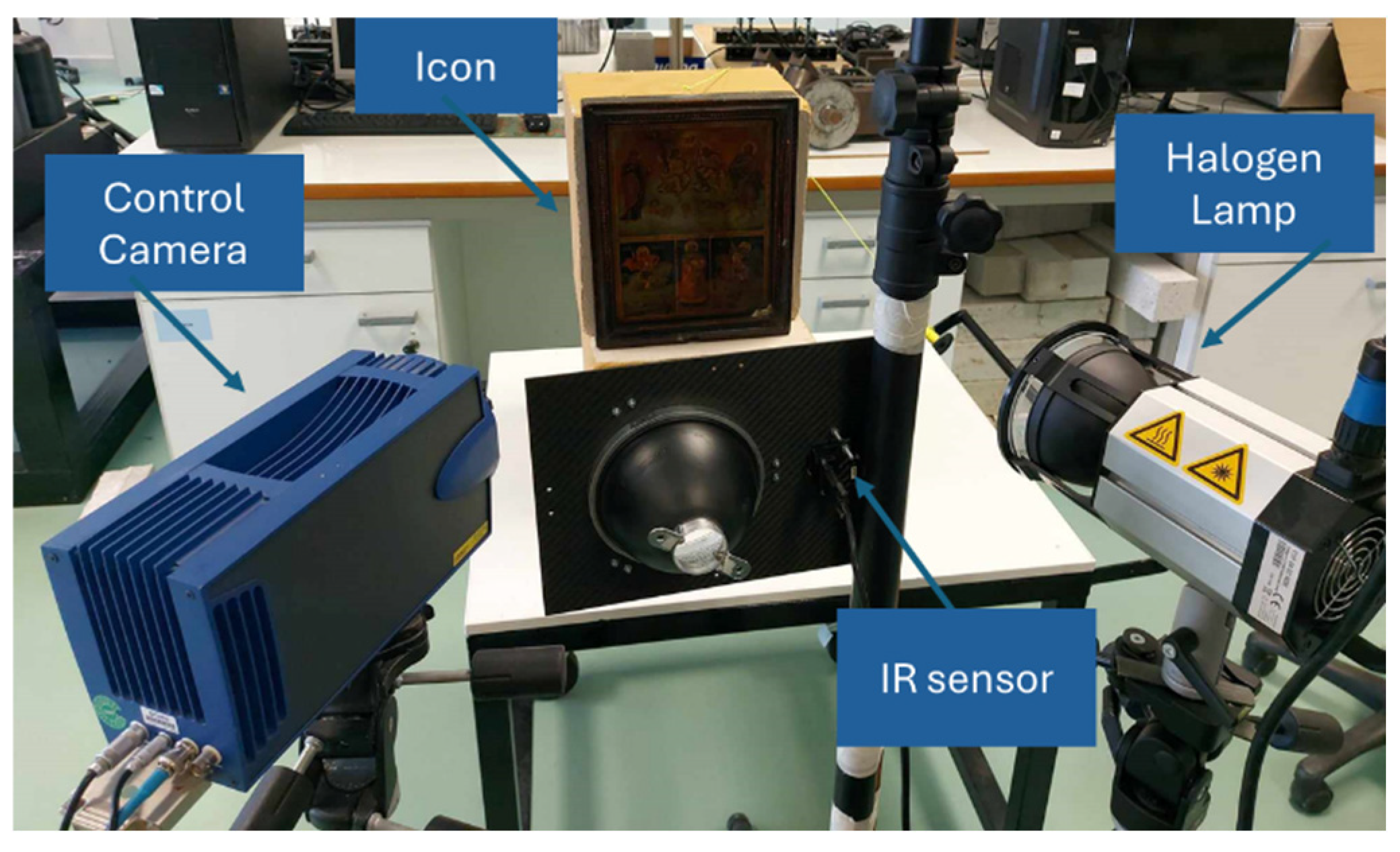
The experimental setup includes an IR sensor, a halogen lamp (300 W) and a temperature control camera. The long-wave IR camera (operates in the infrared longwave band) with a thermal sensitivity of less than 50 mK, spectral range of 8–14 μm and a resolution of 640 × 480 pixels was used to conduct the IRT experiments. As an external thermal excitation source, a halogen lamp was used for continuous and uniform heat flux (amplitude varied between 40% to 80%, and the Lock-in frequency ranged from 0.01 to 0.1 Hz). Both were positioned approximately 45 cm away from the icon to ensure optimal focus and resolution, as shown in Figure 1. Additionally, a second mid-wave IR camera continuously monitored the icon’s temperature to prevent it from exceeding 30 °C, as recommended by the conservator.

Figure 1.
Experimental set-up of infrared thermography.
2.2.2. Multispectral Imaging
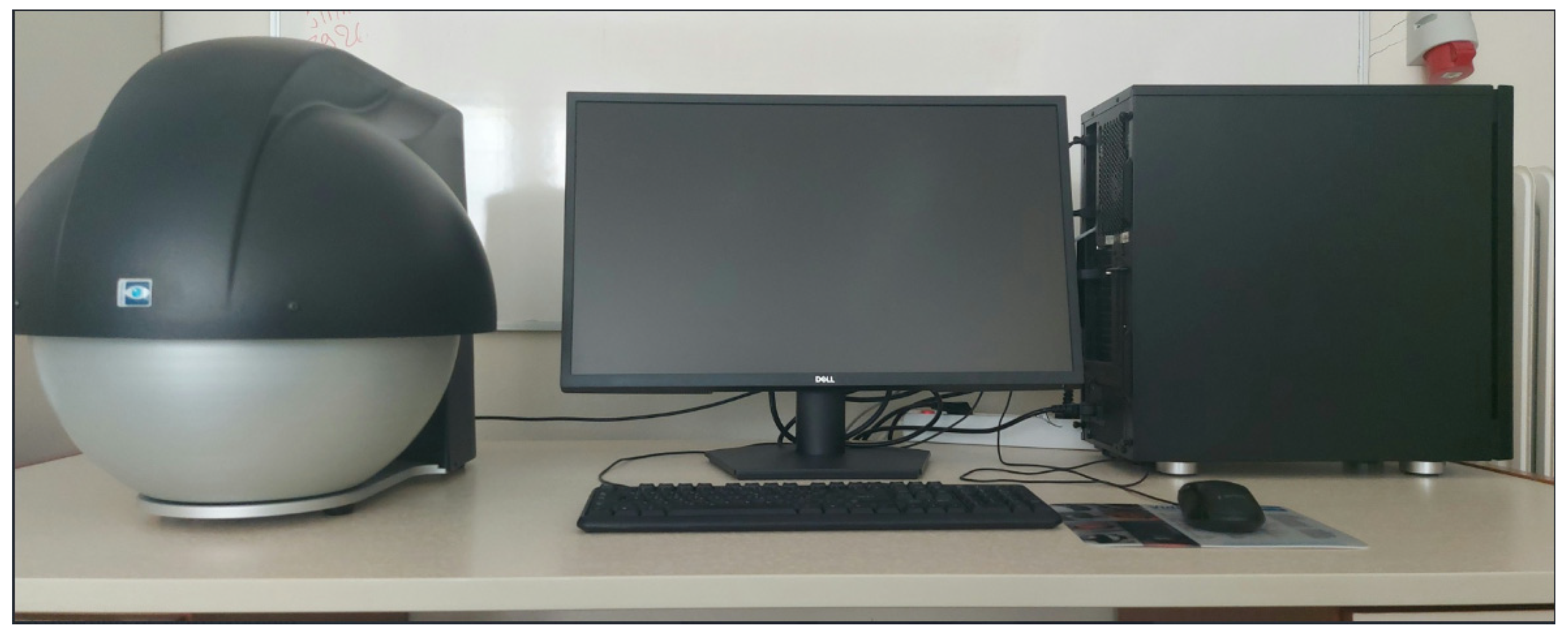
The experimental set-up involved the utilization of a multispectral instrument (VideometerLab 4, Videometer A/S, Herlev, Denmark, Figure 2) connected to a computer, facilitating the acquisition and analysis of the captured images. Each image has specific dimensions (95 mm × 95 mm), so in case of wider specimens, multiple and successive images must be acquired. This advanced spectral imaging system utilizes strobed LED technology, enabling measurements up to 19 distinct wavelengths, integrated into a single high-resolution spectral image. The spectral image resolution spans from 365 to 970 nm, encompassing the UV to NIR spectra. The calibration of the instrument, as well as the data processing, was performed by means of the software provided by the company.

Figure 2.
Experimental set-up of multispectral imaging.
2.2.3. MA-XRF
The icon’s MA-XRF scanning was conducted by using the M6-Jetstream scanner (Bruker Nano GmbH, Berlin, Germany), which is equipped with a 30 W rhodium anode X-ray tube (maximum high voltage: 50 kV) with polycapillary optics and a silicon drift detector (active area: 30 mm2, energy resolution: 139 eV at Mn Kα). The polycapillary full lens focuses the X-ray radiation from the tube on the target. The X-ray tube was operated in voltage and current settings of 50 kV and 600 mA for the icon’s MA-XRF scanning study, respectively.
To examine the painting on the front side of the icon (recto), the beam spot size was set to 100 μm in diameter. The area of the painting is 186 × 217 mm2. The pixel size was set to 150 × 150 μm2, and the dwell time per pixel was 20 ms. These parameters resulted in an overall scanning time of 14 h and the collection of 2,240,680 individual spectra from the icon’s scanned area. For the decorated frame study (25 × 122 mm2), the beam spot size was set to a diameter of 100 μm, the pixel size to 250 × 250 μm2, while the dwell time was set to 25 ms per pixel. These parameters resulted in an overall scanning time of 48 min and the collection of 76,860 individual spectra. The collected spectra were processed and visualized (i.e., transformed into elemental distribution maps) using the built-in M6-Jetstream ESPRIT software (version 1.6.621.0).
3. Results and Discussion
Multispectral imaging, infrared thermography, and MA-XRF are the three nondestructive methods implemented to fulfill this study’s aim. By means of MSI, interesting information was revealed from the icon’s painting surface and the wooden decorated frame. The images acquired within the spectral range of 365 nm to 970 nm (UV to NIR), significantly improved the clarity of Figure 3, Figure 4, Figure 5 and Figure 6, allowing the study of the details of the painting surface. Moreover, exciting hints were revealed using infrared thermography, as depicted in Figure 7 and Figure 8, and by MA-XRF in Figure 9, Figure 10 and Figure 11.

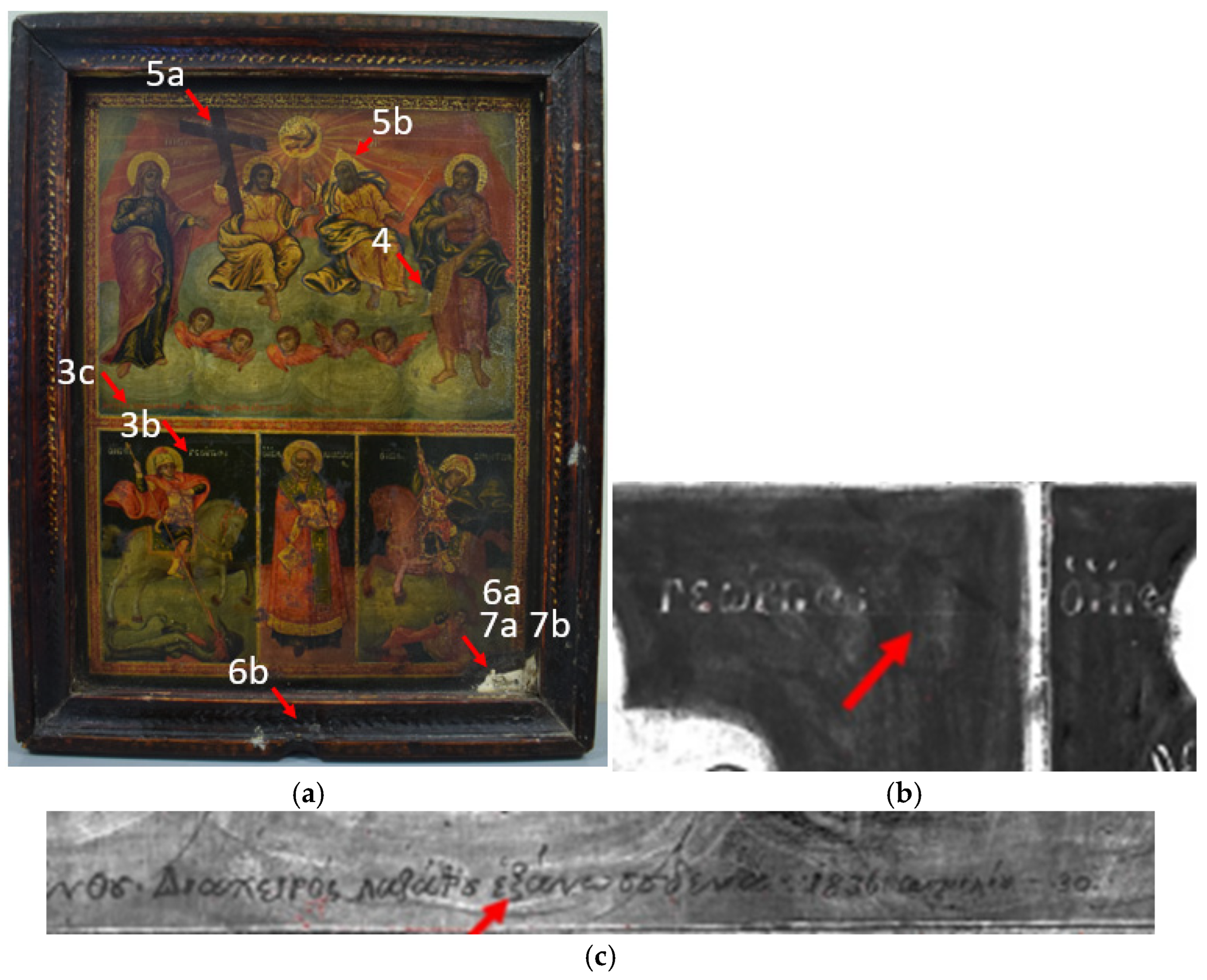
Figure 3.
(a) Post-Byzantine Icon. (b) Boundary lines of the letters. (c) Inscription.
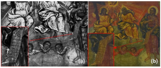
Figure 4.
Improvement of the readability of the Saint John’s scroll. (a) MSI (660 nm) improved picture; (b) Actual image. Arrows indicate the scroll under magnification.

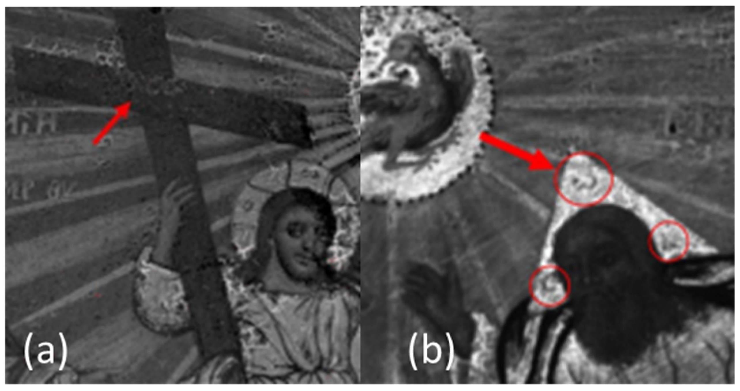
Figure 5.
(a) Crown of thorns on the Cross, as revealed via MSI (590 nm). (b) Revelation of the letters on the halo of the Father (MSI image capture at 590 nm).
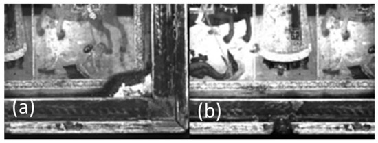
Figure 6.
Multispectral images revealing the loss of the painting surface and preparation layer in (a) the right lower corner and (b) around the perimeter of the frame.

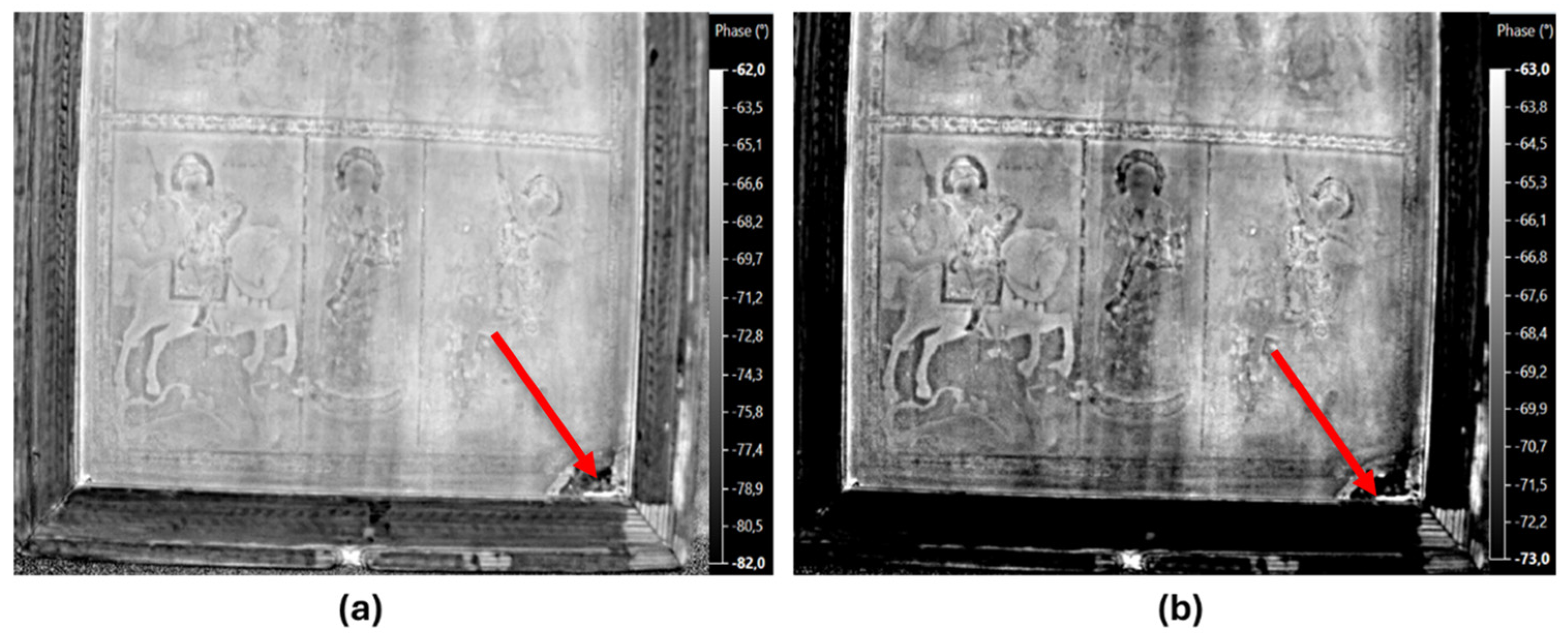
Figure 7.
IRT result of the front side of the icon. Loss of paint surface and the preparation layer/absorbed moisture (a) at the lower right corner of the icon and (b) in the middle of the basis of the frame.
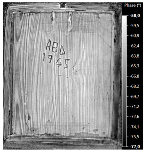
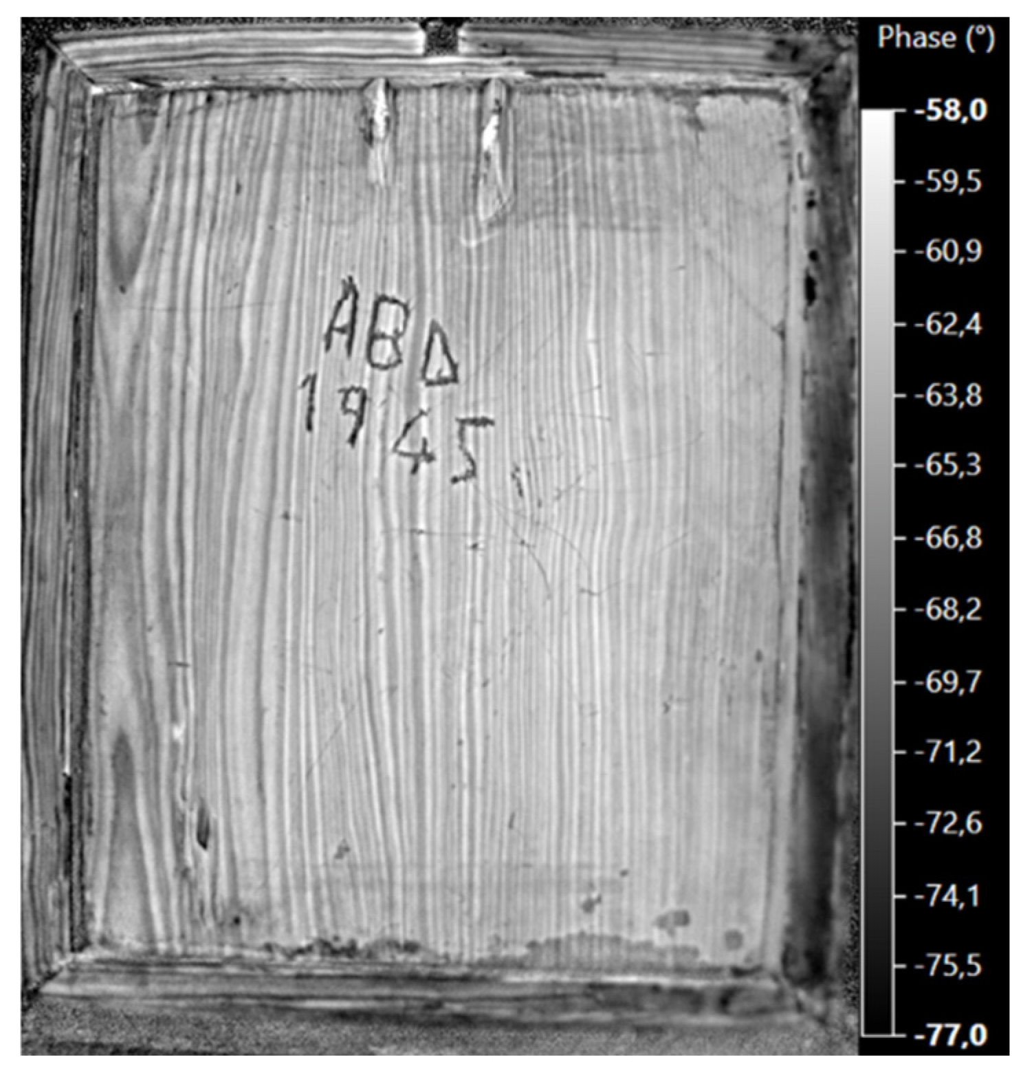
Figure 8.
IRT result of the back side of the icon.
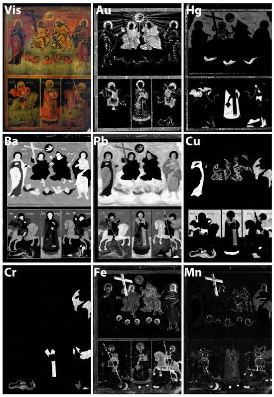
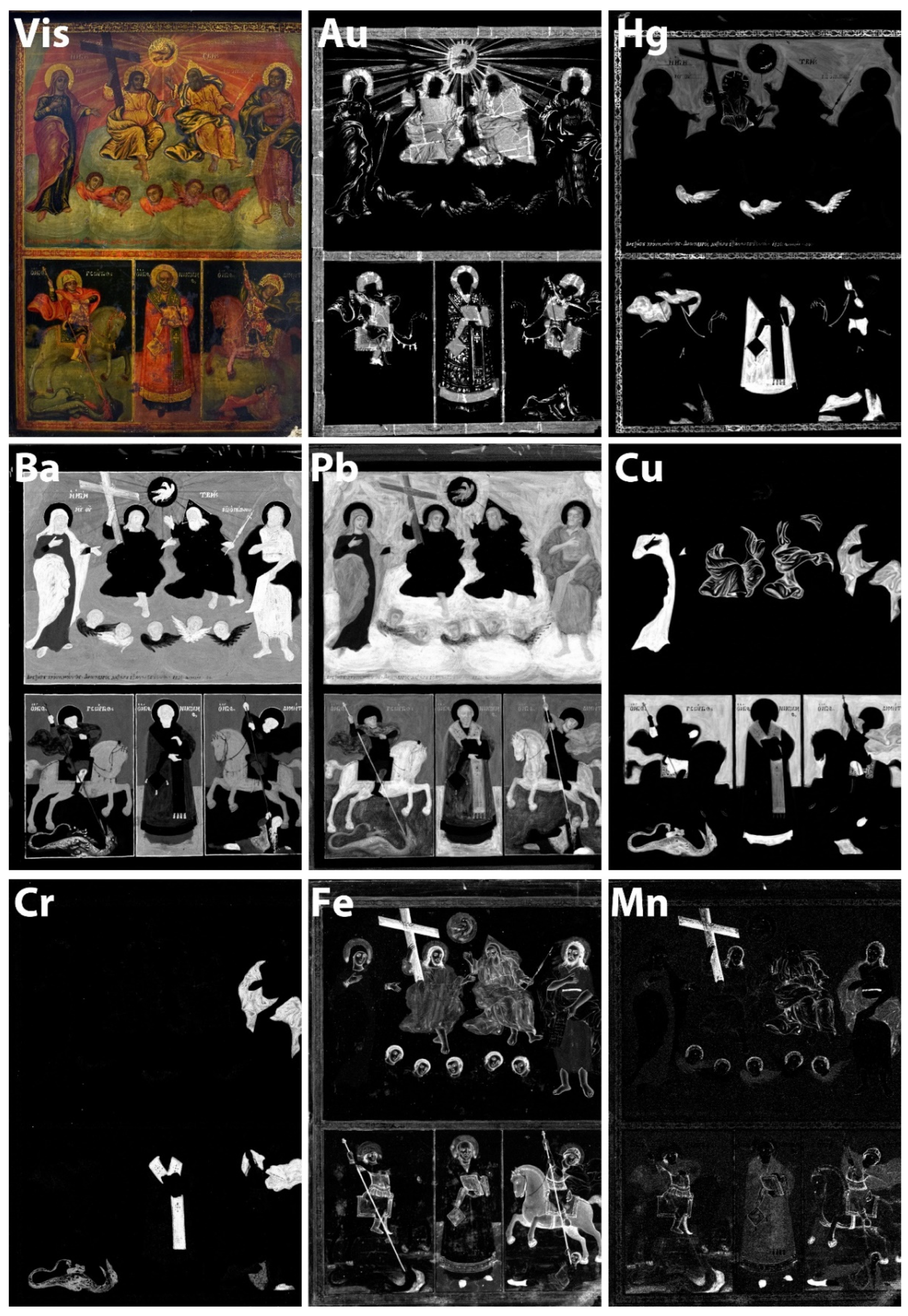
Figure 9.
The scanned area of the recto of the icon and the intensity distribution maps of Au Lα, Hg Lα, Ba Lα, Pb Lα, Cu Κα, Cr Κα, Fe Κα, and Mn Κα.
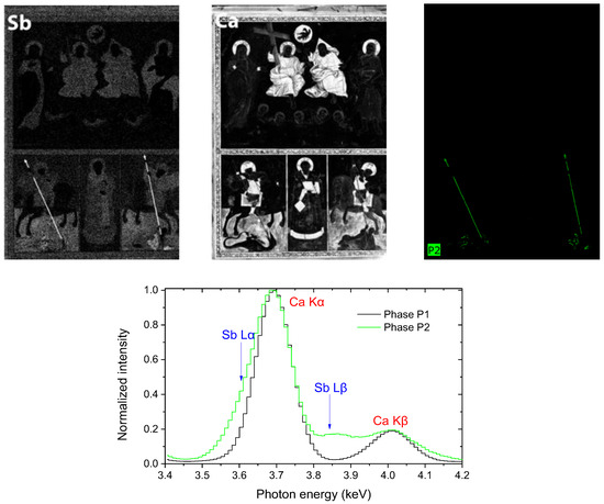
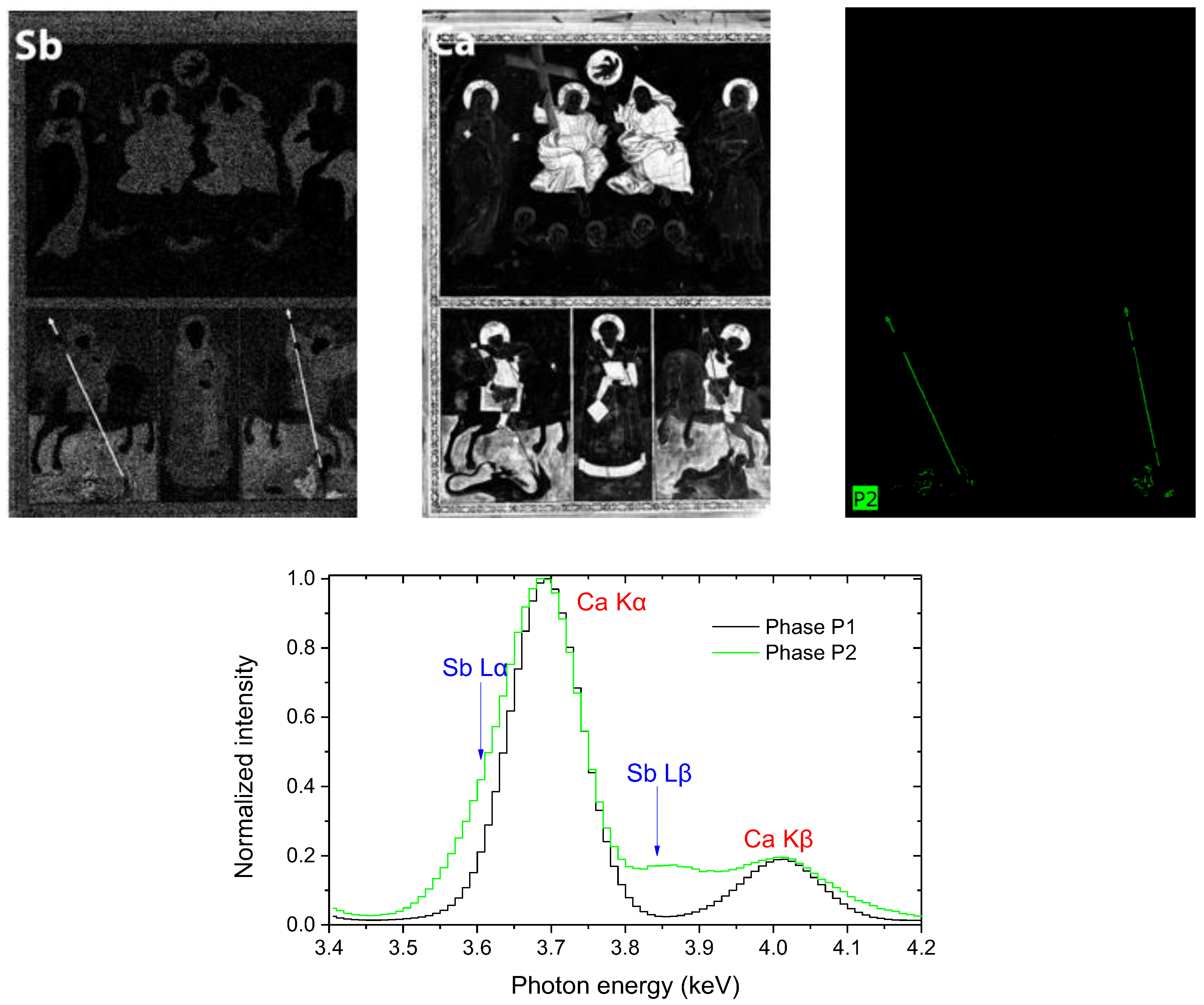
Figure 10.
(Upper) The intensity distribution maps of Sb Lα (Left), Ca Kα (middle), and the spatial distribution of the Sb Lα phase “P2” (right, see text). (Bottom) XRF mean spectra of the “P1” and “P2” phases, normalized to the Ca Kα peak intensity.
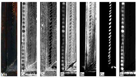
Figure 11.
Results of the MA-XRF analysis of the frame (detail). From left to right: the scanned areas (visible) and the elemental distribution maps of Ca Kα, Zn Κα, Pb Lα, Ba Lα, Cu Κα, and As Κα.
Figure 3a depicts the under-investigation icon, where a wooden frame is attached to its perimeter, while the painting is divided into two horizontal zones. In the upper zone, there is a depiction of the Holy Trinity, on the left of the Son Virgin Mary is depicted, while on the right side of the Father stands St John the Baptist. Beneath the Holy Trinity there are five angels. The lower zone is divided into three subsections by perpendicular margins. On the left is Saint Georgios the Dragon Slayer, in the middle is Saint Nikolaos the Bishop of Myra, and on the right is Saint Demetrius killing Lyaios.
In Figure 3b,c, we observe the first results of the MSI. Figure 3b was acquired at the 14th band (690 nm), where deep red light is incident. More specifically, the boundary lines of the letters next to the Saint’s head can be observed in the lower zone of the icon, which belong to the underdrawings of the painting. Additionally, in Figure 3c, that was acquired at the 8th band (540 nm) where green light is incident, is presented the inscription that is written on the bottom of the upper zone of the painting (on the left corner), in which the readability is significantly improved in comparison to the naked eye observation.
Improved readability is also evident at Saint John’s scroll (Figure 4). In Figure 4a, the image acquired at 660 nm (incident light: red) is presented, while the corresponding visible light image is presented in Figure 4b. This MSI image (Figure 4a) is of great importance, as the pigment that the painter used for the inscription of the scroll is organic, so it could not be deduced from the elemental analysis (XRF).
Figure 5a,b present the two most impressive hints that the MSI disclosed. In particular, Figure 5a, which was captured at 590 nm (incident light: amber), depicts the crown of thorns on the wooden cross, a revelation that was also validated by the XRF results. Moreover, in Figure 5b, the Greek letters OΩΝ in the Father’s halo were revealed at 690 nm (incident light: deep red light), it is worth noting that these letters could hardly be seen even after the cleaning of the icon.
Furthermore, the images shown in Figure 6a,b were acquired at 780 nm (using deep red incident light). In Figure 6a the loss of the painting surface and preparation layer can be clearly seen in the right lower corner and around the perimeter of the frame. Similarly, in Figure 6b, the loss of the painting surface and preparation layer is depicted on the basis of the icon in the central part. These results were also validated by infrared thermography.
As aforementioned, loss of the painting surface and preparation layer due to thermal degradation was also indicated by the results of infrared thermography as shown in Figure 7a and in Figure 7b, in particular at the lower right corner of the icon and in the middle of the basis of the frame, respectively. In detail, the first two thermographs in addition to the loss of paint surface and the preparation layer indicate the absorbed moisture in the areas where the delaminations occur. These areas are depicted in brighter hues (white), as in the right and lower border of the frame. Additional information about the wooden panel was collected through thermography and the corresponding results are presented in Figure 8.
More specifically IRT that was used for the study of the wooden panel (back side of the icon) clearly revealed the curvature on the left side of the icon and the open frame on the upper and especially on the bottom edge (Figure 8). The thermal image indicates the presence of knobs on the upper and the lower edge of the wooden panel. This is a very important finding concerning the wooden panel, as the knobs can cause mechanical stresses between the icon and the frame, due to expansion and contraction (different thermal coefficient), depending on the ambient conditions [19,20,49,50,51,52,53,54]. Along with these results, it is worth noting that in the upper and the lower border of the frame, the wood grain texture is perpendicular, in contrary to the wood grain of the left border, the right border and the wooden panel that are vertical. This can also affect the expansion and contraction of the icon, causing also undesirable results like the curvature of the panel and the frame.
MA-XRF was applied to study the pictorial elements, and returned interesting information concerning the painting itself (recto of the icon) and its decorated frame. Figure 9 depicts a photograph of the icon’s recto while showing the elemental distribution map of gold (Au) that resulted from the MA-XRF scanning of the major part of the painting’s recto. The distribution of Au indicates rather extensive employment of gold, while a detailed examination of the corresponding elemental map reveals the use of two distinct Au-based materials. Indeed, for the gilding of extensive and continuous areas like the haloes, the vestments of the Father and the Son, the borderlines of the icon, etc., gold leaves have been used as the even distribution of Au can deduce it. Note that these areas are periodically interrupted by well-defined, thin bands of higher Au-peak intensity, which correspond either to areas where adjacent leaves are overlapped or to areas of Au-leaf foldings; both the latter are common features observed in Au-distribution maps of pertinent artifacts [36,39]. However, in several other areas, e.g., on the highlights of Virgin Mary and St John vestments, the angels’ wings, etc., the distribution of Au indicates a free/brush application of a pigment-like Au component; this seemingly corresponds to a gold-leaf powder plus organic binder/ medium (“shell gold”), a painting material commonly used by Greek icon painters, after the 17th century [55,56].
The distribution of Mercury (Hg) on the scanned area reveals the rather extensive employment of the pigment cinnabar/vermilion (HgS) in the painting under investigation (Figure 9). In particular, the major peak intensities of Hg are detected on some of the bright red areas like the vestment of the Hierarch (bottom scene—middle), St Nikolaos’ cloak, the date-bearing inscription, the decorated borderlines, etc. Interestingly, Hg is detected only on the wings of three out of the five angels that appear below the Holy Trinity; on the wings of the other two angels, Barium is primarily detected, a finding which suggests that they were rendered in an organic-based red lake fixed on a Ba-containing inorganic substrate. Besides, the presence of Ba is most probably related to the employment of barium sulfate (BaSO4), a (primarily synthetic) substance that has been used as a filler/extender for colors and as a lake substrate since its introduction into pigments’ manufacturing industry on early 19th century [57]. It is worth noting that, being a rather low-cost, bright white pigment, barium sulfate was often used to adulterate the more highly prized lead white, and this seems to be the case of the white pigment used in the icon in consideration as Ba and Pb show a strong correlation (Figure 9). It shall also be noted that Pb is detected throughout the painting and in particular in whitish areas (e.g., clouds), hinting thus to the application of Lead white (2PbCO3•Pb(OH)2). However, Pb is also detected in the red sky. Hence, the employment of a mixture of red lead/minium (Pb3O4) and vermilion (detection of mercury), a mixture commonly used in the framework of post-Byzantine paintings, is inferred [58].
Copper (Cu) is detected on all the pictorial elements that have been rendered in green and blue color [59]. Furthermore, a careful inspection of the corresponding elemental distribution maps reveals the employment of three distinct –in terms of elemental composition—Cu-based pigments in these very areas, namely, one blue and two green. In detail, all the blue areas show primarily Cu accompanied by minor Ba, indicating using a single, Cu-based blue pigment mixed with minor barium sulfate. On the contrary, green areas are differentiated based on the detection of pertinent elements like Chromium (Cr) and Pb: in the green vestments that have received gilded highlights (e.g., St John’s cloak), Cu is accompanied by Cr and minor Pb, revealing thus that a—possibly blue—Cu-based pigment mixed with Chrome yellow (PbCrO4) was used [60]. On the contrary, the green pigment used to render pictorial elements over gold leaves (e.g., the saddles of the horses) shows neither chrome nor lead; the use of a “pure” Cu-based green pigment is inferred (presumably copper resinate), see (Figure 7) [61].
Iron (Fe) is primarily detected on the dark brown and reddish pictorial elements (see e.g., the Cross held by Jesus Christ, the hair of the figures, etc.), implying employment of ochres, i.e., Fe-rich, earthy pigments (Figure 9). Additionally, Fe is detected on the gilded areas indicating that the gold leaves have been attached by using a clayey/bole based gluing agent which is typically used in the framework of the “water gilding” technique [55,60,62]. Manganese (Mn) seems to accompany Fe in the darker brown tones, hinting thus towards the use of umber or sienna-type pigments; the latter is, in fact, generic terms describing Fe-rich earthy pigments that contain variable amounts of Mn compounds that impart brown tones [59,63]. Note also that the aforementioned earthy pigments have—in most cases—been mixed with other pigments (see, e.g., the distribution of Ba and Pb), a practice dictated by the painting manuals used by post-Byzantine painters [58,64].
Of particular interest is the detection of significant Antimony (Sb) at the spears held by St. Georgios and St. Demetrius (Figure 10, upper/left). In fact, the distribution map of Sb Lα that was initially generated by the employed software indicated that this element shows a solid correlation to Ca Kα (Figure 10, upper/middle). However, this is partially an artifact that pertains to the proximity of the energies of the major transitions of these elements, as the Kα transition of Ca lays about 90 eV above the energy of the Lα transition of Sb (3.69 and 3.60 keV respectively); therefore, partial overlap of the two elemental peaks is inevitable [39]. To confirm the presence of Sb, the data were re-analyzed using the “Phases” analytical tool provided by the ESPRIT analysis software. The “Phases” tool groups together pixels with similar compositions into the same cluster (phase). Figure 10, upper/right shows the scanned icon divided into two clusters based on the intensity of the Sb Lα. The pixels of phase “P2” contain significantly higher Sb Lα intensity than those of phase “P1”. Phase “P2” contains the pixels at the spears held by St. Georgios and St. Demetrius (Figure 10, upper/right). The mean XRF spectra of phases “P1” and “P2” are shown in Figure 10, bottom. The Sb L X-ray transitions in the spectrum corresponding to the “P2” distribution confirm the presence of Sb. The presence of Sb indicates that the painter applied a mixture of Naples Yellow (lead antimony oxide–Pb2Sb2O7) with a Fe-based pigment (ochre?) for rendering the spears [60,64].
Finally, in the framework of the MA-XRF investigation of the icon in consideration, the lower left part of its decorated frame was analyzed, revealing the presence of various elements like Ca, Cu, Zn, As, Ba and Pb (Figure 11). The somewhat even distribution of Ca and Zn suggests that these elements pertain to the preparation material/gesso that was applied before decorating the wooden frame; employment of a mixture of calcium sulfate and zinc oxide seems a possibility. On the contrary, Pb and Ba appear only on the inner part of the frame; since the whitish spots that are merely visible in this very area show the maximum intensity of Pb spectral peaks, it is assumed that the background was probably rendered using a mixture of red lead/minium and barium sulfate. Moreover, on a mirrored position to the Pb-rich whitish spots, there lay spots rendered in a—presumably blue—Cu pigment, which probably contributed to the decoration of the frame. Finally, of particular interest is the detection of Arsenic on the dots that embellish the outer margin of the frame (left). This element indicates the employment of Orpiment pigment (As2S3), which is of special interest considering its use in the post-Byzantine Greek religious painting.
4. Conclusions
Three complementary, non-destructive techniques were applied to thoroughly investigate a Greek religious wooden panel painting (“icon”) and the decorated frame. multispectral imaging (MSI) was applied to render information about the underdrawings, improving the readability of the inscription and the text on Saint John’s scroll, and revealing hints about the painting surface and the extent of damage due to degradation through the ages. Infrared thermography helped validate the results of MSI and revealed hints as to the internal structure of the wooden panel. Furthermore, MA-XRF provided exceptional information about the pigments and various materials used by the painter, revealing the employment of a rather rich palette comprising more than ten pigments and two types of gilded decorations. The complementary examination of the results of the three methods provides results that reveal findings, that none of the methods can on its own. It is thus demonstrated that the combined application of the aforementioned non-destructive techniques leads to a thorough investigation of paintings’ materials, techniques, and state of preservation, proving to be a valuable tool for proper conservation interventions and accurate planning of them.
Author Contributions
Conceptualization, T.E.M.; methodology, T.E.M., G.T.V., D.A.E. and D.F.A.; software, G.T.V., S.F. and A.A.; investigation, G.T.V., S.F., G.P.M. and A.A.; data curation, G.T.V., S.F., A.A. and G.P.M.; writing—original draft preparation, G.T.V. and G.P.M.; writing—review and editing, G.T.V., S.F., G.P.M., D.A.E., D.F.A. and T.E.M.; supervision, T.E.M. All authors have read and agreed to the published version of the manuscript.
Funding
This research received no external funding.
Data Availability Statement
The raw data supporting the conclusions of this article will be made available by the authors on request.
Conflicts of Interest
The authors declare no conflicts of interest.
References
- Colombini, M.P.; Andreotti, A.; Bonaduce, I.; Modugno, F.; Ribechini, E. Analytical Strategies for Characterizing Organic Paint Media Using Gas Chromatography/Mass Spectrometry. Acc. Chem. Res. 2010, 43, 715–727. [Google Scholar] [PubMed]
- Marengo, E.; Manfredi, M.; Zerbinati, O.; Robotti, E.; Mazzucco, E.; Gosetti, F.; Bearman, G.; France, F.; Shor, P. Development of a technique based on multi-spectral imaging for monitoring the conservation of cultural heritage objects. Anal. Chim. Acta 2011, 706, 229–237. [Google Scholar] [PubMed]
- Issa, Y.M.; Abdel-Maksoud, G.; Ibrahim, M.; Magdy, M. A combination of analytical methods to evaluate the effect of humidity aging on the painting materials of icon models. Vib. Spectrosc. 2020, 107, 103010. [Google Scholar]
- Tavakolian, P.; Shokouhi, E.; Sfarra, S.; Gargiulo, G.; Mandelis, A. Non-destructive imaging of ancient marquetries using active thermography and photothermal coherence tomography. J. Cult. Herit. 2020, 46, 159–164. [Google Scholar]
- Laureti, S.; Colantonio, C.; Burrascano, P.; Melis, M.; Calabrò, G.; Malekmohammadi, H.; Sfarra, S.; Ricci, M.; Pelosi, C. Development of integrated innovative techniques for paintings examination: The case studies of The Resurrection of Christ attributed to Andrea Mantegna and the Crucifixion of Viterbo attributed to Michelangelo’s workshop. J. Cult. Herit. 2019, 40, 1–16. [Google Scholar]
- Trentelman, K.; Bouchard, M.; Ganio, M.; Namowicz, C.; Patterson, C.S.; Walton, M. The Examination of Works of Art Usingin SituXRF Line and Area Scans. X-Ray Spectrom. Int. J. 2010, 39, 159–166. [Google Scholar]
- Borg, B.; Dunn, M.; Ang, A.; Villis, C. The application of state-of-the-art technologies to support artwork conservation: Literature review. J. Cult. Herit. 2020, 44, 239–259. [Google Scholar]
- Yao, Y.; Sfarra, S.; Ibarra-Castanedo, C.; You, R.; Maldague, X. The multi-dimensional ensemble empirical mode decomposition (MEEMD). J. Therm. Anal. Calorim. 2017, 128, 1841–1858. [Google Scholar]
- Alexakis, E.; Delegou, E.T.; Mavrepis, P.; Rifios, A.; Kyriazis, D.; Moropoulou, A. A novel application of deep learning approach over IRT images for the automated detection of rising damp on historical masonries. Case Stud. Constr. Mater. 2024, 20, e02889. [Google Scholar]
- Wu, D.; Busse, G. Lock-in thermography for nondestructive evaluation of materials. Rev. Générale Therm. 1998, 37, 693–703. [Google Scholar]
- Grinzato, E. IR Thermography Applied to the Cultural Heritage Conservation. Recent Advances in Non-Destructive Inspection. In Proceedings of the 18th World Conference on Nondestructive Testing, Durban, South Africa, 16–20 April 2012. [Google Scholar]
- Ambrosini, D.; Daffara, C.; Di Biase, R.; Paoletti, D.; Pezzati, L.; Bellucci, R.; Bettini, F. Integrated reflectography and thermography for wooden paintings diagnostics. J. Cult. Herit. 2010, 11, 196–204. [Google Scholar]
- Mercuri, F.; Cicero, C.; Orazi, N.; Paoloni, S.; Marinelli, M.; Zammit, U. Infrared Thermography Applied to the Study of Cultural Heritage. Int. J. Thermophys. 2015, 36, 1189–1194. [Google Scholar]
- Mercuri, F.; Zammit, U.; Orazi, N.; Paoloni, S.; Marinelli, M.; Scudieri, F. Active infrared thermography applied to the investigation of art and historic artefacts. J. Therm. Anal. Calorim. 2011, 104, 475. [Google Scholar]
- Mercuri, F.; Buonora, P.; Cicero, C.; Helas, P.; Manzari, F.; Marinelli, M.; Paoloni, S.; Pasqualucci, A.; Pinzari, F.; Romani, M.; et al. Metastructure of illuminations by infrared thermography. J. Cult. Herit. 2018, 31, 53–62. [Google Scholar]
- Mercuri, F.; Orazi, N.; Paoloni, S.; Cicero, C.; Zammit, U. Pulsed Thermography Applied to the Study of Cultural Heritage. Appl. Sci. 2017, 7, 1010. [Google Scholar]
- Gavrilov, D.; Maeva, E.; Grube, O.; Vodyanoy, I.; Maev, R. Experimental Comparative Study of the Applicability of Infrared Techniques for Non-destructive Evaluation of Paintings. J. Am. Inst. Conserv. 2013, 52, 48–60. [Google Scholar]
- Yao, Y.; Sfarra, S.; Lagüela, S.; Ibarra-Castanedo, C.; Wu, J.-Y.; Maldague, X.P.V.; Ambrosini, D. Active thermography testing and data analysis for the state of conservation of panel paintings. Int. J. Therm. Sci. 2018, 126, 143–151. [Google Scholar]
- Kordatos, E.Z.; Exarchos, D.; Matikas, T.E.; Stavrakos, C.; Moropoulou, A. Application of IR thermography to damage characterization of structures and the diagnosis of historic monuments. In Emerging Technologies in Non-Destructive Testing V, Proceedings of the 5th Conference on Emerging Technologies in NDT, Ioannina, Greece, 19–21 September 2011; Routledge: London, UK, 2011; pp. 77–81. [Google Scholar]
- Kordatos, E.Z.; Exarchos, D.A.; Stavrakos, C.; Moropoulou, A.; Matikas, T.E. Infrared thermographic inspection of murals and characterization of degradation in historic monuments. Constr. Build. Mater. 2013, 48, 1261–1265. [Google Scholar]
- Varfi, G.T.; Asvestas, A.; Exarchos, D.A.; Farmaki, S.; Mastrotheodoros, G.; Anagnostopoulos, D.F.; Matikas, T.E. Nondestructive Assessment of Post-Byzantine Icon. In Advanced Nondestructive and Structural Techniques for Diagnosis, Redesign and Health Monitoring for the Preservation of Cultural Heritage; Osman, A., Moropoulou, A., Eds.; Springer Proceedings in Materials; Springer: Cham, Switzerland, 2022; Volume 16. [Google Scholar]
- Varfi, G.T.; Farmaki, S.; Gerodimos, T.; Exarchos, D.A.; Mastrotheodoros, G.; Anagnostopoulos, D.F.; Matikas, T.E. Nondestructive Assessment of Hidden Information in a Post-Byzantine Icon. In Advanced Nondestructive and Structural Techniques for Diagnosis, Redesign and Health Monitoring for the Preservation of Cultural Heritage; Osman, A., Moropoulou, A., Lampropoulos, K., Eds.; TMM 2023; Springer Proceedings in Materials; Springer: Cham, Switzerland, 2024; Volume 33. [Google Scholar] [CrossRef]
- Exarchos, D.; Farmaki, S.; Tragazikis, I.; Mpalaskas, A.; Mitsikostas, A.; Vasios, A.; Papadopoulou, V.; Matikas, T.E. Development of innovative 3D representation techniques and nondestructive evaluation tools for the sustainability of antiquities. In Proceedings of the SPIE 12046, Sensors and Smart Structures Technologies for Civil, Mechanical, and Aerospace Systems, Long Beach, CA, USA, 6 March–11 April 2022. [Google Scholar]
- Exarchos, D.A.; Farmaki, S.G.; Tragazikis, I.K.; Mpalaskas, A.C.; Vasios, A.; Papadopoulou, V.; Matikas, T.E. Documenting Artifacts Using 3D Representation and Nondestructive Evaluation Tools. In Advanced Nondestructive and Structural Techniques for Diagnosis, Redesign and Health Monitoring for the Preservation of Cultural Heritage; Osman, A., Moropoulou, A., Eds.; Springer International Publishing: Cham, Switzerland, 2022. [Google Scholar]
- Bonizzoni, L. ED-XRF analysis for Cultural Heritage: Is quantitative evaluation always essential? J. Phys. Conf. Ser. 2015, 630, 012001. [Google Scholar]
- Andrić, V.; Gajić-Kvaščev, M.; Korolija Crkvenjakov, D.; Marić-Stojanović, M.; Gadžurić, S. Evaluation of pattern recognition techniques for the attribution of cultural heritage objects based on the qualitative XRF data. Microchem. J. 2021, 167, 106267. [Google Scholar]
- Ha, J.-W.; Lee, S.-J. Identification of natural inorganic pigments used on 18th century Korean traditional mural paintings by using a portable X-ray fluorescence. J. Ind. Eng. Chem. 2015, 28, 328–333. [Google Scholar]
- Galli, A.; Gargano, M.; Bonizzoni, L.; Bruni, S.; Interlenghi, M.; Longoni, M.; Passaretti, A.; Caccia, M.; Salvatore, C.; Castiglioni, I.; et al. Imaging and spectroscopic data combined to disclose the painting techniques and materials in the fifteenth century Leonardo atelier in Milan. Dye. Pigment. 2021, 187, 109112. [Google Scholar]
- Kokiasmenou, E.; Caliri, C.; Kantarelou, V.; Karydas, A.G.; Romano, F.P.; Brecoulaki, H. Macroscopic XRF imaging in unravelling polychromy on Mycenaean wall-paintings from the Palace of Nestor at Pylos. J. Archaeol. Sci. Rep. 2020, 29, 102079. [Google Scholar]
- Molari, R.; Appoloni, C.R. A PXRF and TXRF study of The Portrait of a young gentleman (1539), by Lucas Cranach the Elder. Radiat. Phys. Chem. 2019, 165, 108413. [Google Scholar]
- Molari, R.; Appoloni, C.R. Pigment analysis in four paintings by Vincent van Gogh by portable X-ray fluorescence (pXRF). Radiat. Phys. Chem. 2021, 181, 109336. [Google Scholar]
- Stratis, J.A.; Makarona, C.; Lazidou, D.; Gómez Sánchez, E.; Koutsoudis, A.; Pamplona, M.; Pauswein, R.; Pavlidis, G.; Simon, S.; Tsirliganis, N. Enhancing the examination workflow for Byzantine icons: Implementation of information technology tools in a traditional context. J. Cult. Herit. 2014, 15, 85–91. [Google Scholar]
- Kriznar, A.; Muñoz, M.d.V.; de la Paz, F.; Respaldiza, M.Á.; Vega, M. Non-destructive XRF analysis of selected Flemish panel paintings in the Fine Arts Museum of Seville. J. Inst. Conserv. 2014, 37, 136–151. [Google Scholar]
- Hoffmann, P.; Flege, S.; Ensinger, W.; Wolf, F.; Weber, C.; Seeberg, S.; Sander, J.; Schultz, J.; Krekel, C.; Tagle, R.; et al. MA-XRF investigation of the Altenberg Retable from 1330. X-Ray Spectrom. 2018, 47, 215–222. [Google Scholar]
- Alberti, R.; Frizzi, T.; Gironda, M.; Occhipinti, M.; Parsani, T.; Seccaroni, C.; Tatì, A. From noise to information. Analysing macro-XRF mapping of strontium impurities in Raphael’s Baglioni Entombment in the Galleria Borghese, Rome. J. Cult. Herit. 2022, 58, 130–136. [Google Scholar]
- Mastrotheodoros, G.P.; Asvestas, A.; Gerodimos, T.; Tzima, A.; Papadopoulou, V.; Anagnostopoulos, D.F. MA-XRF investigation of a 17th century icon by the renowned painter Theodoros Poulakis. J. Archaeol. Sci. Rep. 2024, 53, 104313. [Google Scholar]
- Gerodimos, T.; Asvestas, A.; Mastrotheodoros, G.P.; Chantas, G.; Liougos, I.; Likas, A.; Anagnostopoulos, D.F. Scanning X-ray Fluorescence Data Analysis for the Identification of Byzantine Icons’ Materials, Techniques, and State of Preservation: A Case Study. J. Imaging 2022, 8, 147. [Google Scholar] [CrossRef]
- Mastrotheodoros, G.P.; Anagnostopoulos, D.F.; Beltsios, K.G. Cleaning Old Icons: The Effect of Varnishes on the XRF Analysis of Portable Panel Paintings. In Advanced Nondestructive and Structural Techniques for Diagnosis, Redesign and Health Monitoring for the Preservation of Cultural Heritage; Springer Nature: Cham, Switzerland, 2024. [Google Scholar]
- Mastrotheodoros, G.P.; Asvestas, A.; Gerodimos, T.; Anagnostopoulos, D.F. Revealing the Materials, Painting Techniques, and State of Preservation of a Heavily Altered Early 19th Century Greek Icon through MA-XRF. Heritage 2023, 6, 1903–1920. [Google Scholar] [CrossRef]
- Jones, C.; Duffy, C.; Gibson, A.; Terras, M. Understanding multispectral imaging of cultural heritage: Determining best practice in MSI analysis of historical artefacts. J. Cult. Herit. 2020, 45, 339–350. [Google Scholar]
- Li, H.; Li, G.; Ye, Y.; Lin, L. A high-efficiency acquisition method of LED-multispectral images based on frequency-division modulation and RGB camera. Opt. Commun. 2021, 480, 126492. [Google Scholar]
- Moropoulou, A.; Delegou, E.T.; Vlahakis, V.; Karaviti, E. Digital processing of SEM images for the assessment of evaluation indexes of cleaning interventions on Pentelic marble surfaces. Mater. Charact. 2007, 58, 1063–1069. [Google Scholar]
- Simon Chane, C.; Mansouri, A.; Marzani, F.S.; Boochs, F. Integration of 3D and multispectral data for cultural heritage applications: Survey and perspectives. Image Vis. Comput. 2013, 31, 91–102. [Google Scholar]
- Hain, M.; Bartl, J.; Jacko, V. Multispectral analysis of cultural heritage artefacts. Meas. Sci. Rev. 2003, 3, 9–12. [Google Scholar]
- Havermans, J.; Aziz, H.A.; Scholten, H. Non Destructive Detection of Iron Gall Inks by Means of Multispectral Imaging Part 1: Development of the Detection System. Restaur. Int. J. Preserv. Libr. Arch. Mater. 2003, 24, 55–60. [Google Scholar]
- Elias, M.; Mas, N.; Cotte, P. Review of several optical non-destructive analyses of an easel painting. Complementarity and crosschecking of the results. J. Cult. Herit. 2011, 12, 335–345. [Google Scholar]
- Cosentino, A. Identification of pigments by multispectral imaging; a flowchart method. Herit. Sci. 2014, 2, 8. [Google Scholar]
- Alexakis, E.; Delegou, E.T.; Lampropoulos, K.C.; Apostolopoulou, M.; Ntoutsi, I.; Moropoulou, A. NDT as a monitoring tool of the works progress and the assessment of materials and rehabilitation interventions at the Holy Aedicule of the Holy Sepulchre. Constr. Build. Mater. 2018, 189, 512–526. [Google Scholar]
- Avdelidis, N.P.; Moropoulou, A. Applications of infrared thermography for the investigation of historic structures. J. Cult. Herit. 2004, 5, 119–127. [Google Scholar]
- Avdelidis, N.P.; Moropoulou, A. Emissivity considerations in building thermography. Energy Build. 2003, 35, 663–667. [Google Scholar]
- Avdelidis, N.P.; Moropoulou, A.; Delegou, E. A thermographic study for the assessment of historic structures. In Proceedings of the 7th Quantitative Infrared Thermography Conference (QIRT), Brussels, Belgium, 1 January 2004. [Google Scholar]
- Moropoulou, A.; Avdelidis, N.P.; Koui, M.; Delegou, E.T.; Tsiourva, T. Infrared Thermographic Assessment of Materials & Techniques for the Protection of Cultural Heritage. In Proceedings of the SPIE—The International Society for Optical Engineering, Wuhan, China, 25 September 2001; Volume 4548, pp. 313–318. [Google Scholar] [CrossRef]
- Moropoulou, A.; Labropoulos, K.C.; Delegou, E.T.; Karoglou, M.; Bakolas, A. Non-destructive techniques as a tool for the protection of built cultural heritage. Constr. Build. Mater. 2013, 48, 1222–1239. [Google Scholar]
- Varfi, G.T.; Farmaki, S.; Moschovas, D.; Exarchos, D.A.; Matikas, T.E. Evaluation of Materials and Techniques of a Nineteenth Century Hieratic Manuscript. In Advanced Nondestructive and Structural Techniques for Diagnosis, Redesign and Health Monitoring for the Preservation of Cultural Heritage; Osman, A., Moropoulou, A., Lampropoulos, K., Eds.; TMM 2023; Springer Proceedings in Materials; Springer: Cham, Switzerland, 2024; Volume 33. [Google Scholar]
- Mastrotheodoros, G.P.; Beltsios, K.G.; Bassiakos, Y.; Papadopoulou, V. On the Metal-Leaf Decorations of Post-Byzantine Greek Icons. Archaeometry 2018, 60, 269–289. [Google Scholar]
- Eastaugh, N.; Walsh, V.; Chaplin, T.; Siddall, R. Pigment Compendium: A Dictionary of Historical Pigments, 1st ed.; Routledge: Abingdon, UK, 2004. [Google Scholar] [CrossRef]
- Feller, R.L. Barium Sulfate—Natural and Synthetic. In Artists’ Pigments: A Handbook of Their History and Characteristics; Feller, R.L., Ed.; National Gallery of Art: Washington, DC, USA, 1986; Volume 1, pp. 47–64. [Google Scholar]
- Mastrotheodoros, G.P.; Beltsios, K.G.; Bassiakos, Y. On the Red and Yellow Pigments of Post-Byzantine Greek Icons*. Archaeometry 2021, 63, 753–778. [Google Scholar]
- Mastrotheodoros, G.P.; Beltsios, K.G.; Bassiakos, Y. On the blue and green pigments of post-Byzantine Greek icons. Archaeometry 2020, 62, 774–795. [Google Scholar]
- Mactaggart, P.; Mactaggart, A. Practical Gilding; Archetype; Archetype Publications: London, UK, 2002. [Google Scholar]
- Mastrotheodoros, G.P.; Beltsios, K.G. Pigments—Iron-based red, yellow, and brown ochres. Archaeol. Anthropol. Sci. 2022, 14, 35. [Google Scholar]
- Mastrotheodoros, G.P.; Beltsios, K.G. The Balpis Expansion of the Technical Part of the Hermeneia Painting Manual by Dionysius of Fourna. Stud. Conserv. 2024, 69, 425–451. [Google Scholar]
- Dionysios of Fourna; Hetherington, P. The ‘Painter’s Manual’of Dionysius of Fourna: An English Translation with Commentary of Cod. Gr. 708 in the Saltykov-Shchedrin State Public Library, Leningrad; Oakwood Publications: Torrance, CA, USA, 1996. [Google Scholar]
- Feller, R.L. (Ed.) Artists’ Pigments: A Handbook of Their History and Characteristics; National Gallery of Art: Washington, DC, USA, 1986. [Google Scholar]


Disclaimer/Publisher’s Note: The statements, opinions and data contained in all publications are solely those of the individual author(s) and contributor(s) and not of MDPI and/or the editor(s). MDPI and/or the editor(s) disclaim responsibility for any injury to people or property resulting from any ideas, methods, instructions or products referred to in the content. |
© 2025 by the authors. Licensee MDPI, Basel, Switzerland. This article is an open access article distributed under the terms and conditions of the Creative Commons Attribution (CC BY) license (https://creativecommons.org/licenses/by/4.0/).