Abstract
Imagining life before the advent of modern medical treatments is challenging. Today, congenital dysplasia is typically diagnosed within the first months of a child’s life, allowing for timely intervention. In the past, however, this condition often went unrecognized and untreated, as evidenced by archaeological findings that document the presence of congenital dysplasia persisting into adulthood. We present the case of the individual recovered from the hypogeal cemetery of Santa Maria Maggiore in Vercelli, Italy, a funerary context dated from the 18th to the 19th century. Using macroscopic and radiographic analyses, various morphological irregularities were identified, consistent with the characteristics of developmental hip dysplasia. The skeletal remains identified as FU12 SU151 include a right os coxa and femur, belonging to an adult female. The femur features a 90-degree femoral head angle and a shortened neck with nodules. The acetabulum shows significant morphological changes, including a triangular shape and absence of lunate surfaces, deviating from the normal structure for femoral articulation. CT scans revealed a void within the acetabulum, indicating an absence of material. Despite preservation challenges that restrict the identification of definitive signs, our findings offer valuable insights into possible developmental dysplasia in historic skeletal remains. This research provides insights into the impact of untreated congenital conditions on past populations, underscoring the importance of preserving and studying such remains to enhance our understanding of historical health issues.
1. Introduction
Paleopathology provides unique opportunities to uncover human narratives from the past, particularly through the study of health and disease, and their broader cultural significance. The analysis of human remains recovered from ancient burial contexts not only documents the presence of past pathologies, but also enhances our understanding of how these conditions impacted individuals’ lives and shaped community responses, such as healthcare and caregiving practices. This integrative approach allows us to explore the intersection of health and heritage, offering insights into the cultural and societal adaptations to disease [1,2,3]. In times when medical care was often inadequate, untreated congenital conditions could significantly affect individuals’ lives. Through the study of these ancient conditions, we can understand the consequences of untreated cases and the developmental disabilities that arose as a result.
Congenital anomalies, or birth defects, refer to structural or functional abnormalities that occur during prenatal development. They can present as either structural malformations or functional impairments, with a significant proportion affecting the skeletal system [4]. In particular, developmental dysplasia of the hip (DDH) progressively leads to disability by disrupting the normal alignment of joint components. Clinically, DDH can range from complete dislocation at birth to acetabular dysplasia in adults. Nowadays early diagnosis through clinical exams in newborns is performed, and prevents cases of untreated DDH, which can progress to childhood and adult dysplasia, eventually requiring total hip replacement [5,6,7]. In fact, both misdiagnosed and undiagnosed cases of DDH can lead to abnormal stress distribution on the acetabulum and femoral head. This results from the impaired alignment and development of these structures, causing hip instability and skeletal deformities. The morphological appearance of the dysplastic acetabulum is significantly influenced by the degree of contact maintained with the femoral head. If dislocation occurs during the neonatal phase, the lack of proper alignment between the femoral head and the acetabulum hinders the normal development of the hip joint. The absence of a proper articulating surface obstructs the correct formation of both the acetabulum and the femoral head. Consequently, the acetabulum takes on a triangular shape with three obtuse margins, which affects the realignment of the femoral head [5]. Therefore, cases that persist into adulthood may result in altered gait, morphological abnormalities of the joint, and early-onset osteoarthritis [6]. The etiology of developmental dysplasia of the hip (DDH) is multifaceted, encompassing both endogenous and exogenous factors. Among these factors, certain variables emerge as particularly influential risk factors, including female sex, a positive family history of DDH, breech presentation, vaginal delivery, and primiparity [8,9,10].
The literature on paleopathology predominantly showcases case studies originating from European nations such as France [11,12,13], England [14,15,16,17,18], Poland [19], Slovakia [20], Romania [21], The Netherlands [22] and Italy [23,24], confirming Europe as a high-prevalence zone for this pathology [13].
Although instances from North America have been documented across diverse temporal epochs, spanning from the 6th century AD [25] to the 12th–15th centuries AD [26,27] and the notably high modern incidence in Native Americans [28], the literature on South American paleopathology remains notably limited. Historical records trace back to Moodie’s work in 1923, which vaguely alludes to occurrences of hip luxation and neoacetabulum formation in a Peruvian skeleton [29]. Additionally, Costa and Llagostera [30] broach the subject with their examination of an individual from the Coyo Occidente 3 site in San Pedro de Atacama, Chile, dated to the conclusion of the Middle Period (10th century AD). Another noteworthy instance arises from the research of Plischuk et al. [9], who document a case in a female individual from Argentina dating between 400 and 1000 AD.
More recently, a notable case of bilateral hip dysplasia emerged from South Africa, attributed to a male individual from the 17th–18th century [31].
Our investigation centers on a skeletal remain displaying indications of hip dysplasia, unearthed during archaeological excavations carried out in Sector III of the hypogeal cemetery of Santa Maria Maggiore in Vercelli, Italy. The preservation of these remains, along with meticulous documentation at every stage of the archaeological process, is crucial, as it allows for the reconstruction of the individuals’ lifestyle. This preservation enables us to connect with historical practices and health challenges, offering invaluable insights into the evolution of skeletal diseases and their impacts on past populations.
2. Materials
2.1. The Burial Site
The co-cathedral of Santa Maria Maggiore in Vercelli, Piedmont, northern Italy, was constructed in the 18th century, replacing an earlier complex and the adjacent ‘Santissima Trinità’ church [32]. After the Jesuit suppression, the Episcopal Curia completed the new church by 1780, transferring the dedication from the former Basilica of Santa Maria Maggiore, demolished in 1776–1777 [33,34].
The hypogeal cemetery, inaugurated in 1777, initially accommodated interments and received relocated remains from the former episcopal complex from 1775 [32,35,36]. Architecturally, the vaulted subterranean space includes masonry structures, burial chambers, private chapels, and two central underground ossuaries.
Since September 2020, the recovery and analysis of the bioarchaeological remains from the cemetery have been ongoing for research purposes, and have revealed several noteworthy cases already published, including the presence of naturally mummified bodies, evidence of scurvy in children’s remains and the discovery of skulls with anatomical dissections [35,36,37].
To facilitate organization and cataloging, the cemetery was divided into sectors I to V, and the tomb structures, referred to as “Funerary Units (FUs)”, were numbered from 1 to 19, with each find being assigned a serial number.
2.2. Sector III, Funerary Unit 12
Funerary Unit 12, Sector III (Figure 1) is situated within a compartment externally delineated by a sealed arch. Its identification was facilitated by a breach in the enclosing wall, through which the presence of a contiguous wooden coffin extending longitudinally within the confines of the burial enclosure was discernible [38].
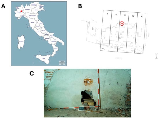
Figure 1.
Funerary Unit 12, Sector III. (A) Map of Italy with the location of the site. (B) Planimetry of the hypogeal cemetery: funerary Unit 12, Sector III in the red circle [37]. (C) Detail of FU12 [38].
The bone fragments discovered within this ossuary, had an overall state of good preservation. Analysis of the tibiae, the most represented skeletal element in the coffin, revealed a minimum count of eleven adult individuals, while among the non-adults there were six individuals identified [37]. The age range for adults spans from middle-aged adults to old adults, based on the auricular surface [39], while for non-adults, it ranges from infants to children, based on the union of epiphyses [40].
The depositional context, the coffin, appears to be a secondary deposition, as evidenced by the disorganized arrangement of the bones and the absence of small bones, such as those from the hands and feet, as well as the lack of anatomical connections. Furthermore, as reported in Tibaldeschi’s article, it is likely that these bones were relocated from the cemetery of the former Basilica at the time it was demolished for the construction of the new Santa Maria Maggiore cathedral [32]. Therefore, relative dating of the remains in this funerary context is particularly challenging, due to their secondary deposition and the probable relocation from the former church.
2.3. FU12 151
The individual identified as FU12 SU151 is represented by a right coxae and its femur (Figure 2). Among the bones found in the wooden coffin, only the two bones examined in this study could be attributed to a single individual, based on the presence of compatible pathological alterations. By examining the anatomical features and pathological characteristics of UF12 SU151, we aim to glean insights into the individual’s mobility, health, and potential life history.
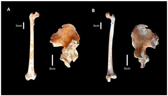
Figure 2.
FU12 SU151. (A) Anterior view of the femur and posterior view of the os coxa individual. (B) Posterior view of the femur and anterior view of the os coxa.
3. Methods
Morphological analysis of the os coxae was employed to estimate both sex and age-at-death of the skeletal remains.
Sex determination was conducted through the examination of specific pelvic features, following the method proposed by Buikstra [40,41]. Due to the state of preservation, it was possible to examine only the greater sciatic notch and the composite arch.
Age-at-death estimation was performed by assessing degenerative changes in the auricular surface [39].
All possible measurements of the bones were conducted using an electronic digital caliper, following the guidelines established in Martin and Saller’s treatise [42].
Macroscopic observations were conducted through both unaided visual inspection and examination under a magnifying glass. Upon examination, abnormalities in the morphological structure of the femora and os coxae were discerned, necessitating a comprehensive assessment of potential pathologies. This evaluative process involved meticulous consideration of the distinctive features and spatial distribution patterns exhibited by the anomalous morphology, in conjunction with a review of pertinent findings within the paleopathological literature [1,4,9,13,16,17,18].
Radiographic Analyses
The individual also underwent comprehensive radiological assessment and computed tomography (CT) in anteroposterior plane, at the Diagnostic Imaging and Interventional Unit of the IRCCS Galeazzi Hospital in Milan. The imaging process involved the utilization of a Revolution Ascend CT scanner (GE Medical, Milwaukee, MI, USA).
The imaging protocol adopted for the study entailed capturing images using a slice thickness ranging from 0.625 mm to 1.25 mm. Furthermore, the field of view (FOV) spanned 500 mm, while the voltage (Kv) was set at 120 kV, maintaining an effective current (mA) ranging from 80 to 125 mA. The measurements obtained from the CT scan were performed using the Weasis DICOM medical viewer software, version number 4.5.1.0.
4. Results
FU12 SU151 is identified as a right coxa and a right femur (Figure 2). Analysis of the skeletal remains indicated that they belonged to an adult female, with an estimated age range of 40 to 49 years. However, this age estimate should be interpreted with caution, due to both the pathological condition observed and the overall state of preservation of the remains. The bone preservation is adequate, notwithstanding the evident signs left by taphonomic processes.
The femur displays a femoral head angle of 90 degrees, accompanied by a shortened neck featuring nodules. The spongy texture is evident within the femoral epiphyses, especially pronounced in the proximal epiphysis, as a consequence of taphonomic processes, along with generalized porosity (Figure 3).
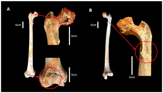
Figure 3.
(A) Anterior view of the femur with magnification of the depression and microporosity in the femur neck and the evident spongy texture, both in the proximal and distal epiphyses. (B) Posterior view of the femur, with the red circle indicating the osteophytic beak.
Notably, the proximal epiphysis lacks trochanters due to taphonomic events, with a small osteophytic beak present in the expected area of the lesser trochanter (Figure 3B). Furthermore, a depression (depth of 7.3 mm) with macroporosity is observed in the portion of the neck closest to the femoral head (Figure 3A). The femoral shaft presents a medial–lateral midshaft diameter of 26.70 mm and anterior-posterior midshaft diameter of 30.40 mm. The latter increases in the portion above the midshaft closest to the proximal epiphysis, to 32.20 mm. The rough line is not particularly pronounced.
The most pronounced morphological changes are observed in the acetabulum. It displays a triangular shape (with a height of 46.50 mm and a depth of 13.60 mm), an irregular base (34.70 mm long) positioned adjacent to the obturator foramen and the absence of the lunate surfaces (Figure 4A). This shape deviates significantly from the normal rounded, cup-like structure necessary for stable articulation with the femoral head. Moreover, the coxa notably exhibits an extended exostosis—a bony growth (74.3 mm long)—positioned proximate to the acetabulum, characterized by osteophytic growth and microporosity (Figure 4A). The greater sciatic notch angle does not appear particularly wider than normal, although Mitchell and Redfern observed that the sciatic notch on the side of the dislocated hip was consistently unusually broad [17] (Figure 4C).
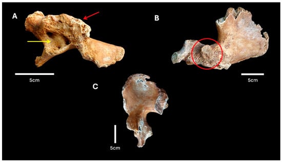
Figure 4.
(A) Lateral view of the coxa, with the yellow arrow indicating the acetabulum and the red arrow indicating the exostosis. (B) Detail of the posterior view of the coxa, with the red circle indicating the raised bony area. (C) Anterior view of the coxa showing the normal width of the greater sciatic notch.
Furthermore, a raised area of bone, a probable accessory articular facet, is identified on the ischium, located posteriorly to the acetabulum (with a height of 22.2 mm and 24.5 mm in width), characterized by the presence of taphonomic microporosity but without evidence of eburnation. A fine layer of irregular and unevenly distributed bone is also observed on the surface of the ilium, just above the raised bony plaque and posterior to the large bony exostosis. No false acetabulum was detected on the iliac wing of the pelvis.
The CT scans of the femur did not reveal significant abnormalities in either the cortical or trabecular bone, and no evidence of fractures was detectable. The CT scans of the os coxa showed no notable abnormalities, except for one observation: within the zone of the acetabulum, the visual representation resembled an area of low bone-mineral density, reminiscent of a black hole (with a length of 31.6 mm and 22.8 mm in width), indicating a notable absence of material (Figure 5).
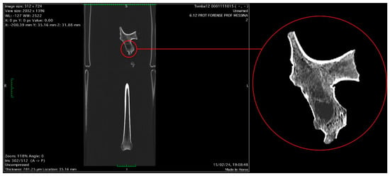
Figure 5.
Anteroposterior plane CT image in anterior view, with the red circle indicating the void visible in the acetabulum zone.
5. Discussion
The skeletal remains, designated as FU12 SU151, presented various morphological irregularities, potentially providing insight into the life and health of the individual under examination.
Despite the limitations and challenges posed by the state of preservation of the skeletal remains, it was still possible to perform a differential diagnosis. The taphonomic alterations primarily affected the cortical bone, preserving the overall morphology and leaving the more diagnostic features largely intact. Numerous studies demonstrate that meticulous examination of both commingled and poorly preserved remains is feasible [24,43]. However, this study’s limitations include the absence of the rest of the skeleton, particularly the contralateral elements that would have been crucial for a more accurate description and an understanding of the individual’s health and pathological conditions.
The most diagnostically significant features were the morphology of the acetabulum and the femoral neck. As part of the differential diagnosis, various conditions affecting these skeletal parts were considered, such as Legg–Calvé–Perthes disease, slipped capital femoral epiphysis, traumatic osteonecrosis of the femoral head, and developmental dysplasia of the hips (DDH) [1,4,17,19,44,45,46].
Legg–Calvé–Perthes disease is a condition caused by avascular necrosis of the femoral head epiphysis during childhood [47]. Notably, the femoral head is flattened and deformed, giving it a ‘mushroom-shaped’ appearance [44]. However, this condition is unlikely in this case, as the CT scans revealed normal trabeculae, and the anatomy of the femoral head did not appear to be significantly mushroom-shaped.
Slipped capital femoral epiphysis (SCFE) is a condition primarily affecting adolescents, typically during periods of rapid growth. It entails the femoral head moving out of alignment with the femoral neck via the epiphyseal plate, commonly happening within the hypertrophic zone of the growth plate. In relative terms, the epiphysis tends to shift backward on the femoral neck, potentially leading to a lasting external rotation deformity [44]. This condition was excluded, due to the absence of external rotation of the femoral shaft and forward displacement of the femoral neck.
In hip fractures and dislocations, the blood vessels that supply the femoral head can be susceptible to tearing. In this case, osteonecrosis is a possible complication. CT scans can detect the changes induced by this condition, particularly in later stages. Typically, there is observable new bone formation in the avascular segment, and in advanced cases, the breakdown of trabecular bone within the necrotic segment becomes apparent [44]. Given the absence of these alterations on the available CT scans, osteonecrosis of the femoral head was ruled out as a diagnosis. Furthermore, morphological examinations and CT scans of the individual showed no visible signs of fractures occurring before or around the time of death.
In paleopathological cases of DDH, morphological features of the acetabulum often include a triangular or oval shape, shallow depth, and smaller size, compared to a normal acetabulum. The femoral neck is typically short and thin. Additionally, the femoral shaft is noticeably smaller than in an unaffected limb, and the markings for muscle attachments are more gracile. In the case of dislocation, a false acetabulum may form on the iliac wing, which Mitchell and Redfern classified into four types [17]. Based on the presence of a shallow, triangular-shaped acetabulum and abnormal angle (<120°) of the femoral neck [48], a probable diagnosis of developmental dysplasia of the hips (DDH) was made. Additionally, evident on computed tomography (CT) scans, the distinctive presentation resembling a ‘black hole’ or a conspicuous void likely indicates the presence of osteolysis [49], deviating from the expected anatomical depiction of a well-defined acetabular socket. This alteration, although not a typical sign of DDH, may indicate a notable aberration in hip joint morphogenesis, potentially precipitating compromised hip functionality, diminished mobility, and predisposition to premature onset of arthritic conditions.
Notably, DDH has been identified in paleopathological studies through both population and individual case studies. This pathological condition is more prevalent in Europe, occurring at a high rate in Northern European populations, compared to the rest of the world [8]. It predominantly affects females, in accordance with contemporary epidemiological data and findings from bioarchaeological literature, and is more commonly seen in a unilateral form, typically affecting the left side [13,16,18,28,31]. Consequently, the presence of the condition on the right side in the studied individual appears to deviate from the expected prevalence.
The individual under examination exhibited a triangular-shaped and shallow acetabulum with an irregular base, consistent with developmental dysplasia of the hip (DDH). Additionally, the femur displayed a femoral head angle of 90 degrees, indicative of coxa vara, an anomaly commonly associated with developmental hip dysplasia [13,49]. However, no evidence of a false acetabulum could be detected on the lateral aspect of the iliac wing of the coxal bone.
Furthermore, an extended exostosis was detected near the acetabulum, likely indicating the development of secondary osteoarthritis. Developmental dysplasia of the hip (DDH) stands as one among several conditions that confer a predisposition to osteoarthritis [50]. In certain instances, a significant osseous collar may form around the acetabulum, enveloping the femoral neck and thereby enhancing femoral stability. It is frequently noted that the augmented bone proliferation surrounding the acetabulum can restrict complete hip mobility [1,45]. This observation potentially applies to the present case, evidenced by the absence of discernible eburnation and the fact that the femoral head was not distinctly dislocated from the joint, as there is no indication of a false acetabulum or a secondary joint forming elsewhere on the pelvis. If this is our case, we can hypothesize a compensatory mechanism that enabled the joint to maintain stability, despite the pathological changes [51]. This could have been achieved through adaptive alterations in the surrounding soft tissues and musculature, as well as adjustments in the contralateral leg. The individual’s gait might have been characterized by a pronounced limp due to the immobility of the right hip, evidenced by the absence of eburnation typical of a used mobile joint. Additionally, it is possible that this individual relied on mobility aids, such as walking sticks or crutches, and received care throughout their life [31]. All these hypotheses cannot be confirmed, due to the lack of the remaining skeletal districts.
Moreover, research has established that developmental dysplasia of the hip (DDH) is characterized by irregular hip joint development, which can lead to symptoms such as joint pain, damage to articular cartilage, functional limitations, and decreased quality of life [7,52]. Although pain and degenerative changes may not manifest during the first decade of life, they often emerge in adulthood, typically at the age of 40 or older [25]. Dysplasia of the hip is a current medical concern in contemporary populations, which frequently necessitates surgical intervention and lifelong management to prevent further complications like osteoarthritis and to maintain optimal hip function. Hip dysplasia can be classified into three types, based on the degree of dislocation and morphological changes: Type I involves a positionally unstable or subluxatable hip, where the femoral head can partially dislocate but remains within the acetabulum; Type II features a subluxated hip, characterized by eversion and inversion of the labrum, with the femoral head partially dislocated; Type III represents the most severe form, where the femoral head is fully dislocated and forms a false acetabulum, with no articulation with the true acetabulum [53]. In our case, the observed findings suggest that the condition may correspond to Type II dysplasia. Clinical treatment approaches vary, based on severity, ranging from conservative measures like physical therapy and bracing for mild cases to surgical procedures such as arthroscopy, osteotomy, or total hip replacement for more advanced conditions [44]. All of this was not possible in past times, when the understanding of developmental dysplasia of the hip (DDH) and its treatment options were far more limited. Historically, many cases went undiagnosed until significant symptoms appeared in adulthood, leading to delayed treatment and poorer outcomes [52]. Surgical techniques and technologies have advanced significantly, allowing for earlier intervention and more precise corrective procedures today. These advancements have revolutionized the management of DDH, enabling healthcare providers to offer better quality of life and improved functional outcomes for patients affected by this condition [7,8].
This study underscores the critical role of ongoing research and meticulous examination of isolated bone specimens to advance our understanding of skeletal diseases and their impact on health outcomes prior to the advent of surgical treatments. Such research enriches our heritage by connecting historical medical practices with modern advancements, demonstrating how past conditions and their management inform current clinical practices and contribute to the broader narrative of human health [52]. The preservation and analysis of these remains, indeed, offer invaluable insights into the past, shedding light on the evolution of these diseases and honoring the individuals they represent. By maintaining and studying these remains, we bridge the gap between past and present, facilitating the transfer of knowledge across generations.
Equally important is the preservation of the archaeological context in which these remains are found. The co-cathedral of Santa Maria Maggiore in Vercelli, Piedmont, Italy, exemplifies this significance. The architecture of this subterranean cemetery, featuring vaulted spaces, masonry structures, burial chambers, private chapels, and central ossuaries, reflects a historical commitment to preserving sacred spaces and honoring the deceased.
By safeguarding both the skeletal remains and their original contexts, we not only protect physical artifacts, but also ensure that future generations can continue to learn and appreciate our shared heritage.
6. Conclusions
Anthropological and paleopathological analysis of the human skeletal remains FU12 SU151 from Sector III of the hypogeal cemetery of Santa Maria Maggiore in Vercelli, Italy, sheds light on the probable presence of developmental dysplasia of the hip (DDH) in an adult female individual. Despite the limitations in the preservation status preventing the detection of all definitive signs proposed by Mitchell and Redfern [17], the findings remain suggestive and informative. This condition, characterized by irregular hip joint development, presents significant implications for the individual’s health and quality of life, potentially leading to joint pain, cartilage damage, and functional limitations later in life.
Furthermore, this investigation prompted us to speculate that this individual’s gait might have been characterized by a visible limp, and may have required assistance with locomotion during their lifetime, offering valuable insights into the challenges faced by past populations in terms of mobility and caregiving practices.
This study also highlights how even isolated bone specimens can provide information into the evolution of bone diseases and their impact on individual health outcomes, addressing the importance of ongoing research and analysis of such collections.
Advancements in surgical techniques and understanding have greatly improved the management of DDH in contemporary medicine, offering earlier intervention and more effective treatment options. This stands in stark contrast to historical times, when the understanding and treatment of such conditions were rudimentary, often resulting in delayed diagnosis and poorer outcomes. Moving forward, continued research and application of these advancements will be crucial in further enhancing patient care and outcomes for individuals affected by developmental dysplasia of the hip.
The preservation of skeletal remains and their archaeological contexts is pivotal for advancing our understanding of historical health and disease. This study highlights the profound value of examining these specimens to unravel the complexities of skeletal diseases and their impact on individual health before modern medical interventions. Equally crucial is the conservation of the archaeological context of sites like the co-cathedral of Santa Maria Maggiore in Vercelli, which offers a tangible link to past practices and beliefs surrounding death and burial. By safeguarding both the physical remains and their historical settings, we not only honor those who came before us, but also ensure that future research and heritage can continue to illuminate our shared past. Through these efforts, we foster a deeper connection with history and a more comprehensive understanding of human experience across generations.
Author Contributions
Conceptualization, N.R. and R.F.; Investigation, N.R.; Writing—original draft, N.R.; Writing—review & editing, R.F., A.V. and M.L.; Data Curation, C.M. and A.V.; Supervision, M.L. All authors have read and agreed to the published version of the manuscript.
Funding
This research received no external funding.
Data Availability Statement
Data available on request.
Acknowledgments
Special thanks are extended to the Fondazione Cassa di Risparmio di Torino for their generous support in financing the bioarchaeological project at the Cemetery of Santa Maria Maggiore in Vercelli. The excavations and associated research endeavors were conducted under the scientific oversight of the Soprintendenza Archeologia Belle Arti e Paesaggio per le Province di Biella, Novara, Verbano-Cusio Ossola e Vercelli, with the invaluable collaboration and assistance provided by the Ufficio Diocesano per i Beni Culturali Ecclesiastici e l’Edilizia di Culto, Arcidiocesi Vercelli. We would like to express our gratitude to the Department of Humanities at the University of Eastern Piedmont for their invaluable contribution in conducting the archaeological component of this research.
Conflicts of Interest
The authors declared no potential conflicts of interest with respect to the research, authorship, and/or publication of this article.
References
- Waldron, T. Paleopathology; Cambridge University Press: Cambridge, UK, 2020. [Google Scholar] [CrossRef]
- Roberts, C.A.; Manchester, K.L. The Archaeology of Disease, 2nd ed.; Cornell University Press: Ithaca, NY, USA, 1997; 243p, ISBN 080148440. [Google Scholar]
- Larsen, C.S. Bioarchaeology: Interpreting Behavior from the Human Skeleton, 2nd ed.; Part of Cambridge Studies in Biological and Evolutionary Anthropology; Cambridge University Press: Cambridge, UK, 2015. [Google Scholar]
- Auferhede, A.C.; Rodriguez-Martin, C. The Cambridge Encyclopedia of Human Paleopathology. Arthur C. Auferhede and Conrado Rodriguez-Martin, including a dental chapter by Odin Langsjoen. 1998. Cambridge University Press, England, xviii + 478 pp., 310 figures, references, index. $100.00 (cloth), ISBN 0-521-55203-6. Am. Antiq. 1999, 64, 562–563. [Google Scholar] [CrossRef]
- Ibrahim, M.M.; Smit, K. Anatomical Description and Classification of Hip Dysplasia. In Hip Dysplasia; Beaulé, P., Ed.; Springer: Cham, Switzerland, 2020. [Google Scholar] [CrossRef]
- Harsanyi, S.; Zamborsky, R.; Krajciova, L.; Kokavec, M.; Danisovic, L. Developmental Dysplasia of the Hip: A Review of Etiopathogenesis, Risk Factors, and Genetic Aspects. Medicina 2020, 56, 153. [Google Scholar] [CrossRef] [PubMed] [PubMed Central]
- Sonoda, K.; Hara, T. “Anterior-shift sign”: A novel MRI finding of adult hip dysplasia. Arch. Orthop. Trauma Surg. 2022, 142, 1763–1768. [Google Scholar] [CrossRef] [PubMed]
- Marras, F.; Asti, C.; Ciatti, C.; Pescia, S.; Locci, C.; Pisanu, F.; Doria, C.; Caggiari, G. Congenital hip dysplasia: The importance of early screening and treatment. Pediatr. Med. Chir. 2022, 44. [Google Scholar] [CrossRef] [PubMed]
- Plischuk, M.; De Feo, M.E.; Desántolo, B. Developmental dysplasia of the hip in female adult individual: Site Tres Cruces I, Salta, Argentina (Superior formative period, 400–1000 AD). Int. J. Paleopathol. 2018, 20, 108–113. [Google Scholar] [CrossRef]
- Spasovski, D.D. Introductory chapter: Five-dimensional approach to the developmental dysplasia of the hip. In Developmental Diseases of the Hip-Diagnosis and Management; Spasovski, D., Ed.; InTech: Takasago, Japan, 2017. [Google Scholar] [CrossRef]
- Arnaud, G.; Arnaud, S. Luxation congenital bilatérale de la hanche et manifesta tions d’hyperostose porotique sur un squelette d’époque paléo-chrétienne. Bull. Mém. Soc. Anthropol. Paris 1975, 2, 307–326, (XIII Série, 10.3406/bmsap). [Google Scholar] [CrossRef]
- Blondiaux, J.; Millot, F. Dislocation of the hip: Discussion of eleven cases from medieval France. Int. J. Osteoarchaeol. 1991, 1, 203–207. [Google Scholar] [CrossRef]
- Mafart, B.; Kéfi, R.; Béraud-Colomb, E. Palaeopathological and palaeogenetic study of 13 cases of developmental dysplasia of the hip with dislocation in a historical population from southern France. Int. J. Osteoarchaeol. 2007, 17, 26–38. [Google Scholar] [CrossRef]
- Dawes, J.; Magilton, J. The Cemetery of St. Helen-on-the-Walls, Aldwark. In The Archaeology of York; Council for British archaeology: London, UK, 1980; Volume 12. [Google Scholar]
- Wakely, J. Bilateral congenital dislocation of the hip, spina bifida occulta and spondylosis in a female skeleton from the medieval cemetery at Abingdon. Engl. J. Paleop. 1993, 1, 37–45. [Google Scholar]
- Mitchell, P.; Redfern, R. The prevalence of dislocation in developmental dysplasia of the hip in Britain over the past thousand years. J. Pediatr. Orthop. 2007, 27, 890–892. [Google Scholar] [CrossRef]
- Mitchell, P.; Redfern, R. Diagnostic criteria for developmental dislocation of the hip in human skeletal remains. Int. J. Osteoarchaeol. 2008, 18, 61–71. [Google Scholar] [CrossRef]
- Mitchell, P.; Redfern, R. Brief communication: Developmental dysplasia of the hip in medieval London. Am. J. Phys. Anthropol. 2011, 144, 479–484. [Google Scholar] [CrossRef] [PubMed]
- Agnew, A.; Justus, H. Developmental Dysplasia of the Hip in a Child from Medieval Poland. Poster on 2013 Conference Paleopathology Association. 2013. Available online: http://www.slavia.org/posters/2013_agnew_ddh.pdf (accessed on 10 July 2024).
- Masnicová, S.; Beňuš, R. Developmental anomalies in skeletal remains from the Great Moravia and Middle Ages cemeteries at Devín (Slovakia). Int. J. Osteoarchaeol. 2003, 13, 266–274. [Google Scholar] [CrossRef]
- Eng, J.; Szöcs, P.; Hagen, C. Developmental dysplasia of the hip in a post-medieval Transylvanian population: Case study and diagnosis. Paleopathol. News 2009, 148, 25–32. [Google Scholar]
- Katzmarzyk, C.; Schats, R. Conflicting Pelvic Morphology in a Pathological Late Medieval Skeleton from the Netherlands. Poster on 2011 Conference of British Association for Human Identification (BAHID). 2011. Available online: https://www.academia.edu/3388868/Conflicting_Pelvic_Morphology_in_a_Pathological_Late_Medieval_Skeleton_from_the_Netherlands (accessed on 17 June 2024).
- Petrella, E.; Piciucchi, S.; Feletti, F.; Barone, D.; Piraccini, A.; Minghetti, C.; Gruppioni, G.; Poletti, V.; Bertocco, M.; Traversari, M. CT Scan of thirteen natural mummies dating back to the XVIXVIII centuries: An emerging tool to investigate living condi tions and diseases in history. PLoS ONE 2016, 11, e0154349. [Google Scholar] [CrossRef]
- Traversari, M.; Feletti, F.; Vazzana, A.; Gruppioni, G.; Frelat, M. Three cases of developmental dysplasia of the hip on partially mummified human remains (Roccapelago, Modena, 18th Century): A study of palaeopathological indicators through direct analysis and 3D virtual models. Bull. Mémoires Société Anthropol. Paris 2016, 28, 202–212. [Google Scholar] [CrossRef]
- Blatt, S.H. To swaddle, or not to swaddle? Paleoepidemiology of developmental dysplasia of the hip and the swaddling dilemma among the indigenous populations of North America. Am. J. Hum. Biol. 2015, 27, 116–128. [Google Scholar] [CrossRef] [PubMed]
- Clabeaux, M. Congenital dislocation of the hip in the prehistoric northeast. Bull. N. Y. Acad. Med. 1977, 53, 338–346. [Google Scholar]
- Ausel, E. Down But Not out: A Probable Case of Congenital Hip Dysplasia in a Late Prehistoric Native American Community. Paleopathology Association Scientific Program. In Proceedings of the 43rd Annual North American Meeting, Atlanta, GA, USA, 18–23 October 2016. [Google Scholar]
- Loder, R.T.; Skopelja, E.N. The epidemiology and demographics of hip dysplasia. ISRN Orthop. 2011, 2011, 238607. [Google Scholar] [CrossRef] [PubMed] [PubMed Central]
- Brothwell, D. Major congenital anomalies of the skeleton: Evidence from earlier population. In Diseases in Antiquity; Brothwell, D., Sandison, A., Eds.; Charles Thomas: Springfield, IL, USA, 1967; Chapter 34; pp. 423–443. [Google Scholar]
- Costa, M.; Llagostera, A. Coyo 3: Momentos finales del Período Medio en San Pedro de Atacama. Estud. Atacameños 1994, 11, 73–107. [Google Scholar] [CrossRef]
- Voegt, C.; Gunston, G.; Nortje, M.; Sealy, J.C.; He, L.; Le Roux, P.; Namayega, C.; Gibbon, V.E. Bilateral hip dysplasia in a South African male: A case study from the 17–18th century. Int. J. Paleopathol. 2023, 42, 27–33, ISSN 1879-9817. [Google Scholar] [CrossRef] [PubMed]
- Tibaldeschi, G. La Chiesa di S. Maria Maggiore di Vercelli e l’Assunzione di Paolo Borroni. Boll. Stor. Vercellese 1996, 2, 131–150. [Google Scholar]
- Sommo, G. Vercelli e la Memoria dell’antico. Schede e Documenti per un Approccio Alla Storia ed ai Problemi Dell’archeologia, Della Tutela e Conservazione in un Centro Della Provincia Piemon-Tese. Edizione Elettronica Archeovercelli.it. Gruppo Archeo-Logico Vercellese. Vercelli, Italy. 2008. Available online: http://www.archeovercelli.it/VERCELLI%20E%20LA%20MEMORIA.pdf (accessed on 27 June 2024).
- Caldano, S. Un cantiere per un capitolo canonicale di prestigio: Santa Maria Maggiore di Vercelli nel XII secolo. Arte Lomb. 2019, 186/187, 71–84. [Google Scholar]
- Fusco, R.; Messina, C.; Tesi, C.; Vanni, A. “Capta est ne Malitia Mutaret Intelletum Eius”. Study on a Natural Mummy from an Underground Cemetery (18–19th Century). Med. Hist. 2023, 7, e2023044. Available online: https://mattioli1885journals.com/index.php/MedHistor/article/view/15140 (accessed on 27 June 2024).
- Vanni, A.; Fusco, R.; Tesi, C.; Licata, M. Autopsy or anatomical dissection? Comparative analysis of an osteoarcheological sample from an 18-19th century hypo-geal cemetery (northern Italy). J. Archaeol. Sci. Rep. 2024, 54, 104418. [Google Scholar] [CrossRef]
- Vanni, A.; Fusco, R. Behind the Wall: A Paleopathological Examination of a Non-Adult Subject from the Cemetery of Santa Maria Maggiore, Vercelli. J. Bioarchaeol. Res. 2024, 2, e2024005. Available online: https://www.mattioli1885journals.com/index.php/JBR/article/view/15809 (accessed on 27 June 2024).
- Bettin, S. Archeologia e antropologia fisica a Santa Maria Maggiore di Vercelli [Corso di Laurea Triennale in Lettere]; Università del Piemonte Orientale: Vercelli, Italy, 2022. [Google Scholar]
- Lovejoy, C.O.; Meindl, R.S.; Pryzbeck, T.R.; Mensforth, R.P. Chronological metamorphosis of the auricular surface of the ilium: A new method for the determination of adult skeletal age at death. Am. J. Phys. Anthropol. 1985, 68, 15–28. [Google Scholar] [CrossRef] [PubMed]
- Buikstra, J.E. Standards for data collection from human skeletal remains. Ark. Archaeol. Surv. Res. Ser. 1994, 44, 18. [Google Scholar]
- Phenice, T.W. A newly developed visual method of sexing the os pubis. Am. J. Phys. Anthropol. 1969, 30, 297–301. [Google Scholar] [CrossRef]
- Martin, R.; Saller, K. 1957–1962. Lehrbuch der Anthropologie, Stuttgart. Martin, R., & Saller, K. (1957). Lehrbuch der Anthropologie in Systematischer Darstellung Mit Besonderer Berücksichtigung der Anthropologischen Methoden (Vol. 1). G. Fischer. Available online: https://archive.org/details/lehrbuchderanthr00mart/page/n7/mode/2up (accessed on 27 June 2024).
- Assis, S.; Henderson, C.Y.; Casimiro, S.; Alves Cardoso, F. Is differential diagnosis attainable in disarticulated pathological bone remains? A case-study from a late 19th/early 20th century necropolis from Juncal (Porto de Mós, Portugal). Int. J. Paleopathol. 2018, 20, 26–37. [Google Scholar] [CrossRef]
- Solomon, L.; Ganz, R.; Leunig, M.; Monsell, F.; Learmonth, I. The hip. In Apley’s System of Orthopaedics and Fractures; Solomon, L., Warwick, D., Nayagam, S., Eds.; CRC Press: Boca Raton, FL, USA, 2010; pp. 493–545. [Google Scholar]
- Ortner, D.J. Identification of Pathological Conditions in Human Skeletal Remains, 3rd ed.; Buikstra, J.E., Ed.; Arizona State University: Tempe, AZ, USA, 2019. [Google Scholar]
- Giovagnorio, F. Manuale di Diagnostica per Immagini nella Pratica Medica, 3rd ed.; Società Editrice Esculapio: Bologna, Italy, 2021; p. 384. ISBN 9788893852548. [Google Scholar]
- Fusco, R.; Tesi, C.; Larentis, O. Paleopathological Evidence of Legg-Calve’-Perthes from the Medieval Cemetery of St. Agostino in Caravate, Northwestern Italy. Med. Hist. 2022, 5, e2021025. Available online: https://www.mattioli1885journals.com/index.php/MedHistor/article/view/12735 (accessed on 12 September 2024).
- Meier, M.E.; Appelman-Dijkstra, N.M.; Collins, M.T.; Geels, R.E.S.; Stanton, R.P.; de Witte, P.B.; Boyce, A.M.; van de Sande, M.A.J. Coxa Vara Deformity in Fibrous Dysplasia/McCune-Albright Syndrome: Prevalence, Natural History and Risk Factors: A Two-Center Study. J Bone Miner Res. 2023, 38, 968–975. [Google Scholar] [CrossRef] [PubMed]
- Parvizi, J.; Kim, G.K. Chapter 61—Coxa Vara. In High Yield Orthopaedics, 1st ed.; Elsevier Health Sciences Publisher: Amsterdam, The Netherlands, 2010; p. 125. ISBN 9781416002369. [Google Scholar] [CrossRef]
- Gül, D.; Orsçelik, A.; Akpancar, S. Treatment of Osteoarthritis Secondary to Developmental Dysplasia of the Hip with Prolotherapy Injection versus a Supervised Progressive Exercise Control. Med. Sci. Monit. 2020, 26, e919166. [Google Scholar] [CrossRef] [PubMed] [PubMed Central]
- Licata, M.; Iorio, S.; Benaglia, P.; Tosi, A.; Borgo, M.; Armocida, G.; Ronga, M.; Ruspi, A.; Verzeletti, A.; Rossetti, C. Biomechanical analysis of a femur fracture in osteoarchaeology: Reconstruction of pathomechanics, treatment and gait. J. Forensic Leg. Med. 2019, 61, 115–121. [Google Scholar] [CrossRef] [PubMed]
- Wenger, D.R.; Bomar, J.D. Historical Aspects of DDH. Indian J. Orthop. 2021, 55, 1360–1371. [Google Scholar] [CrossRef] [PubMed] [PubMed Central]
- Lafferty, C.M.; Sartoris, D.J.; Tyson, R.; Resnick, D.; Kursunoglu, S.; Pate, D.; Sutherland, D. Acetabular alterations in untreated congenital dysplasia of the hip: Computed tomography with multiplanar re-formation and three-dimensional analysis. J. Comput. Assist. Tomogr. 1986, 10, 84–91. [Google Scholar] [CrossRef] [PubMed]
Disclaimer/Publisher’s Note: The statements, opinions and data contained in all publications are solely those of the individual author(s) and contributor(s) and not of MDPI and/or the editor(s). MDPI and/or the editor(s) disclaim responsibility for any injury to people or property resulting from any ideas, methods, instructions or products referred to in the content. |
© 2024 by the authors. Licensee MDPI, Basel, Switzerland. This article is an open access article distributed under the terms and conditions of the Creative Commons Attribution (CC BY) license (https://creativecommons.org/licenses/by/4.0/).