

The Book of Uí Mhaine: An Interdisciplinary Analysis of the Materiality of the Gaelic Manuscript Tradition

 , , ,
, , , 
Abstract
1. Introduction
2. Materials and Methods
3. Results
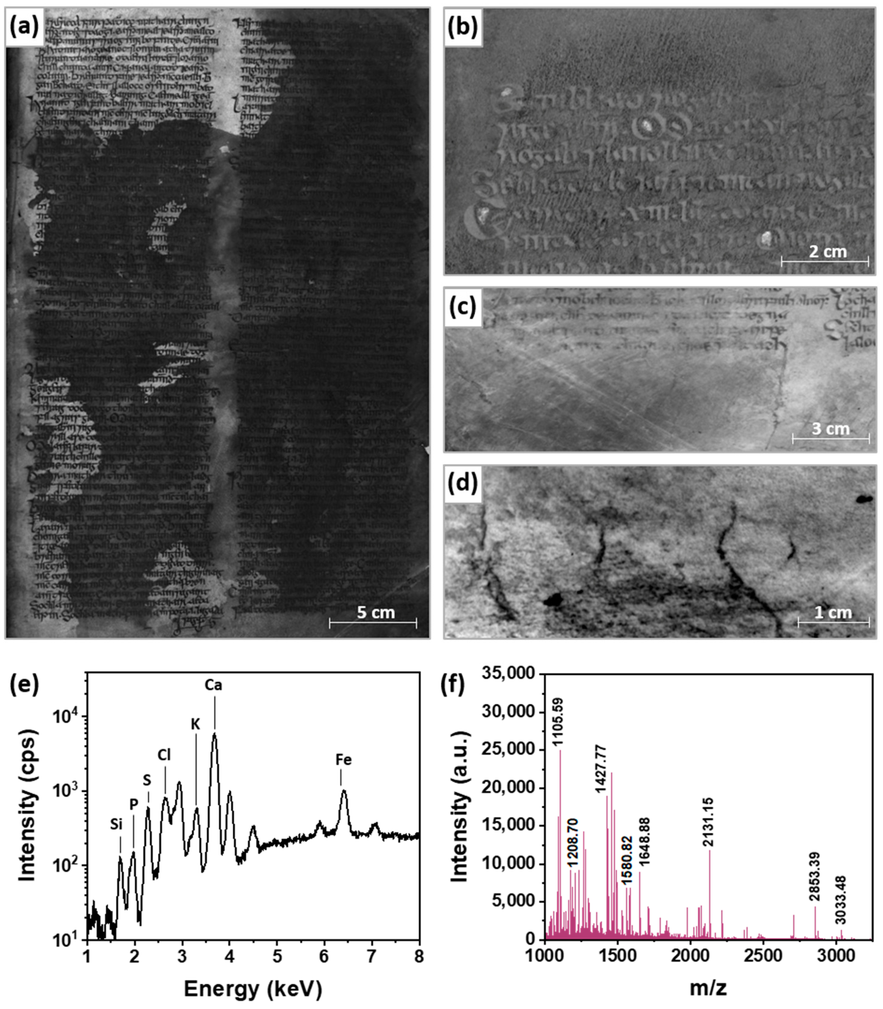
3.1. Parchment
3.2. Ruling
3.3. Writing Inks
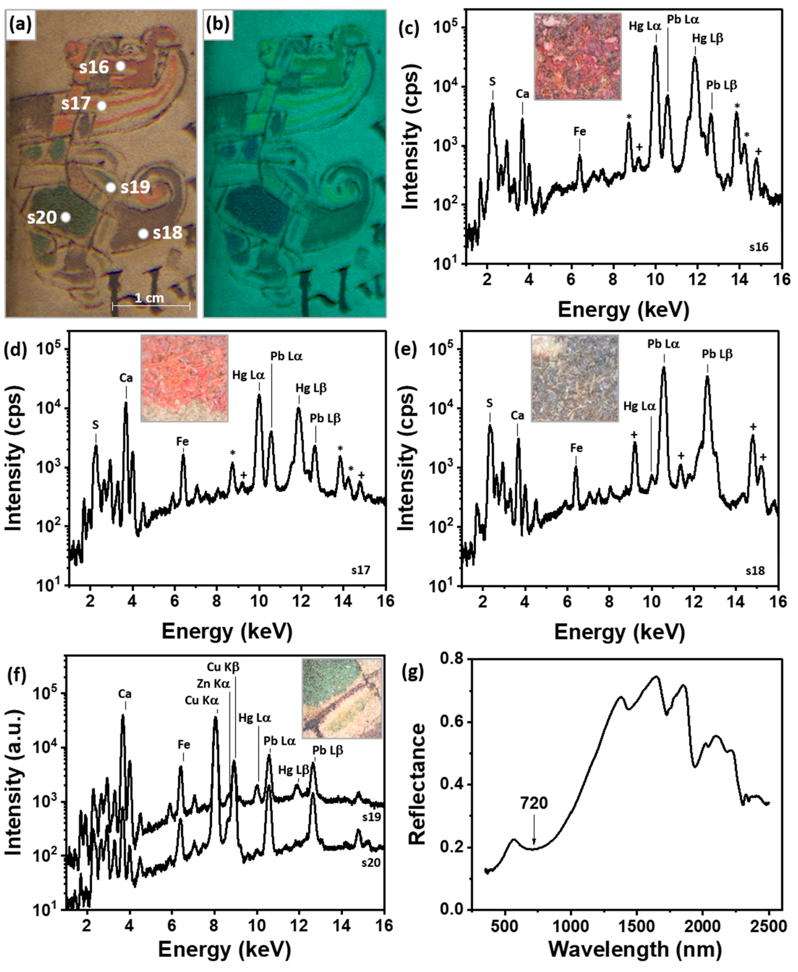
3.4. Pigments
3.5. Pigment Degradation
4. Conclusions
Supplementary Materials
Author Contributions
Funding
Data Availability Statement
Acknowledgments
Conflicts of Interest
References
- Library, R.I.A. Catalogue of Irish Manuscripts in the Royal Irish Academy; Catalogue of Irish manuscripts in the Royal Irish Academy (28, fasc.); Royal Irish Academy: Dublin, Ireland; Figgis & Co.: Hodges, FL, USA; Williams & Norgate: London, UK, 1926; p. 3314. [Google Scholar]
- O’Sullivan, W. The Book of Uí Maine formerly the Book of Ó Dubhagáin: Scripts and structure. Éigse 1989, 23, 151–166. [Google Scholar]
- Lettner, M.; Sablatnig, R. Multispectral imaging for analyzing ancient manuscripts. In Proceedings of the 2009 17th European Signal Processing Conference, Glasgow, UK, 24–28 August 2009. [Google Scholar]
- Hollaus, F.; Diem, M.; Fiel, S.; Kleber, F.; Sablatnig, R. Investigation of Ancient Manuscripts based on Multispectral Imaging. In Proceedings of the 2015 ACM Symposium on Document Engineering, in DocEng ’15, Lausanne, Switzerland, 8–11 September 2015; Association for Computing Machinery: New York, NY, USA, 2015; pp. 93–96. [Google Scholar] [CrossRef]
- Weiskott, E. Multispectral Imaging and Medieval Manuscripts. In The Routledge Research Companion to Digital Medieval Literature; Boyle, J.E., Burgess, H.J., Eds.; Routledge: England, UK, 2018; pp. 186–196. Available online: https://www.academia.edu/35291754/Multispectral_Imaging_and_Medieval_Manuscripts (accessed on 6 April 2023).
- Beeby, A. Spectral imaging of medieval manuscripts. In Hyperspectral Imaging and Applications; SPIE: Bellingham, WA, USA, 2020. [Google Scholar] [CrossRef]
- Cohen, Z.; Kindzorra, E.; Hahn, O.; Glaser, L.; Łojewski, T.; Rabin, I.; Cohen. 2015-Composition of the primary inks in medieval palimpsests–effects of ink removal. Opusc. Musealia 2016, 23, 75–82. [Google Scholar] [CrossRef]
- Cohen, Z.; Olszowy-Schlanger, J.; Hahn, O.; Rabin, I. Composition Analysis of Writing Materials in Geniza Fragments: New Perspectives. In Jewish Manuscript Cultures; De Gruyter: Berlin, Germany, 2017. [Google Scholar] [CrossRef]
- Aceto, M.; Agostino, A.; Fenoglio, G.; Capra, V.; Demaria, E.; Cancian, P. Characterisation of the different hands in the composition of a 14th century breviary by means of portable XRF analysis and complementary techniques: Different hands in a 14th-century breviary by means of XRF analysis. X-ray Spectrom. 2017, 46, 259–270. [Google Scholar] [CrossRef]
- Melo, M.; Otero, V.; Vitorino, T.; Araújo, A.R.D.S.D.; Muralha, V.S.; Lemos, A.; Picollo, M. A Spectroscopic Study of Brazilwood Paints in Medieval Books of Hours. Appl. Spectrosc. 2014, 68, 434–444. [Google Scholar] [CrossRef]
- Leona, M.; Winter, J. Fiber Optics Reflectance Spectroscopy: A Unique Tool for the Investigation of Japanese Paintings. Stud. Conserv. 2001, 46, 153. [Google Scholar] [CrossRef]
- Liu, W.; Li, M.; Wu, N.; Liu, S.; Chen, J. A new application of Fiber optics reflection spectroscopy (FORS): Identification of ‘bronze disease’ induced corrosion products on ancient bronzes. J. Cult. Heritage 2021, 49, 19–27. [Google Scholar] [CrossRef]
- Ricciardi, P.; Delaney, J.K.; Facini, M.; Glinsman, L.D. Use of Imaging Spectroscopy and in situ Analytical Methods for the Characterization of the Materials and Techniques of 15th Century Illuminated Manuscripts. J. Am. Inst. Conserv. 2013, 52, 13–29. [Google Scholar] [CrossRef]
- Aceto, M.; Agostino, A.; Fenoglio, G.; Idone, A.; Gulmini, M.; Picollo, M.; Ricciardi, P.; Delaney, J.K. Characterisation of colourants on illuminated manuscripts by portable fibre optic UV-visible-NIR reflectance spectrophotometry. Anal. Methods 2014, 6, 1488–1500. [Google Scholar] [CrossRef]
- Fiddyment, S.; Holsinger, B.; Ruzzier, C.; Devine, A.; Binois, A.; Albarella, U.; Fischer, R.; Nichols, E.; Curtis, A.; Cheese, E.; et al. Animal origin of 13th-century uterine vellum revealed using noninvasive peptide fingerprinting. Proc. Natl. Acad. Sci. USA 2015, 112, 15066–15071. [Google Scholar] [CrossRef]
- Teasdale, M.D.; Fiddyment, S.; Vnouček, J.; Mattiangeli, V.; Speller, C.; Binois, A.; Carver, M.; Dand, C.; Newfield, T.P.; Webb, C.C.; et al. The York Gospels: A 1000-year biological palimpsest. R. Soc. Open Sci. 2017, 4, 170988. [Google Scholar] [CrossRef]
- Fiddyment, S.; Teasdale, M.D.; Vnouček, J.; Lévêque Binois, A.; Collins, M.J. So you want to do biocodicology? A field guide to the biological analysis of parchment. Heritage Sci. 2019, 7, 35. [Google Scholar] [CrossRef]
- Toniolo, L.; D’Amato, A.; Saccenti, R.; Gulotta, D.; Righetti, P.G. The Silk Road, Marco Polo, a bible and its proteome: A detective story. J. Proteom. 2012, 75, 3365–3373. [Google Scholar] [CrossRef]
- Cucci, C.; Bracci, S.; Casini, A.; Innocenti, S.; Picollo, M.; Stefani, L.; Rao, I.G.; Scudieri, M. The illuminated manuscript Corale 43 and its attribution to Beato Angelico: Non-invasive analysis by FORS, XRF and hyperspectral imaging techniques. Microchem. J. 2017, 138, 45–57. [Google Scholar] [CrossRef]
- De Viguerie, L.; Rochut, S.; Alfeld, M.; Walter, P.; Astier, S.; Gontero, V.; Boulc’h, F. XRF and reflectance hyperspectral imaging on a 15th century illuminated manuscript: Combining imaging and quantitative analysis to understand the artist’s technique. Heritage Sci. 2018, 6, 11. [Google Scholar] [CrossRef]
- Calà, E.; Agostino, A.; Fenoglio, G.; Capra, V.; Porticelli, F.; Manzari, F.; Fiddyment, S.; Aceto, M. The Messale Rosselli: Scientific investigation on an outstanding 14th century illuminated manuscript from Avignon. J. Archaeol. Sci. Rep. 2019, 23, 721–730. [Google Scholar] [CrossRef]
- Strohalm, M.; Kavan, D.; Novák, P.; Volný, M.; Havlíček, V. mMass 3: A Cross-Platform Software Environment for Precise Analysis of Mass Spectrometric Data. Anal. Chem. 2010, 82, 4648–4651. [Google Scholar] [CrossRef]
- Buckley, M.; Collins, M.; Thomas-Oates, J.; Wilson, J.C. Species identification by analysis of bone collagen using matrix-assisted laser desorption/ionisation time-of-flight mass spectrometry. Rapid Commun. Mass Spectrom. 2009, 23, 3843–3854. [Google Scholar] [CrossRef]
- Buckley, M.; Kansa, S.W.; Howard, S.; Campbell, S.; Thomas-Oates, J.; Collins, M. Distinguishing between archaeological sheep and goat bones using a single collagen peptide. J. Archaeol. Sci. 2010, 37, 13–20. [Google Scholar] [CrossRef]
- Lowe, E.A. Codices Latini Antiquiores: A Palaeographical Guide to Latin Manuscripts Prior to the 9th Century. Part 2, Great Britain and Ireland, 2nd ed.; Clarendon Press: Oxford, UK, 1972. [Google Scholar]
- Moog, G.; Danforth, C. Skins and Hides for Making Parchment: An Overview of Parchment’s Histologic Features that are Helpful when Identifying Raw Source Material. Art Transl. 2021, 13, 419–431. [Google Scholar] [CrossRef]
- Duran, A.; López-Montes, A.; Castaing, J.; Espejo, T. Analysis of a royal 15th century illuminated parchment using a portable XRF–XRD system and micro-invasive techniques. J. Archaeol. Sci. 2014, 45, 52–58. [Google Scholar] [CrossRef]
- Cains, A. The Vellum of the Book of Kells. Pap. Conserv. 1992, 16, 50–61. [Google Scholar] [CrossRef]
- Samidurai, S.; Khambhaty, Y.; Alagamuthu, T.S. Bio-preservation of raw hides/skins: A review on greener substitute to conventional salt curing. Environ. Sci. Pollut. Res. 2022, 29, 64513–64535. [Google Scholar] [CrossRef] [PubMed]
- Reed, R. Ancient Skins, Parchments and Leathers. In Studies in Archaeological Science; Seminar Press: London, UK, 1972; Available online: http://catalogue.bnf.fr/ark:/12148/cb353652597 (accessed on 28 February 2023).
- Tanimoto, S.; Verri, G. A Note on the Examination of Silverpoint Drawings by Near-Infrared Reflectography. Stud. Conserv. 2009, 54, 106–116. [Google Scholar] [CrossRef]
- Balbas, D.Q.; Fovo, A.D.; Montalbano, L.; Fontana, R.; Striova, J. Non-invasive contactless analysis of an early drawing by Raffaello Sanzio by means of optical methods. Sci. Rep. 2022, 12, 1–10. [Google Scholar] [CrossRef]
- Havermans, J.; Aziz, H.A.; Scholten, H. Non Destructive Detection of Iron Gall Inks by Means of Multispectral Imaging Part 1: Development of the Detection System. Restaurator 2003, 24, 55–60. [Google Scholar] [CrossRef]
- Colini, C.; Hahn, O.; Bonnerot, O.; Steger, S.; Cohen, Z.; Ghigo, T.; Christiansen, T.; Bicchieri, M.; Biocca, P.; Krutzsch, M.; et al. The Quest for the Mixed Inks. H. Siegfried Stiehl (Eds.), Proceedings of the Second International Conference on Natural Science and Technology in Manuscript Analysis, Manuscript Cultures 11 (2018), 41–48. Available online: https://www.academia.edu/38120349/The_Quest_for_the_Mixed_Inks (accessed on 9 February 2022).
- Fichera, G.V.; Malagodi, M.; Cofrancesco, P.; Weththimuni, M.L.; Guglieri, C.; Olivi, L.; Ruffolo, S.; Licchelli, M. Study of the copper effect in iron-gall inks after artificial ageing. Chem. Pap. 2018, 72, 1905–1915. [Google Scholar] [CrossRef]
- Edwards, H.G.M. Chapter 1. Analytical Raman Spectroscopy of Inks; Royal Society of Chemistry: Washington, DC, USA, 2018; pp. 1–15. [Google Scholar] [CrossRef]
- Piantanida, G.; Menart, E.; Bicchieri, M.; Strlič, M. Classification of iron-based inks by means of micro-Raman spectroscopy and multivariate data analysis. J. Raman Spectrosc. 2013, 44, 1299–1305. [Google Scholar] [CrossRef]
- Pallipurath, A.R.; Skelton, J.M.; Ricciardi, P.; Elliott, S.R. Estimation of semiconductor-like pigment concentrations in paint mixtures and their differentiation from paint layers using first-derivative reflectance spectra. Talanta 2016, 154, 63–72. [Google Scholar] [CrossRef]
- Cheng, H.; Zhou, Y.; Frost, R.L. Structure comparison of Orpiment and Realgar by Raman spectroscopy. Spectrosc. Lett. 2017, 50, 23–29. [Google Scholar] [CrossRef]
- Hayem-Ghez, A.; Ravaud, E.; Boust, C.; Bastian, G.; Menu, M.; Brodie-Linder, N. Characterizing pigments with hyperspectral imaging variable false-color composites. Appl. Phys. A 2015, 121, 939–947. [Google Scholar] [CrossRef]
- Villafana, T.; Edwards, G. Creation and reference characterization of Edo period Japanese woodblock printing ink colorant samples using multimodal imaging and reflectance spectroscopy. Heritage Sci. 2019, 7, 1–14. [Google Scholar] [CrossRef]
- Miguel, C.; Claro, A.; GonÃ, A.P.; Muralha, V.S.F.; Melo, M.J. A study on red lead degradation in a medieval manuscript Lorvão Apocalypse (1189). J. Raman Spectrosc. 2009, 40, 1966. [Google Scholar] [CrossRef]
- Zhao, Y.; Tang, Y.; Tong, T.; Sun, Z.; Yu, Z.; Zhu, Y.; Tong, H. Red lead degradation: Monitoring of color change over time. New J. Chem. 2016, 40, 3686–3692. [Google Scholar] [CrossRef]
- Carvalho, I.; Casanova, C.; Araújo, R.; Lemos, A. Colour Identification, Degradation Processes and Findings in a Fifteenth-Century Book of Hours: The Case Study of Cofre n.o 31 from Mafra National Palace. Herit. Sci. 2018, 6. Available online: https://www.academia.edu/36720833/Colour_identification_degradation_processes_and_findings_in_a_fifteenth_century_Book_of_Hours_the_case_study_of_Cofre_n_o_31_from_Mafra_National_Palace (accessed on 28 July 2022). [CrossRef]
- Perino, M.; Pronti, L.; Di Forti, L.G.; Romani, M.; Taverna, C.; Massolo, L.; Manzari, F.; Cestelli-Guidi, M.; Nucara, A.; Felici, A.C. Revealing Artists’ Collaboration in a 14th Century Manuscript by Non-Invasive Analyses. Minerals 2021, 11, 771. [Google Scholar] [CrossRef]


Disclaimer/Publisher’s Note: The statements, opinions and data contained in all publications are solely those of the individual author(s) and contributor(s) and not of MDPI and/or the editor(s). MDPI and/or the editor(s) disclaim responsibility for any injury to people or property resulting from any ideas, methods, instructions or products referred to in the content. |
© 2023 by the authors. Licensee MDPI, Basel, Switzerland. This article is an open access article distributed under the terms and conditions of the Creative Commons Attribution (CC BY) license (https://creativecommons.org/licenses/by/4.0/).
Share and Cite
Biolcati, V.; Wilson, M.; Fiddyment, S.; Unitt, R.; Ryan, C.C.; Hoffmann, A.G.; Gillis, J.; France, F.; Ó Macháin, P.; Iacopino, D. The Book of Uí Mhaine: An Interdisciplinary Analysis of the Materiality of the Gaelic Manuscript Tradition. Heritage 2023, 6, 5393-5409. https://doi.org/10.3390/heritage6070284
Biolcati V, Wilson M, Fiddyment S, Unitt R, Ryan CC, Hoffmann AG, Gillis J, France F, Ó Macháin P, Iacopino D. The Book of Uí Mhaine: An Interdisciplinary Analysis of the Materiality of the Gaelic Manuscript Tradition. Heritage. 2023; 6(7):5393-5409. https://doi.org/10.3390/heritage6070284
Chicago/Turabian StyleBiolcati, Veronica, Meghan Wilson, Sarah Fiddyment, Richard Unitt, Cynthia Connelly Ryan, Anna Grace Hoffmann, John Gillis, Fenella France, Pádraig Ó Macháin, and Daniela Iacopino. 2023. "The Book of Uí Mhaine: An Interdisciplinary Analysis of the Materiality of the Gaelic Manuscript Tradition" Heritage 6, no. 7: 5393-5409. https://doi.org/10.3390/heritage6070284
APA StyleBiolcati, V., Wilson, M., Fiddyment, S., Unitt, R., Ryan, C. C., Hoffmann, A. G., Gillis, J., France, F., Ó Macháin, P., & Iacopino, D. (2023). The Book of Uí Mhaine: An Interdisciplinary Analysis of the Materiality of the Gaelic Manuscript Tradition. Heritage, 6(7), 5393-5409. https://doi.org/10.3390/heritage6070284