Article

A Hydroalcoholic Gel-Based Disinfection System for Deteriogenic Fungi on the Contemporary Mixed Media Artwork Poesia by Alessandro Kokocinski

1
Conservation Laboratory of Cultural Heritage, Department for Innovation in Biological, Agro-Food and Forest Systems (DIBAF), University of Tuscia, 01100 Viterbo, Italy
2
Department of Ecological and Biological Sciences (DEB), University of Tuscia, 01100 Viterbo, Italy
3
Department of Economy, Engineering, Society and Business Organization (DEIM), University of Tuscia, 01100 Viterbo, Italy
4
Department of Chemical Science and Technologies (DSTC), University of Rome “Tor Vergata”, 00133 Rome, Italy
5
Department for Innovation in Biological, Agro-Food and Forest Systems (DIBAF), University of Tuscia, 01100 Viterbo, Italy
*
Author to whom correspondence should be addressed.
Heritage 2023, 6(3), 2716-2734; https://doi.org/10.3390/heritage6030144
Submission received: 16 January 2023 / Revised: 20 February 2023 / Accepted: 4 March 2023 / Published: 6 March 2023

Abstract

:
The disinfection of deteriogenic microorganisms and the removal of induced chromatic alterations in artworks are still open challenges in the field of conservation. For this purpose, a new alcoholic hydrogel was tested to remove an extensive fungal attack from a multimaterial collage by the artist Alessandro Kokocinski and to mitigate chromatic changes caused by the contamination of its poster paper and plywood support layers. A Gellan gum-based hydrogel was used, which was modified by adding a high concentration of alcohol (66.7% ethanol), to give the system an effective disinfecting agent in addition to the detergent capacity of the gel for water-sensitive works of art. It was successfully tested on samples mimicking the complex stratigraphy of the artwork under study. To create replica mock-ups, the artwork materials and stratigraphy were investigated through diagnostic and laboratory techniques such as multispectral imaging, X-ray fluorescence spectroscopy, Fourier transform infrared spectroscopy, and pyrolysis coupled with gas-chromatography-mass spectrometry. The treatment was shown to have a disinfecting effect on the test samples and did not alter their structure, allowing us to apply the method to the artwork. Here, the hydrogel successfully removed and inhibited fungal proliferation in addition to mitigating the color changes caused by fungi.

1. Introduction

Multidisciplinary approaches are crucial for the sustainability of cultural heritage, as they enable the complete characterization of objects, from artistic technique to the analysis of the conservation status, and the definition and application of the best conservation protocols [1]. This is especially true when dealing with complex works of art created using mixed media, particularly when they are in a highly degraded and fragile state. This was the case of the contemporary multi-material media collage Poesia by the artist Alessandro Kokocinski (Figure 1, Figures S1 and S2) [2]. In these cases, one wrong step in the conservation process can seriously compromise the artwork under study.
A multidisciplinary team was involved in the characterization of the collage materials through a targeted diagnostic campaign, which also identified the fungal species involved in its degradation. The correct conservation methodology for the cleaning and disinfection of the deteriogenic fungi was corroborated by laboratory testing.
Wet cleaning represents a fundamental step in the restoration of paper and wooden artifacts. Several hydrogels have been suggested for this purpose in the past ten years [3,4,5,6]. They all have different water retention properties and cleaning capabilities (including the removal of waxes, hydrophobic contaminants, animal glue, starch paste, and synthetic polymeric adhesives) [7,8,9]. Additionally, “green” approaches for cleaning water-sensitive works of art have recently received a lot of attention; among these, innovative systems include twin-chain polymer hydrogels or nanostructured fluids [10]. At the same time, trial approaches have been tested to identify systems that can be used for biocidal treatments, such as enzymes [11,12] or essential oils [13,14]. However, to the best of our knowledge, no biocidal treatment system is yet available in the literature.
This study aims to define a methodology for the conservation of mixed media collages. Wet cleaning was used principally because the idea was to test a method that would have a cleaning action as well as a disinfection action with a substance commonly used for the disinfection of paper artifacts. A new methodology, based on the use of the well-known Gellan gum hydrogel [4,6,7,8], here prepared in a hydroalcoholic solution, capable of cleaning and disinfecting the mixed collage at the same time, is proposed. Its efficacy was tested using laboratory specimens, prepared according to the results of a multi-analytical investigation of the artwork under study, obtained through different imaging, spectroscopic, chromatographic, and microscopy techniques, and then applied to the artwork by Alessandro Kockocinki, which was seriously affected by the microbiological attack.

2. Materials and Methods

2.1. Diagnostic Study

A diagnostic campaign was performed using non-invasive imaging and analytical techniques, such as Hypercolorimetric Multispectral Imaging (HMI) and X-ray fluorescence spectroscopy (XRF), to define and characterize the variety of different constituent materials of the collage Poesia.
HMI is a proprietary system developed by the society Profilocolore that consists of an image acquisition apparatus, a calibration software (Spectrapick©), and a processing software (PickViewer©) [15]. The acquisition system consists of a digital camera Nikon D800, modified in order to be full-range, two modified Neewer flashes Speedlight NW 620 that cover the entire spectral range, from 300 to 1000 nm, and two filters named A and B: the first for the UV-vis part, and the second for the IR. Two shots are sufficient to obtain the images required to collect, after calibration, the seven monochromatic bands and the visible image in a single folder that can be uploaded to PickViewer for the processing step [16]. In the case of Poesia, four shots were acquired: two shots with filter A (with and without colorchecker) and two with filter B (with and without colorchecker). The calibration software automatically calibrates the images without colorchecker based on those with the colorchecker. It is essential that the two images (with and without colorchecker) are acquired under the exact same conditions.
A further shot was taken to obtain the ultraviolet fluorescence image. Acquisition was performed in a dark room with an irradiation source consisting of two CR230B-HP 10W LED projectors. In front of the camera lens, two coupled filters were added: the above-mentioned filter A and a UV-IR cut filter whose spectra are reported in [17]. The acquired files were uploaded into the calibration software that, based on artificial intelligence algorithms, produces seven images centered at the following wavelengths (in nm): 350 (reflected UV), 450 (blue), 550 (green), 650 (red), 750 (IR1), 850 (IR2), and 950 (IR3). The folder containing the seven calibrated images can be uploaded to the processing software to perform color analysis, spectral characterization, and comparison, to create for example, the false color infrared image.
In situ, non-invasive diagnostics were completed with X-ray fluorescence spectroscopy, using a portable instrument supplied by Assing (Surface Monitor II). The XRF spectrometer is equipped with a Ag anode as the X-ray source, operating at 40 kV and 300 µA, and a silicon detector. The acquisition time was set to 60 s. The points of XRF analysis are shown in the Supplementary Materials (Figure S3).
Following the non-invasive analysis, the diagnostic approach was aimed at characterizing the organic materials using Fourier transform infrared spectroscopy (FTIR) on micro-samples in the laboratory. The sampling points were chosen respecting the principle of minimal invasiveness. FTIR spectroscopic analysis was carried out by a Nicolet Avatar 360 instrument, equipped with a Diffuse reflectance accessory (DRIFT), operating in the medium infrared region (400–4000 cm−1), and a deuterated triglycine sulphate (DTGS) detector. For each sample, 128 scans were set to acquire spectra with a resolution of 4 cm−1. The spectra were processed using the Omnic 8.0 software supplied by Thermo Scientific.
The characterization of organics was augmented by pyrolysis coupled with gas chromatography-mass spectrometry (Py-GC-MS), specifically used to define the origin of synthetic resins used as pigment binders and of the adhesive used to glue the paper to the wood substrate, as well as to attach different materials to the collage. This analysis was carried out through a Pyrojector II (SGE) microfurnace pyrolysis chamber operating at 600 °C. Pyrolysis products were separated in a Shimadzu GC-MS QP5050A gas chromatograph-mass spectrometer endowed with a Restek Rtx®-1701 (30 m × 0.25 mm I.D., 0.25 μm film thickness) capillary separative column. Helium was the carrier gas at a pressure of 100 kPa in the pyrolyzer and 70 kPa in the GC injector (280 °C, 1:20 split ratio, 38 cm/sec rate flow). The temperature in the oven was held initially at 45 °C for 4 min, then increased to 240 °C with a heating rate of 4 °C/min and finally to 280 °C at a rate of 39 °C/min. The mass spectrometer operated in EI mode at 70 eV and scans from m/z 35 to m/z 500 were run in 0.7 s/scan. The pyrolysis products were identified by interpretation of the mass spectra and by comparison with NIST and Wiley computer libraries and reference literature [18].
Moreover, pH measurements were performed to register the pH values on the artwork surface before and after treatments so as to monitor the effects of the cleaning and disinfection processes. pH measurements were carried out on both the front and the back of the artwork and on the test samples by using a portable pH meter model HI 8424 (Hanna Instrument, Woonsocket, RI, USA) equipped with a flat surface electrode.
Colorimetric measurements were carried out on the test samples to observe possible colorimetric variations due to the application of alcohol hydrogels. Furthermore, five measurements were carried out on the artwork to evaluate whether the alcohol hydrogel would mitigate the color changes caused by fungi. Five points were measured, as shown in Figure 2, using an X-Rite CA22 spectrophotometer operating in the visible range (400–700 nm), according to the CIELAB color system [19]. The characteristics of the measuring instrument are the following: 45°/0° geometry; diameter of the measured area equal to 4 mm; standard observer 10°; light source D65; resolution 10 nm. In both cases, changes in color were evaluated using ΔL*, Δa*, Δb*, and ΔE* as parameters. ΔE* is the total color change calculated using the formula:
ΔE*2,1 = [(ΔL*)2 + (Δa*)2 + (Δb*)2]½
where ΔL* represents a lightness difference after treatment, Δa* the redness or greenness difference after treatment, and Δb* the blueness–yellowness difference after treatment [20].

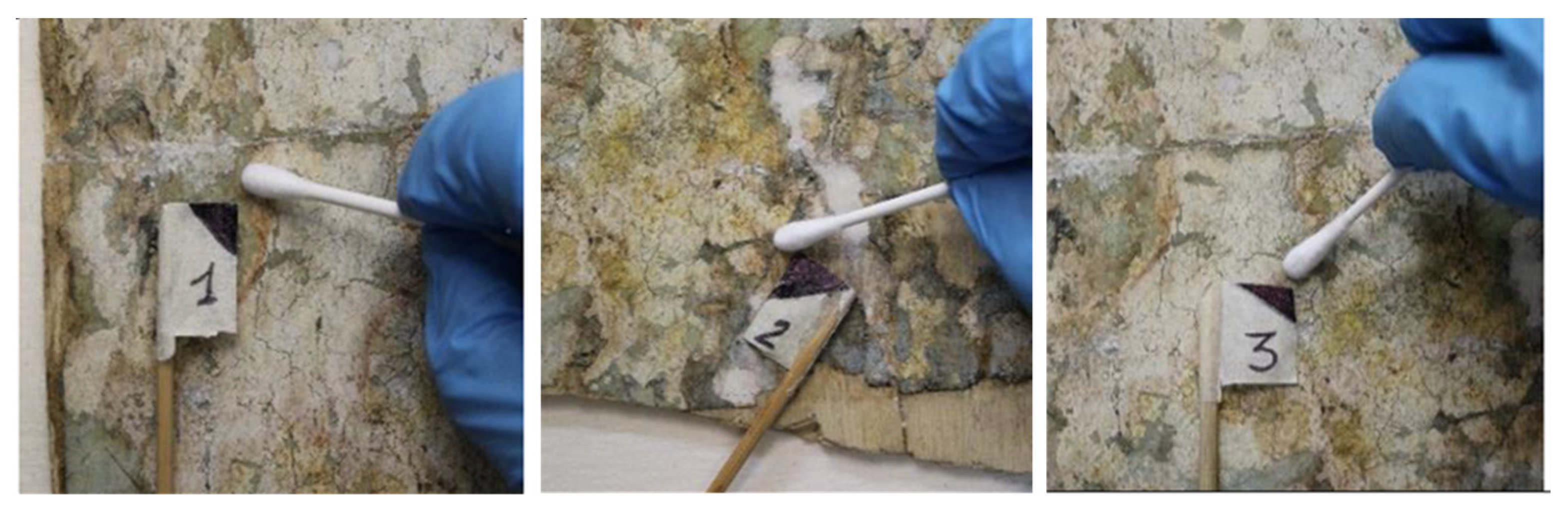
2.2. Microbiological Analyses

The damage produced by the presence of fungal growth on the support of the paper was documented. To isolate and identify the strains involved in the deterioration of the artwork, which will be used to test the efficacy of disinfestation treatments, samples were collected from both the front and reverse of the lower left edge of the collage, where the HMI technique showed a lack of homogeneity on the surface due to increased biological proliferation. Three samples of biological origin ranging from black to reddish brown were taken by wiping the surfaces with dry sterile cotton swabs (Figure 3). Each swab was used for microbial cultivation in triplicate on Malt Agar (Malt extract 30 g/L, agar 15 g/L; MA). The plates were then incubated at room temperature (22 °C) and inspected daily for one month.
The strains responsible for deterioration patterns were selected and determined using diagnostic keys (two strains) [21,22] or molecular techniques (four strains) [23]. For molecular identification, DNA was extracted from the mycelium that actively grows on MA, using Nucleospin Plant kit, following a protocol optimized for fungi. The fungal internal transcribed spacer (ITS) region of the nuclear-encoded ribosomal RNA was amplified using the ITS5 and ITS4 primers. PCR reactions were performed using the BioMix by BioLine (GmbH, Luckenwalde, Germany). PCR reactions consisted of 5 ng of DNA template, 5 pmol of each primer, and Milli-Q sterile water to a final volume of 25 µL. Amplifications were carried out through the MyCycler™ Thermal Cycler (Bio-Rad Laboratories, GmbH, Munich, Germany). The PCR reactions consisted of 3 min at 95 °C, 35 cycles of denaturation at 95 °C for 30 s, annealing at 55 °C for 30 s, and extension at 72 °C for 30 s, followed by a final extension at 72 °C for 5 min. Amplicons were sequenced by Macrogen Inc. (Seoul, Republic of Korea). Sequences were manually inspected using ChromasPro v1.32 (Technelysium, Southport, Queensland, Australia). Obtained ITS sequences were compared with sequences present in the National Center for Biotechnology Information (NCBI) GenBank database using the BLASTN software (https://blast.ncbi.nlm.nih.gov/Blast.cgi, accessed on 9 January 2023). ITS sequences have been deposited in the NCBI GenBank (accession numbers OQ231607-OQ231610). The strains have been deposited in a metabolically inactive state in the Culture Collection of Fungi from Extreme Environments (CCFEE), Tuscia University, Viterbo, Italy.
The biocidal effects of lipases by Candida rugosa (Antares S.r.l.) and chitinases by Trichoderma viride (Sigma Aldrich, St. Louis, MO, USA) were assessed both for each enzyme and in combination of them. The biocidal effect of ethyl and isopropyl alcohols have been tested as well. Due to the inefficiency of enzyme function in this case (see Supplementary Materials, S4), only the results obtained using the alcohols are reported here. The latter were assessed against two selected strains: CCFEE6674 and CCFEE6317, by culture approaches.
Fungal biomasses from well-grown and strongly sporulating colonies were harvested, gently crushed with a pestle, and diluted in alcoholic solutions (ethanol 95% and 70%, isopropyl alcohol 100 and 70%) at concentrations of 170 mg/mL and 500 mg/mL for strain CCFEE6674 and strain CCFEE6317, respectively. The effects were evaluated, in triplicate, by microscopic observations and cultural tests, after 1, 3, and 5 h, and compared with controls consisting of the same concentrations of mycelium in a saline solution. Saline solutions were used to preserve cell integrity and fungal activity. The biomasses were collected, rinsed with sterile water, suspended in saline solution using the same volume of the starting solution, spread on MA (Malt Agar, 100 µL for dishes), incubated at room temperature, and monitored for 2 weeks. Cultural tests were carried out on five replicates for each condition and strain.

2.3. Hydrogel Preparation

Given the obtained biocide effects of ethylic and isopropyl alcohols (see Section 3.3), they were incorporated into rigid Gellan gels (Kelcogel®) to combine biocidal activity with the adsorptive activity of the gel. Given the greater rigidity of the isopropylic Gellan gel as well as its lower homogeneity and tonicity compared to the ethylic one, the latter was selected for the successive tests. The recipe used for its formulation was as follows: Gellan (Kelcogel®) at 2% (w/w) in 66.7% ethanol and 33.3% water. This is the highest possible concentration of alcohol to water usable, because at higher percentages of ethanol to water, the gelification process was inhibited.
The preparation of hydroalcoholic Gellan gels followed the procedure previously described in the literature [7] with slight modifications. Briefly, Gellan powder was dispersed in demineralized water on a heating plate, and, under constant stirring, 0.04% w/w calcium acetate was added. The temperature was raised to 100 °C in order to simmer the solution and achieve maximum hydration of the polymer chain. While waiting for the temperature to drop to 65 °C, the alcoholic solvent was slowly added, under strong agitation, using a Pasteur pipette to allow homogeneous diffusion in the liquid. Gelation occurred after the solution was cooled at room temperature.
Hydrogel tablets were first applied on mock-ups, prepared ad hoc as described below, and in a second step, on the artist’s artwork.

2.4. Mockups and Treatments

Stratigraphic mock-up samples replicating the collage execution technique were made using the same materials as the original artwork.
Squares (5 × 5 cm) of poplar plywood (the secondary support) were used as the support, on which blue-back paper printed with the same type of typographic inks used for the collage was adhered. The glue used was polyvinyl acetate (Bindan-P266) because it was chemically similar to the original (see results and discussion). After the samples had dried, two different pictorial films were applied: the first using natural earths and the second using black acrylic colors, in order to replicate two chromatic media used by the artist (see results and discussion). The stratigraphic samples were prepared in order to have on the mock-up all the layers of the artwork, i.e., the paper, the paper with the velatura (thinly painted glaze layer), and the paper with the painting.
A set of mock-up samples was subjected to artificial aging as defined by ISO 5630/30 [24] to simulate natural aging of the artwork. They were used to observe the interaction between the hydrogel and the substrate, verify the absence of side effects, and select the best application mode of the alcoholic Gellan tablets. Alcohol hydrogel tablets were applied in contact with the surface of aged samples for 1 h, covered with a transparent glass slide to monitor any interference with the substrate, and held under a light weight to ensure their adhesion to the test sample (Figure 4).
A second set of mock-up samples was inoculated with fungi and used to verify the disinfecting power of the alcohol hydrogel tablets as follows: the samples were oven sterilized at 140 °C for 3 h and inoculated by spreading 1 mL of strain CCFEE6317, CCFEE6674, and CCFEE6678 suspensions in triplicate. For each strain, suspensions were obtained by harvesting mycelia from healthy colonies grown in MA at 22 °C and diluting the biomass in 10% Czapek liquid in distilled water at the same final concentration (200 colonies fungal units, CFUs). The mock-up samples were then incubated at 25 ° C. To check the disinfecting power of gels on inoculated samples, culture tests were performed by rubbing sterile swabs on the samples after 1 h and 2 h of application of the 2% ethyl alcohol hydrogel. The swabs were immediately rubbed onto MA plates, incubated at 22 °C for 1 month, and checked daily to exclude possible delayed growth due to inhibition by alcohol treatment.
Finally, to evaluate the effects of the disinfection treatment experiment on the Poesia collage and possible modifications at the micro-morphological level of the fibers constituting the support, images were acquired through scanning electron microscopy (SEM) with the backscattered electron detector (EDS). For SEM-EDS analysis, a Jeol JSM 6010 LA scanning electron microscope was used, operating at 20 kV as the acceleration voltage of the electrons. Before SEM-EDS analysis, the samples were placed on aluminum stubs, fixed with carbon tape, and metallized with gold through a Balzers Union MED10 sputter coater, operating under a vacuum.

3. Results

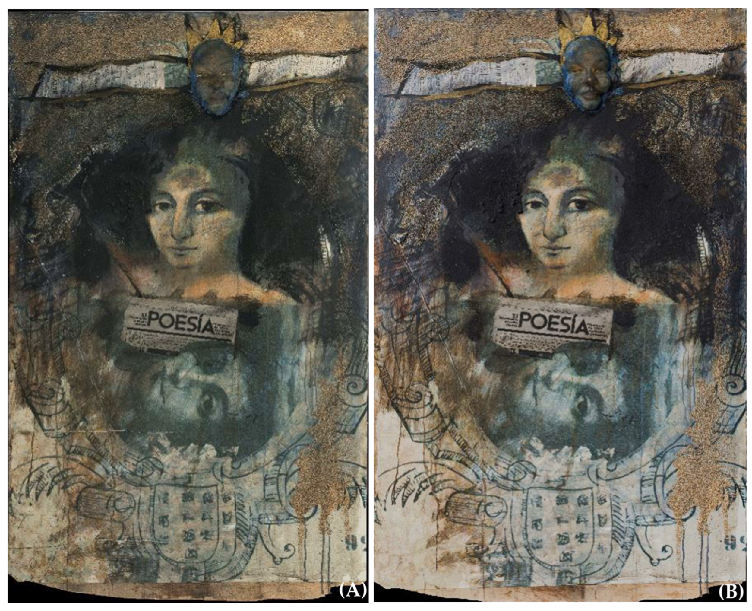
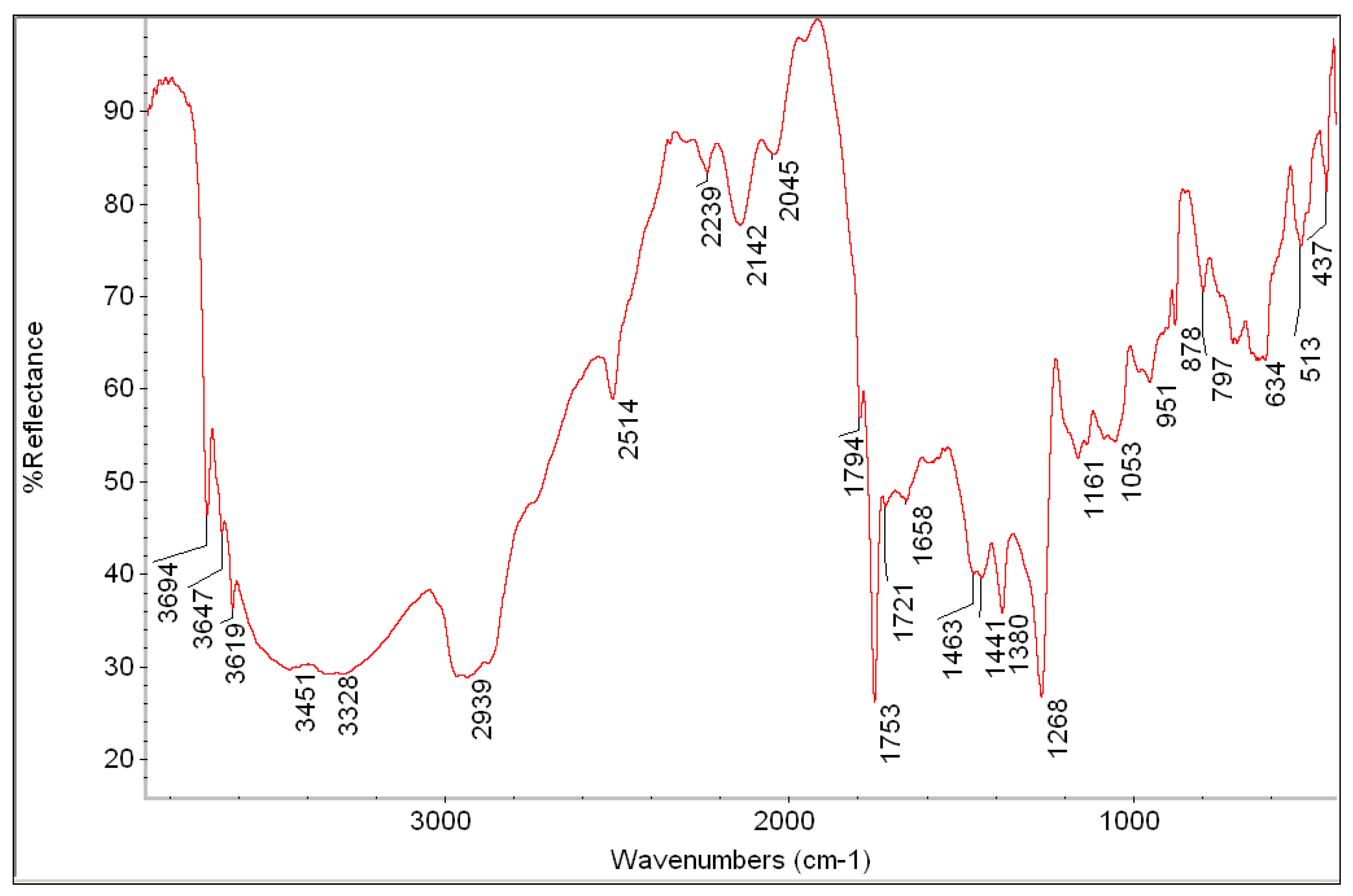
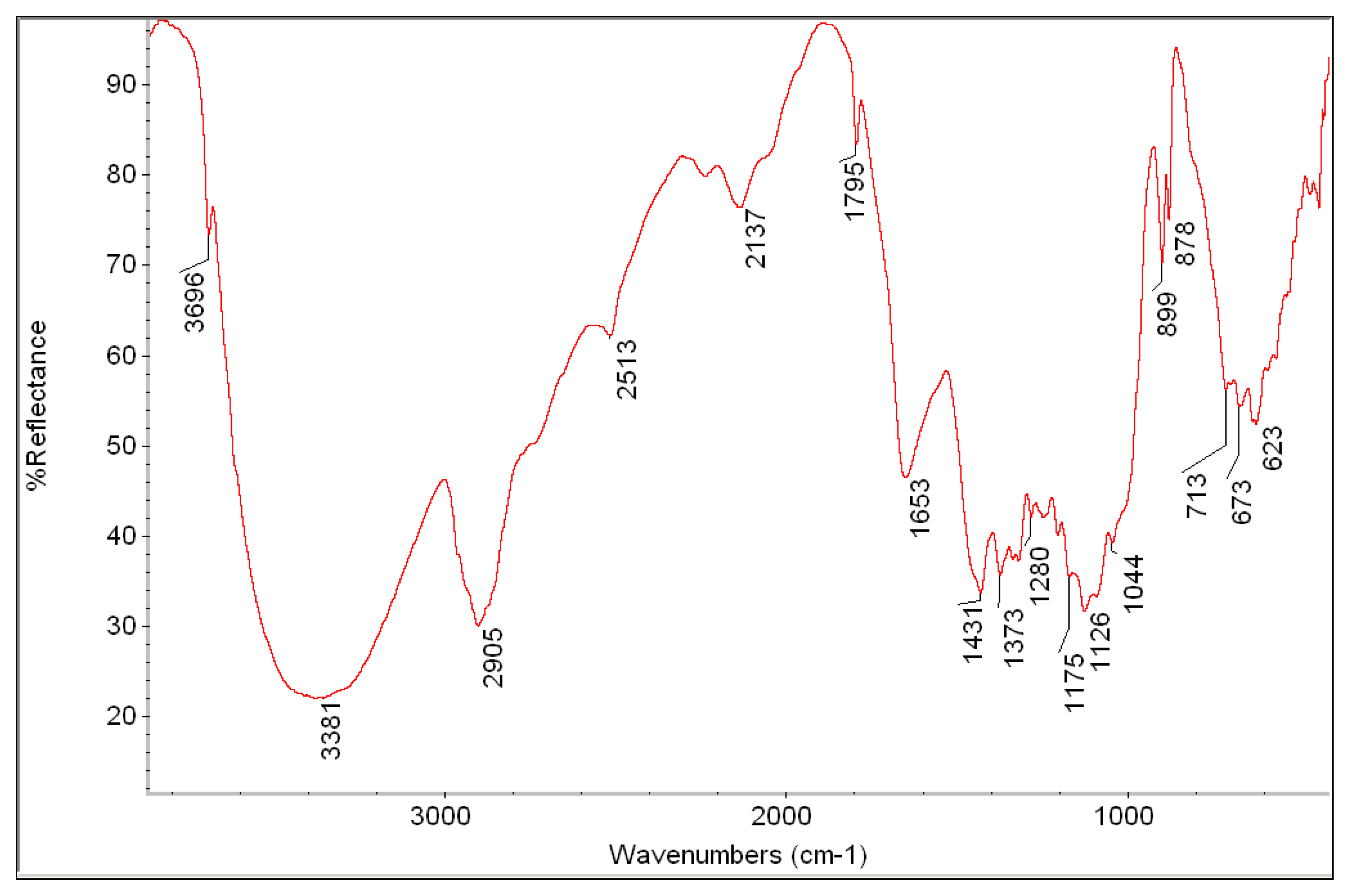
3.1. Diagnostic Analysis

The diagnostic campaign aimed at identifying materials and techniques used by the artist to prepare suitable mock-ups for testing the disinfection methodology before applying it to the artwork, beginning with non-invasive imaging (HMI) and pinpoint (XRF) analyses. HMI is an important multispectral tool that has been shown to be very useful in investigating different types of artworks [16,25,26], but it had never been used in contemporary collages like Poesia.
By applying a specific tool of the HMI processing software, it was possible to obtain the infrared false-color (IRFC) image (Figure 5B), which showed a well-visible reddish response in the centerer and upper part of the surface. Different blue pigments could give such a response, i.e., ultramarine blue, indigo, and cobalt blue [27,28].
Another useful output obtained by HMI analysis was the acquisition of ultraviolet fluorescence (UVF), which allowed us to map the conservation status of the surface. In fact, UVF provided a map of the fungal attack, which was diffused mainly in the lower part of the artwork (Figure 5C). The fluorescence was attributed to fungal mycelium that extended over large surface areas, even under the pictorial layers. When irradiated with UV light, the spread of the colonies was also detectable in non-discolored parts, due to autofluorescence of the components of the fungal cell wall [29,30].
This imaging analysis was useful in determining the target points for the XRF analysis (Figure S3), the results of which are reported in Table 1. The presence of Ca and Fe in all points examined suggests their use in paper production [31,32]. The blue paint film contains Cu, suggesting the use of a copper-based pigment together with another blue (reddish in IRFC) that does not contain elements detectable by portable XRF instruments. According to the IRFC response, the most probable pigments are artificial ultramarine blue and indigo.
The gold color was obtained with mica pigment characterized by the combination of iron oxide and titanium oxide, which gives the metallic appearance of gold [33]. In particular, the gold hue can be obtained by applying an iron oxide layer over a coating of titanium oxide [34,35].
Organic materials were investigated by FTIR spectroscopy performed on micro-samples picked up from the artwork. Specifically, FTIR analysis was used to characterize the typologies of the adhesives used for attaching the various parts of the multi-material artwork and of the binder of the painting material. The first sample examined was of the paper and the adhesive used to attach it to the plywood support (Figure 6 and Figure 7) [36]. The main signatures in the infrared spectrum were due to polyesters (cm−1: 2939, 1753, 1721, 1268) (Figure 6), to industrial paper (whose spectrum was also acquired separately) revealing the cellulose bands at 2905, 1653, 1373, 1175, 1126, 899, and 623 cm−1 (Figure 7), and to some inorganics such as calcium carbonate (2514, 1794, 1441, and 878 cm−1) and silicates (3694, 3647, 3619, 1053, and 951 cm−1) (Figure 6) [37,38,39,40].
The inorganic materials can be from industrial paper preparation [31,32]. The signatures in the range of 2000–2250 cm−1 are probably due, indeed, to sulphates, such as sodium sulphate, employed in the industrial processes for producing paper.
The FTIR analysis of the surface resin (used to adhere the sand to the blue-back paper) and that of the fiberglass mask allowed us to detect the presence of a polyester as the main component (Figure 8 and Figure 9). The spectra of the two adhesives are very similar to the sharp and medium-intensity signals of C-H stretching vibrations, suggesting the use of a polyester resin [41]. The intense signals at 1633–1635 cm−1 can be attributed to the presence of aromatic compounds.
The black’s FTIR spectrum was very similar to that shown in Figure 8 and Figure 9, suggesting that the binder had the same polyester composition (spectrum not shown). The spectrum of the black painting sample had, in addition to those of adhesives, the presence of the typical signatures of calcium carbonate (at 2515, 1796, 1443, 877, and 712 cm−1), which was a probable filler of the pigment.
The composition of the adhesives was further investigated by analytical pyrolysis coupled with gas chromatography and mass spectrometry (Py-GC/MS). Chromatograms are reported in the Supplementary Materials, Section S.4.
From the chromatogram relative to the surface adhesive used to adhere the sand to the paper (Figure S5), it was evident that it mainly consists of diisobutylphthalate, an external plasticizer usually added to the already preformed polymer. An internal plasticizer, such as “vinyl poured” or “VeoVa”, is also present. Furthermore, the presence of acetic acid, although at a lower concentration, led to the assumption that the material investigated could be a polyvinyl acetate (PVA) emulsion [42,43,44].
The pyrolysis products related to the paper adhesive used in creating the collage were very similar to those already highlighted in the previous sample, and the presence of phthalic anhydride and acetic acid as typical pyrolysis products of vinyl emulsions confirmed the hypothesis. The presence of levo-glucosan, the main byproduct of cellulose pyrolysis, indicated that there were paper residues in the sample that could not be separated from the adhesive (Figure S6).
In the chromatogram of the fiberglass mask adhesive, the abundant presence of phthalic anhydride was highlighted, which often identifies the class of alkyd resins [43] (pp. 225–241). The presence of phthalic anhydride was confirmed by the detection of traces of benzoic acid and dimethyl phthalate, commonly produced by the anhydride itself and the initial polyester (Figure S7). In this case, the high levels of styrene and phthalic anhydride in the analyzed sample indicate the presence of unsaturated polyester resins consisting of orthophthalic acid and styrene, thermosetting resins used in the field of contemporary art, and often accompanying fiberglass [45].
The Py-GC/MS analysis of a sample of black color showed the presence of diisobutyl phthalate and acetic acid, indicating the vinyl nature of the binder used in the formulation of the color in the tube. From the chromatogram (Figure S8), traces of butyl acrylate (BA) and butyl methacrylate (BMA) were detected (which identified acrylic resins), as well as an intense styrene signal. This result, taking also into account the presence of vinyl versatate (VeoVa) in the color, leads to the hypothesis that the binder used for the creation of the tube color was a polyvinyl acetate/vinyl versatate copolymer (PVA/VeoVa), softer than pure PVA [43] (pp. 225–242).
The data obtained by FTIR and Py-GC/MS were highly useful in obtaining information about the materials used by the artist, such as adhesives and binders, and in understanding the technical construction of the collage so that the replicated laboratory mock-ups were usable for testing the effectiveness of alcoholic hydrogels in removing the fungi.

3.2. pH and Color Measurements

Ten pH values were measured in the areas of the artwork where the fungal colonization was more evident to evaluate its eventual effect on the collage materials. The measured values on the front side of the artwork ranged from 6.48 to 6.93, indicating that no acidification process occurred; on the contrary, the values on the back are from 4.53 to 5.64 (see Figure S9 and Table S1). In the case of the plywood support, it seems that the fungal attack, more evident in the back of the artwork, led to a decrease in the pH values of the material [46].
The pH values were also measured from the mock-up samples to be used for the hydrogel application. In this case, the initial pH values ranged between 5.68 on the back of the paper and 7.70 on the front (Table 2). After artificial aging of the samples, the pH values showed a general decrease. After the application of the 2% gel with ethyl alcohol on aged samples, a slight increase in pH values was observed (Table 2).
Colorimetric measurements performed before and after accelerated aging treatment of the test samples showed a variation in the color coordinates L*, a*, and b*, which was higher in the case of paper and lower for the painting layer. The main evidence is a decrease in L* and an increase in b*, suggesting a darkening and yellowing of the paper as usually occurs for paper and painting materials exposed to UV light [47]. The values obtained are reported in the Supplementary Materials, Table S2.
Treatment with 2% Gellan hydrogel in ethanol on the test samples (data not shown) caused little color variation in all areas examined in the sample. The main changes occurred in the applied painting film as thinly painted glaze (velatura), where a value of ΔE* equal to 4.12 was obtained, mainly due to an increase in lightness. In all other points, the total color differences are always below 3, indicating that the gel mixture formulated does not significantly affect the color of the samples.
Color changes on the artwork (the five points shown in Figure 2) after the hydrogel application are reported in Table 3. The highest color change occurs in point 1 (light blue color) due to a decrease in lightness. In the other points, total color changes are less relevant and variations are generally due to the lightness.

3.3. Microbiological Analyses

Different types of damage produced by the microbial growth on the support of the paper were documented [48,49]: (i) embrittlement, which resulted in the loss of some fragments of paper due to the enzymatic metabolization of the substrate used as a nutrient source; (ii) chromatic variations caused, in addition to intrinsic pigmentation of some melanized microfungi, by the release of metabolic products around the infection site; (iii) growth and penetration of fungal hyphae in and under the paper.
Six fungal phylotypes were identified and selected among those mainly responsible for the observed biological alterations, due to the presence of strongly pigmented hyphae and conidia. They were determined by molecular approaches, except for two strains of Cladosporium. The six strains were identified as Cladosporium sp. 1, Cladosporium sp. 2 (strains CCFEE6673 and CCFEE6674, respectively), a black meristematic fungus (strain CCFEE6317) producing punctiform microcolonies ascribed to Teratosphaeriaceae (it had as its closest neighbor a strain described as Mycocalicium victoriae), Penicillium sp. cfr. vinaceum (CCFEE6677), having a 99.82% similarity with strain MT312765, Chaetomium globosum (CCFEE6678), having a 100% similarity with strain MZ724876, and Penicillium sp. cfr. brevicompactum (CCFEE6679), having a 99.81% similarity to CBS210.28.
Because lipases and chitinases did not affect CCFEE6674 cell integrity and survival (see Supplementary Materials), ethyl and isopropyl alcohols were investigated for their biocidal potential. Microscopic examination of strains CCFEE6674 and CCFEE6317, selected for the tests among the isolated strains, revealed no morphological damage after treatments with both alcohols. Despite this finding, cultural tests demonstrated a complete inhibitory effect on the growth of these strains, even after the shortest treatment interval tested (1 h), corroborating the treatment procedure involving the loading of an alcohol solution into a rigid Gellan system (Figure 10). Changes in the biomass consistency were observed even after 1 h of treatment; after this time, the suspension indeed becomes gelatinous, and in the case of strain CCFEE6317, the release of pigment occurred.
After selecting the 2% Gellan gel in ethanol/water, due to its superior performance over the others tested, culture tests were carried out to assess its fungicidal effect after application on mock-up samples, and fungal colonies ranging from 0 to 1 per sample were recorded.
SEM images of samples before the treatments showed the presence of diffuse fungal hyphae on the paper fibers, with diameters ranging from 1.13 and 1.60 μm; images after treatment showed the presence of only residual collapsed fungal structures anchored to the cellulose fibers (Figure 11).

4. Discussion

The restoration of the mixed media collage Poesia by Alessandro Kokocinski was a complex process involving different steps to overcome several problems linked to the presence of various layers in the collage. In the present paper, we report procedures related to the setup of an appropriate approach for the removal of a biological attack. For this purpose, an innovative method based on Gellan gum hydrogel with the incorporation of ethyl alcohol was effective in the disinfection process. Preliminary diagnostics were necessary to characterize the materials and the techniques used for the manufacture of artwork and the discussed laboratory mock-ups. The usability of artificially aged mock-ups as valid samples for testing the alcoholic hydrogels was assessed by color and pH measurements.
The diagnostic project began with non-invasive investigations and continued with characterizing, using semi-invasive diagnostic techniques, all of the materials used for the creation of the collage as well as its state of preservation.
The biological contribution to this multidisciplinary approach involved the characterization of the main fungal agents responsible for the biological degradation and the subsequent testing of different biocidal treatments, in order to define the most effective protocol to be applied for the disinfection. Among the isolated fungal strains, six were identified as most likely responsible for the observed damage, due to their dominance in all collected samples, their morphological characteristics (mainly strong pigmentation), and their potential degradative activities. In particular, the main fungal strains belonged to Penicillium, Cladosporium, a black meristematic species within Teratosphaeriaceae, and Chaetomium globosum. The latter is known as a cellulolytic agent and as the etiological agent of soft wood caries [22]. After testing, with little success, the biocidal effect of enzymes, despite their use in the restoration field since the 1970s [50,51], we focused our attention on alcoholic solutions, which are widely used for the disinfection of paper and library materials [52,53]. Their use against fungi and bacteria has also been widely reported in biomedical applications, and their effectiveness depends on the concentrations used, the application time, and the species tested [54]. Our study confirmed that a hydroalcoholic solution is a good tool to counteract fungal growth and dispersion on cellulosic substrates.
The diffuse fungal attack and the stratigraphic succession of the paper bound to the wooden support did not allow the application of traditional methods of cleaning and disinfecting paper artwork. In fact, the blue-back paper, from which the artwork was made, resulted sensitive to the prolonged use of water or free hydroalcoholic solutions that, dispersing unevenly on the surface, caused chromatic variations and deformations of the supports. Furthermore, the vulnerability of the graphic medium and painting materials to mechanical stress and rubbing prevented the use of traditional methods to remove biological structures.
As aqueous immersion methods could not be used, it was decided to study gelled compounds that would limit water release. Once the biocidal effect of alcohols on the main fungal isolates responsible for the damage on the artwork was tested, it was decided to test the effectiveness of Gellan-based hydrogel loaded with ethanol for the disinfection and removal of the microorganisms and their deposits, and, at the same time, ensure the absence of substrate alterations. We initially began investigating the usage in the Gellan-based gel of isopropyl or ethyl alcohols, since they have both been identified in bibliography as antifungal agents [53,55]. At about 70% in a water solution, the ethanol has already been shown to have a biocidal effect. Moreover, between the two organic solvents, we ultimately chose ethylic alcohol, because a more uniform and maneuverable gel is obtained with its use. Following the encouraging findings of these preliminary tests, we opted to use it as a microbial agent in the rigid Gellan gel, without adding a biocide.
By inhibiting fungal regrowth and eliminating a large portion of the fungal structures that had infiltrated the wood fibers of the paper and semi-finished wood, the use of Gellan gels demonstrated excellent results and ensured the survival of Kokocinski’s artwork for future generations. Furthermore, this gel revealed not only a fungicidal but also a cleaning effect, which is significant in minimizing the risk of surface recolonization.
pH measurements are a significant outcome for art conservation. Indeed, pH values play a significant role in determining the chemical stability of organic cellulose-based materials because they have an impact on the hydrolysis process that leads to the degradation of cellulose and, as a result, the fragility of paper and wood. The use of the hydroalcoholic gel on the aged paper and pictorial layers resulted in a slight pH value increase. Therefore, the restoration of pre-aging pH values following hydrogel application appears to be another reason in support of this cleaning and disinfecting approach. Moreover, the high alcohol content in the gel minimizes the amount of water used and it is still effective in removing surface deposits. Finally, colorimetric measurements on the samples showed that, after the application of the alcoholic hydrogel, there were no significant color variations, indicating that the gels did not affect the chromatic characteristics of the collage layers. In the case of color measurements on the artwork, a slight increase in the yellow component (∆b* with positive values) was observed. It could be associated with the fungicidal action of the disinfectant solution capable of damaging the structure of the cell wall, resulting in minimal release of the fungal pigment. Changes were observed mainly in the coordinate L*, representing the lightness, which increased in two of the measured points and decreased in the other three. The decrease in L* can be related to the removal of surface bleaching, whereas the increase in lightness may be associated with the removal of black fungi. The color changes were, however, sufficiently low to be considered acceptable for the disinfection process.
Based on these results, we can conclude that the hydroalocholic Gellan gel could be used for both disinfecting action and cleaning action. This represents an improvement in the performance of the Gellan-based hydrogels, which, to the best of our knowledge, have been used only as cleaning agents [7,8]. It should be noted, finally, that Gellan-based hydrogels could be applied on artwork more times, depending on the condition and nature of the artwork and the nature of the biological attack.

5. Conclusions

The restoration of Poesia involved different expertise, with a biological contribution which allowed us to characterize the main biodeteriogens and test different treatments to define an effective protocol for their removal. Among the isolated fungal strains, six of them were defined as the main strains responsible for the observed damages. The hydroalcoholic Gellan gel (containing 66.7% ethanol) resulted in an effective removal of both fungi and stains from the artwork thanks to the adsorbent capacity of the polysaccharidic gel component. From a practical point of view, this gel results in enough rigidity to be easily applied and removed from paper, without leaving surface residues. It is able to uniformly adhere to and clean the paper surface. The high alcohol percentage in the hydrogel allowed us to limit the amount of water, obviating serious side effects on water-sensitive materials. Tests performed on aged specimens showed that the use of the proposed Gellan-based gel increases the pH values of the materials, probably by removing the water-soluble cellulose and lignin byproducts responsible for paper acidity. This result confirms that the proposed method is appropriate.
The alcoholic hydrogel studied here proved to be a valid tool in cases where the polymeric composition of the artifacts makes it difficult to apply traditional chemical biocides, either because of the risk of interference with some of the artwork components or because the degradation of the artwork is in a very advanced state. Designed in this case for the conservation of a mixed media collage, it is suitable for the restoration of different types of water-sensitive works of art [56,57,58].
Finally, in addition to being effective for fungal disinfection of a modern paper that is characterized by typographic inks and painted with different chromatic mixtures, this new gel based on Gellan and alcohols has the advantage of being ecological, economical, and easy to prepare.
As a whole, the restoration project of Poesia represented a long and stimulating path that has led to its conservation treatment, thanks to the contribution of experts in different fields. They have worked together to contextualize the artwork, characterize the materials used by the artist, define previous restoration interventions, define the ‘health status’ of the object, and, as the final objective of their research, apply the best conservation treatments.

Supplementary Materials

The following supporting information can be downloaded at: https://www.mdpi.com/article/10.3390/heritage6030144/s1, Figure S1: Stratigraphic scheme of the artwork Poesia by Alessandro Kokocinski: (a) plywood panel; (b) blue-back poster paper; (c) paper fragments of magazine and newspaper; (d) medium-grain sand; (e) pictorial layer; (f) fiberglass mask with cardboard crown; Figure S2: Photographs of the artwork with the colorckecher: (A) the RGB image obtained through HMI system before the restoration; (B) the RGB image obtained through a professional camera after the restoration; Figure S3: Photograph of the artwork with the points of XRF analysis whose results are reported in the main document; Figure S4: Results of culture tests after 2 h treatments of Cladosporium sp. 2 with lipases and chitinases, alone and in combination. (a) control; (b) lipase treated plate; (c) chitinase treated plate; (d) lipase and chitinase treated plate. Figure S5. Chromatogram of the surface adhesive; Figure S6: Chromatogram of the paper adhesive; Figure S7: Chromatogram of the fiberglass mask adhesive; Figure S8: Chromatogram of the black colour binder; Figure S9: Points of superficial pH measurements before restoration, on front and back sides; Table S1. pH values measured on front and back sides of Poesia, in the points shown in the Figure S9. Table S2. Chromatic differences before and after artificial ageing on the three replicates of test sample A.

Author Contributions

Conceptualization, E.W., L.Z., C.M., M.M. and C.P.; methodology, F.A., E.W., M.M., L.Z. and C.P.; software, C.P. and L.L.; validation, C.M., E.W., M.M., F.A, L.Z. and F.C.; formal analysis, V.V., F.C., C.P. and L.L.; investigation, V.V., C.P., L.L., F.C. and F.A.; resources, L.Z., C.P., V.V. and C.M.; data curation, F.A., C.P., F.C., C.M. and V.V.; writing—original draft preparation, L.Z. and C.P.; writing—review and editing, all authors.; visualization, C.P. and L.L.; supervision, E.W., C.P., L.Z. and C.M.; project administration, E.W., C.P. and L.Z.; funding acquisition, C.P., C.M., V.V. and L.Z. All authors have read and agreed to the published version of the manuscript.

Funding

This research was developed within the activities of EVER Project (Groups of Research call), funded by Lazio Region, grant number G04014, 13 April 2021, CUP J85F21000520002. POR FESR Lazio 2014–2020.

Institutional Review Board Statement

Not applicable.

Informed Consent Statement

Not applicable.

Data Availability Statement

Not applicable.

Acknowledgments

The work presented in the paper was the subject of a conservation work in the restoration laboratories of University of Tuscia as part of the requirements for a master’s degree. The authors would like to thank Giorgia Agresti for having supported Francesca Ambroselli during the development of her thesis work, specifically for the pH and color measurements, Daniela Isola for her suggestions in microbiological analyses, Ottaviano Caruso for his support in the HMI analysis, and Gaetano Alfano for the photos after the restoration. The SEM-EDS analysis was performed by Anna Rita Taddei at the Center of Microscopy, as part of the Large Equipment Center at the University of Viterbo.

Conflicts of Interest

The authors declare no conflict of interest.

References

  1. Hirsenberger, H.; Ranogajec, J.; Vucetic, S.; Lalic, B.; Gracanin, D. Collaborative projects in cultural heritage conservation–management challenges and risks. J. Cult. Herit. 2019, 37, 215–224. [Google Scholar] [CrossRef]
  2. Ambroselli, F. Poesia di Alessandro Kokocinski. Un approccio Multidisciplinare al Restauro di un Collage Polimaterico Affetto da Degrado Biologico. Master’s Thesis, University of Tuscia, Viterbo, Italy, 2021. [Google Scholar]
  3. Baglioni, M.; Poggi, G.; Chelazzi, D.; Baglioni, P. Advanced Materials in Cultural Heritage Conservation. Molecules 2021, 26, 3967. [Google Scholar] [CrossRef]
  4. Mazzuca, C.; Poggi, G.; Bonelli, N.; Micheli, L.; Baglioni, P.; Palleschi, A. Innovative Chemical Gels Meet Enzymes: A Smart Combination for Cleaning Paper Artworks. J. Colloid Interface Sci. 2017, 502, 153–164. [Google Scholar] [CrossRef] [PubMed]
  5. Mazzuca, C.; Carbone, M.; Cancelliere, R.; Prati, S.; Sciutto, G.; Mazzeo, R.; Tositti, L.; Regazzi, R.; Mostacci, D.; Micheli, L. A new analytical approach to characterize the effect of γ-ray sterilization on wood. Microchem. J. 2018, 143, 493–502. [Google Scholar] [CrossRef]
  6. Mazzuca, C.; Severini, L.; Missori, M.; Tumiati, M.; Domenici, F.; Micheli, L.; Titubante, M.; Bragaglia, M.; Nanni, F.; Paradossi, G.; et al. Evaluating the Influence of Paper Characteristics on the Efficacy of New Poly(Vinyl Alcohol) Based Hydrogels for Cleaning Modern and Ancient Paper. Microchem. J. 2020, 155, 104716. [Google Scholar] [CrossRef]
  7. Mazzuca, C.; Micheli, L.; Carbone, M.; Basoli, F.; Cervelli, E.; Iannuccelli, S.; Sotgiu, S.; Palleschi, A. Gellan Hydrogel as a Powerful Tool in Paper Cleaning Process: A Detailed Study. J. Colloid Interface Sci. 2014, 416, 205–211. [Google Scholar] [CrossRef] [Green Version]
  8. Mazzuca, C.; Micheli, L.; Lettieri, R.; Cervelli, E.; Coviello, T.; Cencetti, C.; Sotgiu, S.; Iannuccelli, S.; Palleschi, G.; Palleschi, A. How to tune a paper cleaning process by means of modified gellan hydrogels. Microchem. J. 2016, 126, 359–367. [Google Scholar] [CrossRef]
  9. Petrella, G.; Mazzuca, C.; Micheli, L.; Cervelli, E.; De Fazio, D.; Iannuccelli, S.; Sotgiu, S.; Palleschi, G.; Palleschi, A. A new sustainable and innovative work for paper artworks cleaning process: Gellan hydrogel combined with hydrolytic enzymes. Int. J. Conserv. Sci. 2016, 7, 273–280. [Google Scholar]
  10. Mastrangelo, R.; Chelazzi, D.; Poggi, G.; Fratini, E.; Buemi, L.P.; Petruzzellis, M.L.; Baglioni, P. Twin-chain polymer hydrogels based on poly (vinyl alcohol) as new advanced tool for the cleaning of modern and contemporary art. Proc. Natl. Acad. Sci. USA 2020, 117, 7011–7020. [Google Scholar] [CrossRef] [Green Version]
  11. De Santis, P.C. Some observations on the use of enzymes in paper conservation. J. Am. Inst. Conserv. 1983, 23, 7–27. [Google Scholar] [CrossRef]
  12. Palla, F. Bioactive molecules: Innovative contributions of biotechnology to the restoration of cultural heritage. Conserv. Sci. Cult. Herit. 2013, 13, 369–373. [Google Scholar]
  13. Fidanza, M.R.; Caneva, G. Natural biocides for the conservation of stone cultural heritage: A review. J. Cult. Herit. 2019, 38, 271–286. [Google Scholar] [CrossRef]
  14. Proto, M.R.; Biondi, E.; Baldo, D.; Levoni, M.; Filippini, G.; Modesto, M.; Di Vito, M.; Bugli, F.; Ratti, C.; Minardi, P.; et al. Essential Oils and Hydrolates: Potential Tools for Defense against Bacterial Plant Pathogens. Microorganisms 2022, 10, 702. [Google Scholar] [CrossRef] [PubMed]
  15. Melis, M.; Miccoli, M.; Quarta, D. Multispectral hypercolorimetry and automatic guided pigment identification: Some master-pieces case studies. In Proceedings of the SPIE 8790, Optics for Arts, Architecture, and Archaeology IV; Munich, Germany, 13–16 May 2013, Pezzati, L., Targowski, P., Eds.; SPIE: Washington, WA, USA, 2013; Volume 8790. [Google Scholar]
  16. Colantonio, C.; Pelosi, C.; D’Alessandro, L.; Sottile, S.; Calabrò, G.; Melis, M. Hypercolorimetric Multispectral Imaging (HMI) system for cultural heritage diagnostics: An innovative study for copper painting examination. Eur. Phys. J. Plus 2018, 133, 526. [Google Scholar] [CrossRef]
  17. Lanteri, L.; Pelosi, C. 2D and 3D ultraviolet fluorescence applications on cultural heritage paintings and objects through a low-cost approach for diagnostics and documentation. In Proceedings of the SPIE 11784, Optics for Arts, Architecture, and Archaeology VIII; Munich, Germany, 21–25 June 2021, Liang, H., Groves, R., Eds.; SPIE: Washington, WA, USA, 2021; Volume 11784, p. 1178417. [Google Scholar]
  18. Learner, T. The Analysis of Synthetic Paints by Pyrolysis-Gas Chromatography-Mass Spectrometry (PyGCMS). Stud. Conserv. 2001, 46, 225–242. [Google Scholar]
  19. UNI EN 15886; Conservation of Cultural Property–Test Methods–Colour Measurement of Surfaces. CEN: Brussels, Belgium, 2010.
  20. Pelosi, C.; Capobianco, G.; Agresti, G.; Bonifazi, G.; Morresi, F.; Rossi, S.; Santamaria, S.; Serranti, S. A methodological approach to study the stability of selected watercolours for painting reintegration, through reflectance spectrophotometry, Fourier transform infrared spectroscopy and hyperspectral imaging. Spectrochim. Acta A 2018, 198, 92–106. [Google Scholar] [CrossRef]
  21. Pitt, J.I. A Laboratory Guide to Common Penicillium Species; Commonwealth Scientific and Industrial Research Organization; Division of Food Processing: North Ryde, Australia, 1991. [Google Scholar]
  22. Domsch, K.H.; Gams, W.; Anderson, T.H. Compendium of Soil Fungi, 2nd ed.; IHW-Verlag: Eching, Germany, 2007. [Google Scholar]
  23. Isola, D.; Zucconi, L.; Onofri, S.; Caneva, G.; De Hoog, G.S.; Selbmann, L. Extremotolerant rock inhabiting black fungi from Italian monumental sites. Fungal Divers. 2016, 76, 75–96. [Google Scholar] [CrossRef]
  24. ISO 5630-3; Paper and Board–Accelerated Ageing–Part 3: Moist Heat Treatment at 80 °C and 65% Relative Humidity. ISO: Genève, Switzerland, 1996.
  25. Laureti, S.; Colantonio, C.; Burrascano, P.; Melis, M.; Calabrò, G.; Malekmohammadi, H.; Sfarra, S.; Ricci, M.; Pelosi, C. Development of integrated innovative techniques for paintings examination: The case studies of The Resurrection of Christ attributed to Andrea Mantegna and the Crucifixion of Viterbo attributed to Michelangelo’s workshop. J. Cult. Herit. 2019, 40, 1–16. [Google Scholar] [CrossRef]
  26. Ricci, M.; Laureti, S.; Malekmohammadi, H.; Sfarra, S.; Lanteri, L.; Colantonio, C.; Calabrò, G.; Pelosi, C. Surface and Interface Investigation of a 15th Century Wall Painting Using Multispectral Imaging and Pulse-Compression Infrared Thermography. Coatings 2021, 11, 546. [Google Scholar] [CrossRef]
  27. Cosentino, A.; Cosentino, A. Identification of pigments by multispectral imaging; a flowchart method. Herit. Sci. 2014, 2, 8. [Google Scholar] [CrossRef] [Green Version]
  28. Boust, C.; Wohlgelmuth, A. DATABASE: Pigment image database under UV and IR radiations. In Scientific Imaging for Cultural Heritage; Hypotheses: Ivry-sur-Seine, France, 2017; Available online: https://copa.hypotheses.org/552 (accessed on 13 February 2023).
  29. Wu, C.H.; Warren, H.L. Natural autofluorescence in fungi, and its correlation with viability. Mycologia 1984, 76, 1049–1058. [Google Scholar] [CrossRef]
  30. Lin, S.J.; Tan, H.Y.; Kuo, C.J.; Wu, R.J.; Wang, S.H.; Chen, W.L.; Jee, S.H.; Dong, C.Y. Multiphoton autofluorescence spectral analysis for fungus imaging and identification. Appl. Phys. Lett. 2009, 95, 043703. [Google Scholar]
  31. Bown, R. Particle size, shape and structure of paper fillers and their effect on paper properties. Pap. Technol. 1998, 39, 44–48. [Google Scholar]
  32. Schabel, S.; Putz, H.-J.; Hamm, U.; Kersten, A.; Bobek, B.; Hirsch, G.; Voss, D. Calcium carbonate in the paper industry-Blessing for coated papermaking and curse for recycling processes. Tappi J. 2014, 13, 47–54. [Google Scholar] [CrossRef]
  33. Bailão, A.; Šustic, S. Retouching with mica pigments. E-Conserv. J. 2013, 1, 45–60. [Google Scholar] [CrossRef]
  34. Pfaff, G. Special Effect Pigments: Technical Basics and Applications, 2nd ed.; Vincentz Network GmbH & Co KG: Hannover, Germany, 2008. [Google Scholar]
  35. Pfaff, G. The world of inorganic pigments. ChemTexts 2022, 8, 15. [Google Scholar] [CrossRef]
  36. Gadhave, R.V.I.; Gadhave, C.R. Adhesives for the Paper Packaging Industry: An Overview. Open J. Polym. Chem. 2022, 12, 55–79. [Google Scholar] [CrossRef]
  37. Doménech-Carbó, M.T.; Doménech-Carbó, A.; Gimeno-Adelantado, J.V.; Bosch-Reig, F. Identification of Syntethic Resins Used in Works of Art by Fourier Transform Infrared Spectroscopy. Appl. Spectrosc. 2001, 55, 1590–1602. [Google Scholar] [CrossRef]
  38. Mitchell, G.; France, F.; Nordon, A.; Leung Tang, P.; Gibson, L.T. Assessment of historical polymers using attenuated total reflectance-Fourier transform infrared spectroscopy with principal component analysis. Herit. Sci. 2013, 1, 28. [Google Scholar] [CrossRef] [Green Version]
  39. IRUG DATABASE. Available online: http://www.irug.org/search-spectral-database (accessed on 13 February 2023).
  40. Poliszuk, A.; Ybarra, G. Analysis of Cultural Heritage Materials by Infrared Spectroscopy. In Infrared Spectroscopy: Theory, Developments and Applications; Cozzolino, D., Ed.; Nova Science Publishers: Hauppauge, NY, USA, 2013; pp. 1–18. [Google Scholar]
  41. Ramírez-Hernández, A.; Aguilar-Flores, C.; Aparicio-Saguilán, A. Fingerprint analysis of FTIR spectra of polymers containing vinyl acetate. DYNA 2019, 86, 198–205. [Google Scholar] [CrossRef] [Green Version]
  42. Crook, J.; Learner, T.J.S. The Impact of Modern Paints; Watson-Guptill publications: New York, NY, USA, 2000. [Google Scholar]
  43. Learner, T.J.S. Analysis of Modern Paints; The Getty Conservation Institute: Los Angeles, CA, USA, 2005. [Google Scholar]
  44. Jablonski, E.; Learner, T.; Hayes, J.; Golden, M. Conservation concerns for acrylic emulsion paints. Stud. Conserv. 2003, 48, 3–12. [Google Scholar] [CrossRef]
  45. López, F.A.; Martín, M.I.; Alguacil, F.J.; Rincón, J.M.; Centeno, T.A.; Romero, M. Thermolysis of fibreglass polyester composite and reutilisation of the glass fibre residue to obtain a glass–ceramic material. J. Anal. Appl. Pyrol. 2012, 93, 104–112. [Google Scholar] [CrossRef] [Green Version]
  46. Mansour, M.M.A.; Hamed, S.A.E.-K.M.; Salem, M.Z.M.; Ali, H.M. Illustration of the Effects of Five Fungi on Acacia saligna Wood Organic Acids and Ultrastructure Alterations in Wood Cell Walls by HPLC and TEM Examinations. Appl. Sci. 2020, 10, 2886. [Google Scholar] [CrossRef]
  47. Hassan, R.R.A.; Mohamed, W.S. Effect of methyl methacrylate/hydroxyethyl methacrylate copolymer on optical and mechanical properties and long-term durability of paper under accelerated ageing. Int. J. Conserv. Sci. 2017, 8, 237–250. [Google Scholar]
  48. Szczepanowska, H.; Lovett, C.M. A study of the removal and prevention of fungal stains on paper. J. Am. Inst. Conserv. 1992, 31, 147–160. [Google Scholar] [CrossRef]
  49. Sterflinger, K.; Pinzari, F. The revenge of time: Fungal deterioration of cultural heritage with particular reference to books, paper and parchment. Environ. Microbiol. 2012, 14, 559–566. [Google Scholar] [CrossRef]
  50. Cremonesi, P. L’uso Degli Enzimi Nella Pulitura di Opera Policrome; Il Prato, Arti Grafiche Padovane: Padova, Italy, 1999. [Google Scholar]
  51. Palla, F. Biotecnologie per i Beni Cultuali. Innovazioni scientifiche Bio-cleaning, Bio-reinforcing. CRPR/InForma Riv. Semest. Del Cent. Reg. Progett. E Restauro 2006, 1, 22–26. [Google Scholar]
  52. Paulus, W. Directory of Microbiocides for the Protection of Materials. A Handbook; Springer Dordrecht: Berlin, Germany, 2005. [Google Scholar]
  53. Sequeira, S.O.; Phillips, A.J.L.; Cabrita, E.J.; Macedo, M.F. Ethanol as an antifungal treatment for paper: Short-term and long-term effects. Stud. Conserv. 2017, 62, 33–42. [Google Scholar] [CrossRef]
  54. Cassanelli, M.; Norton, I.; Mills, T. Effect of alcohols on gellan gum gel structure: Bridging the molecular level and the three-dimensional network. Food Struct. 2017, 14, 112–120. [Google Scholar] [CrossRef]
  55. Sequeira, S.O.; Cabrita, E.J.; Macedoequeira, M.F. Fungal biodeterioration of paper: How are paper and book conservators dealing with it? An international survey. Restaurator. Int. J. Preserv. Libr. Arch. Mater. 2014, 35, 181–199. [Google Scholar] [CrossRef]
  56. Florian, M.L.; Purinton, N. Determination of location of stains in fungal spots and enzymatic removal of pigmented hyphae in paper. In Biodeterioration of Cultural Property 3, Proceedings of the 3rd International Conference on Biodeterioration of Cultural Property, Bangkok, Thailand, 4–7 July 1995; The Conservation Science Division, The Fine Arts Department: Bangkok, Thailand, 1995; pp. 414–425. [Google Scholar]
  57. Di Vito, M.; Vergari, L.; Mariotti, M.; Proto, M.R.; Barbanti, L.; Garzoli, S.; Sanguinetti, M.; Sabatini, L.; Peduzzi, A.; Bellardi, M.G.; et al. Anti-Mold Effectiveness of a Green Emulsion Based on Citrus aurantium Hydrolate and Cinnamomum zeylanicum Essential Oil for the Modern Paintings Restoration. Microorganisms 2022, 10, 205. [Google Scholar] [CrossRef] [PubMed]
  58. Zmeu, N.; Bosch-Roig, P. Risk analysis of biodeterioration in contemporary art collections: The poly-material challenge. J. Cult. Herit. 2022, 58, 33–48. [Google Scholar] [CrossRef]
Figure 1. The mixed media collage Poesia by Alessandro Kokocinski before (A) (image supplied by Ottaviano Caruso) and after (B) (photo by Gaetano Alfano) the restoration. The main evidence after restoration is the removal, especially in the lower part of the artwork, of the biological dark patina.
Heritage 06 00144 g001
Figure 2. A detail of the front side of the collage Poesia before treatment (left side, bottom corner) with the five points of color measurements. A perforated film of rigid acetate was used to guarantee that the measurements before and after treatments would be taken at the same points.
Heritage 06 00144 g002
Figure 3. Details of the three samples taken with sterile dry cotton swabs used for investigating the fungal attack on the collage’s surface.
Heritage 06 00144 g003
Figure 4. Application of alcohol hydrogel tablets on the replica samples. Tablets were applied directly to the surface of aged samples (on the left) and covered with a transparent glass slide under light weight to ensure their adhesion to the test sample (on the right).
Heritage 06 00144 g004
Figure 5. Multispectral images of the collage Poesia obtained by HMI software before the restoration. (A) RGB (red, green, and blue) image; (B) IRFC (infrared false color) image; and (C) UVF (ultraviolet fluorescence) image.
Heritage 06 00144 g005
Figure 6. FTIR spectrum of the adhesive of the paper.
Heritage 06 00144 g006
Figure 7. FTIR spectrum of the paper.
Heritage 06 00144 g007
Figure 8. FTIR spectrum of the surface adhesive.
Heritage 06 00144 g008
Figure 9. FTIR spectrum of the fiberglass adhesive.
Heritage 06 00144 g009
Figure 10. Biocidal effect of ethanol on CCFEE6674; treated samples in the red frame, control samples in the blue one (data on the isopropanol effect are not shown).
Heritage 06 00144 g010
Figure 11. SEM Images of fungal hyphae and conidium before the hydrogel application (A) and apparent residua after the treatment (B), as indicated by the white arrows.
Heritage 06 00144 g011
Table 1. Results of XRF analysis in terms of detected chemical elements.
Measured PointDetected ElementsHypothesis about Pigments and Materials
X1, gold color of the crownTi (main), Fe, CaMica pigment based on Ti-oxide and Fe-oxide
X2, gold color of the crownTi (main), Fe, CaMica pigment based on Ti-oxide and Fe-oxide
X3, blue colorCa (main), Cu, Zn, FeCu-based pigment
X4, black colorCa (main), FeFe-based ink
X5, brown glazingCa (main), Fe, Zn, TiFe-based pigment
X6, sandCa, Fe
X7, white of the paperCa (main), FeMaterial used in paper treatment
X8, brown on the shoulderCa (main), FeIron-based pigment
Table 2. pH measurements before and after aging and after treatment on different mock-ups reproducing the stratigraphy of the artwork: paint layer, paper plus thinly painted glaze layer (velatura), front and back of the paper.
Sample Nr.LayerOriginalAfter AgingAfter Treatment
1Paint layer6.645.455.98
Paper + velatura7.446.676.58
Paper7.646.898.09
Plywood5.755.485.67
2Paint layer7.036.436.60
Paper + velatura7.446.447.12
Paper7.707.637.39
Plywood6.686.276.59
3Paint layer7.076.616.62
Paper + velatura7.507.018.75
Paper7.656.527.97
Plywood5.686.165.80
4Paint layer6.936.706.79
Paper + velatura7.527.478.30
Paper7.687.686.71
Plywood6.336.286.22
Table 3. Color changes in the five points selected on the collage (see Figure 2) after the application of 2% alcoholic Gellan hydrogel.
Measured PointΔL*Δa*Δb*ΔE*
1−5.000.821.575.30
22.32−0.94−0.0832.50
32.78−0.91−0.302.94
4−0.910.010.721.17
5−2.770.370.622.86
Disclaimer/Publisher’s Note: The statements, opinions and data contained in all publications are solely those of the individual author(s) and contributor(s) and not of MDPI and/or the editor(s). MDPI and/or the editor(s) disclaim responsibility for any injury to people or property resulting from any ideas, methods, instructions or products referred to in the content.

