Article

History and Techniques of a Polychrome Wooden Statue, How an Integrated Approach Contributes to Resolving Iconographic Inconsistencies

1
Department of Agriculture and Forest Science (DAFNE), University of Tuscia, 01100 Viterbo, Italy
2
Department of Economics, Engineering, Society and Business Organization (DEIM), University of Tuscia, 01100 Viterbo, Italy
3
High Equipment Centre, Section of Electron Microscopy, University of Tuscia, 01100 Viterbo, Italy
*
Author to whom correspondence should be addressed.
Heritage 2022, 5(3), 2488-2503; https://doi.org/10.3390/heritage5030129
Submission received: 5 August 2022 / Revised: 26 August 2022 / Accepted: 30 August 2022 / Published: 1 September 2022

Abstract

:
The object of this paper is the diagnostic campaign performed on the polychrome wooden statue, revered as St. John, stored in the church dedicated to the “Beata Vergine d’Itria” (Blessed Virgin of Itria), (Noragugume town, Sardinia, Italy). The restoration of the statue gave the occasion to investigate micro-samples from the wooden support and from the polychromy in order to gather information that could be useful for the intervention. In fact, there was limited information on this fine carved statue and its iconographic pattern appeared to be not strictly canonical. Moreover, these kinds of artworks, having religious significant for the faithful, have been generally modified over time to satisfy possible changes in artistic or religious needs or ecclesiastical reforms. The aims of this study are: (i) support the restorers in defining the conservation status of the statue, and (ii) perform a series of analyses to identify the wood and the materials of the painting layers. Specifically, wood micro-sample was examined under the microscope in order to investigate the anatomical characteristics useful to identify the species; pigments were studied through X-ray fluorescence spectroscopy (XRF) and scanning electron microscopy coupled with energy dispersive spectroscopy (SEM-EDS); organic materials were analysed by Fourier transform infrared spectroscopy (FTIR); and, lastly, the panting technique was investigated through micro-stratigraphy (MS). The analysis performed on the micro-sample taken from the support allowed for identifying the wood as poplar. XRF detected chemical elements that could be associated to pigments such as azurite, blue smalt, red lead, white lead and ochres, and Au for the gilding. SEM-EDS confirmed these pigments and allowed to find also other elements useful to suggest the presence of further materials such as Ag in the metal foil. FTIR detected proteinaceous binder and siccative oils that were mapped in the micro-stratigraphy by using histochemical tests. All this information was used by the restorers to address the intervention from the cleaning to the final retouching.

1. Introduction

The polychrome wooden statue object of this study, revered as St. John (Figure 1), is stored in the main altar of the church dedicated to the “Beata Vergine d’Itria” (Blessed Virgin of Itria). The church is located in the main square of Noragugume, a small town in the Marghine, a geographical area of central Sardinia in the province of Nuoro (Italy). The wooden altar is positioned in the presbytery, and it is one of the four that retain the original location out of the 43 examples distributed throughout Sardinia [1]. The altar, dated back to the first half of the seventeenth century [2], is characterised by three niches. In the central niche, a wooden sculptural group, representing the Virgin looking at the faithful, with the Child, the freed slave and the Turkish master, is located. The iconography of this sculpture group is typical of the Virgin Hodegetria (the one who shows the way) [3]. The other niches contain the statue of Saint Anna (left niche) and that of Saint John (right niche).
The exhibition of polymaterial simulacra during liturgical events was aimed at making the sacred event present and alive: Sacred Representations are known from the early centuries of Christianity up to the entire Middle Ages [4,5]. There are still living traditions that combine the theatricality of the representation with liturgical events to underline the significance for the Christians local community and to involve the faithful in the narration of sacred events during the celebration of the main liturgical feasts [6,7,8]. For example, it is interesting to cite the “acchianata da Madonna” (the Virgin’s Ascent) at Gangi town in Sicily (Italy) [9]. On that occasion, during the liturgical celebration, the altar machine slowly makes the statue of the Virgin rise and then suddenly masks it at the sight of the faithful. Moreover, in Noragugume town the faithful celebrate the “Beata Vergine d’Itria” with a gallop race on Pentecost Tuesday.
Religious cultural heritage artworks had and have often a profound significance for the faithful so that they were and are frequently modified in order to maintain a pleasant aspect. Maintenance and restoration of such a kind of objects was usually addressed to restore the functionality of the artifact without taking into account the materials, the techniques and the conservative aspect making very difficult to know the original appearance and the subsequent modifications [10,11]. In addition, artworks having a religious significance may have been modified to satisfy changing in artistic taste or requirements due to ecclesiastical reforms [12,13].
The sacred and devotional character of the statue of St John inevitably required restoration and re-painting interventions aimed to maintain a pleasing appearance (Figure 1), recovering bright colours, so as to give the impression of care to the faithful [14]. The documentary research in the local archives did not provide useful information on the statue of St. John, whose iconography is not strictly canonical, and on the interventions it underwent over time. However, the archive research revealed the presence, in the church of the Beata Vergine d’Itria, of a statuary group entitled “Annunciation”, with the Virgin and announcing Angel. In the second half of the nineteenth century, this group was modified and the Virgin of the Annunciation was adapted into Addolorata (Sorrowed), but no information about the Angel was found. It may be supposed that the Saint John revered today was originally the announcing Angel.
On the occasion of the restoration, recently performed, the statue was investigated in order to obtain information on the constituent materials: micro-samples were supplied by restorers to characterize the wooden support and the materials of the painting layers through different techniques based on optical and electronic microscopes, and spectroscopic methods. These analyses carried out during the restoration were used to develop restoration strategies and to draw hypotheses about the events that affected this interesting statue.

2. Materials and Methods

An interdisciplinary and multidisciplinary approach was adopted for the investigation of the St. John little statue during the restoration activities, even if it was not possible to perform analysis on site due to the difficulty of moving instruments and operators at the time of the intervention. So, the only possibility to make analysis was on samples taken from the restorers and sent to our laboratory. The conservation status was detected by a carefully observation performed before and during the restoration. This critical inspection made it possible to identify the construction technique and to select the sampling points for studying executive technique and constituent materials. This last step and the subsequent laboratory analysis were performed taking into account the minimum intervention criteria in order to obtain as much information as possible with the minimum effort in terms of analysis and techniques used [15,16,17].
Following this well-consolidated approach of our research group, to investigate the presumably original executive technique and to characterize the constituent materials, it was possible to select eleven samples, about 1–2 mm, from the wooden statue after the removal of the surface layer covering the original painting (see Table 1 for the description of the micro-fragments and Table S1 for the micro-fragments images).
The identification of the botanical taxon closest to the species was carried out macroscopically on the parts not covered by pictorial layers and microscopically on a wood sample taken from the statue. The forearms were examined macroscopically. The head was not examined, because no surface was uncovered of plaster and painting layers. The transverse surface of the wood was observed to find macroscopic features and thin sections were observed under a light microscope (Zeiss Axioskop, Zeiss S.p.a., Milan, Italy) equipped with a Zeiss AxioCam (Zeiss S.p.a., Milan, Italy) digital camera to capture photomicrographs of anatomical details. Anatomical features were compared with identification keys [18,19] through established practices [20]. The description of the anatomical features followed the terms codified in International Association of Wood Anatomists Association list (IAWA) [21].
The hypothetical palette used to create the colour schemes and decorations of the mantle and robe of St. John was investigated by portable X-ray Fluorescence spectroscopy (p-XRF). The analysis of the chemical elements in the samples (Table 1) was performed through XRF Surface Monitor II spectrometer (Assing S.p.a., Monterotondo, Italy). The XRF spectrometer was equipped with a silver anode tube and Amptek X-123 Si-PIN detector, resolution 145 to 260 eV at 5.9 KeV, optimum energy range 1–50 KeV. The measuring conditions were voltage 50 kV, distance of 94 mm from the analysed surface, spot of 2 mm, current beam 76 μA and acquisition time 60 s for each measured point. Spectra were collected by Gonio software by Assing, which was also used for gathering the relative amount of each detected element. The used instrument detects element from S (atomic number 16) onwards.
To further investigate the pigment composition and the distribution of the chemical elements, scanning electron microscopy coupled with Energy Dispersive Spectroscopy (SEM-EDS) and back scattered electrons detection were performed. For this analysis, a Jeol JSM 6010 LA scanning electron microscope was used, operated at 20 kV as the acceleration voltage of the electrons. Before the SEM-EDS analysis, the samples were placed on aluminium stubs, fixed with carbon tape, and metallized with gold through a sputter coater Balzers Union MED10, operating under vacuum.
Cross sections were prepared from the samples according to traditional techniques by embedding micro fragments in polyester resin and, after polymerisation, by polishing the surface with micro-abrasive paper (P4000). The cross-section samples were observed and photographed using a Zeiss Axioskop polarizing microscope (Zeiss S.p.a., Milan, Italy) equipped with a Zeiss AxioCam digital camera (Zeiss S.p.a., Milan, Italy). The cross-sections were also studied under UV lighting, using a mercury vapour lamp directly connected to the microscope to observe the fluorescence of the materials. A filter with the following characteristics was interposed between the mercury lamp and the sample: excitation BP 365/12, beam splitter FT 395 and emission LP 397.
The SG4 and SG5 cross section were treated by Acid Fuchsine (Fuchsine S Natriumsalz C.I. 42685 Acid Violet 19, Aldrich, Darmstadt, Germany) to mapping animal glues, egg white and egg yolk. A drop of Fuchsine aqueous solution 1% was applied on sample for 15 min, then washing with a solution of acetic acid/water (1/99 v/v) [22].
The organics were confirmed through Fourier transform infrared spectroscopy in diffuse reflectance modality. A Nicolet Avatar 360 FTIR spectrometer was used operating in the 4000–400 cm−1 spectral range with a resolution of 4 cm−1. For each sample about 10 mg were mixed with potassium bromide (KBr) FTIR grade, that was used also as background reference.

3. Results and Discussion

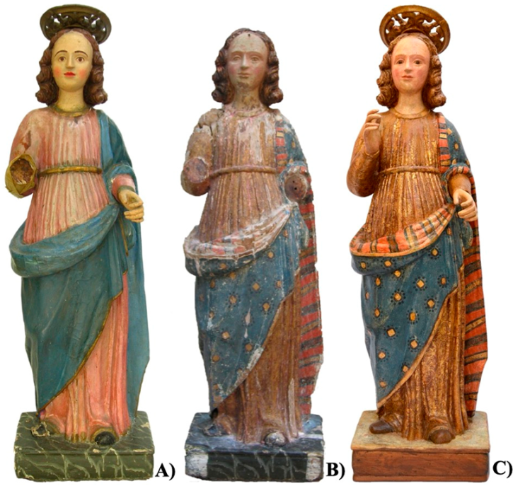
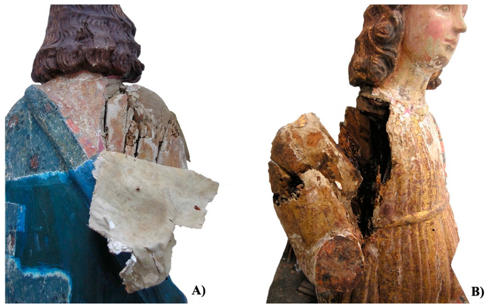
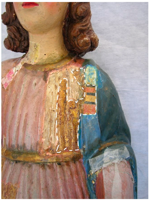
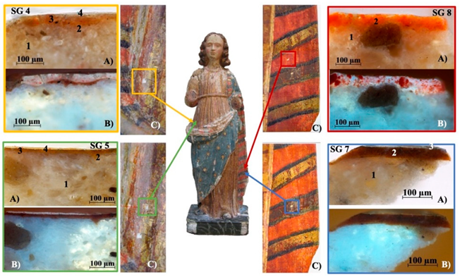
3.1. Description and Conservation State

The statue shows a young man standing and blessing with his right hand. The left hand makes the gesture of holding a thin object, probably a lily or a rod.
The sculpture (height 104 cm) rests on a wooden base having quadrangular shape (31.5 × 23 cm). A single wooden trunk carved in the round was used for the figure, hollowed to make it lighter and easier to transport, and also less susceptible to breakage due to angular shrinkage. The projecting elements, such as the forearms and hands, as well as the head, were carved separately and placed with wooden dowels. The base was also excavated by using gouges whose traces are clearly visible. The carved wooden surface was covered with a layer of plaster to define and refine the details of the sculpture, thicker than that applied on the back of the statue. The hair is covered with a thin layer of plaster and a coloured-ochre pigment. The dress and the mantle are finely carved with a realistic rich drapery.
The visual observation revealed a rather compromised state of conservation. The probable unsuitable conditions of usage and conservation environment caused obvious damages to both the wooden structure and the painted surfaces. Galleries of xylophagous insects were found mainly in the back and lower parts of the statue. A conspicuous fracture was present in correspondence of the right shoulder (Figure 2). In the back area a reinforcement based on a cotton tissue was added to cover the fracture. The detached wooden parts were glued with a commercial adhesive. The entire surface appeared covered by both coherent and incoherent deposits, accumulated over time. The surface appeared evidently repainted, as previously discussed: the decorations of the mantle and the robe were, respectively, blue and pink; the base was clearly re-painted to imitate marble. Detachment, abrasion and lacunae were detected on the shoulders, the coat lapel, the forearms, the hands and the basement.
As usual in restoration, some cleaning tests were performed to evaluate the solubility of the repainting layer (Figure 3) and, in this case, also to confirm the presence of underlying polychromy that appeared partially visible in correspondence of lacunae of the surface. As visible in the Figure 1C, after the removal of the surface layers, the pictorial decoration of the blue mantle appears characterized by a rhombus (gold on the front and silver on the back) surrounded by small dark blue dots. Particularly interesting is the turn-up of the mantle with alternate bands of red, gold, green and brown colours. The dress shows a decoration with floral motifs inserted on trilobate tiles with a Gothic style [23].
The removal of the repainting made possible to ascertain the extension of original colour layers preserved on the statue. The dress is almost intact at the front, being the largest gaps on the back; the mantle remained largely original, whereas the original flesh tones reduced to small areas.

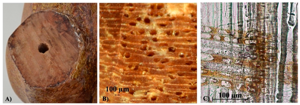
3.2. Wood Identification

Visual observation revealed a light-coloured wood of an angiosperm tree, with distinct rings, diffuse porosity and no rays visible to the naked eye (Figure 4), in the body and in the forearms. The general characteristics seem to fit poplar wood, also due to its frequent use in cultural heritage objects. However, willow also has similar macroscopic features, but differs anatomically in the characteristics of the parenchyma ray cells. For this reason, microscopic identification was carried out.
The microscopic observation on thin section confirmed the wood is heteroxylous, with diffuse porosity and elliptical solitary vessels or in short radial chains of two to four elements; the rays were monoseriate. In the radial section, all ray cells were procumbent with large, simple ray vessel pits (Figure 4C). The characteristics of the ray cells allow the wood to be identified as poplar (Populus sp.). The anatomical features to distinguish the genus Populus from the genus Salix are the procumbent parenchyma cells in the rays observable on radial section [19,24,25]. Different species of the genus Populus cannot be distinguished based on anatomical features of the wood, so identification remains at the genus level [19,24].
Poplar wood is frequently used in Italian artworks for panel paintings and statuary [26,27,28]. It has been underlined that one of the conditions for choosing wood for the support is linked to the availability of wood of a certain species in the size needed, to the aesthetical appearance and to the characteristics of workability [29,30,31,32]. The trees of the genus Populus, which grow on the Italian peninsula, have a widespread range in central and southern Europe, reaching as far as northern Africa and central Asia [33]. Poplar wood is a widely available material, today as in the past. It has good workability characteristics, a density of low entity, and is not easily attacked by xylophagous insects [29]. The undifferentiated heartwood, which is not rich in extractives, also allows for easy use in preparing the support for painting layers. Giordano [24] emphasizes the propensity to split. Therefore, the greater difficulty in obtaining well-defined details was obviated modelling the fine details, directly on the painting ground layer [31] as in the case of this statue.
The knowledge of the wood species, or the taxon closest to it, is an informative basis for any activity carried out in a philological approach, since the general characteristics of the wood are already outlined [20,32,34], as density, mechanical strength and durability can vary greatly with the species or genus. Wooden object properties and durability can be declined in the specific cases, observing the original defects of the wood and the conditions of conservation [31,35].

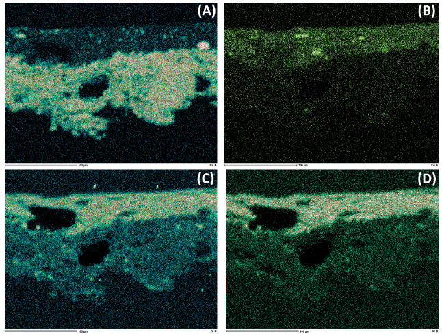
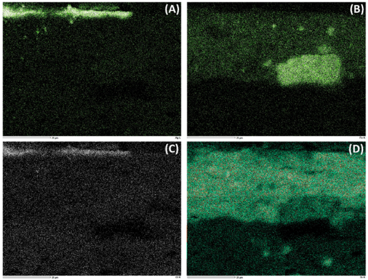
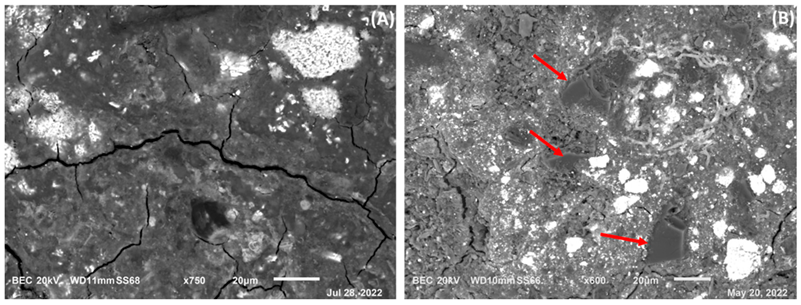
3.3. Identification of Pigments and Executive Technique

Information on pigments’ composition has been derived from X-ray fluorescence spectroscopy and SEM-EDS applied on the micro-samples. The overall results of XRF analysis are reported in the Table 2. The order of elements is reported as relative amounts from the highest to the lowest. From the data of Table 2, it can be defined a quite complete frame of the materials used for polychromy of the statue. The blue colour of the basement (sample SG1 and SG3) was obtained through copper-based pigments, probably azurite, and cobalt-based pigment, probably blue smalt, respectively. These two pigments are probably related to different layers applied in different periods to restore the surface of the devotional statue. In the other blue samples (SG9 and SG10) from the mantle, cobalt and arsenic have been detected in SG9 and only As in SG10. Cobalt associated with arsenic and traces of Ni allows to identify blue smalt [36,37]. Clearly, it cannot be excluded the presence of other blue pigments such as indigo or ultramarine blue that do not contain elements detectable by p-XRF used in the present work, as, for example, in SG10 that represents the dark blue visible on the surface before the cleaning.
Lead is revealed in all samples probably due to the use of Pb-based pigments in the priming. Pb can also be associated to red lead (minium) a pigment characterised by a red-orange hue, as visible in the detail of samples SG2 (the quadrangular base) and SG8 (turn-up of the mantle) [36].
Iron is the chromophore element of earths and ochre that are used for brown and red-brown colours. Traces of Ti can be ascribed to impurities of the earths or to non-original pigment due to re-painting [36,38]. This last assessment can be made also for Zn (found in sample SG2) another element generally associated with 19th painting materials [36,37,39].
The presence of gold confirms the gilding on the St. John garment; Cu is probably a metal associated with gold [40].
Calcium is also present in all samples, and it may be associated with the ground layer generally made of gypsum (calcium sulphate) and glue.
In order to deepen the characterisation of pigments and ground materials, some selected samples were further investigated through SEM-EDS analysis that was performed both on micro-fragments (SG2, SG3, SG9 and SG10) and cross-sections (SG6, SG7 and SG8). In the case of SEM-EDS analysis on cross-sections, a general information was obtained through backscattered electron (BSE) images of the entire sample (Figure 5).
BSE observation allows for highlighting areas with chemical elements of different atomic number in respect to the matrix made of binders, both in the ground and paintings layers. The areas having lighter response indicate the presence of chemical elements with higher atomic number. In sample SG6 (brown from the robe), the EDS analysis revealed the presence of the following main elements: Ca and S concentrated in the ground layer; Fe, Si and Al concentrated in the painting layer (see Figure 6 for the chemical maps of the main detected elements). This result confirms the use of red earth for the painting layer and gypsum for the ground layer.
In the sample SG7 (brown, turn-up of the mantle) cross-section, there is an evident surface layer very light in BSE, suggesting the presence of chemical elements with high atomic number. SEM-EDS mapping revealed the presence of Ag as main element of the surface layer (Figure 7 and Supplementary Figure S1). From the map shown in the Figure 7C there is evidence of the presence of Cl associated with Ag. This implies the corrosion of the silver used. In fact, silver halides are common corrosion products of the metal and are often due to past restoration treatment [41]. Under this Ag layer, the brown colour is based on silicon, as main element, with presence of Fe (Figure 7B). This layer is a preparatory ground for the metal foil, and, from its composition, it can be supposed to have been made of bolus. The presence of Pb in the sample can be associated with the use of lead-based pigments added to the ground layer as siccative for oil.
Lastly, SG8 (orange, turn-up of the mantle) cross-section was examined with SEM-EDS. The chemical mapping shows the presence of Pb, as the main element, in the painting layer, whose detail is highlighted in the Figure 5D. This result confirms the XRF analysis and allows for establishing that the orange-red pigment used for the St. John mantle is minium. The ground layer is made of gypsum due to the presence, also in this sample cross-section, of Ca and S.
SEM-EDS was applied to micro-fragments of blue and red-orange colours, specifically SG2, SG3, SG9 and SG10, to better understand the composition of the pigments used for obtaining these hues (Figure 8). In the sample SG2, SEM-EDS confirmed the main elements detected by XRF: Pb (main element), Ca and As, this last one probably associated with traces of blue smalt, or more probably with residues of disinfection treatment made in the past on the statue (Supplementary Figure S2). This last hypothesis seems to be confirmed by the presence of As in several examined points (not only the ones showed in the paper and in the supplementary material) [42]. The white areas in BSE image of sample SG2 are rich of lead and are probably grains of red lead (Figure 8A and Supplementary Figure S3). In the samples SG3 and SG9, the presence of Co and As, combined with high counts of Si in the examined points of the surface of the micro-fragments, confirmed the use of blue smalt (See Supplementary Figures S4 and S5 and Figure 8B where the grey fragments exhibit the typical conchoidal fracture and thin, sharp edges of glass splinters characteristic of smalt [43], p. 115). In sample SG10, SEM-EDS revealed several elements in the examined points. The main are: Pb, Si, Ca and As with other minor elements, such as Cl, Na, K, Mg, Al, P and Zr (Supplementary Figures S6 and S7). The presence of As associated with Si suggests smalt as pigment in the probable original layer. The absence of other chemical elements that could be responsible of the blue colour suggest that the dark blue on the surface may be indigo.
The presence of Pb in the samples SG3, SG9 and SG10 is due to the addition of lead white to the blue pigment.
Another observation derived from the SEM-EDS is the presence, on the surface of sample SG10 and partially in SG9, of fibrous material that appears bright in BSE images (Supplementary Figures S5–S7). In these points the EDS micro-chemical analysis detected Pb associated with smaller quantities of other elements. It can be supposed that this morphology of the surface is due to past restoration treatments such as aggressive cleaning [44].
The morphological analysis of the layers pointed out in the cross sections (Figure 9) identifies the executive technique used by skilled workers to obtain colour schemes and metal leaf decorations [45,46]. In all examined samples, it was observed a ground layer based on gypsum, as highlighted by SEM-EDS analysis and mapping, and sparse mineral aggregates (Figure 9 layer 1 in A and B), added to a protein substance identified by the pink colour scattered over the entire layer following treatment with fuchsin (Figure 10(A,A1). The same test was performed on a sample from the orange area of the turn-up of the mantle (SG8), and on one characterized by the gilding of the robe (SG5). The histochemical test performed is based on the electrostatic interaction between the acid fuchsin dye and the specific functional groups of the proteins present in the sample layers [22]. The absence of colouring following treatment with fuchsin in the orange painting layer of the turn-up of the mantle (SG8) suggests that the binder used is not of a protein nature.
The gilding of the robe is characterized by a layer of bolus with a fine grain size (Figure 9, layer 2, samples SG4 and SG5) on which the gold leaf rests (visible only in the sample SG5, layer 3 in Figure 9) giving it a warm hue. Above, a layer of intense fluorescence is identified. It is characterized by grains with a purple colour that can be linked at organic dye. The dark pink colour assumed by the ground layer after the fuchsin test suggests the use of a protein binder (Figure 10(B,B1)).
The layers identified in the gilding reflect the methodology adopted by the skilled workers for the realization of the “graffio” technique as defined by Cennino Cennini [47] in the fourteenth century (Chapters CXLI and CXLII) or “estofado de oro” in the seventeenth century by Pacheco [48]. The technique consists of applying an egg tempera paint layer on the gold leaf with the precaution to avoid the moving off the brush from the surface to prevent an excessive roughness. The surface was then scratched with a pointed object until the underlying gilding was discovered according to the desired pattern. This technique is commonly used also in Sardinia to imitate precious fabrics. In the archive documents on the gilding and polychrome decoration of the sculptures, it is referred as «indoratura, graffiatura, coloritura» or by defining the method of execution, which consisted in the removal of part of the colour with a tip to reveal the underlying gold [2,45,46,47,48,49]. The metal decoration technique also characterizes the brown parts of the turn-up of the mantle of which the SG7 sample is representative. In fact, the cross section shows an oil/resin layer on the ground with sparse pigment grains (Figure 9, layer 2 of the SG7 sample) which could be associated to the medium used for adhering the silver foil (see SEM-EDS mapping Figure 7 and Supplementary Figure S1), now lost. Also in this case, the reference to the sources confirms such a technique; in fact, in the Libro dell’arte by Cennino Cennini (Cap CLI) [47], the author recommends the preparation of an oil/resin mixture to attach the metal foil that had to decorate the garments.
The presence of organics was confirmed by Fourier transform infrared spectroscopy performed on samples SG2, SG3, SG4, SG6, SG7, SG8 and SG10 where the available micro-fragment residues were enough for obtaining significant IR spectra. The spectrum of sample SG8 is reported in the Figure 11 whereas those of the other ones are shown in the Supplementary Materials (Figures S8–S13).
From the IR spectra, it can be derived the evident presence of protein glue with the characteristic signatures at 1659 cm−1 (C=O stretching), 1546 cm−1 (C-N-H bending) and 1448–1406 cm−1 (C-H bending). These signatures are in all recorded spectra (Figures S4–S9) confirming the use of protein glue in the ground layer together with gypsum whose main signatures are well-visible in the FTIR spectra at cm−1: 3549, 2238, 2133, 1157, 674, 615, 596 (Figure 10).
Some signatures can be associated with a lipid binder that can be supposed a siccative oil, i.e., the two signals at 2921 cm−1 and 2852 cm−1 due to C-H stretching in oils, and the weak signal at 1728 cm−1 due to C=O in esters. Other bands are covered by gypsum and glue that are the main constituent of the sample.
The presence of lipids can be supposed also in samples SG2, SG3, SG4 and SG7, where similar bands can be observed (Figures S7–S9 and S11).
The presence in the church of a statue representing Our Lady of Sorrows, disfigured by interventions to allow dressing with finely embroidered clothes, also drew attention to the statue of St. John whose diagnostic tests are reported here. The hieratic figure of a young man in rich robes was not entirely consonant with a young Saint John. The documents of the Parish Archive of Noragugume allow us to hypothesize that the statue is the representation of the archangel Gabriel, rather than of St. John. The documents, in fact, report a group of statues representing the Annunciation to Mary and the celebration, every 25th of March from the year 1880 to 1885, of the feast of the Annunciation. However, from the year 1886, only the cult of Our Lady of Sorrows is documented, during the rites of the Passion. On the back of the statue venerated as St. John, no holes, that could accommodate the wings, were found. Perhaps the artist was inspired by the description of the apocryphal Gospels in which the Angel has human features: “[...] while working the purple with her fingers, a young man of inexpressible beauty entered her” [Pseudo Matthew 9,1—2].

4. Conclusions

The statue, the object of the present paper, has a deep devotional meaning as young St. John. Therefore, the restoration work was carried out in order to: (i) restore an image recognizable to the faithful, (ii) while also recover the original aspects and (iii) pay attention to the preservation of the original materials.
Similar to many other artistic Italian artifacts, the statue was carved from poplar, and the identification of the taxon remains at the genus level. A philological approach needs to know the botanical taxon belonging the artifacts. In addition, the knowledge of the taxon closest to species might be useful for creating a register of the woods used for artistic objects to better understand the artists themselves and their production.
The attention to the details of the robe finds a precise choice in the implementation of the decoration layers to achieve precise final chromatic effects. The palette used for the realization of the wooden sculpture of St. John is made up of pigments based on lead and iron and blue of different nature certainly referred to different phases of the polychromy: smalt was probably the original blue used by the artist and copper-based pigment seems applied in a subsequent layer. Both “guazzo” and “graffito” techniques for the application of the gold and silver leaf were highlighted. The analysis combined with the close observation of the surfaces by restorers helped in the treatment choices during the intervention allowing to show, at the end of the intervention, the probable original appearance of the statue polychromy and asset.
The representation of the young man in rich robe using fine pictorial techniques and materials, and the documents in the parish archives on the feast of the Annunciation, solemnly celebrated until 1855, help to clarify and resolve the iconographic inconsistency highlighted at a first sight.

Supplementary Materials

The following supporting information can be downloaded at: https://www.mdpi.com/article/10.3390/heritage5030129/s1, Table S1: Micro samples images and points of sampling. Figure S1: Chemical map of sample SG7 section (brown, turn-up of the mantle). Figure S2: Sample SG2 (red from the quadrangular shaped base), magnification 750×; point analysis with the chemical ele-ments found. Figure S3: Sample SG2 (red from the quadrangular shaped base), magnification 750×; point analysis (very bright area) with the chemical elements found. Figure S4: Sample SG3 (blue from the quadrangular shaped base), magnification 1500×; point analysis with the chemical elements found. Figure S5: Sample SG9 (blue from the mantle), magnification 600×; point analy-sis with the chemical elements found. Figure S6: Sample SG10 (blue from the mantle’s embroi-dery), magnification 750×; point analysis with the chemical elements found. Figure S7: Sample SG10 (blue from the mantle’s embroidery), magnification 1500×; point analysis with the chemi-cal elements found. Figure S8: FTIR spectrum of sample SG2 (red from the quadrangular shaped base) in DRIFT modality. Figure S9: FTIR spectrum of sample SG3 (blue from the quadrangular shaped base) in DRIFT modality. Figure S10: FTIR spectrum of sample SG4 (orange from the robe mantle) in DRIFT modality. Figure S11: FTIR spectrum of sample SG6 (brown from the robe) in DRIFT modality. Figure S12: FTIR spectrum of sample SG7 (brown, turn-up of the man-tle) in DRIFT modality. Figure S13: FTIR spectrum of sample SG10 (blue from the mantle’s em-broidery) in DRIFT modality.

Author Contributions

Conceptualization, A.L.M. and C.P. methodology, A.L.M., G.A., G.S., A.R.T. and C.P.; validation, A.L.M., G.A. and C.P.; formal analysis, A.L.M.; investigation, A.L.M., G.A., G.S., A.R.T. and C.P.; resources, A.L.M. and C.P.; data curation, A.L.M., G.A., A.R.T. and C.P.; writing—original draft preparation, A.L.M., G.A. and C.P.; writing—review and editing, A.L.M., G.A. and C.P.; visualization, A.L.M., G.A., A.R.T. and C.P.; supervision, A.L.M.; project administration, A.L.M.; funding acquisition, A.L.M. and C.P. All authors have read and agreed to the published version of the manuscript.

Funding

This research received no external funding.

Institutional Review Board Statement

Not applicable.

Informed Consent Statement

Not applicable.

Data Availability Statement

Not applicable.

Acknowledgments

This work was partially supported by the individual financial support to the basic research activities (FABR 2017 by Law 232/2016 to A. Lo Monaco and C. Pelosi). This research was in part carried out within the framework of the MIUR (Italian Ministry for Education, University and Research) initiative “Departments of Excellence” (Law 232/2016), WP 4 (Angela Lo Monaco), which financed the Department of Agriculture and Forest Science at the University of Tuscia.

Conflicts of Interest

The authors declare no conflict of interest.

References

  1. Masala, C. Il culto di Nostra Signora d’Itria in Sardegna. La storia, le tradizioni, le località; Aìsara: Cagliari, Italy, 2008; pp. 1–696. ISBN 886104025X. (In Italian) [Google Scholar]
  2. Porcella, M.F. Iconografia e culto di Nostra Signora D’Itria nella Sardegna Spagnola. ArcheoArte 2012, 1, 687–701. [Google Scholar]
  3. Scano, M.G. Pittura e Scultura del ‘600 e del ‘700; Ilisso: Nuoro, Italy, 1991; ISBN 88-85098-13-4. (In Italian) [Google Scholar]
  4. Artoni, P. Il Santuario della Beata Vergine delle Grazie: Inedite carte d’archivio per la storia dell’impalcato ligneo. In Accademia Nazionale Virgiliana di Scienze Lettere e Arti, Atti e memorie; Nuova Serie LXXIII: Mantova, Italy, 2005; pp. 27–80. (In Italian) [Google Scholar]
  5. Agresti, G.; Castorina, R.; Genco, G.; Giagnacovo, C.; Lo Monaco, A.; Pelosi, C. Wood of chestnut in Cultural Heritage. Acta Hortic. 2010, 866, 51–57. [Google Scholar] [CrossRef]
  6. Di Lodovico, D. Revising Devotion: The Role of Wooden Sculptures in Affecting Painting and Devotion in the Late Medieval Period in Italy (XII-XV century). Ph.D. Thesis, University of Washington, Seattle, WA, USA, 2016; 394p. [Google Scholar]
  7. Lo Monaco, A.; Giagnacovo, C.; Falcucci, C.; Pelosi, C. The triptych of the Holy Saviour in the Tivoli cathedral: Diagnosis, conservation and religious requirements. Eur. J. Sci. Theol. 2015, 11, 73–84. [Google Scholar]
  8. Lo Monaco, A.; Micheli, M.; Orellana, J.A.; Schirone, A. Consagrada imagen de Jesus Nazareno of Sonsonate. A xylological study of the wooden sculpture support. Eur. J. Sci. Theol. 2017, 13, 41–49. [Google Scholar]
  9. Lo Monaco, A.; Marabelli, M.; Pelosi, C.; Salvo, M. The Altar Machine in the Church Mother of Gangi (Palermo, Italy). Interpretation of the past uses, scientific investigation and preservation challenge. Chem. Cent. J. 2012, 6, 47. [Google Scholar] [CrossRef]
  10. Avanzati, D.; Saccuman, R.; Agresti, G.; Faieta, R.; Caliano, E.; Pelosi, C.; Lo Monaco, A. Multidisciplinary approach for the conservation of the Casoli tryptich. Eur. J. Sci. Theol. 2020, 16, 207–216. [Google Scholar]
  11. Kumar, S.V.; Singh, M.; Wagh, S.W.; Mahajan, N.E. Polychrome Sculptures of St. Francis of Assisi Church, Old Goa: A challenge in scientific conservation. Int. J. Conserv. Sci. 2015, 6, 465–472. [Google Scholar]
  12. Pelosi, C.; Lo Monaco, A.; Bernabei, M.; Agresti, G.; Colantonio, C.; Perri, A.; Comelli, D.; Valentini, G.; Manzoni, C. Beyond the visible: The Viterbo Crucifixion panel painting attributed to Michelangelo Buonarroti. Microchem. J. 2020, 154, 1046368. [Google Scholar] [CrossRef]
  13. Paoletti, J.T. Wooden Sculpture in Italy as Sacral Presence. Artibus Hist. 1992, 13, 85–100. [Google Scholar] [CrossRef]
  14. Auffret, S.; Nikolaus, S.B. Cleaning of Wooden Gilded Surfaces an Experts Meeting Organized by the Getty Conservation Institute, 12–14 March 2018; Getty Conservation Institute: Los Angeles, CA, USA, 2019. [Google Scholar]
  15. Pelosi, C.; Balletti, F.; Agresti, G.; Lo Monaco, A. Diagnostic investigation through minimal sampling for religious artworks knowlwdge. Eur. J. Sci. Theol. 2019, 15, 223–235. [Google Scholar]
  16. Burgio, L.; Gregory, T. Protocol for the Analysis of Cross-Sections from Gilded Surfaces. Heritage 2021, 4, 2416–2430. [Google Scholar] [CrossRef]
  17. Franquelo, M.L.; Duran, A.; Castaing, J.; Arquillo, D.; Perez-Rodriguez, J.J. XRF, μ-XRD and μ-spectroscopic techniques for revealing the composition and structure of paint layers on polychrome sculptures after multiple restorations. Talanta 2012, 89, 462–469. [Google Scholar] [CrossRef] [PubMed]
  18. Nardi Berti, R. Contributi scientifico-pratici per una migliore conoscenza ed utilizzazione del legno. In La Struttura Anatomica del Legno ed il Riconoscimento dei Legnami Italiani di Più Corrente Impiego; CNR-Istituto del Legno: Firenze, Italy, 1993; p. 155. (In Italian) [Google Scholar]
  19. Schweingruber, F.H. Microscopic Wood Anatomy: Structural Variability of Stems and Twigs in Recent and Subfossil Woods from Central Europe; Swiss Federal Institute for Forest, Snow and Landscape Research: Birmensdorf, Switzerland, 1990; p. 226. [Google Scholar]
  20. Lo Monaco, A.; Balletti, F.; Pelosi, C. Wood in Cultural Heritage. Properties and conservation of historical wooden artefacts. Eur. J. Sci. Theol. 2018, 14, 161–171. [Google Scholar]
  21. Wheeler, E.A.; Baas, P.; Gasson, P.E. IAWA List of microscopic features for Hardwood identification. IAWA Bullettin. 1989, 10, 219–332. [Google Scholar] [CrossRef]
  22. Sandu, I.C.A.; Schäfer, S.; Magrini, D.; Bracci, S.; Roque, C.A. Cross-Section and Staining-Based Techniques for Investigating Organic Materials in Painted and Polychrome Works of Art: A Review. Microsc. Microanal. 2012, 18, 860–875. [Google Scholar]
  23. Pasolini, A. Contributo allo studio della statuaria in ‘estofado de oro’. In “Oltre Longhi”: Ai confini dell’Arte Scritti per gli ottant’anni di Francesco Abbate; Cleopazzo, N., Panarello, M., Eds.; Centro Studi sulla civiltà artistica dell’Italia meridionale “Giovanni Previtali”: Roccagloriosa (SA), Italy, 2020; pp. 85–92. (In Italian) [Google Scholar]
  24. Giordano, G. Tecnologia del legno. In Vol I–La Materia Prima, Vol III-I legnami del Mondo; UTET: Torino, Italy, 1981. (In Italian) [Google Scholar]
  25. Jacquiot, C.; Trenard, Y.; Dirol, D. Atlas D’anatomie des Bois des Angiospermes; CTDB and CNRS: Paris, France, 1973. (In French) [Google Scholar]
  26. Marette, J. Connaissance des Primitifs par L’étude du Bois du XII au XVI Siècle; Editions A. & J. Picard: Paris, France, 1961; pp. 1–383. (In French) [Google Scholar]
  27. Fidanza, G. Caratteristiche tecnologiche e formali delle specie legnose: Una verifica su statue e intagli di età moderna. In Statue di Legno. Caratteristiche Tecnologiche e Formali delle Specie Legnose; Istituto Poligrafico e Zecca dello Stato: Roma, Italy, 2008; pp. 33–57. (In Italian) [Google Scholar]
  28. Macchioni, N.; Fachechi, G.M.; Lazzeri, S.; Sozzi, L. Timber species and provenances of wooden sculptures. Information from the collections of the National Museum of “Palazzo di Venezia” in Rome. J. Cult. Herit. 2015, 16, 57–64.
  29. Borghini, G.; Massafra, M.G. Legni da Ebanisteria; De Luca Editori d’Arte: Roma, Italy, 2002. (In Italian) [Google Scholar]
  30. Mazzanti, P.; Togni, M.; Uzielli, L. Drying shrinkage and mechanical properties of poplar wood (Populus alba L.) across the grain. J. Cult. Herit. 2012, 13, S85–S89. [Google Scholar] [CrossRef]
  31. Macchioni, N.; Lazzeri, S.; Sozzi, L.; Vitiello, R. Wooden sculptures from XVII and XVIII centuries in the region of Asti (Italy): Scientific identification of the species. Int. J. Conserv. Sci. 2011, 2, 251–260. [Google Scholar]
  32. Bernabei, M.; Capra, N.; Giovannini, L.; Tomasi, M.L. Flying on wood and fabric: The tree species used for the construction of the Ansaldo A.1 “Balilla” biplane (1918). Archaeometry 2021, 64, 256–264. [Google Scholar] [CrossRef]
  33. Gellini, R.; Grossoni, P. Botanica Forestale, Vol. 2: Angiosperme; CEDAM: Firenze, Italy, 1997. (In Italian) [Google Scholar]
  34. Corona, E. Xylology: Technical, historical, philological involvements. Ann.-Accad. Ital. Sci. For. 1991, 10, 211–236. (In Italian) [Google Scholar]
  35. Bernabei, M.; Macchioni, N.; Pizzo, B.; Sozzi, L.; Lazzeri, S.; Fiorentino, L.; Pecoraro, E.; Quarta, G.; Calcagnile, G. The wooden foundations of Rialto Bridge (Ponte di Rialto) in Venice: Technological characterisation and dating. J. Cult. Herit. 2019, 36, 85–93. [Google Scholar] [CrossRef]
  36. Larsen, R.; Coluzzi, N.; Cosentino, A. Free XRF Spectroscopy Database of Pigments Checker. Int. J. Conserv. Sci. 2016, 7, 659–668. [Google Scholar]
  37. Hochleitnera, B.; Desnica, V.; Mantlera, M.; Schreinerb, M. Historical pigments: A collection analyzed with X-ray diffraction analysis and X-ray fluorescence analysis in order to create a database. Spectrochim. Acta B 2003, 58, 641–649. [Google Scholar] [CrossRef]
  38. Helwig, K. Iron oxide pigments: Natural and synthetic. In Artists’ Pigments, a Handbook of their History and Characteristics; Berrie, B.H., Ed.; Archetype Publication: London, UK, 2007; Volume 4, pp. 39–109. [Google Scholar]
  39. Croll, S. Oview of Developments in the Paint Industry since 1930. In Modern Paints, Uncovered Proceeding of Modern Paints Uncovered Symposium, London, United Kingdom, 16–19 May 2006; Learner, T.J.S., Smihen, P., Kruger, J.W., Schillig, M.R., Eds.; The Getty Conservation Institute: Los Angeles, CA, USA, 2006; pp. 17–29. [Google Scholar]
  40. Lalli, C.G.; Innocenti, F. La doratura nelle tecniche artistiche. OPD Restauro 2016, 48, 340–348. (In Italian) [Google Scholar]
  41. Costa, V. The deterioration of silver alloys and some aspects of their conservation. Stud. Conserv. 2013, 46, 18–34. [Google Scholar] [CrossRef]
  42. Unger, A. Decontamination and “deconsolidation” of historical wood preservatives and wood consolidants in cultural heritage. J. Cult. Herit. 2012, 13S, S196–S202. [Google Scholar] [CrossRef]
  43. Mühlethaler, B.; Thissen, J.S. Artists’ Pigments, a Handbook of Their History and Characteristics; Roy, A., Ed.; National Gallery of Art: Washington, DC, USA, 1993; Volume 2, pp. 113–130. [Google Scholar]
  44. Burnstock, A.; Learne, T. Changes in the surface characteristics of artificially aged mastic varnishes after cleaning using alkaline reagents. Stud. Conserv. 1992, 37, 165–184. [Google Scholar]
  45. Sandu, I.C.A.; de Sa, M.H.; Pereirac, M.C. Ancient ‘gilded’ art objects from European cultural heritage: A review on different scales of characterization. Surf. Interface Anal. 2011, 43, 1134–1151. [Google Scholar] [CrossRef]
  46. Sandu, I.C.A.; Afonso, L.U.; Murta, E.; De Sa, M.H. Gilding techniques in religious art between East and West 14th-18th centuries. Int. J. Conserv. Sci. 2010, 1, 47–62. [Google Scholar]
  47. Cennini, C. The Craftsman’s Handbook—The Italian “Il libro dell’arte”; Thompson, D.V., Translator; Dover Publications: New York, NY, USA, 1954. [Google Scholar]
  48. Cerasuolo, A. Estofado e policromie: Osservazioni sulla tecnica attraverso la testimonianza di Francisco Pacheco. Boll. D’arte 2011, 8, 147–160. (In Italian) [Google Scholar]
  49. Scano, M.G.; Messina, M.G. Estofado de Oro: La Statuaria Lignea Nella Sardegna Spagnola, Exhibition Catalogue (Cagliari-Sassari, Italy, 2001–2002); Exmà: Cagliari, Italy, 2001. (In Italian) [Google Scholar]
Figure 1. St. John, polychrome wooden statue. (A) Before restoration; (B) during restoration, and (C) after restoration.
Heritage 05 00129 g001
Figure 2. St. John, fracture at the right shoulder. (A) Before restoration and (B) during restoration.
Heritage 05 00129 g002
Figure 3. St. John, cleaning test made it possible to highlight precious details and brilliant colours of the original painting.
Heritage 05 00129 g003
Figure 4. St. John, wood identification. (A) Arm, sampling point. (B) Cross section under stereomicroscope: diffuse-porous heteroxylous wood, with small vessels, uniformly distributed in the growth ring, monoseriate rays. (C) Microscopic radial section: homocellular ray.
Heritage 05 00129 g004
Figure 5. BSE images of cross-sections from samples SG6, brown from the robe (A) at 430× (scale bar 100 µm), SG7, brown, turn-up of the mantle (B) at 1700× (scale bar 20 µm), and SG8, orange, turn-up of the mantle (C,D), this last shown at two different magnifications, i.e., 140× and 800×.
Heritage 05 00129 g005
Figure 6. Chemical mapping of the main elements detected through SEM-EDS analysis on SG6 sample (brown from the robe) (scale bar 100 µm). Magnification 430×. (A) map of Ca; (B) map of Fe; (C) map of Si; and (D) map of Al.
Heritage 05 00129 g006
Figure 7. Chemical mapping of the main elements detected through SEM-EDS analysis on SG7 (brown, turn-up of the mantle) cross-section (scale bar 20 µm). Magnification 1700×. (A) map of Ag; (B) map of Fe; (C) map of Cl; and (D) map of Si.
Heritage 05 00129 g007
Figure 8. BSE images of samples SG2 (A) at 750× and SG9 (B) at 600× (scale bar 20 µm). The red arrows in (B) indicate grains of smalt with the typical sharp edges of glass splinters.
Heritage 05 00129 g008
Figure 9. Samples SG4, SG5, SG8 and SG7. The figure shows the images under the polarizing microscope, 200×, reflected light (A) and ultraviolet radiation (B), the detail of the sampling area (C) and the location on the wooden statue of St. John after removal the layers of later painting.
Heritage 05 00129 g009
Figure 10. Cross section fuchsin treatment, 100×. (A) Samples SG5, gilding from the robe; (A1) Samples SG5 after fuchsin treatment; (B) Samples SG8, turn-up of the mantle; (B1) Samples SG8 after fuchsin treatment.
Heritage 05 00129 g010
Figure 11. FTIR spectrum of sample SG8, orange, turn-up of the mantle, in DRIFT modality.
Heritage 05 00129 g011
Table 1. List and description of the samples taken from the wooden statue of St. John and scientific investigation performed.
SampleDescriptionScientific Investigations
SG1Blue from the quadrangular shaped baseXRF
SG2Red from the quadrangular shaped baseXRF, FTIR
SG3Blue from the quadrangular shaped baseXRF, FTIR, SEM-EDS
SG4Orange from the robe mantleXRF, cross section, FTIR
SG5Gilding from the robeXRF, cross section
SG6Brown from the robeXRF, cross section, FTIR, SEM-EDS
SG7Brown, turn-up of the mantleXRF, cross section, FTIR, SEM-EDS
SG8Orange, turn-up of the mantleXRF, cross section, FTIR, SEM-EDS
SG9Blue from the mantleXRF, SEM-EDS
SG10Blue from the mantle’s embroideryXRF, FTIR, SEM-EDS
SG11Wood, arm, not paintedOptic Microscope
Table 2. Results of XRF analysis reported as decreasing order of the detected elements.
SampleDetected ElementsNote on Probable Pigment
SG1Cu (main), Pb, Fe, CaCu-based pigment
SG2Pb (main), Ca, Zn, K, As, Cu (tr)Pb-based red, probably minium
SG3Pb (main), Ca, Fe, Co, Ni, Ti, Cl, S (tr)Probable smalt blue with lead white
SG4Ca (main), Pb, Fe, Cl, Cu, Ti, Au (tr)Gold on priming
SG5Fe (main), Pb, Ca, Au, Ti (tr)Gold on priming
SG6Fe (main), Ti, CaIron-based pigments
SG7Ca (main), Fe, Pb, Ti, CuIron-based pigments with traces of probable repainting
SG8Pb (main), Ca, Ti (tr)Pb-based pigment probably minium
SG9Pb (main), Co, As, Ni, FeBlue smalt and lead white
SG10Pb (main), Ca, Fe, AsPb based pigment, traces of smalt
Publisher’s Note: MDPI stays neutral with regard to jurisdictional claims in published maps and institutional affiliations.

