Review

Antihypertensives’ Rock around the Clock

Department of Pharmaceutical Technology, Faculty of Pharmacy, University of Sarajevo, Zmaja od Bosne 8, 71000 Sarajevo, Bosnia and Herzegovina
*
Author to whom correspondence should be addressed.
J 2021, 4(1), 62-81; https://doi.org/10.3390/j4010005
Submission received: 4 March 2021 / Revised: 12 March 2021 / Accepted: 16 March 2021 / Published: 18 March 2021
(This article belongs to the Section Medicine & Pharmacology)

Abstract

:
Although homeostasis is a commonly accepted concept, there is incontrovertible evidence that biological processes and functions are variable and that variability occurs in cycles. In order to explain and understand dysregulation, which has not been embraced by homeostatic principles, the allostatic model has emerged as the first serious challenge to homeostasis, going beyond its homeostatic roots. Circadian rhythm is the predominant variation in the body, and it is a pattern according to which many physiological and pathological events occur. As there is strong experimental and clinical evidence that blood pressure fluctuations undergo circadian rhythm, there is equally strong evidence that targeted time therapy for hypertension provides a better outcome of the disease. The research has gone even further throughout the development and approval process for the use of pulsatile drug release systems, which can be considered as an option for an even more convenient dosage regimen of the medicines needed.

1. Introduction

There is a well-known and generally accepted assumption that homeostasis is a basic concept in biology and medicine. However, over the past fifty years, it has been clearly proven that biological processes and functions are not constant but are characterized by variability. This variability can be predicted as it occurs in cycles, which are of endogenous origin and occur even without external stimuli [1].
Many researchers mistakenly confused the homeostatic model with the one that defines and maintains the constant conditions in the body. This misunderstanding may be due to the terms used by Bernard (1870) and Cannon (1929), the creators of the homeostasis model, who used the French word “fixité” which was wrongly translated as consistency rather than stability. This notion, conceived as the “wisdom of the body” led many scientists to believe that homeostasis is the model for maintaining constancy in the body, which can be misleading up to a certain point. The purpose of homeostasis is to explain the maintenance of a steady-state in the organism by maintaining vital functions. On the other hand, the main disadvantage of a homeostatic model is the lack of prediction and elaboration of the ability to learn and anticipate and accordingly respond to stressful states [2]. Therefore, the homeostatic model had to be modified and adapted to explain as many situations as the body could be confronted with. In view of these assumptions, the allostatic model was introduced by Sterling and Eyer, according to which “stability is achieved through change” [3]. Allostasis is now accepted by many scientists who have recognized its undeniable advantages over homeostasis [2]. This approach to a case goes hand in hand with the maxim that “the only constant in life is change” (Heraclitus). According to Sterling and Eyer, the main difference between homeostasis and allostasis is in “waiting” as opposed to “preventing”. Homeostasis waits for errors and then reacts and corrects them, while allostasis waits for nothing. Allostasis uses either intrinsic or learned prior knowledge to prevent and reduce errors [3].
It is well known that under the effect of stressors and in anticipation of stressors, also known as allostatic challenges, the activation of the sympathetic adrenal system is strongly related to our urge to survive (Figure 1). The task of this system is to adjust blood pressure, glucose levels, body temperature, etc., so that their level responds to emergencies [4]. In other words, when people are confronted with emergencies or stress, a so-called “fight or flight response” occurs, which prepares the body to become active. People become physically and mentally alert when their heart and breathing rate rise, enabling them to act in situations that require a rapid response.
However, apart from allostatic challenges, the sympathetic adrenal system is activated in everyday activities, such as eating, speaking, exercise, etc. [4]. The allostatic response is shown in another way, apart from activating the body’s reaction to adapt to changes. This other way is shown by shutting down this reaction when the threat is over [5,6]. This means that the allostatic reaction is characterized by resilience, that is, the body is able to respond to various external challenges and to efficiently terminate the reaction as soon as challenges are over and recover from these negative events [7].
The process described requires mediators, including cortisol, vegetative nervous system (sympathetic, parasympathetic activity), metabolic hormones, and the immune system, i.e., pro- and anti-inflammatory cytokines. These mediators regulate each other and thus form a nonlinear network. Prolonged dysregulation of allostatic mediators, either continuous or intermittent, as in the case of chronic stress can have harmful, even pathophysiological consequences, a phenomenon known as allostatic load [4,7,8]. This means that the body remains in a heightened state of arousal even though there is no immediate threat.
The allostatic load might evoke different adaptation responses (altered phenotype) due to four events (Figure 1). The first is recurrent stress. For example, an increase in blood pressure can trigger myocardial infarction in susceptible individuals. In the second type of allostatic load, there is no adaptation to repeated stressors of the same type, which leads to longer exposure to stress hormones, as was the case in some people with chronic health problems or involved in difficult relationships with family members, partners, colleagues, or others. With the third type of allostatic load (Figure 1), there is an inability to switch off allostatic reactions after the end of stress. It is speculated that allostatic load over a lifetime may cause the allostatic systems to wear out or exhaust themselves. With the fourth type of allostatic load, inadequate reactions of some allostatic systems trigger compensatory increases in others. If one system does not react satisfactorily to a stressful stimulus, the activity of other systems increases because the underactive system does not provide the usual counter-regulation. If, for example, cortisol secretion does not increase in response to stress, the secretion of inflammatory cytokines increases, which are counteracted by cortisol [5].
Expectation and worry can also favor allostatic load. Expectation participates in the reflex that prevents us from fainting when we get out of bed in the morning and is also part of worry, anxiety, and cognitive preparation for a threat. Expectation anxiety can lead to the release of mediators like corticotropin, cortisol, and epinephrine, and for this reason, persistent anxiety and expectation result in allostatic load [5]. However, at this point, we must emphasize that expectation and worry are part of the “prior knowledge” in the allostatic model. Nevertheless, one of the most important features of allostasis is prediction and perceived control. This is an ability to track events, which leads to psychobiological adaptation, and is particularly pronounced under stress. At this moment, the state of permanent expectation develops, which can lead to allostatic load and pathological arousal [11].
Since allostasis involves the achievement of stability through change, including the specific rhythms of various functions in the organism, this narrative review aimed to briefly introduce the circadian rhythm of blood pressure in health and disease and to present the proven and novel possibilities in the treatment of hypertension through the art of pulsatile drug release systems.

2. Methods

Our aim in this topic review was to give a brief introduction to the concept of allostasis and to contrast it with the outdated but very familiar concept of homeostasis. Allostasis was of interest to us because the circadian variations of numerous physiological and pathophysiological processes undergo this type of rhythmic change. Since allostatic load and overload can lead to various health disorders, including elevated blood pressure, it was of particular interest to us to present physiological and pathophysiological variations in blood pressure in order to justify the chronotherapeutic approach to the treatment of hypertension, since chronotherapy involves targeted delivery of drugs at a specific time and studies presented later in the paper support the application of chronotherapeutic principles in the treatment of hypertension. Since we are pharmaceutical technologists, our goal was to provide an insight into the formulation aspects of drug delivery systems that will be able to adapt to circadian fluctuations in blood pressure. We present the drug delivery systems with the pulsatile release that are already approved for use. In the Future Directions section, we outline novel approaches in the development of such drug delivery systems. We conclude that a chronotherapeutic approach is a promising tool in managing hypertension and that many new approaches to formulating such systems are looming on the horizon.
Therefore, we reviewed data from Medline, Embase, Science Direct, and the public digital archive PubMed, including reference texts related to the field of allostasis, circadian variations of blood pressure and hypertension, and pulsatile antihypertensives delivery systems approved for use. Regarding Section 7. Future Directions, we searched the above databases for novel, biodegradable polymer-based, and nanotechnology-based drug delivery systems for pulsatile release that had been published before March 2021.

3. Types of Rhythmic Cycles
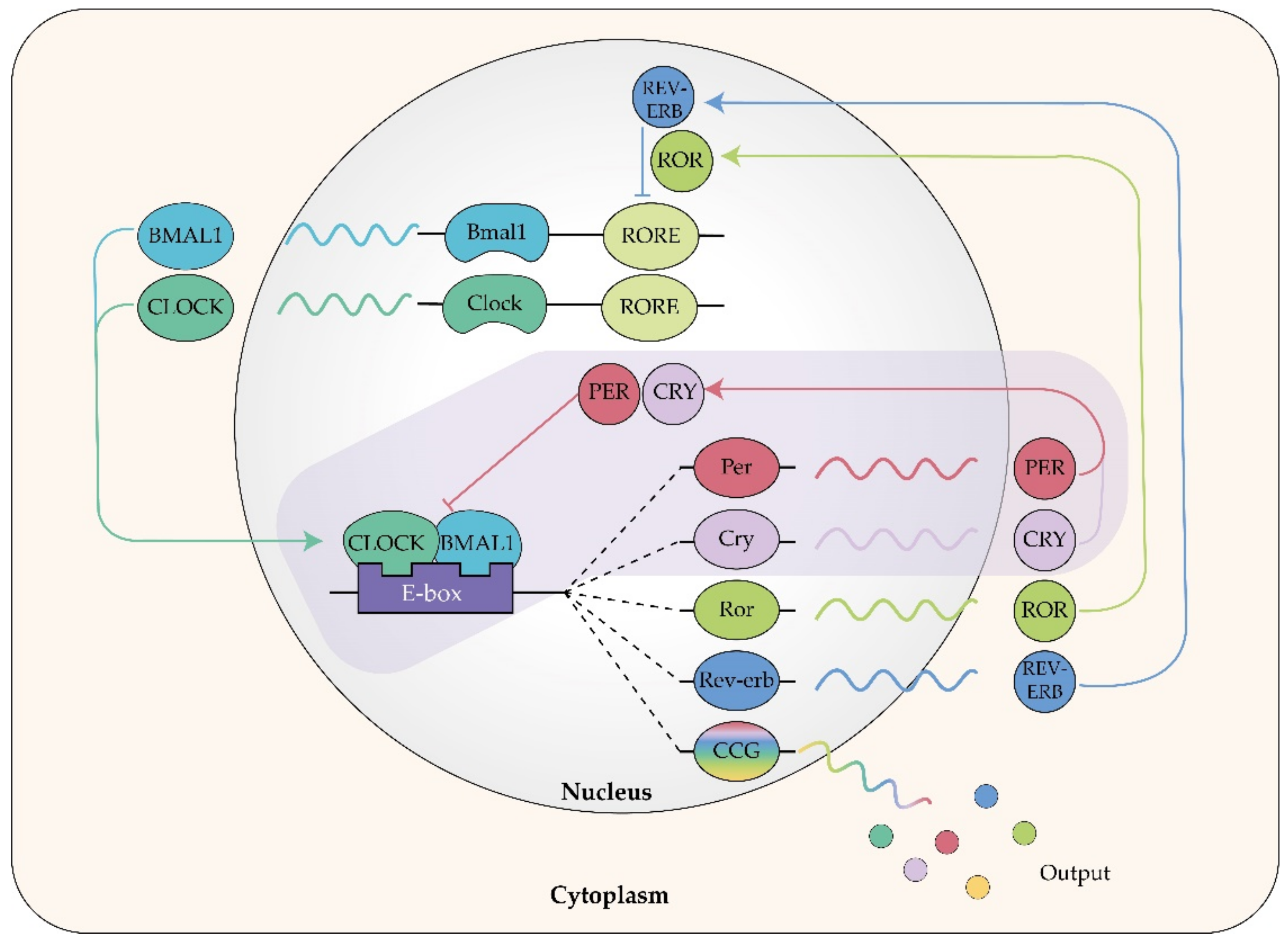
The frequency of the cycles that our body undergoes can be different. There are short cycles that vary every second (i.e., electroencephalogram) or every few seconds (breathing rhythm, heart rate). Intermediate and long cycles are characterized by frequencies ranging from a few hours to a few days, years, decades, centuries, etc. These cycles include the ultradian, circadian, and infradian cycles [12].
The ultradian cycle is characterized by a frequency of seconds, minutes, or hours (i.e., cell division, human rapid eye movement (REM)-non-REM (NREM) sleep cycles, food intake cycles) and occurs several times a day. Moszczynski and Murray [13] conducted one of the first ultradian cycle studies in humans. They studied the rhythms of the REM-NREM sleep cycle, which alternates every 90 min, and thus one has three to five of these cycles during a sleep period. Apart from this, it has been proven that the rhythmicity of sleep follows not only ultradian rhythm but also circadian rhythm [14].
It is not uncommon for biological processes to follow different rhythms. Hormones, which are the most energetically advantageous secretions in a pulsatile way, are one of these examples [15]. Glucocorticoids, for instance, are secreted under stress in an ultradian rhythm, while under basal, unstressed conditions they are secreted in a circadian rhythm [16]. The frequency of 24 h is characteristic of the circadian cycle which will be discussed in detail later. The infradian cycles have frequencies of more than 24 h and range from days to weeks, months, or years, with the example of the menstrual cycle in women [12,17].

Circadian Rhythm

The predominant variation in the body is 24-h and occurs as a result of the change between day and night. Phenomena that have this frequency are said to be subject to a circadian rhythm. The name circadian comes from the Latin words circa, which means about, and diem, which means day [18]. Above mentioned allostatic mediators show a rhythmic activity that is regulated according to the time of day, i.e., it is dependent on the sleep-wake cycle and therefore obeys circadian rhythm [19,20].
The so-called circadian clock has control over circadian rhythms. The circadian clock is a biological clock, and the most important attribute of a biological clock is temperature compensation. This means that the oscillation frequency must remain virtually unchanged over a wide temperature range [17]. The main circadian clock in mammals, which is also the biological master clock, is located in the suprachiasmatic nuclei (SCN) (lat. nuclei suprachiasmatici) of the hypothalamus and drives all rhythms in physiology and behavior [1,19]. The SCN has all the necessary characteristics of the biological clock: intrinsic rhythmicity, constant frequency, and temperature compensation. The proof that SCN is a master biological clock lies in the fact that SCN lesions terminate the circadian rhythmicity of locomotor activity, feeding, drinking, and hormonal secretion. Moreover, transplantation of SCN cells leads to a repair of the circadian rhythms in laboratory animals [17]. Circadian clocks have also been found in other parts of the brain, but also in some peripheral tissues where, together with SCN, they affect many of the systems involved in the mediation of allostasis. Therefore, disturbance of the circadian system can put the organism into a state of high allostatic load and ultimately overload [7,19].
The relationship between circadian rhythm disturbance and allostatic load can be explained by the role of the hypothalamic-pituitary-adrenal (HPA) axis in these processes. The HPA axis plays a crucial role in physiological stress response and its efficient regulation is a symbol of a “healthy” response. Maintaining the circadian rhythmicity of the HPA axis is important not only for the defense against stressors but also for the appropriate physiological regulation of glucocorticoid-sensitive target tissues throughout the body [20,21]. In case of repeated disturbance of glucocorticoid rhythms due to the inappropriate engagement of the HPA axis in the reaction to chronic or repeated stressors, physiological “wear and tear” as a synonym for allostatic load occurs [7].
Circadian clocks found in the brain and in the body, known as “peripheral clocks”, are designed to set the local time. SCN delivers synchronicity to peripheral clocks via multiple signals, including glucocorticoids, which can “reset” some peripheral clocks in the brain and body [7,19,22]. In addition to receiving inputs from SCN, peripheral clocks receive signals from feeding [23], glucocorticoids [24], temperature [25], metabolic state [26], and sleep [27,28]. The exact mechanism of interaction between these inputs and peripheral clocks is unknown.
Stokkan et al. (2001) used a transgenic rat model and demonstrated in vitro that feeding cycles can entrain the liver independently of the SCN and the light cycle. They also pointed out that it is possible that peripheral circadian clocks, such as those in the liver, can be linked to the SCN primarily through rhythmic behavior, such as feeding [23]. To support the claim that glucocorticoids can produce signals for peripheral clocks, Balsalobre et al. showed that the glucocorticoid hormone analog dexamethasone induces circadian gene expression in the liver, kidney, and heart, which leads to phase delays or phase advances throughout the 24-h cycle, while gene expression in SCN is not affected [24].
The influence of temperature on peripheral clocks was demonstrated by Morf et al. who proved that simulated body temperature cycles, but not peripheral oscillators, controlled the rhythmic expression of cold-inducible RNA-binding protein in fibroblasts [25]. On the other hand, Ramsey et al. suggested that the circadian feedback loop through nicotinamide phosphoribosyltransferase (NAMPT)-mediated NAD+ biosynthesis could work to fine-tune the daily cycles of energy storage and utilization, as NAD+ could have a cascade of effects on downstream pathways, including chromatin regulation, metabolism, and aging [26].
The effects of sleep were investigated by Yamakawa et al. and Davies et al. In brief, Yamakawa et al. showed that sleep deprivation or arousal activates cholinergic cells, which are then projected onto the circadian clock in the SCN [27]. Davies et al. identified plasma metabolites (lipids, serotonin, tryptophan, taurine), which were significantly altered during sleep deprivation, as well as 24-h variations of amino acids, biogenic amines, acylcarnitines, glycerophospholipids, and sphingolipids in the presence and absence of night’s sleep [28].
In the same way that cellular organelles are used for the spatial compilation of metabolic reactions, peripheral clocks serve for the temporal compilation of physiological functions. For example, hepatic and gastric clocks are reset by a food intake at a certain time of day, with a delay of 1 h between these two clocks. This delay enables the activation of metabolic pathways and enzymatic systems in the liver, which can then reach their peak and thus prepare for the increase in nutrient levels coming from the stomach [29].
The circadian clock does not generate a cycle that only corresponds to a solar day but must be able to align the phases of the cycle with the phases of the day. This process of optimal synchronization with the environment is called entrainment and takes place through periodic stimuli that act on the circadian clock. The literature lists periodic stimuli called “zeitgeber” [30], which would literally mean “time giver” in German. The most important “zeitgeber” is light, since light gives a photo signal for day or night, as well as for changing seasons (Figure 2).
The information about the light is transmitted to SCN neurons via intrinsically photosensitive retinal ganglion cells, which contain the pigment melanopsin [31]. The signal is transmitted through the retinohypothalamic tract (RHT) [32]. Furthermore, there is evidence that rods and cone opsins, as well as heme proteins, biliverdin, and bilirubin, may play a role in the transmission of light information to SCN [33]. The information from the retina does not contribute to the vision. The main task of “zeitgebers” is to induce phase shifts in the body rhythms, either promoting or obstructing them [34].
The main circadian clock consists of two oscillatory systems that respond to the appearance of dawn or dusk [35]. In oscillatory systems, there is a different expression pattern of specific genes called “clock genes”, as shown in Figure 2. “Clock genes” are located both in brain cells and in most peripheral tissues, including skin and mucosal cells [36].
Core circadian clock genes encode the synthesis of proteins that are omnipresent in the production and regulation of circadian rhythms. Clock proteins achieve this through interdependent transcriptional/translational feedback loops that involve the rhythmic transcription of specific “clock” genes and the interaction of the proteins they encode [37].
There are four important proteins (Figure 3) in the cytoplasm of SCN neurons: aryl hydrocarbon receptor nuclear translocator-like (ARNTL; also known as BMAL1) and circadian locomotor output cycles coat (CLOCK) which are activators, while period circadian protein homolog 1, 2, and 3 (PER1, PER2, and PER3, respectively) and cryptochrome 1 and 2 (CRY1 and CRY2, respectively) [38] are transcription inhibitors. Additional components, such as retinoic acid-related orphan receptor ROR1 and ROR2 and REV-ERBs (also known as NR1D1), form secondary feedback loops [39,40]. Circadian rhythm gene feedback maintains circadian oscillations in a single cell at transcriptional and posttranscriptional levels, and these oscillations are stimulated by the alternation of light and darkness. The entire process of gene activation and expression within the loop takes about 24 h, and transcription factors are involved that affect gene expression and trigger a series of physiological changes driven by gene oscillations, which are controlled by the clock in SCN [41,42]. Heterodimers of the transcription factors ARNTL and CLOCK bind to the regulatory elements of the promoters and enhancers (E-regions of DNA) of the PER and CRY genes and stimulate their expression and expression of other clock-controlled genes (CCGs). While the amount of PER and CRY proteins slowly increases in the cytoplasm at night, their heterodimers are formed. Phosphorylated PER-CRY heterodimers are transferred to the nucleus, where they inhibit the ARNTL-CLOCK protein complex [43]. In this way, the transcription of PER and CRY genes is reduced during the day, while the level of PER and CRY proteins is reduced due to their degradation by the ubiquitin system. PER-CRY heterodimers bind directly to the ARNTL-CLOCK complex, and since PER2 contains histone deacetylase, the structure of chromatin changes leading to interruption of transcription. Furthermore, the PER-CRY heterodimer interacts with RNA-binding proteins and helicases, which are important for stopping transcription regardless of interaction with the ARNTL-CLOCK complex. Moreover, PER-CRY heterodimers regulate the transcription of various nuclear hormone receptors [44,45].
On the other hand, the DNA-binding domains of the nuclear receptors REV-ERB are closely related to those of the ROR subfamily nuclear receptors and bind to the same target sequences (Figure 3) [46]. BMAL1-CLOCK heterodimers activate ROR and REV-ERB. Since these receptors have the same target sequences, they compete for the ROR response element (RORE), a binding site located in the promoter region of BMAL1. Depending on which of the competing protein wins, this binding leads to either activation or inhibition of BMAL1 transcription. The activation of BMAL1 transcription occurs when RORs are bound, while REV-ERBs suppress BMAL1 transcription [47,48]. This secondary feedback loop is essential for the rhythmic expression of BMAL1 [48,49].
In addition to light, food intake, activity, as well as social factors (such as an alarm clock) also act as a “zeitgeber”. Zeitgebers allow people to function optimally at certain periods of the day because the predictability of certain activities during the day allows our brain and body to prepare for them in appropriate cycles. Figure 4 summarizes the most pronounced activities of individual processes and secretions of substances in the human body.
Problems with circadian rhythm alignment can be observed in night shift workers, resulting in an increased risk of errors and accidents, reduced productivity, health risks, such as the increased risk of cancer, depression, sleep disorders, gastrointestinal, metabolic, and cardiovascular disorders, weakened immune response, and even shortened life expectancy [50]. Shift workers have been shown to very often develop glucose intolerance, diabetes, hypertension, and in some cases even cancer [51,52].
Given the increased risk of chronic disease associated with shift work, it is critical to access and understand helpful interventions that can minimize the effect of risk factors. A literature review by Neil-Sztramko et al. [53] showed that pharmacological interventions, such as the use of hypnotics do not provide better outcomes of chronic disease in shift workers. On the other hand, Neil-Sztramko et al. [53] as well as Crowther et al. [54] suggested that non-pharmacological interventions, such as fast-forward rotating shifts, timed use of bright light and light-blocking glasses, and targeted health behaviors including physical activity and nutrition, resulted in favorable outcomes [53]. Furthermore, subjective sleep quality is improved by complementary therapies such as massage, touch therapy, and transcutaneous electrical acupoint stimulation [54]. It is extremely important to maintain the synchronicity of biological clocks, which means being exposed to appropriate zeitgebers at the right time and for long enough to maintain optimal functioning and adequate sleep-wake cycles. Maintaining mood, cognitive processing, brain, and behavior function also depend heavily on appropriate biological clock function [55].

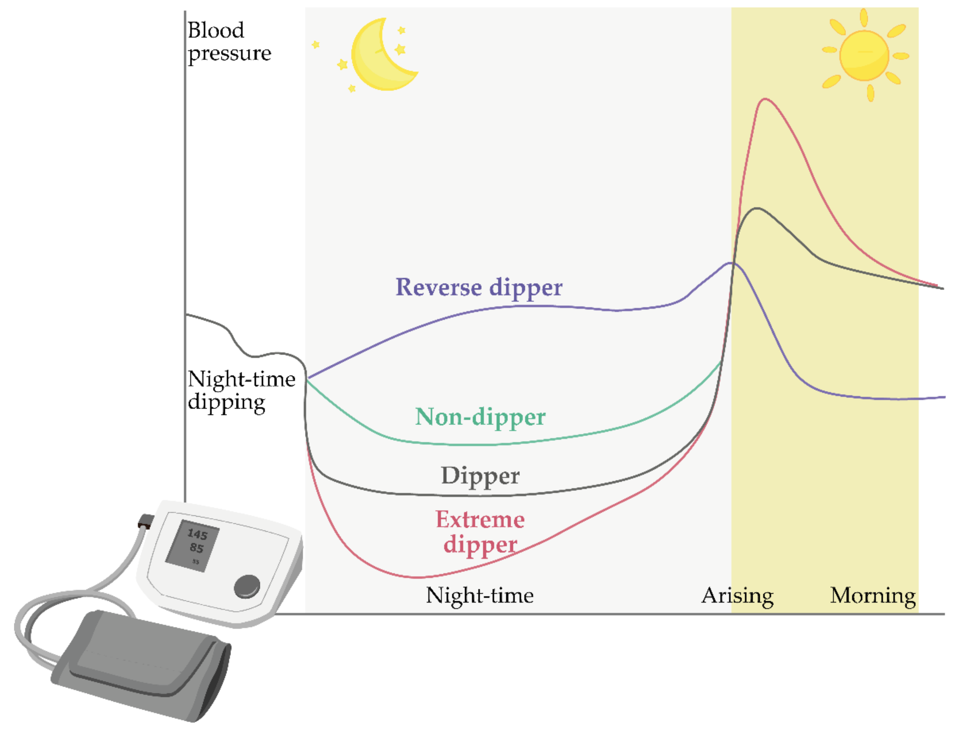
4. Circadian Blood Pressure Fluctuations

Until fifteen years ago, the conventional homeostatic assumption was valid that blood pressure and heart rate have a constant value for 24 h unless the body is exposed to exercise, stress, or other environmental influences. However, studies involving outpatient all-day blood pressure measurement [56] indicated the existence of significant fluctuations in blood pressure during the day in both normotensive and hypertensive patients [57,58,59,60].
When it was introduced, allostasis was explained by physiological fluctuations in blood pressure, as the fluctuation of blood pressure within 24 h is indeed the perfect example of allostatic control [3]. On a 24-h basis, blood pressure changes dramatically and continuously to adapt individuals to everyday environmental conditions. Therefore, there is not only one “homeostatic” blood pressure, but many stable blood pressure states [61], which is precisely the property of blood pressure that defines its hidden adaptability [62]. Blood pressure fluctuations are seamless and are influenced by temperature [63], temporary physicochemical stressors, such as work requirements and childcare [64], or when people think of emotionally charged memories [65]. Changes in blood pressure also occur in response to the intake of salt [66], alcohol, nicotine, and caffeine [67].

Models of Circadian Blood Pressure Fluctuations

With the development and application of ambulatory blood pressure measurement, the presence of significant fluctuations in blood pressure levels during the day was detected. Furthermore, it was observed that in normotensive patients, but also in hypertensive patients, both systolic and diastolic blood pressure decreases during the night [59,60,68]. Blood pressure drop values range from 10% to 20% and are estimated at an average of 15%. In relation to this phenomenon, all patients can be divided into two groups, i.e., their so-called dipping status is determined (Figure 5) [59,60,61,62].
The first group consists of patients whose blood pressure drops by more than 10% at night, and this group of patients is collectively referred to as “dippers”. On the other hand, some patients have no pressure drop at night or who have a pressure drop below 10%; these patients are referred to as “non-dippers”. More recently, the classification has been extended to include two further groups: “extreme-dippers” who have a nocturnal blood pressure drop of more than 20% and “rising/inverse dippers” in which systolic blood pressure is higher at night than during the day [69]. A schematic representation of blood pressure fluctuations in these categories is given in Figure 5.

5. Chronotherapy and Chronopharmaceutics

Chronotherapy can be defined as the targeted administration of drugs at a given time, regardless of whether they are drugs with the time-modified or immediate release of the active pharmaceutical ingredient (API). The timing of administration of such drugs is adjusted so that the concentrations of their API in serum and tissues are in accordance with the known circadian rhythm of the disease or the symptoms for which they are intended. In this way, it is possible to increase the effectiveness and to reduce or eliminate the side effects of the drug [70].
Chronotherapeutics or chronotherapeutic drug delivery systems (CDDSs) can release the required amount of API at the appropriate site of action and at an exact time according to chronobiology and inherent mechanisms. CDDSs are primarily formulated for bedtime administration since diseases affected by circadian rhythms are usually worse in the middle of the night or early morning. CDDSs release API following sigmoid release profile with a specific lag time adapted to the condition [71]. An ideal chronotherapeutic dosage form should have an integrated, time-controlled, and site-specific drug delivery system, regardless of the site of administration [12].
Chronotherapy of hypertension should be adjusted to the specific circadian rhythm of the patient’s hypertension. Particular attention should be paid to the treatment of a sudden rise in blood pressure in the morning to try to normalize high blood pressure during the day and at night, and to correct the “non-dipper” status to dipper status, as the latter is associated with a reduced risk of severe hypertension on peripheral organs. Numerous studies have been conducted on the efficacy of various groups of antihypertensives at certain times of the day. Angiotensin-converting enzyme (ACE) inhibitors include benazepril, captopril, enalapril, imidapril, lisinopril, perindopril, quinapril, trandolapril, and zofenopril and have a greater effect on blood pressure during sleep than during waking. Furthermore, this group of antihypertensives normalizes circadian blood pressure rhythm by influencing the normalization of the patient’s dipping status when administered instead of the morning before bedtime [18,68,72,73,74,75]. Better efficacy in lowering blood pressure when administered at bedtime instead of in the morning has also been shown for angiotensin-II receptor blockers, of which irbesartan, olmesartan, telmisartan, and valsartan have been investigated so far [68,76,77,78,79]. Similar results have been shown for α-adrenergic receptor antagonists [80]. When it comes to β-adrenoreceptor antagonists, findings were similar, but it has also been shown that a decrease of a sudden rise in blood pressure in the morning may occur when an additional dose of carvedilol is administered in the evening. The same effect was not observed when an additional dose of this antihypertensive was administered in the morning [81]. A similar result was found for nebivolol [82].
In contrast to other groups of antihypertensives, calcium channel blockers (amlodipine [83], cilnidipine [84], diltiazem [85], isradipine [86], nifedipine [87], nisoldipine [88], and nitrendipine [89]) are the only ones that reduce blood pressure equally, regardless of the time of administration.
CDDSs can be one of the solutions for the chronotherapy of hypertension apart from the application of conventional dosage forms at a specific time of day. They enable more uniform control of blood pressure for 24 h. In addition, the administration of these drugs once a day has a particular advantage in terms of the convenience of administration to patients. Chronotherapeutics are designed to regulate blood pressure over 24 h and to harmonize the circadian rhythm of hypertension with the circadian rhythm of normal blood pressure in humans.
In addition to these advantages, the chronopharmaceutical approach to the treatment of hypertension, but also of other diseases, has certain disadvantages. The most common deficiency is the unpredictable and decreased bioavailability, the possibility of lack of action due to technological errors in the development of these forms, as well as higher economic costs in their development and production [12].
When formulating dosage forms, several approaches ensure the release of the API in accordance with the chronotherapeutic requirements. Systems with the pulsatile release of the API are particularly suitable for use in chronotherapy.

6. Pulsatile Antihypertensives Delivery Systems Approved for Use

There are currently four pulsatile release antihypertensives approved for use by the Food and Drug Administration (FDA). These are COVERA-HS® and Verelan® PM (containing verapamil), Cardizem® LA (containing diltiazem), and Innopran XL® (containing propranolol) [12]. The first approved system for the chronotherapeutic treatment of hypertension and stable angina pectoris with the pulsatile release was COVERA-HS® (Pfizer Inc., New York, NY, USA) [90,91] (Figure 6a). It contains verapamil, a calcium channel blocker, as an API. The FDA approved this drug in 1996. Release of an API is delayed and occurs 4–5 h after ingestion and is recommended to be taken in the evening, before bedtime. The drug release phase is prolonged with the peak plasma concentration (Cmax) occurring approximately 11 h after administration, with the lowest concentrations occurring approximately 4 h after bedtime dosing while the patient is sleeping. Steady-state pharmacokinetics determined in healthy volunteers is reached by the third or fourth day of dosing. Consumption of a high-fat meal just prior to dosing at night has no effect on the pharmacokinetics of COVERA-HS. The pharmacokinetics were also not affected by whether the volunteers were supine or ambulatory for the 8 h following dosing [92]. This is a formulation that works on the principle of an osmotic pump. This formulation is based on OROS® Push-PullTM technology (ALZA Corporation, Mountain View, CA, USA). The osmotic formulation consists of a two-part tablet core, one containing a “pushing” polymer and the other an API. The tablet core is completely coated with a semipermeable membrane, containing tiny openings made by a laser, that connects the core to an external medium. A hydrophilic polymer coating the core, located below the semipermeable membrane, helps to prolong the lag time. As water penetrates, the API dissolves, and the “push” compartment swells. Consequently, the drug solution is pumped at a constant rate through the openings of the semipermeable membrane, as shown in Figure 6a [93].
Verelan® PM (Lannett Company Inc., Philadelphia, PA, USA) is another pulsatile release system of verapamil (Figure 6b). The FDA approved the use of Verelan® PM in 1999 [76]. This formulation releases verapamil after 4–5 h but uses CODASTM technology (Elan Drug Technologies, Athlone, Ireland). This dosage form consists of capsules filled with pellets, which are coated with polymers to control the release of the API. The coating consists of a combination of water-soluble and water-insoluble polymers. Whereas the hydrosoluble polymer forms a channel system after dissolution through which the drug is released, the hydrophobic polymer represents a release barrier and thus controls it [12].
Cardizem® LA (Bausch Health US LLC, Bridgewater, NJ, USA) (Figure 6c) is a system with a pulsatile release of diltiazem. Diltiazem is also a calcium channel blocker. Cardizem® LA was approved for use by the FDA in 2003. It is dosed once a day, either morning or evening [76]. This system consists of two types of pellets. Some are uncoated and allow immediate release of diltiazem, while others are coated and achieve delayed release of diltiazem. The pellets are coated with a polymer mixture of Eudragit® S100 and Eudragit® L100 (Evonik Industries AG, Darmstadt, Germany). The pellets are then compressed into a tablet that releases diltiazem with a lag period, recording the maximum plasma concentration 11–18 h after administration [94].
Innopran XL® (ANI Pharmaceuticals, Inc., Baudette, MN, USA) (Figure 6d) contains propranolol, a non-selective β-adrenergic receptor blocker. This drug was approved by the FDA in 2003 [76]. Diffucaps® technology (Adare Pharmaceuticals, Inc., Vandalia, OH, USA) was used to make this system. Innopran XL® consists of a capsule filled with pellets. The pellets consist of an inert core onto which a layer of API has been applied. Then, two coatings are applied to the API layer. The outer coating delays the release of propranolol, while the inner coating controls the release of this drug. This chronotherapeutic approach allows the plasma drug concentrations to vary throughout the day according to physiological needs, mimicking circadian rhythms and reaching maximum concentrations when disease symptoms are most pronounced and most dangerous [12].

7. Future Directions

Since hypertension is such a serious health problem worldwide, research into novel therapeutic approaches for hypertension is continuing with some exciting and promising new options. It can be said that research is focused in two directions, one “targeting the clock”, the second “targeting the time” [40].
Targeting the molecular clock in humans is complicated, especially in the case of acute, unexpected diseases where interventions can only be initiated after the index event, but some studies have shown promising results. Small-molecule modifiers, such as REV-ERB inhibitors, can put the molecular circadian clock into a positive state and prevent cardiac damage in animal models [95,96]. Many substances are available that influence the phase, amplitude, and period of the circadian rhythm, which hopefully will allow therapies for clock-targeting in the near future [97].
In terms of “targeting the time”, some notable studies on pulsatile release formulations have been published in the literature. Researchers have taken the advantage of the possibilities of using silica-based ordered mesoporous materials (SMMs). SMMs have a high surface area, high pore volume, and narrow mesopore channels that allow the uptake of drugs into their mesostructures, which can be locally released afterward. These channels can serve as reservoirs for drugs and can be closed by various stimuli-responsive release systems [98].
Qu et al. used three hexagonal silica materials, SBA-15, MCM-4116, and MCM-4112, with different pore sizes and morphologies. MCM-4116 with the highest surface area could be loaded with the highest amounts of the drug (33.99% w/w). Release rates could be controlled by pore sizes and morphologies of SMMs as well as channel lengths. This is of particular importance as captopril is readily soluble and therefore susceptible to dose dumping and burst release [99]. Another study used silylation of MCM-41 by trimethylsilyl groups to sustain the release of captopril. Results showed that as silylation decreased, the drug release rate decreased proportionally [100].
Mesoporous silica nanoparticles (MSNs) were tested as nanocarriers for poorly water-soluble valsartan (VAL) to enhance its oral bioavailability and thus antihypertensive activity. Synthetized MSNs were functionalized by post-synthesis with aminopropyl groups (AP-MSN) and coated with pH-sensitive polymer Eudragit L100-55 (AP-MSN-L100-55) for pH-dependent sustain release of anionic VAL. Functionalized MSNs showed the highest entrapment efficiency (59.77%) due to strong ionic interaction with VAL. In vitro dissolution of M-MSN (MSN-VAL and AP-MSN-VAL-L100-55 mixed equally) at physiological conditions demonstrated immediate release (MSN-VAL fraction) followed by sustained release (AP-MSN-VAL-L100-55 fraction) of 96% VAL in 960 min. The dramatic improvement in dissolution was attributed to the amorphization of crystalline VAL by MSNs as demonstrated by DSC and PXRD studies. MSN, AP-MSN, and AP-MSN-L100-55 showed no noticeable cytotoxicity. Pharmacokinetic study of M-MSN confirmed a 1.82-fold increase in bioavailability compared to commercial VAL tablets in fasting male rabbits. Blood pressure measurement in rats showed that the morning dosing of commercial VAL tablets effectively controlled blood pressure for just over 360 min whereas the effect of M-MSN lasted for over 840 min [101].
Another approach in trying to improve the bioavailability of VAL included the use of nanocrystals as nanocarriers. VAL nanocrystals were prepared by sonication—anti-solvent precipitation method and lyophilized to obtain a dry powder. Nanocrystals were directly compressed to minitablets and coated to achieve pulsatile VAL release. Pharmacokinetic profiles of optimized and commercial formulations were compared in a rabbit model. The dissolution extent of VAL was markedly enhanced with both nanocrystals and minitablets as compared to pure VAL irrespective of the pH of the medium. Core minitablet V4F containing 5% w/w polyplasdone XL showed the quickest release of VAL, over 90% within 15 min. Coated formulation CV4F showed two spikes in release profile after successive lag times of 235 and 390 min. The pharmacokinetic study revealed that the bioavailability of optimized formulation (72.90%) was significantly higher than the commercial VAL tablet (30.18%). The accelerated stability studies showed no significant changes in physicochemical properties, release behavior, and bioavailability of CV4F formulation. The formulation was successfully designed to achieve enhanced bioavailability and dual pulsatile release. Bedtime dosing will more efficiently control the circadian spikes of hypertension in the morning [102].
The solutions presented in formulating VAL speak in favor of nanocrystals, as nanocrystals provide better bioavailability compared to MSNs (2.41-fold and 1.82-fold, respectively). Nanocrystals offered better release control with dual pulsatile release compared to MSNs. Therefore, nanocrystals could be considered a more promising option in the future if we are talking about nanotechnology.
Another future direction for CDDSs is the use of biodegradable polymers in the formulation of dosage forms of antihypertensives. Pasparakis and Bouropoulos [103] prepared calcium-alginate beads, chitosan-coated alginate beads, and alginate-chitosan mixed beads of water-soluble verapamil. The drug-release kinetics were investigated on both wet beads and dried beads. In all cases, the presence of chitosan significantly slowed the verapamil release rate from the wet beads; on the other hand, the release from the dry alginate and alginate/chitosan mixed particles were not influenced by chitosan in the early stage of the release but increased in the later stage due to the higher swelling of chitosan-containing beads. In vivo studies were not conducted [103]. Furthermore, Thampi et al. prepared calcium alginate-based floating pulsatile captopril beads by ionotropic gelation technique. Results of in vitro drug release in both acidic and phosphate buffer showed a minimum percentage cumulative drug release of 11.13% in the sixth hour in an acidic buffer and a sudden release of 96.49% in the phosphate buffer within 1 h corresponding to sigmoidal release profile [104].
Xylitol, xanthan gum, and guar gum have been used in formulating a combination of amlodipine (AMLO) and telmisartan (TELMI) for antihypertensive drug therapy. This drug combination is commonly available on the market as conventional tablets for bedtime or morning dosing but does not reach peak concentrations in times of crisis in the early morning. AMLO has a tmax of 6–7 h and TELMI 0.5–1.0 h. Therefore, both drugs should release with a lag time of 6–7 h to achieve the desired peak plasma concentration. In this study, the authors successfully designed and developed pulsatile capsules of two antihypertensive drugs to mimic the circadian rhythm of blood pressure and counteract its early morning rise, without causing a steep drop of blood pressure at night. In vitro and ex vivo studies indicated that the designed pulsatile capsules were suitable as a chronotherapeutic delivery system for timed release of antihypertensive drugs and could be tailored to synchronize drug release as needed. This study opens up new possibilities for similar systems that can be used as a platform technology for various other diseases that follow circadian patterns [105].
Two other biodegradable polymers, gellan gum, and low methoxy pectin were used to formulate hollow/porous floating beads to achieve a pulsatile release of captopril. To achieve floating, sodium bicarbonate was added to the formulation. Floating beads showed a two-phase release pattern with initial lag time in the acidic medium followed by rapid pulse release in the phosphate buffer medium with an in vitro release of 96.77% for almost 8 h. The in vivo gastric residence of optimized formulation was subjected to gamma scintigraphy on rabbits to determine the retention of beads for up to 6 h. This approach suggested the use of hollow sodium bicarbonate microparticles as promising floating-pulsatile drug delivery systems for the site- and time-specific release of antihypertensive drugs [106].
In the field of biopolymers, β-cyclodextrin (βCD) occupies a special place, particularly in connection with the formulation of poorly water-soluble drugs and the improvement of their solubility and thus bioavailability. Losartan is a widely used angiotensin II receptor antagonist that has low bioavailability and needs to be taken once or twice daily. To improve its bioavailability, Washington et al. used the host-guest strategy based on βCD. The results suggest that losartan included in βCD showed a typical pulsatile release pattern after oral administration to rats, with an increase in plasma levels of losartan. Furthermore, the inclusion substance showed oral efficacy for 72 h, compared to losartan alone, which shows antagonist effect for only 6 h. In transgenic (mREN2)L27 rats, the losartan/βCD complex lowered blood pressure for about six days while losartan alone lowered blood pressure for only two days. In addition, with this host-guest strategy, a greater hypotensive effect, peaking at day 1 after the administration was achieved. While losartan alone lowered blood pressure from 146 ± 3.2 mmHg to 129 ± 4.2 mmHg, the losartan/βCD complex reduced blood pressure from 145 ± 3.0 mmHg to 117 ± 3.6 mmHg. The proposed formulation increased efficacy by reducing the dose or dosing frequency [107].
Similarly, Roy et al. used βCD to formulate a multiparticulate pulsatile release system from the solid dispersion of ramipril to minimize the risk of cardiovascular events associated with an early morning surge in blood pressure. Solid dispersion was prepared by solvent evaporation technique using βCD and polyvinyl alcohol (PVA) as carriers. Release rates showed that the addition of PVA resulted in significant improvement in the solubility of ramipril. The core containing drug was coated with an inner swelling layer of HPMC E5 and Ac-Di-Sol®, which was subsequently coated by an outer enteric coat of ethyl cellulose and Eudragit® L100-55. A higher level of the swelling layer has been observed to improve rapid release, while a higher level of the enteric coat layer increases lag time but delays drug release. In vitro optimized formulation showed 7.42 ± 2.6% drug releases in the first 5 h (lag time) followed by rapid drug release 96.55 ± 2.28% in 5 h. In vivo data showed that Cmax and AUC of optimized formulation significantly increased by 1.43-fold and 8.07-fold, respectively, compared to marketed tablets. Thus, the system may be a promising approach to managing early morning surge in blood pressure with increased solubility and bioavailability of ramipril [108].
Microneedles (MNs) are very interesting and promising for future hypertension therapy. MNs are collections of micrometer-sized needles that can aid the transdermal drug delivery by painlessly and minimally invasively penetrating the protective skin barrier [109]. Of five types of MNs (hollow, solid, coated solid, dissolving, hydrogel [109]), only solid and dissolving MNs have been used to develop a new therapeutic strategy for hypertension. Solid stainless steel MNs have been selected as an option to assist transdermal delivery of propranolol [110], atenolol [111], bisoprolol hemifumarate [111], and amlodipine [112]. Polyvinylpyrrolidone dissolving MNs were used as aids for sodium nitroprusside delivery [113]. Furthermore, MN rollers assisted the delivery of verapamil [112], perindopril erbumine [114], diltiazem hydrochloride [114], atenolol [111], and bisoprolol hemifumarate [111]. All studies performed permeation tests on porcine or rat skin and showed better penetration with MNs. Although great progress has been made with the use of MNs research must continue to ensure the delivery of therapeutic doses of antihypertensives.
The story of the future directions would not be over without the introduction of additive manufacturing technology. In our humble opinion, this is one of the most interesting options for hypertension therapy. In recent years, there has been a great deal of interest in the development of three-dimensional printed (3DP) pharmaceutical products. Of the available 3DP technologies, including powder-based 3D printing, selective laser sintering (SLS), stereolithography (SLA), and fused-filament fabrication, or fused deposition modeling (FFF or FDM), the FFF has proven to be the most promising in the fabrication of pharmaceuticals. FFF printers are inexpensive, portable, and easy to use [115]. FFF works with polymer filaments that move through the printer head and are melted by heating, allowing the device to print objects layer by layer [116].
Kadry et al. [117] used FFF 3DP technology to develop chrono- and pulsatile release tablets of diltiazem. To this end, the authors obtained hydroxypropyl methylcellulose (HPMC) filaments and diltiazem-impregnated HPMC filaments by hot-melt extrusion (HME). 3DP-chrono tablets had diameters of 11.95–13.15 mm and thicknesses of 4.10–5.30 mm. In these tablets, both the caps and bases of the tablets had drug-free layers, and thus the drug-laden fillings were completely encapsulated by a drug-free shell. For pulsatile tablets, the authors printed a tablet with 50% infill density within another tablet, separated by a 0.6 mm thick drug-free shell. The diameters and thicknesses of these tablets were 12.90 mm and 5.75 mm, respectively. The drug was distributed homogeneously across the filament, and the tablet-to-tablet variation of the drug content was within the recommended range of content variability. The observations that set this study apart from published studies are that, by intelligently designing drug-free and drug-laden filaments from a single polymer, 3D printers can be used to produce tablets with desired release profiles, rather than changing polymers every time we want to achieve a specific drug-release pattern. Compared to the published studies, this study is unique in that the oral absorption profiles of the drug from 3D printed tablets accurately mimic the in vitro drug-release profiles, suggesting that the tablets were robust enough to withstand the harsh environment in the stomach and release the drug without losing the built-in structure for controlled drug release. Thus, if translated into clinical products, this approach should bring the future of personalized drug therapy closer to home [117].

8. Conclusions

Research in the area of chronotherapy of hypertension continues, with recent studies supporting a chronotherapeutic approach to treating hypertension with conventional antihypertensives at bedtime [118]. However, the administration of systems with pulsatile release may be even more convenient for patients, having in mind that these delivery systems are specifically designed to adjust drug release to the circadian rhythm of blood pressure. The only disadvantage of using pulsatile release systems is that there are only four approved ones for the treatment of hypertension that contain either calcium channel blockers or β-adrenergic receptor blockers, narrowing down patients for whom they could be used. However, the light at the end of the tunnel brings novel therapeutic approaches in targeting the clock, but more importantly in targeting the time using biodegradable polymers or nanotechnology. Innovation in research is certainly the use of 3DP technologies to obtain tailored release profiles of antihypertensive drugs. It might as well be said that the future of hypertension therapy is in safe hands.

Author Contributions

Conceptualization, O.R. and J.H.; writing—original draft preparation, O.R. and A.T.; writing—review and editing, O.R., J.H., A.T., M.S., and L.H.; supervision, J.H.; drawing figures, A.T. All authors have read and agreed to the published version of the manuscript.

Funding

This research received no external funding.

Institutional Review Board Statement

Not applicable.

Informed Consent Statement

Not applicable.

Data Availability Statement

Not applicable.

Conflicts of Interest

The authors declare no conflict of interest.

References

  1. Duguay, D.; Cermakian, N. The crosstalk between physiology and circadian clock proteins. Chronobiol. Int. 2009, 26, 1479–1513. [Google Scholar] [CrossRef] [PubMed]
  2. Ramsay, D.S.; Woods, S.C. Clarifying the roles of homeostasis and allostasis in physiological regulation. Psychol. Rev. 2014, 121, 225–247. [Google Scholar] [CrossRef] [PubMed] [Green Version]
  3. Sterling, P.; Eyer, J. Allostasis: A new paradigm to explain arousal pathology. In Handbook of Life Stress, Cognition and Health; Fisher, S., Reason, J., Eds.; John Wiley & Sons: New York, NY, USA, 1988; pp. 629–649. [Google Scholar]
  4. Goldstein, D.S.; Mcewen, B. Allostasis, homeostats, and the nature of stress. Stress 2002, 5, 55–58. [Google Scholar] [CrossRef]
  5. McEwen, B.S. Protective and Damaging Effects of Stress Mediators. N. Engl. J. Med. 1998, 338, 171–179. [Google Scholar] [CrossRef] [Green Version]
  6. McEwen, B.S.; Gianaros, P.J. Stress- and Allostasis-Induced Brain Plasticity. Annu. Rev. Med. 2011, 62, 431–445. [Google Scholar] [CrossRef] [PubMed] [Green Version]
  7. Karatsoreos, I.N.; McEwen, B.S. Psychobiological allostasis: Resistance, resilience and vulnerability. Trends Cogn. Sci. 2011, 15, 576–584. [Google Scholar] [CrossRef]
  8. Marón, F.J.M.; Ferder, L.; Saraví, F.D.; Manucha, W. Hypertension linked to allostatic load: From psychosocial stress to inflammation and mitochondrial dysfunction. Stress 2019, 22, 169–181. [Google Scholar] [CrossRef]
  9. McEwen, B.S. Physiology and neurobiology of stress and adaptation: Central role of the brain. Physiol. Rev. 2007, 87, 873–904. [Google Scholar] [CrossRef] [Green Version]
  10. Adobe Inc. Adobe Illustrator. Available online: https://www.adobe.com/products/illustrator.html (accessed on 11 March 2021).
  11. Schulkin, J.; McEwen, B.S.; Gold, P.W. Allostasis, amygdala, and anticipatory angst. Neurosci. Biobehav. Rev. 1994, 18, 385–396. [Google Scholar] [CrossRef]
  12. Youan, B.B.C. Chronopharmaceutics; Youan, B.B.C., Ed.; John Wiley & Sons Inc.: Hoboken, NJ, USA, 2009; ISBN 9780470498392. [Google Scholar]
  13. Moszczynski, A.; Murray, B.J. Neurobiological Aspects of Sleep Physiology. Neurol. Clin. 2012, 30, 963–985. [Google Scholar] [CrossRef]
  14. Dijk, D.-J.; Czeisler, C.A. Contribution of the Circadian Pacemaker and the Sleep Homeostat to Sleep Propensity, Sleep Structure, Electroencephalographic Slow Waves, and Sleep Spindle Activity in Humans. J. Neurosci. 1995, 15, 3526–3528. [Google Scholar] [CrossRef] [PubMed]
  15. Lahav, G. The strength of indecisiveness: Oscillatory behavior for better cell fate determination. Sci. STKE 2004, 2004, pe55. [Google Scholar] [CrossRef] [PubMed]
  16. Spiga, F.; Pooley, J.; Russell, G.; Lightman, S.L. Ultradian Rhythms. In Stress: Neuroendocrinology and Neurobiology; Elsevier Inc.: Amsterdam, The Netherlands, 2017; Volume 2, pp. 429–437. ISBN 9780128024232. [Google Scholar]
  17. Lamont, E.W.; Amir, S. Circadian and Ultradian Clocks/Rhythms. In Reference Module in Neuroscience and Biobehavioral Psychology; Koob, G., Thompson, R.F., Le Moal, M., Eds.; Elsevier Ltd.: Oxford, UK, 2016; pp. 257–261. ISBN 9780128093245. [Google Scholar]
  18. Lemmer, B. The importance of biological rhythms in drug treatment of hypertension and sex-dependent modifications. ChronoPhysiology Ther. 2012, 2, 9. [Google Scholar] [CrossRef]
  19. McEwen, B.S.; Karatsoreos, I.N. Sleep deprivation and circadian disruption: Stress, allostasis, and allostatic load. Sleep Med. Clin. 2015, 10, 1–10. [Google Scholar] [CrossRef] [PubMed]
  20. Rao, R.; Androulakis, I.P. The physiological significance of the circadian dynamics of the HPA axis: Interplay between circadian rhythms, allostasis and stress resilience. Horm. Behav. 2019, 110, 77–89. [Google Scholar] [CrossRef] [PubMed]
  21. Koch, C.E.; Leinweber, B.; Drengberg, B.C.; Blaum, C.; Oster, H. Interaction between circadian rhythms and stress. Neurobiol. Stress 2017, 6, 57–67. [Google Scholar] [CrossRef] [Green Version]
  22. Cuninkova, L.; Brown, S.A. Peripheral Circadian Oscillators. Ann. N. Y. Acad. Sci. 2008, 1129, 358–370. [Google Scholar] [CrossRef] [Green Version]
  23. Stokkan, K.A.; Yamazaki, S.; Tei, H.; Sakaki, Y.; Menaker, M. Entrainment of the circadian clock in the liver by feeding. Science 2001, 291, 490–493. [Google Scholar] [CrossRef] [Green Version]
  24. Balsalobre, A.; Brown, S.A.; Marcacci, L.; Tronche, F.; Kellendonk, C.; Reichardt, H.M.; Schutz, G.; Schibler, U. Resetting of circadian time in peripheral tissues by glucocorticoid signaling. Science 2000, 289, 2344–2347. [Google Scholar] [CrossRef] [PubMed] [Green Version]
  25. Morf, J.; Rey, G.; Schneider, K.; Stratmann, M.; Fujita, J.; Naef, F.; Schibler, U. Cold-inducible RNA-binding protein modulates circadian gene expression posttranscriptionally. Science 2012, 338, 379–383. [Google Scholar] [CrossRef]
  26. Ramsey, K.M.; Yoshino, J.; Brace, C.S.; Abrassart, D.; Kobayashi, Y.; Marcheva, B.; Hong, H.K.; Chong, J.L.; Buhr, E.D.; Lee, C.; et al. Circadian clock feedback cycle through NAMPT-Mediated NAD+ biosynthesis. Science 2009, 324, 651–654. [Google Scholar] [CrossRef] [PubMed] [Green Version]
  27. Yamakawa, G.R.; Basu, P.; Cortese, F.; Macdonnell, J.; Whalley, D.; Smith, V.M.; Antle, M.C. The cholinergic forebrain arousal system acts directly on the circadian pacemaker. Proc. Natl. Acad. Sci. USA 2016, 113, 13498–13503. [Google Scholar] [CrossRef] [PubMed] [Green Version]
  28. Davies, S.K.; Ang, J.E.; Revell, V.L.; Holmes, B.; Mann, A.; Robertson, F.P.; Cui, N.; Middleton, B.; Ackermann, K.; Kayser, M.; et al. Effect of sleep deprivation on the human metabolome. Proc. Natl. Acad. Sci. USA 2014, 111, 10761–10766. [Google Scholar] [CrossRef] [PubMed] [Green Version]
  29. Mazzoccoli, G.; Francavilla, M.; Pazienza, V.; Benegiamo, G.; Piepoli, A.; Vinciguerra, M.; Giuliani, F.; Yamamoto, T.; Takumi, T. Differential patterns in the periodicity and dynamics of clock gene expression in mouse liver and stomach. Chronobiol. Int. 2012, 29, 1300–1311. [Google Scholar] [CrossRef]
  30. Wirz-Justice, A. How to measure circadian rhythms in humans. Medicographia 2007, 29, 84–90. [Google Scholar]
  31. Provencio, I.; Rodriguez, I.R.; Jiang, G.; Hayes, W.P.; Moreira, E.F.; Rollag, M.D. A novel human opsin in the inner retina. J. Neurosci. 2000, 20, 600–605. [Google Scholar] [CrossRef] [Green Version]
  32. Johnson, R.F.; Moore, R.Y.; Morin, L.P. Loss of entrainment and anatomical plasticity after lesions of the hamster retinohypothalamic tract. Brain Res. 1988, 460, 297–313. [Google Scholar] [CrossRef]
  33. Oren, D.A.; Koziorowski, M.; Desan, P.H. SAD and the not-so-single photoreceptors. Am. J. Psychiatry 2013, 170, 1403–1412. [Google Scholar] [CrossRef]
  34. Aschoff, J.; Pohl, H. Phase relations between a circadian rhythm and its zeitgeber within the range of entrainment. Naturwissenschaften 1978, 65, 80–84. [Google Scholar] [CrossRef]
  35. Moore, R.Y. Circadian Rhythms: Basic Neurobiology and Clinical Applications. Annu. Rev. Med. 1997, 48, 253–266. [Google Scholar] [CrossRef]
  36. Bjarnason, G.A.; Jordan, R.C.K.; Wood, P.A.; Li, Q.; Lincoln, D.W.; Sothern, R.B.; Hrushesky, W.J.M.; Ben-David, Y. Circadian expression of clock genes in human oral mucosa and skin: Association with specific cell-cycle phases. Am. J. Pathol. 2001, 158, 1793–1801. [Google Scholar] [CrossRef]
  37. Selfridge, J.M.; Gotoh, T.; Schiffhauer, S.; Liu, J.J.; Stauffer, P.E.; Li, A.; Capelluto, D.G.S.; Finkielstein, C.V. Chronotherapy: Intuitive, Sound, Founded…But Not Broadly Applied. Drugs 2016, 76, 1507–1521. [Google Scholar] [CrossRef] [PubMed] [Green Version]
  38. Shearman, L.P.; Sriram, S.; Weaver, D.R.; Maywood, E.S.; Chaves, I.; Zheng, B.; Kume, K.; Lee, C.C.; Van Der Horst, G.T.J.; Hastings, M.H.; et al. Interacting molecular loops in the mammalian circadian clock. Science 2000, 288, 1013–1019. [Google Scholar] [CrossRef]
  39. Preitner, N.; Damiola, F.; Lopez-Molina, L.; Zakany, J.; Duboule, D.; Albrecht, U.; Schibler, U. The orphan nuclear receptor REV-ERBα controls circadian transcription within the positive limb of the mammalian circadian oscillator. Cell 2002, 110, 251–260. [Google Scholar] [CrossRef]
  40. Crnko, S.; Du Pré, B.C.; Sluijter, J.P.G.; Van Laake, L.W. Circadian rhythms and the molecular clock in cardiovascular biology and disease. Nat. Rev. Cardiol. 2019, 16, 437–447. [Google Scholar] [CrossRef] [PubMed]
  41. Kelleher, F.C.; Rao, A.; Maguire, A. Circadian molecular clocks and cancer. Cancer Lett. 2014, 342, 9–18. [Google Scholar] [CrossRef]
  42. Antypa, N.; Mandelli, L.; Nearchou, F.A.; Vaiopoulos, C.; Stefanis, C.N.; Serretti, A.; Stefanis, N.C. The 3111TC polymorphism interacts with stressful life events to influence patterns of sleep in females. Chronobiol. Int. 2012, 29, 891–897. [Google Scholar] [CrossRef]
  43. Škrlec, I. Circadian rhythm and myocardial infarction. Med. Flum. 2019, 55, 32–42. [Google Scholar] [CrossRef] [Green Version]
  44. Takeda, N.; Maemura, K. Cardiovascular disease, chronopharmacotherapy, and the molecular clock. Adv. Drug Deliv. Rev. 2010, 62, 956–966. [Google Scholar] [CrossRef]
  45. Langmesser, S.; Tallone, T.; Bordon, A.; Rusconi, S.; Albrecht, U. Interaction of circadian clock proteins PER2 and CRY with BMAL1 and CLOCK. BMC Mol. Biol. 2008, 9. [Google Scholar] [CrossRef] [Green Version]
  46. Akashi, M.; Takumi, T. The orphan nuclear receptor RORα regulates circadian transcription of the mammalian core-clock Bmal1. Nat. Struct. Mol. Biol. 2005, 12, 441–448. [Google Scholar] [CrossRef]
  47. Solt, L.A.; Kojetin, D.J.; Burris, T.P. The REV-ERBs and RORs: Molecular links between circadian rhythms and lipid homeostasis. Future Med. Chem. 2011, 3, 623–638. [Google Scholar] [CrossRef] [Green Version]
  48. Ikeda, R.; Tsuchiya, Y.; Koike, N.; Umemura, Y.; Inokawa, H.; Ono, R.; Inoue, M.; Sasawaki, Y.; Grieten, T.; Okubo, N.; et al. REV-ERBα and REV-ERBβ function as key factors regulating Mammalian Circadian Output. Sci. Rep. 2019, 9, 1–9. [Google Scholar] [CrossRef] [Green Version]
  49. Beytebiere, J.R.; Greenwell, B.J.; Sahasrabudhe, A.; Menet, J.S. Clock-controlled rhythmic transcription: Is the clock enough and how does it work? Transcription 2019, 10, 212–221. [Google Scholar] [CrossRef] [PubMed]
  50. Pawar, V.K.; Awasthi, R. Chronotherapy: An Approach to Synchronize Drug Delivery with Circadian Rhythm. J. Chronother. Drug Deliv. 2010, 1, 1–8. [Google Scholar]
  51. Drake, C.L.; Roehrs, T.; Richardson, G.; Walsh, J.K.; Roth, T. Shift Work Sleep Disorder: Prevalence and Consequences Beyond that of Symptomatic Day Workers. Sleep 2004, 27, 1453–1462. [Google Scholar] [CrossRef]
  52. Czeisler, C.A.; Walsh, J.K.; Roth, T.; Hughes, R.J.; Wright, K.P.; Kingsbury, L.; Arora, S.; Schwartz, J.R.L.; Niebler, G.E.; Dinges, D.F. Modafinil for Excessive Sleepiness Associated with Shift-Work Sleep Disorder. N. Engl. J. Med. 2005, 353, 476–486. [Google Scholar] [CrossRef] [Green Version]
  53. Neil-Sztramko, S.E.; Pahwa, M.; Demers, P.A.; Gotay, C.C. Health-related interventions among night shift workers: A critical review of the literature. Scand. J. Work. Environ. Health 2014, 40, 543–556. [Google Scholar] [CrossRef]
  54. Crowther, M.E.; Ferguson, S.A.; Vincent, G.E.; Reynolds, A.C. Non-Pharmacological Interventions to Improve Chronic Disease Risk Factors and Sleep in Shift Workers: A Systematic Review and Meta-Analysis. Clocks Sleep 2021, 3, 9. [Google Scholar] [CrossRef]
  55. Postolache, T.T.; Raheja, U.K. Body Rhythms/Biological Clocks. In Encyclopedia of Mental Health; Friedman, H.S., Ed.; Academic Press: Waltham, MA, USA, 2016; Volume 1, pp. 193–203. ISBN 9780123970459. [Google Scholar]
  56. Hermida, R.C.; Ayala, D.E.; Portaluppi, F. Circadian variation of blood pressure: The basis for the chronotherapy of hypertension. Adv. Drug Deliv. Rev. 2007, 59, 904–922. [Google Scholar] [CrossRef]
  57. Casagrande, M.; Favieri, F.; Langher, V.; Guarino, A.; Di Pace, E.; Germanò, G.; Forte, G. The night side of blood pressure: Nocturnal blood pressure dipping and emotional (dys)regulation. Int. J. Environ. Res. Public Health 2020, 17, 1–11. [Google Scholar] [CrossRef]
  58. Pena-Hernandez, C.; Nugent, K.; Tuncel, M. Twenty-Four-Hour Ambulatory Blood Pressure Monitoring. J. Prim. Care Community Health 2020, 11. [Google Scholar] [CrossRef] [PubMed]
  59. Rhoads, M.K.; Balagee, V.; Thomas, S.J. Circadian Regulation of Blood Pressure: Of Mice and Men. Curr. Hypertens. Rep. 2020, 22. [Google Scholar] [CrossRef]
  60. Smolensky, M.H.; Hermida, R.C.; Portaluppi, F. Circadian mechanisms of 24-hour blood pressure regulation and patterning. Sleep Med. Rev. 2017, 33, 4–16. [Google Scholar] [CrossRef]
  61. James, G.D. Ambulatory blood pressure variation: Allostasis and adaptation. Auton. Neurosci. Basic Clin. 2013, 177, 87–94. [Google Scholar] [CrossRef]
  62. James, G.D. Continuous Blood Pressure Variation: Hidden Adaptability. In Biological Measures of Human Experience across the Lifespan: Making Visible the Invisible; Sievert, L.L., Brown, D.E., Eds.; Springer International Publishing: Cham, Switzerland, 2016; pp. 143–169. ISBN 9783319441030. [Google Scholar]
  63. Modesti, P.A.; Morabito, M.; Bertolozzi, I.; Massetti, L.; Panci, G.; Lumachi, C.; Giglio, A.; Bilo, G.; Caldara, G.; Lonati, L.; et al. Weather-related changes in 24-hour blood pressure profile: Effects of age and implications for hypertension management. Hypertension 2006, 47, 155–161. [Google Scholar] [CrossRef] [Green Version]
  64. James, G.D.; Gates, E.M.; Pickering, T.G.; Laragh, J.H. Parity and Perceived Job Stress Elevate Blood Pressure in Young Normotensive Working Women. Am. J. Hypertens. 1989, 2, 637–639. [Google Scholar] [CrossRef]
  65. Gerin, W.; James, G.D. Psychosocial determinants of hypertension: Laboratory and field models. Blood Press. Monit. 2010, 15, 93–99. [Google Scholar] [CrossRef]
  66. Van Berge-Landry, H.; James, G.D. Serum electrolyte, serum protein, serum fat and renal responses to a dietary sodium challenge: Allostasis and allostatic load. Ann. Hum. Biol. 2004, 31, 477–487. [Google Scholar] [CrossRef]
  67. James, G.D.; Brown, D.E. The Biological Stress Response and Lifestyle: Catecholamines and Blood Pressure. Annu. Rev. Anthropol. 1997, 26, 313–335. [Google Scholar] [CrossRef]
  68. Hermida, R.C.; Ayala, D.E.; Fernández, J.R.; Mojón, A.; Smolensky, M.H.; Fabbian, F.; Portaluppi, F. Administration-Time Differences in Effects of Hypertension Medications on Ambulatory Blood Pressure Regulation. Chronobiol. Int. 2013, 30, 280–314. [Google Scholar] [CrossRef] [PubMed]
  69. Chugh, A.R.; Loughran, J.H.; Slaughter, M.S. Circadian variations in blood pressure in health and disease: Implications for patient management. ChronoPhysiology Ther. 2011, 1, 17–31. [Google Scholar] [CrossRef] [Green Version]
  70. Smolensky, M.H.; Siegel, R.A.; Haus, E.; Hermida, R.; Portaluppi, F. Biological rhythms, drug delivery, and chronotherapeutics. In Fundamentals and Applications of Controlled Release Drug Delivery; Springer: New York, NY, USA, 2012; pp. 359–443. ISBN 9781461408819. [Google Scholar]
  71. Gowthami, B.; Krishna, S.V.G.; Rao, D.S. Application of coating technology to chronotherapeutic drug delivery systems: Recent publications and patents. Curr. Res. Pharmacol. Drug Discov. 2021, 2, 100015. [Google Scholar] [CrossRef]
  72. Hermida, R.C.; Ayala, D.E.; Smolensky, M.H.; Portaluppi, F. Chronotherapy in hypertensive patients: Administration-time dependent effects of treatment on blood pressure regulation. Expert Rev. Cardiovasc. Ther. 2007, 5, 463–475. [Google Scholar] [CrossRef]
  73. Myburgh, D.P.; Verho, M.; Botes, J.H.; Erasmus, T.P.; Luus, H.G. 24-hour blood pressure control with ramipril: Comparison of once-daily morning and evening administration. Curr. Ther. Res. 1995, 56, 1298–1306. [Google Scholar] [CrossRef]
  74. Palatini, P.; Racioppa, A.; Raule, G.; Zaninotto, M.; Penzo, M.; Pessina, A.C. Effect of timing of administration on the plasma ACE inhibitory activity and the antihypertensive effect of quinapril. Clin. Pharmacol. Ther. 1992, 52, 378–383. [Google Scholar] [CrossRef]
  75. Witte, K.; Weisser, K.; Neubeck, M.; Mutschler, E.; Lehmann, K.; Hopf, R.; Lemmer, B. Cardiovascular effects, pharmacokinetics, and converting enzyme inhibition of enalapril after morning versus evening administration. Clin. Pharmacol. Ther. 1993, 54, 177–186. [Google Scholar] [CrossRef] [PubMed]
  76. Hermida, R.C.; Ayala, D.E.; Calvo, C.; Portaluppi, F.; Smolensky, M.H. Chronotherapy of hypertension: Administration-time-dependent effects of treatment on the circadian pattern of blood pressure. Adv. Drug Deliv. Rev. 2007, 59, 923–939. [Google Scholar] [CrossRef]
  77. Hermida, R.C.; Calvo, C.; Ayala, D.E.; Fernández, J.R.; Covelo, M.; Mojón, A.; López, J.E. Treatment of non-dipper hypertension with bedtime administration of valsartan. J. Hypertens. 2005, 23, 1913–1922. [Google Scholar] [CrossRef]
  78. Smolensky, M.H.; Hermida, R.C.; Portaluppi, F. Comparison of the efficacy of morning versus evening administration of olmesartan in uncomplicated essential hypertension. Chronobiol. Int. 2007, 24, 171–181. [Google Scholar] [CrossRef]
  79. Fukuda, M.; Yamanaka, T.; Mizuno, M.; Motokawa, M.; Shirasawa, Y.; Miyagi, S.; Nishio, T.; Yoshida, A.; Kimura, G. Angiotensin II type 1 receptor blocker, olmesartan, restores nocturnal blood pressure decline by enhancing daytime natriuresis. J. Hypertens. 2008, 26, 583–588. [Google Scholar] [CrossRef]
  80. Hermida, R.C.; Calvo, C.; Ayala, D.E.; Domínguez, M.J.; Covelo, M.; Fernández, J.R.; Fontao, M.J.; López, J.E. Administration-time-dependent effects of doxazosin GITS on ambulatory blood pressure of hypertensive subjects. Chronobiol. Int. 2004, 21, 277–296. [Google Scholar] [CrossRef]
  81. Koga, H.; Hayashi, J.; Yamamoto, M.; Kitamoto, K. Prevention of morning surge of hypertension by the evening administration of carvedilol. Jpn. Med. Assoc. J. 2005, 48, 398–403. [Google Scholar]
  82. Hermida, R.C.; Calvo, C.; Ayala, D.; Rodríguez, M.; Chayán, L.; López, J. Administration time-dependent effects of nebivolol on the diurnal/nocturnal blood pressure ratio in hypertensive patients. J. Hypertens. 2006, 24, S89. [Google Scholar]
  83. Hermida, R.C.; Calvo, C.; Ayala, D.E.; Lopez, J.; Rodriguez, M.; Covelo, M. Administration time-dependent effects of amlodipine on ambulatory blood pressure in patients with essential hypertension. Am. J. Hypertens. 2005, 18, A61. [Google Scholar] [CrossRef] [Green Version]
  84. Kitahara, Y.; Saito, F.; Akao, M.; Fujita, H.; Takahashi, A.; Taguchi, H.; Hino, T.; Otsuka, Y.; Kushiro, T.; Kanmatsuse, K. Effect of Morning and Bedtime Dosing with Cilnidipine on Blood Pressure, Heart Rate, and Sympathetic Nervous Activity in Essential Hypertensive Patients. J. Cardiovasc. Pharmacol. 2004, 43, 68–73. [Google Scholar] [CrossRef]
  85. Kohno, I.; Iwasaki, H.; Okutani, M.; Mochizuki, Y.; Sano, S.; Satoh, Y.; Ishihara, T.; Ishii, H.; Mukaiyama, S.; Ijiri, H.; et al. Administration-time-dependent effects of diltiazem on the 24-hour blood pressure profile of essential hypertension patients. Chronobiol. Int. 1997, 14, 71–84. [Google Scholar] [CrossRef]
  86. Portaluppi, F.; Vergnani, L.; Manfredini, R.; Degli Uberti, E.C.; Fersini, C. Time-dependent effect of isradipine on the nocturnal hypertension in chronic renal failure. Am. J. Hypertens. 1995, 8, 719–726. [Google Scholar] [CrossRef]
  87. Hermida, R.C.; Ayala, D.E.; Mojón, A.; Alonso, I.; Fernández, J.R. Reduction of morning blood pressure surge after treatment with nifedipine GITS at bedtime, but not upon awakening, in essential hypertension. Blood Press. Monit. 2009, 14, 152–159. [Google Scholar] [CrossRef]
  88. White, W.B.; Mansoor, G.A.; Pickering, T.G.; Vidt, D.G.; Hutchinson, H.G.; Johnson, R.B.; Noveck, R. Differential effects of morning and evening dosing of nisoldipine ER on circadian blood pressure and heart rate. Am. J. Hypertens. 1999, 12, 806–814. [Google Scholar] [CrossRef] [Green Version]
  89. Meilhac, B.; Mallion, J.M.; Carre, A.; Chanudet, X.; Poggi, L.; Gosse, P.; Dallocchio, M. Study of the influence of the time of administration on the antihypertensive effect and nitrendipine tolerance in mild to moderate essential hypertensive patients. Value of ambulatory recording of blood pressure on 24 hours. Therapie 1992, 47, 205–210. [Google Scholar] [PubMed]
  90. Cutler, N.R.; Anders, R.J.; Jhee, S.S.; Sramek, J.J.; Awan, N.A.; Bultas, J.; Lahiri, A.; Woroszylska, M. Placebo-controlled evaluation of three doses of a controlled-onset, extended-release formulation of verapamil in the treatment of stable angina pectoris. Am. J. Cardiol. 1995, 75, 1102–1106. [Google Scholar] [CrossRef]
  91. Frishman, W.H.; Glasser, S.; Stone, P.; Deedwania, P.C.; Johnson, M.; Fakouhi, T.D. Comparison of controlled-onset, extended-release verapamil with amlodipine and amlodipine plus atenolol on exercise performance and ambulatory ischemia in patients with chronic stable angina pectoris. Am. J. Cardiol. 1999, 83, 507–514. [Google Scholar] [CrossRef]
  92. FDA; CDER. COVERA-HS ® (Verapamil Hydrochloride) Extended-Release Tablets Controlled-Onset; FDA: Silver Spring, MA, USA, 2011. [Google Scholar]
  93. Maroni, A.; Foppoli, A.; Palugan, L.; Gazzaniga, A. Drug Delivery: Pulsatile Systems. In Encyclopedia of Pharmaceutical Science and Technology; Swarbrick, J., Ed.; CRC Press: Boca Raton, FL, USA, 2013; pp. 1173–1182. [Google Scholar]
  94. Davar, N.; Ghosh, S. Oral Controlled Release-Based Products for Life Cycle Management. In Oral Controlled Release Formulation Design and Drug Delivery; John Wiley & Sons Inc.: Hoboken, NJ, USA, 2010; pp. 305–320. [Google Scholar]
  95. Montaigne, D.; Marechal, X.; Modine, T.; Coisne, A.; Mouton, S.; Fayad, G.; Ninni, S.; Klein, C.; Ortmans, S.; Seunes, C.; et al. Daytime variation of perioperative myocardial injury in cardiac surgery and its prevention by Rev-Erbα antagonism: A single-centre propensity-matched cohort study and a randomised study. Lancet 2018, 391, 59–69. [Google Scholar] [CrossRef]
  96. Woldt, E.; Sebti, Y.; Solt, L.A.; Duhem, C.; Lancel, S.; Eeckhoute, J.; Hesselink, M.K.C.; Paquet, C.; Delhaye, S.; Shin, Y.; et al. Rev-erb-α modulates skeletal muscle oxidative capacity by regulating mitochondrial biogenesis and autophagy. Nat. Med. 2013, 19, 1039–1046. [Google Scholar] [CrossRef] [PubMed]
  97. Chen, Z.; Yoo, S.H.; Takahashi, J.S. Small molecule modifiers of circadian clocks. Cell. Mol. Life Sci. 2013, 70, 2985–2998. [Google Scholar] [CrossRef] [PubMed] [Green Version]
  98. Manzano, M.; Colilla, M.; Vallet-Reg, M. Drug delivery from ordered mesoporous matrices. Expert Opin. Drug Deliv. 2009, 6, 1383–1400. [Google Scholar] [CrossRef]
  99. Qu, F.; Zhu, G.; Huang, S.; Li, S.; Sun, J.; Zhang, D.; Qiu, S. Controlled release of Captopril by regulating the pore size and morphology of ordered mesoporous silica. Microporous Mesoporous Mater. 2006, 92, 1–9. [Google Scholar] [CrossRef]
  100. Qu, F.; Zhu, G.; Huang, S.; Li, S.; Qiu, S. Effective Controlled Release of Captopril by Silylation of Mesoporous MCM-41. ChemPhysChem 2006, 7, 400–406. [Google Scholar] [CrossRef]
  101. Biswas, N. Modified mesoporous silica nanoparticles for enhancing oral bioavailability and antihypertensive activity of poorly water soluble valsartan. Eur. J. Pharm. Sci. 2017, 99, 152–160. [Google Scholar] [CrossRef]
  102. Biswas, N.; Kuotsu, K. Chronotherapeutically Modulated Pulsatile System of Valsartan Nanocrystals—an In Vitro and In Vivo Evaluation. AAPS PharmSciTech 2017, 18, 349–357. [Google Scholar] [CrossRef] [PubMed]
  103. Pasparakis, G.; Bouropoulos, N. Swelling studies and in vitro release of verapamil from calcium alginate and calcium alginate-chitosan beads. Int. J. Pharm. 2006, 323, 34–42. [Google Scholar] [CrossRef] [PubMed]
  104. Thampi, N.K. Formulation, Optimization and Evaluation of Floating Pulsatile Beads of Captopril. World J. Pharm. Pharm. Sci. 2017, 6, 1619–1637. [Google Scholar] [CrossRef] [Green Version]
  105. Das, S.; Varma Vegesna, N.S.K.; Shivakumar, H.G. Design and development of a dual-drug loaded pulsatile capsule for treatment of hypertension—In vitro and ex vivo studies. RSC Adv. 2015, 5, 100424–100433. [Google Scholar] [CrossRef]
  106. Gadad, A.P.; Reddy, A.D.; Dandagi, P.M.; Masthiholimath, V.S. Design and characterization of hollow/porous floating beads of captopril for pulsatile drug delivery. Asian J. Pharm. 2012, 6, 137–143. [Google Scholar] [CrossRef]
  107. De Paula, W.X.; Denadai, Â.M.L.; Braga, A.N.G.; Shastri, V.P.; Pinheiro, S.V.B.; Frezard, F.; Santos, R.A.S.; Sinisterra, R.D. A long-lasting oral preformulation of the angiotensin II AT1 receptor antagonist losartan. Drug Dev. Ind. Pharm. 2018, 44, 1498–1505. [Google Scholar] [CrossRef] [PubMed]
  108. Roy, S.K.; Das, P.; Mondal, A.; Mandal, A.; Kuotsu, K. Design, formulation and evaluation of multiparticulate time programmed system of ramipril for pulsed release: An approach in the management of early morning surge in blood pressure. J. Drug Deliv. Sci. Technol. 2021, 62, 102344. [Google Scholar] [CrossRef]
  109. Tucak, A.; Sirbubalo, M.; Hindija, L.; Rahić, O.; Hadžiabdić, J.; Muhamedagić, K.; Čekić, A.; Vranić, E. Microneedles: Characteristics, materials, production methods and commercial development. Micromachines 2020, 11, 961. [Google Scholar] [CrossRef]
  110. Kelchen, M.N.; Brogden, N.K. In Vitro Skin Retention and Drug Permeation through Intact and Microneedle Pretreated Skin after Application of Propranolol Loaded Microemulsions. Pharm. Res. 2018, 35, 1–12. [Google Scholar] [CrossRef]
  111. Ita, K.; Hatsakorzian, N.; Tolstikov, V. Microneedle-Mediated Delivery of Atenolol and Bisoprolol Hemifumarate. J. Nanopharm. Drug Deliv. 2013, 1, 38–44. [Google Scholar] [CrossRef] [Green Version]
  112. Kaur, M.; Ita, K.B.; Popova, I.E.; Parikh, S.J.; Bair, D.A. Microneedle-assisted delivery of verapamil hydrochloride and amlodipine besylate. Eur. J. Pharm. Biopharm. 2014, 86, 284–291. [Google Scholar] [CrossRef]
  113. Li, Y.; Liu, F.; Su, C.; Yu, B.; Liu, D.; Chen, H.J.; Lin, D.-A.; Yang, C.; Zhou, L.; Wu, Q.; et al. Biodegradable Therapeutic Microneedle Patch for Rapid Antihypertensive Treatment. ACS Appl. Mater. Interfaces 2019, 11, 30575–30584. [Google Scholar] [CrossRef]
  114. Luu, E.; Ita, K.B.; Morra, M.J.; Popova, I.E. The Influence of Microneedles on the Percutaneous Penetration of Selected Antihypertensive Agents: Diltiazem Hydrochloride and Perindopril Erbumine. Curr. Drug Deliv. 2018, 15, 1449–1458. [Google Scholar] [CrossRef] [PubMed]
  115. Alhnan, M.A.; Okwuosa, T.C.; Sadia, M.; Wan, K.W.; Ahmed, W.; Arafat, B. Emergence of 3D Printed Dosage Forms: Opportunities and Challenges. Pharm. Res. 2016, 33, 1817–1832. [Google Scholar] [CrossRef] [PubMed]
  116. Goyanes, A.; Buanz, A.B.M.; Basit, A.W.; Gaisford, S. Fused-filament 3D printing (3DP) for fabrication of tablets. Int. J. Pharm. 2014, 476, 88–92. [Google Scholar] [CrossRef] [PubMed]
  117. Kadry, H.; Al-Hilal, T.A.; Keshavarz, A.; Alam, F.; Xu, C.; Joy, A.; Ahsan, F. Multi-purposable filaments of HPMC for 3D printing of medications with tailored drug release and timed-absorption. Int. J. Pharm. 2018, 544, 285–296. [Google Scholar] [CrossRef]
  118. Hermida, R.C.; Crespo, J.J.; Domínguez-Sardiña, M.; Otero, A.; Moyá, A.; Ríos, M.T.; Sineiro, E.; Castiñeira, M.C.; Callejas, P.A.; Pousa, L.; et al. Bedtime hypertension treatment improves cardiovascular risk reduction: The Hygia Chronotherapy Trial. Eur. Heart J. 2020, 41, 4565–4576. [Google Scholar] [CrossRef]
Figure 1. The stress response and development of allostatic load. The top panel shows the perception of stress and the factors influencing it. The bottom panel illustrates normal allostatic response and allostatic load. Modified from McEwen, 2007 [9]. The image was created with the Adobe Illustrator CC (Version 23.0.1.; Adobe Inc., San Jose, CA, USA, 2019) [10].
J 04 00005 g001
Figure 2. Photic and nonphotic zeitgebers in humans and their role on the circadian clock, which consists of peripheral and cellular oscillatory systems. The image was created with the Adobe Illustrator CC (Version 23.0.1.; Adobe Inc., 2019) [10].
J 04 00005 g002
Figure 3. Transcriptional feedback loops of the mammalian circadian clock. Circadian locomotor output cycles coat (CLOCK) and brain-muscle-aryl hydrocarbon receptor nuclear translocator-like protein 1 (BMAL1) are transcription factors that form heterodimer and bind to E-boxes in the nucleus to promote the expression of the period circadian protein (PER) and cryptochrome (CRY) genes. Resulting PER and CRY proteins heterodimerize and return to the nucleus to inhibit their expression by binding to and inactivating BMAL1/CLOCK. The image was created with the Adobe Illustrator CC (Version 23.0.1.; Adobe Inc., 2019) [10].
J 04 00005 g003
Figure 4. A human circadian clock—an overview of the most pronounced activities of individual processes and secretions of substances in the human body (ACTH—an adrenocorticotropic hormone, FSH—a follicle-stimulating hormone, LH—a luteinizing hormone, PRA—plasma renin activity, TSH—a thyroid-stimulating hormone, GH—growth hormone, PRL—prolactin). The image was created with the Adobe Illustrator CC (Version 23.0.1.; Adobe Inc., 2019) [10].
J 04 00005 g004
Figure 5. Schematic representation of the dipping status of patients during the night, including dippers (black), non-dippers (green), extreme dippers (purple), and reverse dippers/risers (violet). The image was created with the Adobe Illustrator CC (Version 23.0.1.; Adobe Inc., 2019) [10].
J 04 00005 g005
Figure 6. A schematic representation of approved pulsatile antihypertensives drug systems: (a) COVERA-HS®, (b) Verelan® PM, (c) Cardizem® LA, and (d) Innopran XL® (API—active pharmaceutical ingredient, SR—sustained-release, IR—immediate-release). The image was created with the Adobe Illustrator CC (Version 23.0.1.; Adobe Inc., 2019) [10].
J 04 00005 g006
Publisher’s Note: MDPI stays neutral with regard to jurisdictional claims in published maps and institutional affiliations.

