Dermoscopic and Reflectance Confocal Microscopic Features of a Primary Cutaneous Anaplastic Large Cell Lymphoma (C-ALCL) of the Eyelid: A Case Report with Histopathologic Correlation

 , ,
, ,  , , and
Abstract
1. Introduction and Clinical Significance
2. Case Presentation
3. Discussion
4. Conclusions
Author Contributions
Funding
Institutional Review Board Statement
Informed Consent Statement
Data Availability Statement
Conflicts of Interest
Abbreviations
| ALK | Anaplastic lymphoma kinase |
| AM | Amelanotic melanoma |
| AR | Androgen receptor |
| AUC | Area under the curve |
| BCC | Basal cell carcinoma |
| BCL2 | B-cell lymphoma 2 |
| BerEP4 | Epithelial cell adhesion molecule (EpCAM) antibody |
| C-ALCL | Primary cutaneous anaplastic large cell lymphoma |
| CD | Cluster of differentiation |
| CK | Cytokeratin |
| CTCL | Cutaneous T cell lymphomas |
| EMA | European Medicines Agency |
| FDA | Food and Drug Administration |
| HMB45 | Human melanoma black 45 |
| H&E | Hematoxylin and eosin |
| INSM1 | Insulinoma-associated protein 1 |
| LPD | Lymphoproliferative disorder |
| LyP | Lymphomatoid papulosis |
| MCC | Merkel cell carcinoma |
| MCPyV | Merkel cell polyomavirus |
| MIB-1 | Antibody against Ki-67 antigen |
| MFs | Mycosis fungoides |
| RCM | Reflectance confocal microscopy |
| SC | Sebaceous carcinoma |
| SCC | Squamous cell carcinoma |
| SOX-10 | SRY (Sex-determining Region Y)-box 10 |
| TCR | T-cell receptor |
| TTF-1 | Thyroid transcription factor-1 |
References
- WHO Classification of Tumors Editorial Board. Hematolymphoid Tumors. Lyon (France): International Agency for Research on Cancer; 2024 (WHO Classification of Tumors Series, 5th ed.; vol. 11). Available online: https://tumourclassification.iarc.who.int/chapters/63 (accessed on 10 April 2026).
- Latzka, J.; Assaf, C.; Bagot, M.; Cozzio, A.; Dummer, R.; Guenova, E.; Gniadecki, R.; Hodak, E.; Jonak, C.; Klemke, C.D.; et al. EORTC consensus recommendations for the treatment of mycosis fungoides/Sézary syndrome. Eur. J. Cancer 2023, 195, 113343. [Google Scholar] [CrossRef] [Scilit]
- Gupta, I.; Ramteke, P.; Narwal, A.; Sharma, M.C.; Bakhshi, S.M.; Mallick, S. ALK-Negative Primary Cutaneous Anaplastic Large Cell Lymphoma With Systemic Involvement or Systemic ALCL With Cutaneous Lesion. A Diagnostic Dilemma. Am. J. Dermatopathol. 2022, 44, 376–379. [Google Scholar] [CrossRef] [Scilit] [PubMed]
- Sławińska, M.; Sokołowska-Wojdyło, M.; Olszewska, B.; Nowicki, R.; Sobjanek, M.; Zalaudek, I. Dermoscopic and trichoscopic features of primary cutaneous lymphomas—Systematic review. J. Eur. Acad. Dermatol. Venereol. 2021, 35, 1470–1484. [Google Scholar] [CrossRef] [Scilit] [PubMed]
- D’onghia, M.; Mendonça-Sanches, M.; Erasti, M.; Cartocci, A.; Calabrese, L.; Sirchio, A.; Tognetti, L.; Batsikosta, A.; Lazzi, S.; Suppa, M.; et al. Non-Invasive Imaging Including Line-Field Confocal Optical Coherence Tomography (LC-OCT) for Diagnosis of Cutaneous Lymphomas. Cancers 2024, 16, 3608. [Google Scholar] [CrossRef] [Scilit]
- Uzuncakmak, T.K.; Akdeniz, N.; Karadag, A.S.; Taskin, S.; Zemheri, E.I.; Argenziano, G. Primary cutaneous CD 30 (+) ALK (-) anaplastic large cell lymphoma with dermoscopic findings: A case report. Dermatol. Pract. Concept. 2017, 7, 59–61. Available online: https://dpcj.org/index.php/dpc/article/view/dermatol-pract-concept-articleid-dp0701a12 (accessed on 10 April 2026). [CrossRef] [Scilit]
- Adya, K.A.; Janagond, A.B.; Inamadar, A.C.; Arakeri, S. Dermoscopy of Primary Cutaneous Anaplastic Lymphoma Kinase Negative Large T-Cell Lymphoma. Indian Dermatol. Online J. 2022, 13, 254–256. [Google Scholar] [CrossRef] [Scilit]
- Geller, S.; Navarrete-Dechent, C.; Myskowski, P.L. Dermoscopy in lymphoproliferative disorders-experience from a cutaneous lymphoma clinic in a tertiary cancer center. J. Am. Acad. Dermatol. 2019, 80, e171–e172. [Google Scholar] [CrossRef] [Scilit]
- Errichetti, E.; Geller, S.; Zalaudek, I.; Longo, C.; Kyrgidis, A.; Akay, B.N.; Piccolo, V.; Myskowski, P.; Vitiello, P.; Russo, T.; et al. Dermatoscopy of nodular/plaque-type primary cutaneous T- and B-cell lymphomas: A retrospective comparative study with pseudolymphomas and tumoral/inflammatory mimickers by the International Dermoscopy Society. J. Am. Acad. Dermatol. 2022, 86, 774–781. [Google Scholar] [CrossRef] [Scilit] [PubMed]
- González, S. Confocal reflectance microscopy in dermatology: Promise and reality of non-invasive diagnosis and monitoring. Actas Dermosifiliogr. 2009, 100, 59–69. [Google Scholar] [CrossRef] [Scilit]
- Boostani, M.; Bozsányi, S.; Suppa, M.; Cantisani, C.; Lőrincz, K.; Bánvölgyi, A.; Holló, P.; Wikonkál, N.M.; Huss, W.J.; Brady, K.L.; et al. Novel imaging techniques for tumor margin detection in basal cell carcinoma: A systematic scoping review of FDA and EMA-approved imaging modalities. Int. J. Dermatol. 2025, 64, 287–301. [Google Scholar] [CrossRef] [Scilit]
- Venturi, F.; Pellacani, G.; Farnetani, F.; Maibach, H.; Tassone, D.; Dika, E. Noninvasive diagnostic techniques in the preoperative setting of Mohs micrographic surgery: A review of the literature. Dermatol. Ther. 2022, 35, e15832. [Google Scholar] [CrossRef] [Scilit]
- Mancebo, S.E.; Cordova, M.; Myskowski, P.L.; Flores, E.S.; Busam, K.; Jawed, S.I.; Skripnik, A.; Rajadhyaksha, M.; Querfeld, C. Reflectance confocal microscopy features of mycosis fungoides and Sézary syndrome: Correlation with histopathologic and T-cell receptor rearrangement studies. J. Cutan. Pathol. 2016, 43, 505–515. [Google Scholar] [CrossRef] [Scilit]
- Koller, S.; Gerger, A.; Ahlgrimm-Siess, V.; Weger, W.; Smolle, J.; Hofmann-Wellenhof, R. In vivo reflectance confocal microscopy of erythematosquamous skin diseases. Exp. Dermatol. 2009, 18, 536–540. [Google Scholar] [CrossRef] [Scilit] [PubMed]
- Li, W.; Dai, H.; Li, Z.; Xu, A.E. Reflectance confocal microscopy for the characterization of mycosis fungoides and correlation with histology: A pilot study. Ski. Res. Technol. 2013, 19, 352–355. [Google Scholar] [CrossRef] [Scilit] [PubMed]
- Yeager, D.G.; Noor, O.; Rao, B.K. Reflectance confocal microscopy as a first-line diagnostic technique for mycosis fungoides. Cutis 2018, 102, 56–58. [Google Scholar]
- Gouveia, B.M.; Wells, J.; Kim, J.; Ardigó, M.; Consuegra, G.; Magdalena, O.S.; Fernández-Penas, P. Reflectance confocal microscopy as a new diagnostic tool in transformed mycosis fungoides. Australas. J. Dermatol. 2020, 61, e358–e363. [Google Scholar] [CrossRef] [Scilit] [PubMed]
- Gouveia, B.M.; Wells, J.; Kim, J.; Consuegra, G.; Longo, C.; Fernandez-Penas, P. Systematic review and proposal of an in vivo reflectance confocal microscopy assessment tool for cutaneous lymphoma. J. Cutan. Pathol. 2020, 47, 295–304. [Google Scholar] [CrossRef] [Scilit]
- Gouveia, B.M.; Wells, J.; Kim, J.; Collgros, H.; Guitera, P.; Longo, C.; Fernandez-Penas, P. Reflectance confocal microscopy role in mycosis fungoides follow-up. Ski. Res. Technol. 2021, 27, 414–421. [Google Scholar] [CrossRef] [Scilit]
- Guitera, P.; Li, L.-X.L.; Scolyer, R.A.; Menzies, S.W. Morphologic features of melanophages under in vivo reflectance confocal microscopy. Arch. Dermatol. 2010, 146, 492–498. [Google Scholar] [CrossRef] [Scilit]
- Uribe, P.; Collgros, H.; Scolyer, R.A.; Menzies, S.W.; Guitera, P. In Vivo Reflectance Confocal Microscopy for the Diagnosis of Melanoma and Melanotic Macules of the Lip. JAMA Dermatol. 2017, 153, 882–891. [Google Scholar] [CrossRef] [Scilit]
- Tan, J.; Lambie, D.; Sinnya, S.; Sahebian, A.; Soyer, H.; Prow, T.; Ardigò, M. Histopathology and reflectance confocal microscopy features of photodamaged skin and actinic keratosis. J. Eur. Acad. Dermatol. Venereol. 2016, 30, 1901–1911. [Google Scholar] [CrossRef] [Scilit]
- Scotti, B.; Cama, E.; Venturi, F.; Veronesi, G.; Dika, E. Clinical and dermoscopic features of Merkel cell carcinoma: Insights from 16 cases, including two reflectance confocal microscopy evaluation and the identification of four representative MCC subtypes. Arch. Dermatol. Res. 2025, 317, 587. [Google Scholar] [CrossRef] [Scilit]
- Pizzichetta, M.; Kittler, H.; Stanganelli, I.; Ghigliotti, G.; Corradin, M.; Rubegni, P.; Cavicchini, S.; De Giorgi, V.; Bono, R.; Alaibac, M.; et al. Dermoscopic diagnosis of amelanotic/hypomelanotic melanoma. Br. J. Dermatol. 2017, 177, 538–540. [Google Scholar] [CrossRef] [Scilit]
- Pizzichetta, M.A.; Polesel, J.; Perrot, J.L.; Rubegni, P.; Fiorani, D.; Rizzo, A.; Stanganelli, I.; Magi, S.; Mazzoni, L.; Medri, M.; et al. Amelanotic/hypomelanotic lentigo maligna: Dermoscopic and confocal features predicting diagnosis. J. Eur. Acad. Dermatol. Venereol. 2023, 37, 303–310. [Google Scholar] [CrossRef] [Scilit]
- D’Onghia, M.; Agueci, S.; Scotti, B.; Falcinelli, F.; Conte, S.L.; Cartocci, A.; Cortez, C.D.; Dika, E.; Tognetti, L.; Rubegni, P.; et al. Dermoscopy of Cutaneous Melanoma Metastases: A Comprehensive Literature Review. Diagnostics 2026, 16, 738. [Google Scholar] [CrossRef] [Scilit]
- Rosendahl, C.; Cameron, A.; Argenziano, G.; Zalaudek, I.; Tschandl, P.; Kittler, H. Dermoscopy of squamous cell carcinoma and keratoacanthoma. Arch. Dermatol. 2012, 148, 1386–1392. [Google Scholar] [CrossRef] [Scilit] [PubMed]
- Reiter, O.; Mimouni, I.; Dusza, S.; Halpern, A.C.; Leshem, Y.A.; Marghoob, A.A. Dermoscopic features of basal cell carcinoma and its subtypes: A systematic review. J. Am. Acad. Dermatol. 2021, 85, 653–664. [Google Scholar] [CrossRef] [Scilit]
- Zaballos, P.; Gómez-Martín, I.; Martin, J.M.; Bañuls, J. Dermoscopy of Adnexal Tumors. Dermatol. Clin. 2018, 36, 397–412. [Google Scholar] [CrossRef] [Scilit] [PubMed]
- Nguyen, K.P.; Peppelman, M.; Hoogedoorn, L.; Van Erp, P.E.; Gerritsen, M.P. The current role of in vivo reflectance confocal microscopy within the continuum of actinic keratosis and squamous cell carcinoma: A systematic review. Eur. J. Dermatol. 2016, 26, 549–565. [Google Scholar] [CrossRef] [Scilit]
- Caviglia, M.; Kaleci, S.; Frascione, P.; Teoli, M.; Fargnoli, M.C.; Pellacani, G.; Mandel, V.D. A Systematic Review and Meta-Analysis of Ocular and Periocular Basal Cell Carcinoma with First-Time Description of Dermoscopic and Reflectance Confocal Microscopy Features of Caruncle Basal Cell Carcinoma. Diagnostics 2025, 15, 1244. [Google Scholar] [CrossRef] [Scilit] [PubMed]
- Carlisle, R.T.; Digiovanni, J. Differential Diagnosis of the Swollen Red Eyelid. Am. Fam. Physician 2015, 92, 106–112. [Google Scholar]
- Martín, J.M.; Ricart, J.M.; Monteagudo, C.; Alcácer, J.; Pinazo, I.; Tomás, L.; Rausell, N.; Jordá, E. Primary cutaneous CD30+ anaplastic large-cell lymphomas mimicking keratoacanthomas. Clin. Exp. Dermatol. 2007, 32, 668–671. [Google Scholar] [CrossRef] [Scilit]
- Price, A.; Miller, J.H.; Junkins-Hopkins, J.M. Pseudocarcinomatous hyperplasia in anaplastic large cell lymphoma, a mimicker of poorly differentiated squamous cell carcinoma: Report of a case and review of the literature. J. Cutan. Pathol. 2015, 42, 863–869. [Google Scholar] [CrossRef] [Scilit]
- Ma, L.L.; Han, S.X.; Fang, M.Y. Primary cutaneous anaplastic large cell lymphoma arising from a long-standing erythema nodosum: A case report. Medicine 2018, 97, e13211. [Google Scholar] [CrossRef] [Scilit]
- Mitteldorf, C.; Kempf, W. Cutaneous pseudolymphoma-A review on the spectrum and a proposal for a new classification. J. Cutan. Pathol. 2020, 47, 76–97. [Google Scholar] [CrossRef] [Scilit]




| Diagnosis | Dermoscopic Features | RCM Hallmarks | IHC | References |
|---|---|---|---|---|
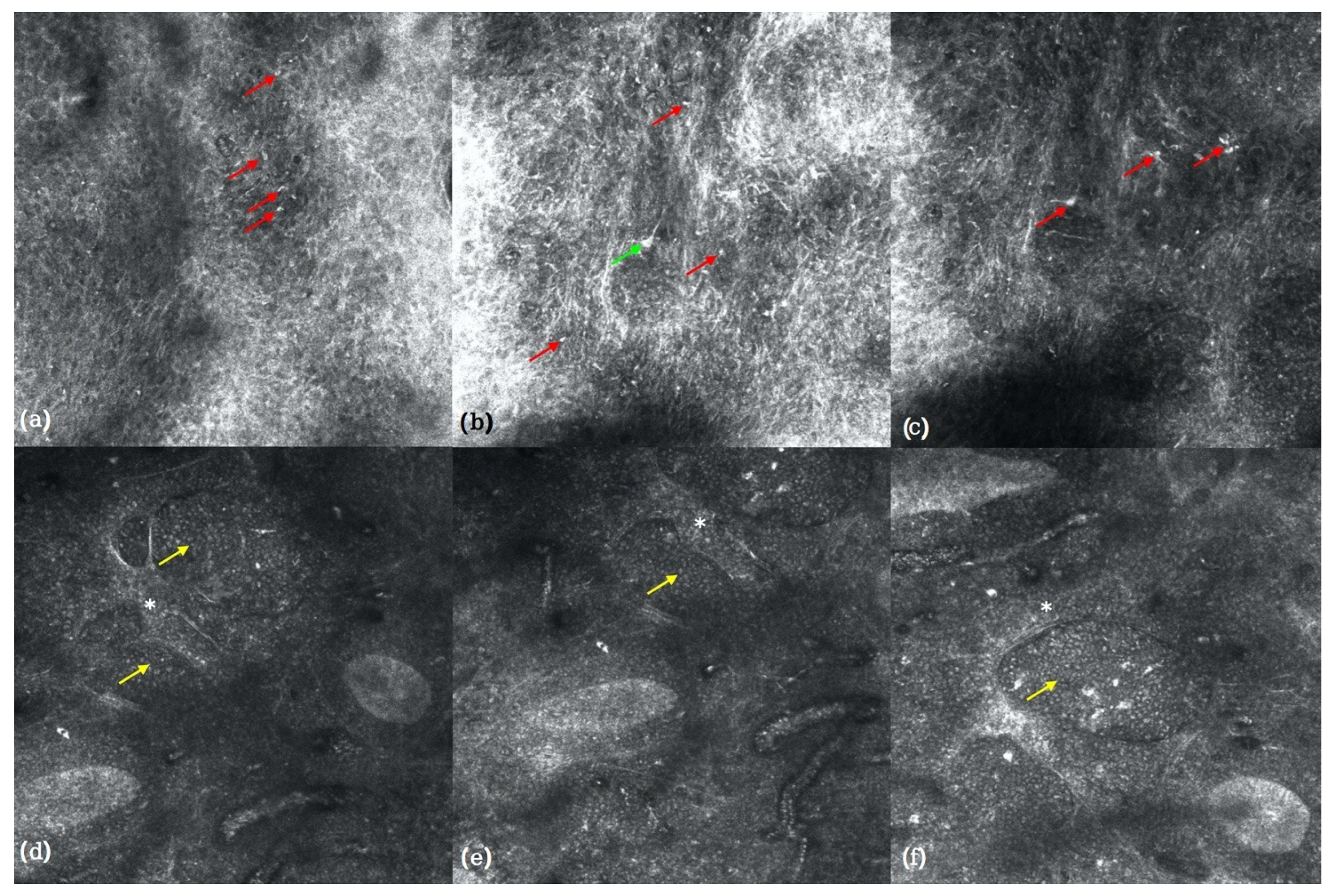
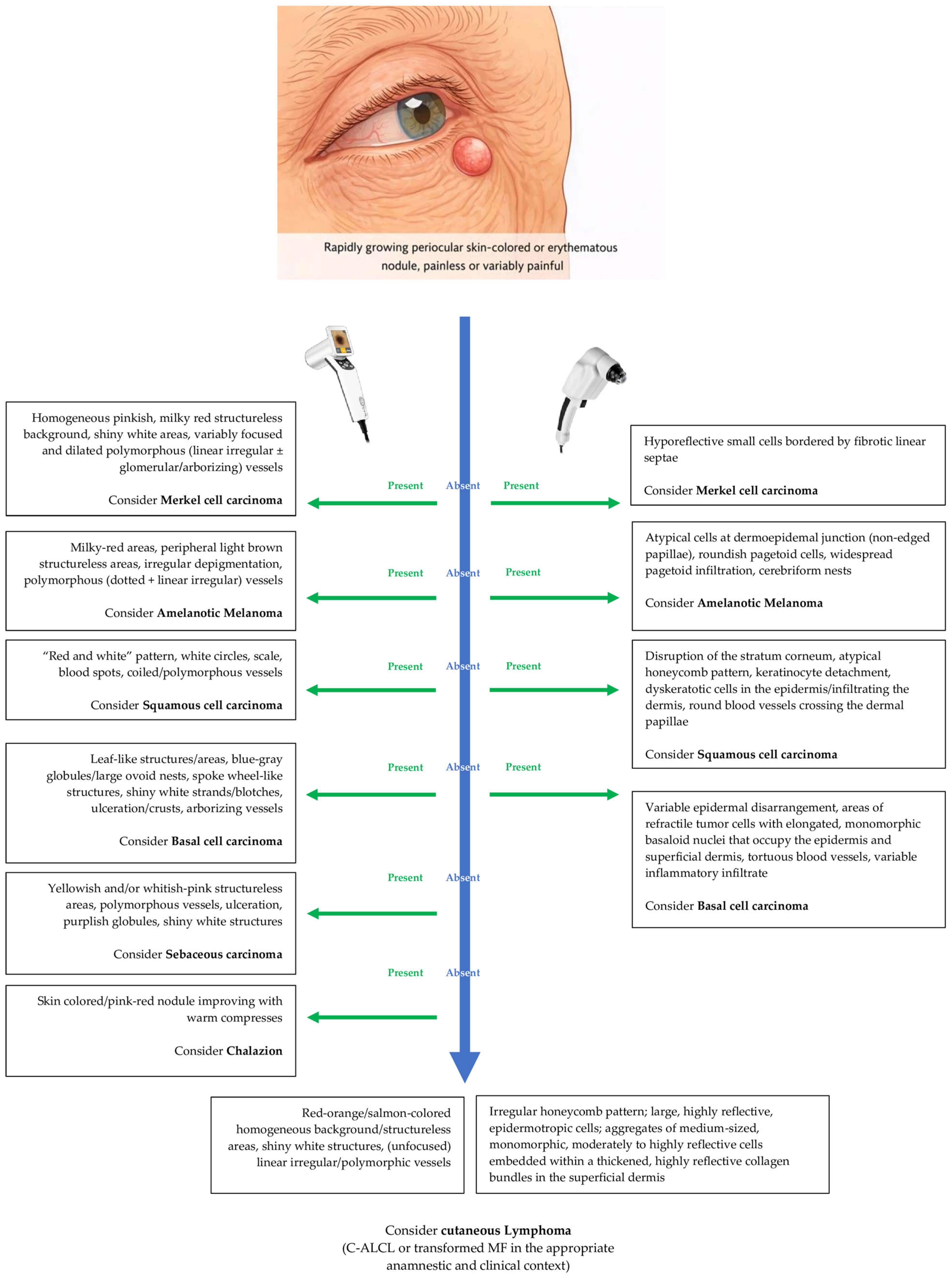
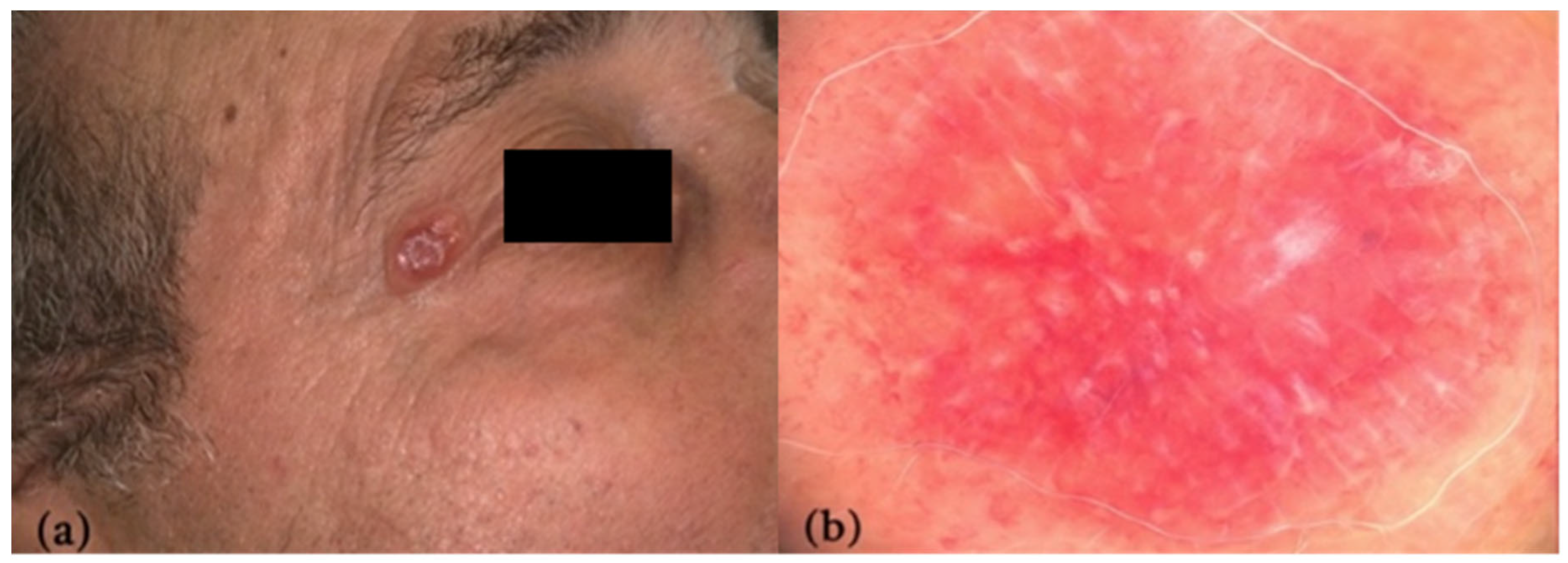
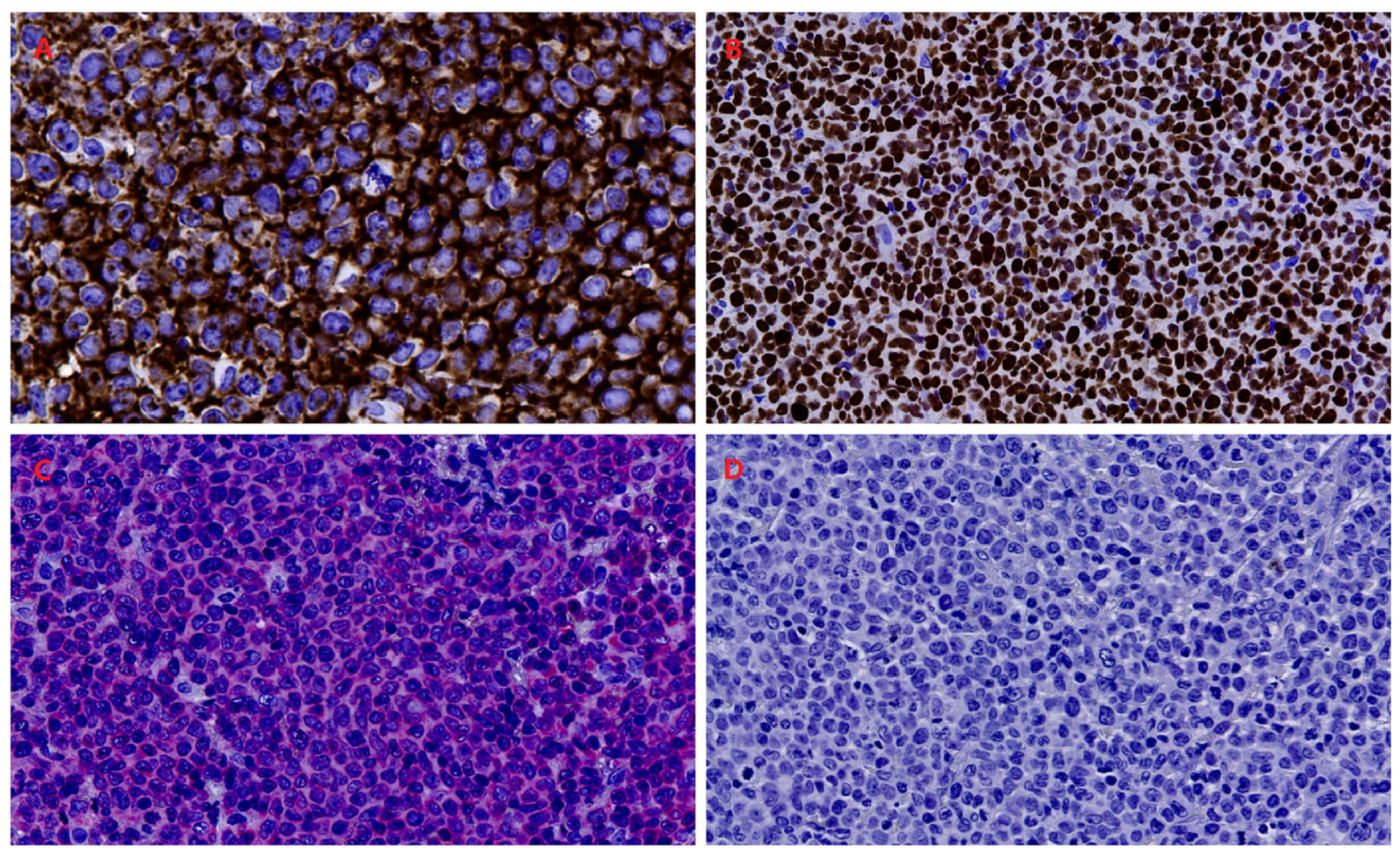
| C-ALCL | Red-orange homogeneous background or structureless areas; shiny white structures; (unfocused) linear, irregular/polymorphic vessels. | Loss of the regular honeycomb pattern; large, highly reflective cells with areas of prominent epidermal involvement within the epidermis and along the dermo-epidermal junction; aggregates of medium-sized, relatively monomorphic, moderately to highly reflective cells embedded within thickened, highly reflective collagen bundles in the superficial dermis | CD30 (>75%), variable T cell phenotype (CD3/CD4/CD8), ALK (−); | [6,7,8,9] |
| Merkel cell carcinoma | Homogeneous pinkish, milky-red structureless background; shiny/not-shiny white areas; variably focused and dilated polymorphous (linear irregular ± glomerular/arborizing vessels). | Hyporeflective small cells bordered by fibrotic linear septae. | CK20 (+, paranuclear dot), TTF-1 (−), synaptophysin, INSM1, chromogranin; MCPyV T-antigen. | [23] |
| Amelanotic melanoma | Milky-red areas; peripheral light brown structureless areas; irregular/subtle depigmentation; polymorphous (dotted + linear irregular) vessels. | Atypical cells at dermoepidemal junction (non-edged papillae); roundish pagetoid cells; widespread pagetoid infiltration; cerebriform nests. | S100 (+), Melan-A (variable), HMB-45 (variable), SOX10 (+), PRAME (+). | [21,24,25,26] |
| SCC/Keratoacanthoma (KA) | Keratin (central in KA); white circles (highest specificity); blood spots; scale; “red and white” pattern; coiled vessels; polymorphous vessels (SCC); radial hairpin vessels (KA). | Disruption of the stratum corneum with evident parakeratosis; alteration of the epidermal pattern with severe keratinocyte detachment; round, nucleated dyskeratotic cells in the epidermis and occasionally infiltrating the dermis; round blood vessels crossing the dermal papillae. | p40 (+), p63 (+), CK5/6 (+). | [27,30] |
| BCC | Leaf-like structures/areas; multiple blue-gray globules (not aggregated); large blue-gray ovoid nests; scattered dots; spoke wheel-like structures; shiny white strands/blotches; ulceration; arborizing vessels. | Variable epidermal disarrangement (loss of the honeycomb pattern and nuclear pleomorphism of the keratinocytes), areas of refractile tumor cells with elongated, monomorphic basaloid nuclei with nuclear streaming occupying the epidermis and superficial dermis; increased dermal vasculature with dilated and tortuous blood vessels; variable inflammatory infiltrate associated with the tumor. | BerEP4 (+), BCL2 (+). | [28,31] |
| Sebaceous carcinoma | Yellowish structures; yellowish structureless areas; whitish-pink areas; polymorphous vessels; ulceration; purplish globules; shiny white structures. | None yet reported. | Adipophilin (100% sensitive), EMA (+), CK7 (+), Ber-EP4 (−/weak), AR | [29] |
| Chalazion | Skin colored/pink-red nodule, variably painful | None yet reported. | Generally not requested | [32] |
Disclaimer/Publisher’s Note: The statements, opinions and data contained in all publications are solely those of the individual author(s) and contributor(s) and not of MDPI and/or the editor(s). MDPI and/or the editor(s) disclaim responsibility for any injury to people or property resulting from any ideas, methods, instructions or products referred to in the content. |
© 2026 by the authors. Licensee MDPI, Basel, Switzerland. This article is an open access article distributed under the terms and conditions of the Creative Commons Attribution (CC BY) license.
Share and Cite
Scotti, B.; Misciali, C.; D’Onghia, M.; Gualandi, A.; Vaccari, S.; Venturi, F.; Magnaterra, E.; Cinotti, E.; Dika, E. Dermoscopic and Reflectance Confocal Microscopic Features of a Primary Cutaneous Anaplastic Large Cell Lymphoma (C-ALCL) of the Eyelid: A Case Report with Histopathologic Correlation. Reports 2026, 9, 164. https://doi.org/10.3390/reports9020164
Scotti B, Misciali C, D’Onghia M, Gualandi A, Vaccari S, Venturi F, Magnaterra E, Cinotti E, Dika E. Dermoscopic and Reflectance Confocal Microscopic Features of a Primary Cutaneous Anaplastic Large Cell Lymphoma (C-ALCL) of the Eyelid: A Case Report with Histopathologic Correlation. Reports. 2026; 9(2):164. https://doi.org/10.3390/reports9020164
Chicago/Turabian StyleScotti, Biagio, Cosimo Misciali, Martina D’Onghia, Alberto Gualandi, Sabina Vaccari, Federico Venturi, Elisabetta Magnaterra, Elisa Cinotti, and Emi Dika. 2026. "Dermoscopic and Reflectance Confocal Microscopic Features of a Primary Cutaneous Anaplastic Large Cell Lymphoma (C-ALCL) of the Eyelid: A Case Report with Histopathologic Correlation" Reports 9, no. 2: 164. https://doi.org/10.3390/reports9020164
APA StyleScotti, B., Misciali, C., D’Onghia, M., Gualandi, A., Vaccari, S., Venturi, F., Magnaterra, E., Cinotti, E., & Dika, E. (2026). Dermoscopic and Reflectance Confocal Microscopic Features of a Primary Cutaneous Anaplastic Large Cell Lymphoma (C-ALCL) of the Eyelid: A Case Report with Histopathologic Correlation. Reports, 9(2), 164. https://doi.org/10.3390/reports9020164