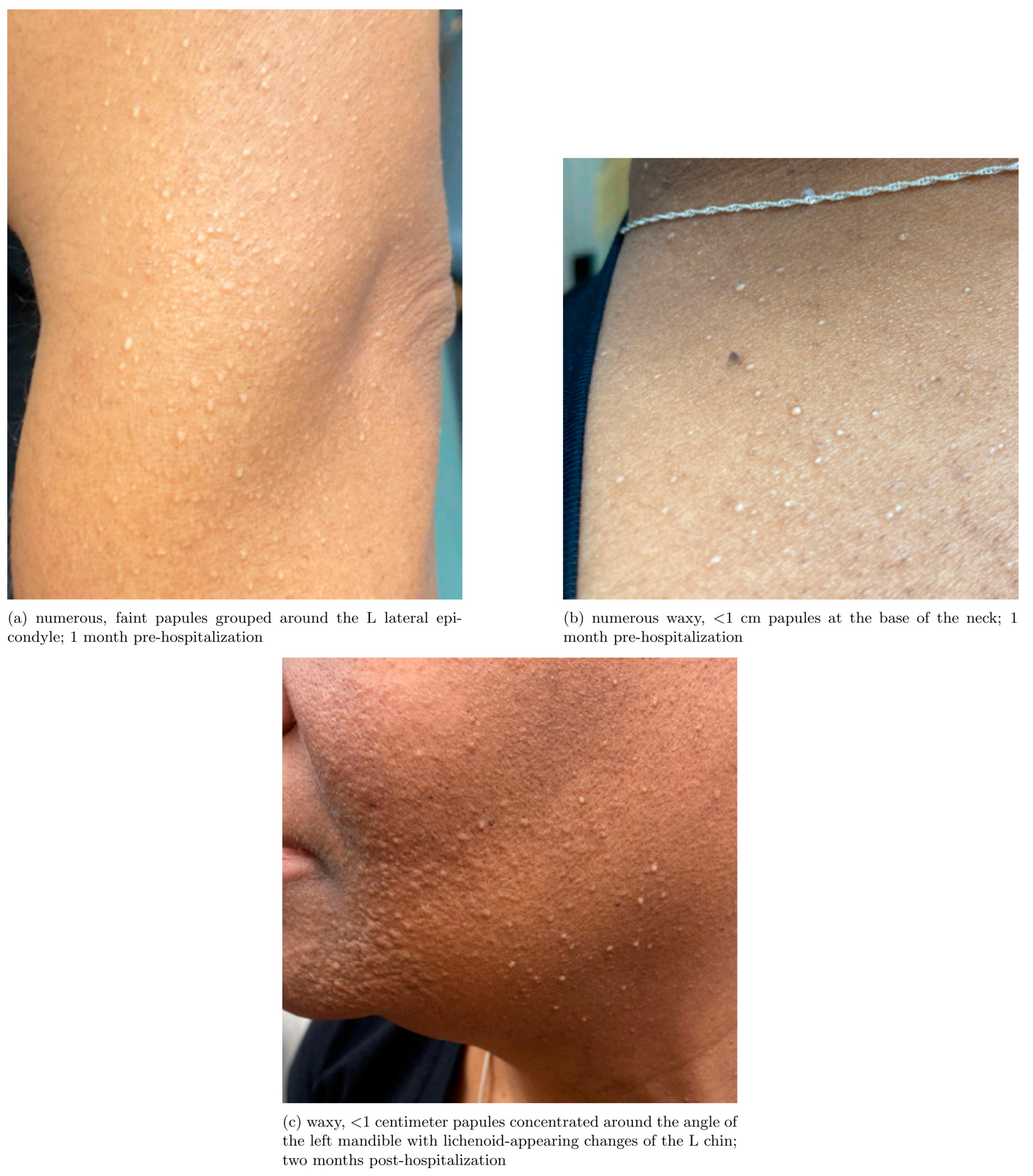
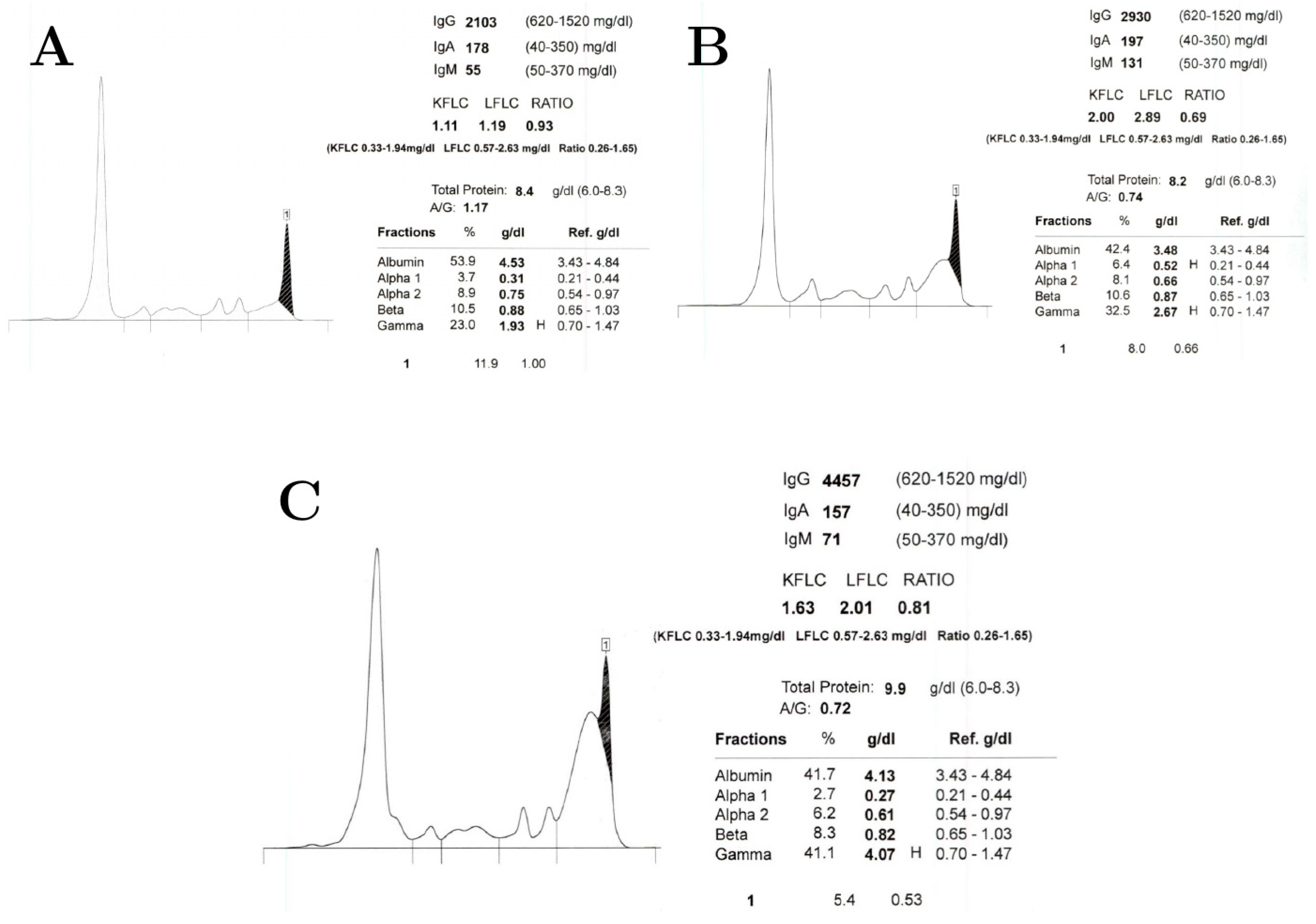
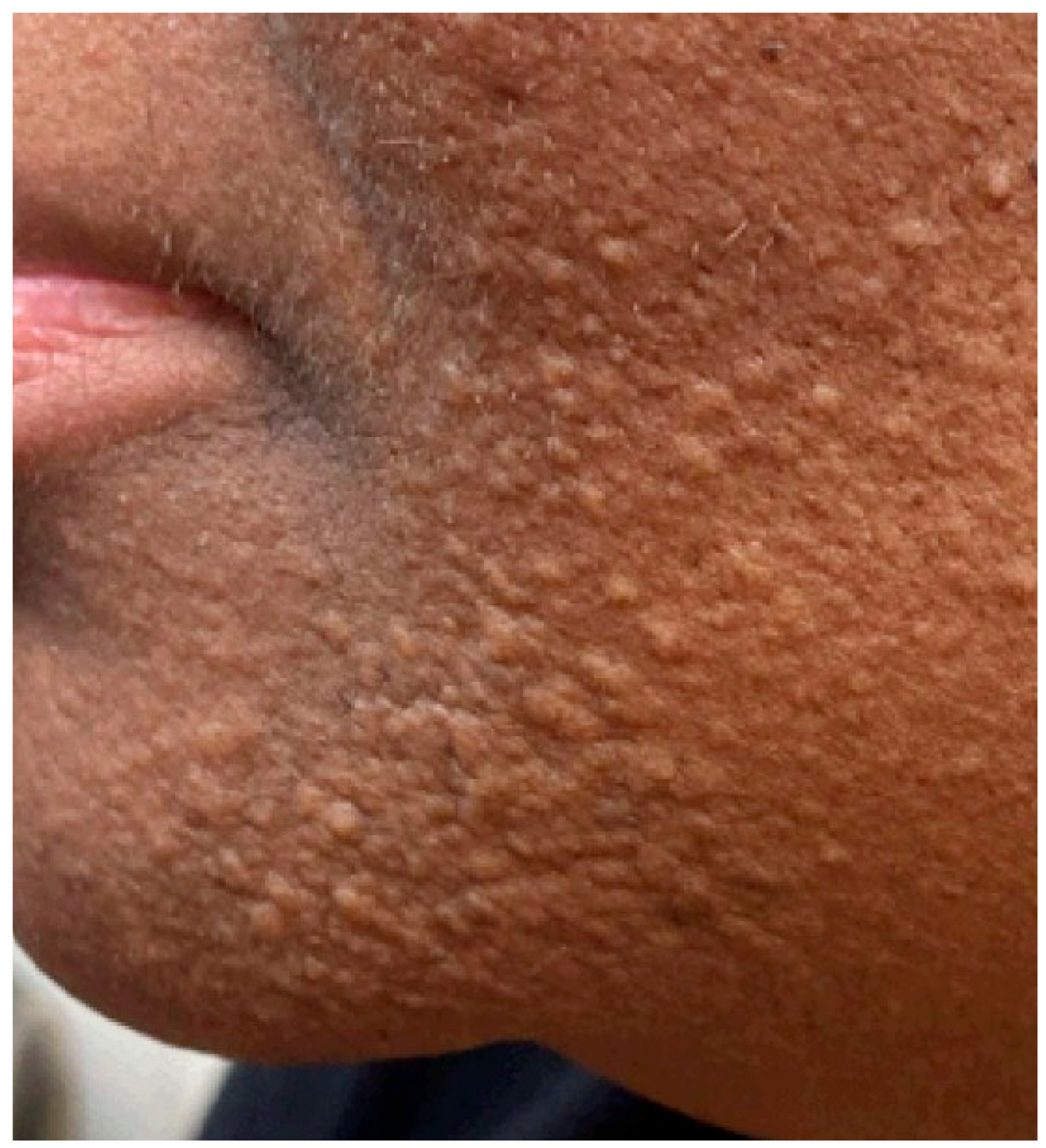
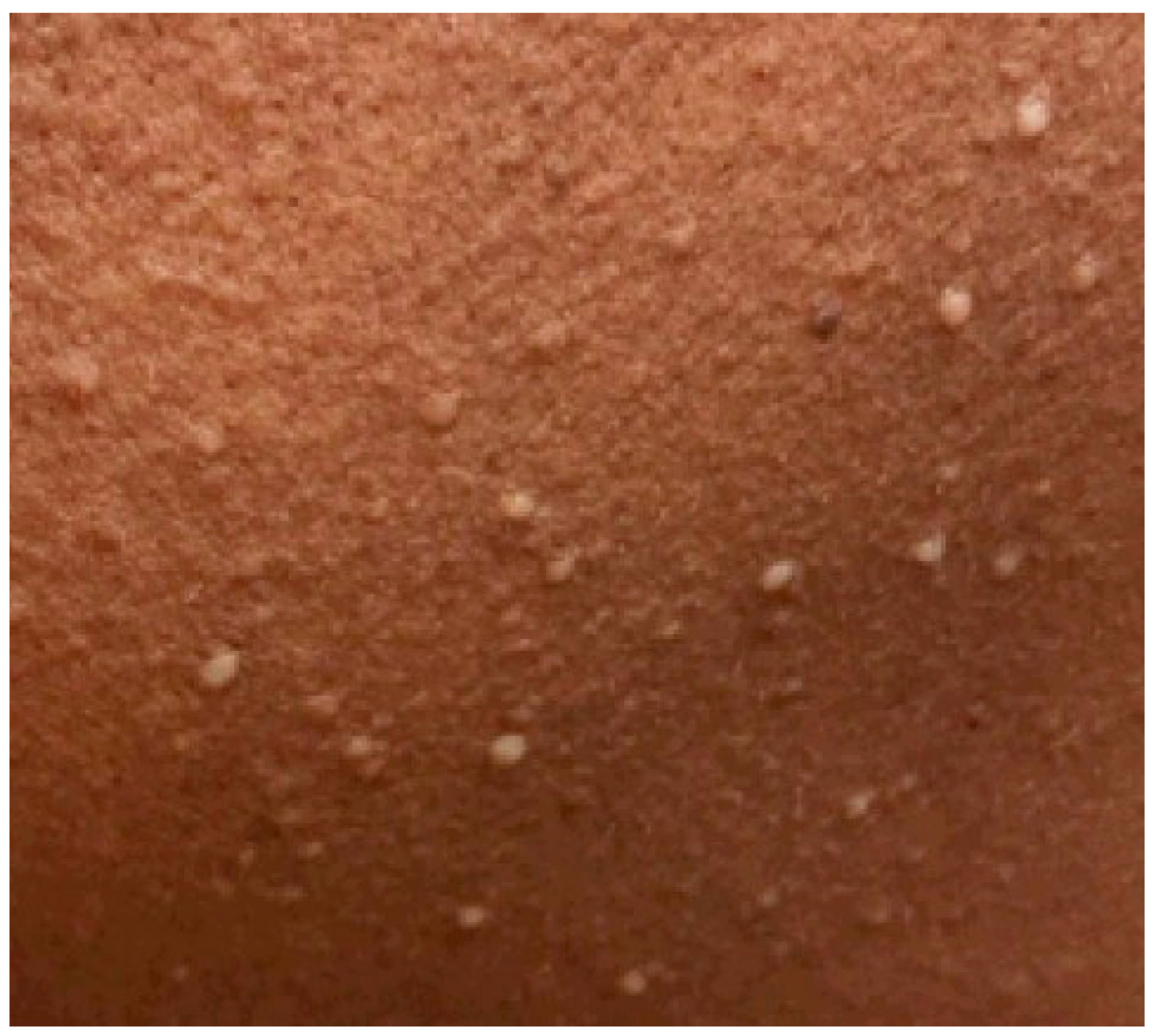
Dermato-Neuro Syndrome After Intravenous Immunoglobulin Infusion: Case Report

Abstract
1. Introduction and Clinical Significance
2. Case Presentation
3. Discussion
4. Conclusions
Author Contributions
Funding
Institutional Review Board Statement
Informed Consent Statement
Data Availability Statement
Conflicts of Interest
Abbreviations
| IVIG | Intravenous Immunoglobulin |
Appendix A


References
- Rongioletti, F.; Merlo, G.; Cinotti, E.; Fausti, V.; Cozzani, E.; Cribier, B.; Metze, D.; Calonje, E.; Kanitakis, J.; Kempf, W.; et al. Scleromyxedema: A multicenter study of characteristics, comorbidities, course, and therapy in 30 patients. J. Am. Acad. Dermatol. 2013, 69, 66–72. [Google Scholar] [CrossRef]
- Rongioletti, F.; Rebora, A. Updated classification of papular mucinosis, lichen myxedematosus, and scleromyxedema. J. Am. Acad. Dermatol. 2001, 44, 273–281. [Google Scholar] [CrossRef]
- Sala, A.C.B.; Cunha, P.R.; Pinto, C.A.L.; de Moraes Alves, C.A.X.; Paiva, I.B.; Araujo, A.P.V. Scleromyxedema: Clinical diagnosis and autopsy findings. An. Bras. Dermatol. 2016, 91, 48–50. [Google Scholar] [CrossRef]
- Marinkovic, A.; Zypchen, L.N.; Chan, J.; Chen, L.Y.; Parkin, S. Monoclonal gammopathy of clinical significance: What the rheumatologist needs to know. Lancet Rheumatol. 2022, 4, e362–e373. [Google Scholar] [CrossRef]
- Atzori, L.; Ferreli, C.; Rongioletti, F. New insights on scleromyxedema. J. Scleroderma Relat. Disord. 2019, 4, 118–126. [Google Scholar] [CrossRef]
- River, Y.; Levy, I.; Gilead, L.; Orbach, H.; Almog, Y. Fever, convulsions and coma in scleromyxedema. Neurology 1996, 46, 1778–1779. [Google Scholar] [CrossRef]
- Mahévas, T.; Arnulf, B.; Bouaziz, J.-D.; Livideanu, C.B.; Osio, A.; Servy, A.; Cribier, B.; Sassolas, B.; Jachiet, M.; Michel, L.; et al. Plasma cell-directed therapies in monoclonal gammopathy-associated scleromyxedema. Blood 2020, 135, 1101–1110. [Google Scholar] [CrossRef]
- Fleming, K.E.; Virmani, D.; Sutton, E.; Langley, R.; Corbin, J.; Pasternak, S.; Walsh, N.M. Scleromyxedema and the dermato-neuro syndrome: Case report and review of the literature. J. Cutan. Pathol. 2012, 39, 508–517. [Google Scholar] [CrossRef]
- Karaman, B.; Guler, A.; Ertam, I.; Celebisoy, N. Dermato-neuro syndrome associated with scleromyxedema. Indian J. Dermatol. Venereol. Leprol. 2015, 81, 519. [Google Scholar] [CrossRef]
- Rey, J.B.; Luria, R.B. Treatment of scleromyxedema and the dermatoneuro syndrome with intravenous immunoglobulin. J. Am. Acad. Dermatol. 2009, 60, 1037–1041. [Google Scholar] [CrossRef]
- Rode, M.; Rigby, M.J.; Schipani, E.; Fugate, J. Influenza-associated encephalopathy mimicking dermatoneuro syndrome. BMJ Case Rep. 2025, 18, e264930. [Google Scholar] [CrossRef]
- Johkura, K.; Susuki, K.; Hasegawa, O.; Kuroiwa, Y.; Komatsumoto, S. Encephalopathy in scleromyxedema. Neurology 1999, 53, 1138. [Google Scholar] [CrossRef]
- Li, Y.; Wang, H.; Wang, A.; Zhao, G. Dermato-neuro syndrome after COVID-19 infection in a patient with scleromyxoedema: Previously successful treatment with intravenous immunoglobulins. J. Dermatol. 2024, 51, 140–144. [Google Scholar] [CrossRef]
- Bhoyrul, B.; Mughal, A.A.; Paulus, J.; Salamat, A.; Howarth, S. Does dermatoneuro syndrome have a viral aetiology? Clin. Exp. Dermatol. 2016, 41, 53–56. [Google Scholar] [CrossRef]
- Liu, A.; Suozzi, K.; Hwang, D.Y.; Moeller, J.J.; Lazova, R.; DiCapua, D. Dermatoneuro syndrome. Neurol. Clin. Pract. 2016, 6, e27–e29. [Google Scholar] [CrossRef]
- Sasaoka, T.; Ujiie, H.; Nishie, W.; Iwata, H.; Ishikawa, M.; Higashino, H.; Natsuga, K.; Shinkuma, S.; Shimizu, H. Intravenous IgG Reduces Pathogenic Autoantibodies, Serum IL-6 Levels, and Disease Severity in Experimental Bullous Pemphigoid Models. J. Investig. Dermatol. 2018, 138, 1260–1267. [Google Scholar] [CrossRef]
- Ibáñez, C.; Suñé, P.; Fierro, A.; Rodríguez, S.; López, M.; Álvarez, A.; De Gracia, J.; Montoro, J.-B. Modulating effects of intravenous immunoglobulins on serum cytokine levels in patients with primary hypogammaglobulinemia. BioDrugs 2005, 19, 59–65. [Google Scholar] [CrossRef]
- Steinberger, B.A.; Ford, S.M.; Coleman, T.A. Intravenous immunoglobulin therapy results in post-infusional hyperproteinemia, increased serum viscosity, and pseudohyponatremia. Am. J. Hematol. 2003, 73, 97–100. [Google Scholar] [CrossRef]
- Mehta, J.; Singhal, S. Hyperviscosity Syndrome in Plasma Cell Dyscrasias. Semin. Thromb. Hemost. 2003, 29, 467–472. [Google Scholar] [CrossRef] [PubMed]
- MacKenzie, M.R.; Fudenberg, H.H.; O’Reilly, R.A. The hyperviscosity syndrome. J. Clin. Investig. 1970, 49, 15–20. [Google Scholar] [CrossRef][Green Version]
- Haber, R.; Bachour, J.; El Gemayel, M. Scleromyxedema treatment: A systematic review and update. Int. J. Dermatol. 2020, 59, 1191–1201. [Google Scholar] [CrossRef]
- Syder, N.C.; Omar, D.; McKenzie, S.; Brown-Korsah, J.B.; Taylor, S.C.; Elbuluk, N. Gaps in medical education curricula on skin of color in medical school, residency, and beyond: Part 1. J. Am. Acad. Dermatol. 2023, 89, 885–892. [Google Scholar] [CrossRef]
- Bellicoso, E.; Quick, S.O.; Ayoo, K.O.; Beach, R.A.; Joseph, M.; Dahlke, E. Diversity in Dermatology? An Assessment of Undergraduate Medical Education. J. Cutan. Med. Surg. 2021, 25, 409–417. [Google Scholar] [CrossRef] [PubMed]



| Test (Unit; Reference Range) | Day 0 | Day 1 | Day 2 | Day 3 | Day 4 | Day 5 | Day 6 | Day 7 | Day 8 | Day 9 | Day 10 |
|---|---|---|---|---|---|---|---|---|---|---|---|
| WBC (×109/L, 4.0–11.0 | 13.0 | 17.0 | 16.8 | 18.8 | 23.8 | 28 | 27.5 | 18.8 | 12.7 | 11.2 | 9.7 |
| Hgb (×109/L; 12.0–16.0) | 11.1 | 8.8 | 9.0 | 8.7 | 9.3 | 8.5 | 7.4 | 8.7 | 8.4 | 8.0 | 8.2 |
| Creatine phosphokinase (U/L; 26–180) | 682 | 2596 | 6789 | 9213 | 12,516 | 9105 | 4610 | 1870 | 1266 | 464 | 256 |
| AST (U/L; <34) | 37 | 92 | 110 | 191 | 247 | 211 | 146 | 88 | 82 | 49 | 36 |
| ALT (U/L; 10–49) | 16 | 22 | 46 | 65 | 76 | 72 | 55 | 61 | 48 | 38 | 31 |
| Serum Protein (g/dL; 6.0–8.3) | 9.4 | 8.5 | 8.5 | 8.5 | 8.3 | 8.2 | 7.4 | 8.4 | 7.6 | 7.2 | 7.3 |
| Sodium (mmol/L; 136–146) | 130 | 132 | 130 | 131 | 133 | 134 | 138 | 135 | 138 | 137 | 136 |
| Glucose (mg/dL; 70–180) | 181 | 132 | 185 | 166 | 133 | 121 | 96 | 118 | 123 | 123 | 132 |
| ESR (mm/h; 0–20) | 73 | 40 | 62 | 56 | 64 | 62 | 49 | 88 | 69 | 87 | - |
| CRP (mg/L; <0.6) | 1.8 | 2.4 | 3.3 | 2.5 | 1.5 | 1.4 | 1.6 | 2.6 | 3.0 | 2.4 | 2.3 |
| Whole Blood Viscosity (cP) | - | - | - | 47.0 | - | - | - | - | - | - | - |
| Plasma Viscosity (cP; 1.35–1.85) | - | - | - | 2.19 | - | - | - | - | - | - | - |
| LDH (U/L; 120–240) | - | 655 | - | - | - | - | - | - | - | - | - |
| Myoglobin, urine (ng/mL; <45) | - | 1340 | - | - | - | - | - | - | - | - | - |
| Thyroid Stimulating Hormone (mIU/L; 0.3–5.5) | 0.89 | - | - | - | - | - | - | - | - | - | - |
| Test (Unit; Reference Range) | Result | Day of Hospitalization Collected |
|---|---|---|
| Microbiology work-up of plasma and urine | Hepatitis C RNA by PCR not detected, Epstein–Barr virus DNA by PCR not detected, cytomegalovirus DNA by PCR not detected, aerobic and anaerobic bacterial cultures showed no growth, acid-fast bacilli culture showed no growth, fungal culture showed no growth | Day 1 |
| Myomarker3 Panel (CU, <20) | Negative Anti-Mi2-Ab, <20 Anti-Jo-1 antibody, <20 Anti-Tif1-gamma antibody | Day 2 |
| HMG-CoA reductase antibody (CU, <20) | <20 | Day 2 |
| MR Femur | No evidence of myositis involving the thighs | Day 2 |
| MR Brain | No evidence of acute process | Day 3 |
| EEG | Background activity was marked by diffuse delta admixed theta frequencies. There was no posterior dominant rhythm, and the AP gradient was not well-formed. During behavioral sleep, there were no sleep architectures. Activation was not performed. There were no epileptiform discharges, focal abnormalities, or seizures recorded during the EEG | Day 3 |
| Cerebrospinal Fluid Studies | Lymphocytic pleocytosis (100 cells counted, 76 lymphocytes), protein 82, glucose 79, red blood cell count 1 | Day 4 |
| Flow cytometry and immunophenotyping of cerebrospinal fluid | Paucicellular specimen with no immunophenotypic evidence of monoclonal B cells or aberrant T cells. B-cell Kappa/lambda ratio: 2.43 | Day 4 |
| Microbiology work-up of cerebrospinal fluid | Human herpes virus 8 DNA by PCR not detected, herpes simplex virus-1 and herpes simplex virus-2 DNA by PCR not detected, varicella DNA by PCR not detected, cryptococcal antigen not detected, syphilis VDRL not detected, bacterial culture yielded no growth, acid-fast bacilli culture yielded no growth, fungal culture yielded no growth | Day 4 |
| Chemokine Ligand 9 (pg/mL, <647) | 2145 | Day 5 |
| Muscle Biopsy | Diffuse type 2 fiber atrophy | Day 8 |
Disclaimer/Publisher’s Note: The statements, opinions and data contained in all publications are solely those of the individual author(s) and contributor(s) and not of MDPI and/or the editor(s). MDPI and/or the editor(s) disclaim responsibility for any injury to people or property resulting from any ideas, methods, instructions or products referred to in the content. |
© 2026 by the authors. Licensee MDPI, Basel, Switzerland. This article is an open access article distributed under the terms and conditions of the Creative Commons Attribution (CC BY) license.
Share and Cite
Kassalow, B.; Kazmi, S.; Shukri, S.; London, Z.N. Dermato-Neuro Syndrome After Intravenous Immunoglobulin Infusion: Case Report. Reports 2026, 9, 148. https://doi.org/10.3390/reports9020148
Kassalow B, Kazmi S, Shukri S, London ZN. Dermato-Neuro Syndrome After Intravenous Immunoglobulin Infusion: Case Report. Reports. 2026; 9(2):148. https://doi.org/10.3390/reports9020148
Chicago/Turabian StyleKassalow, Bryce, Soha Kazmi, Said Shukri, and Zachary N. London. 2026. "Dermato-Neuro Syndrome After Intravenous Immunoglobulin Infusion: Case Report" Reports 9, no. 2: 148. https://doi.org/10.3390/reports9020148
APA StyleKassalow, B., Kazmi, S., Shukri, S., & London, Z. N. (2026). Dermato-Neuro Syndrome After Intravenous Immunoglobulin Infusion: Case Report. Reports, 9(2), 148. https://doi.org/10.3390/reports9020148