Case Report

Intradural Lumbar Disc Herniation: Updated Report of a Case with Literature Review

1
Spinal Surgery Department, Viet Duc University Hospital, Hanoi 100000, Vietnam
2
School of Medicine and Pharmacy, Vietnam National University, Hanoi 100000, Vietnam
3
Institute for Preventive Medicine and Public Health, Hanoi Medical University, Hanoi 100000, Vietnam
4
Institute of Orthopaedic and Trauma Surgery, Viet Duc University Hospital, Hanoi 100000, Vietnam
*
Author to whom correspondence should be addressed.
These authors contributed equally to this work.
Submission received: 15 February 2022 / Revised: 28 February 2022 / Accepted: 7 March 2022 / Published: 11 March 2022

Abstract

:
Intradural lumbar disc herniation is a rare complication of disc disease. The mechanism by which a herniated disc tears the dura matter remains unknown. The preoperative diagnosis of an intradural lumbar disc herniation is still difficult. We report our experience dealing with a case of intradural lumbar disc herniation at level L3/4 in a 34-year-old man. Based on current experience, we found that attachment of the ventral dura to the posterior longitudinal ligament can be a favorable factor indicating intradural lumbar disc herniation. One should pay attention to those with cauda equina syndrome, as it can thereby promptly suggest a preoperative diagnosis of intradural lumbar disc herniation. Surgeons need to avoid omitting intraoperative lesions by palpating the dura mater during surgery for suspected tumor cases.

1. Introduction

Intradural lumbar disc herniation is a serious complication of herniated discs and is very rare in the clinical treatment of lumbar disc herniation. The proportion of intradural lumbar disc herniation was estimated to be 0.3% of all disc herniations, occurring most commonly among middle-aged adults [1]. Diagnosing this condition preoperatively is still challenging, and diagnosis may be missed [2,3,4,5]. Here, we report our experience dealing with a case of intradural lumbar disc herniation at level L3/4. Accordingly, we sought to review the literature and discuss the pathogenesis, methods for definitive diagnosis and treatment for this particular hernia.

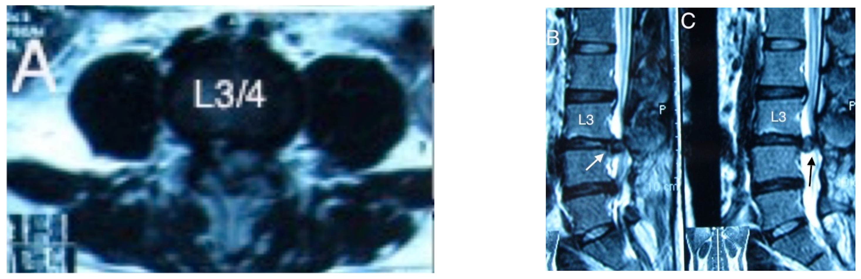
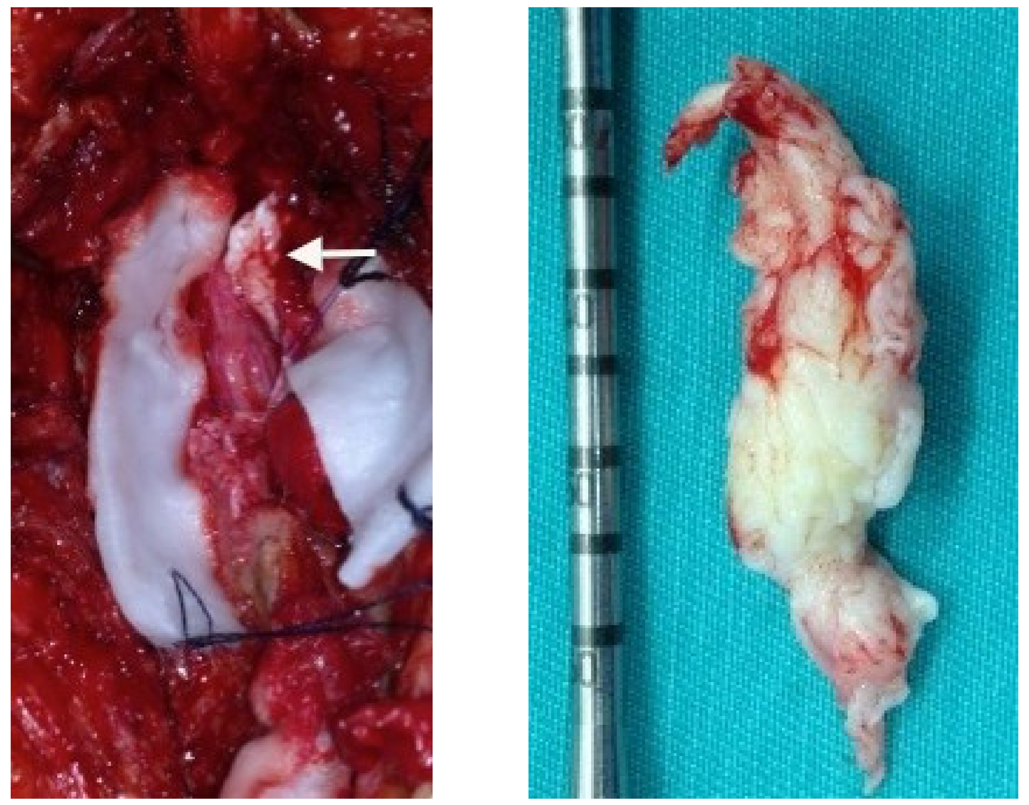
2. Case Presentation Section

A 34-year-old man had a history of lumbar disc herniation 10 years earlier. After surgical treatment, the patient sometimes had back pain without leg pain when the weather changed. For the 10 days before admission, the patient presented with sudden and increasing back pain radiating to the left leg. On admission, he presented to us with ankle paralysis (left ankle and foot movement: 0/5 points) and numbness for the L4, L5 and S1 spinal nerve root, with bladder and bowel dysfunction.
Non-contrast magnetic resonance image (MRI) revealed a 2.5-cm intradural extramedullary mass which was round, demonstrated a co-signal with the intervertebral disc, and had a uniform signal. Additionally, we saw that this mass had consecutive pins with the L3-L4 intervertebral disc. MRI showed abrupt disruption of the posterior longitudinal ligament. On cross-sectional MRI, this mass occupied most of the spinal canal (Figure 1). Beak signs on the film were unknown.
Our patient underwent surgery for the L3 location. The L3-L4 intervertebral disc was examined, and the dura was opened to check. We proceeded to remove the herniated mass, then irrigated and sutured the dura when there was a herniated disc fragment located in the dura, and the inflamed nerve roots stuck to the mass (Figure 2).

3. Discussion

Intradural lumbar disc herniation was first described by Dandy in 1942 [6]. Since then, this condition of disc herniation has been reported in research articles around the world, showing that this is a rare lesion with only 151 cases [7] which accounted for about 0.04–0.33% of all disc herniations [7,8,9]. In patients with intradural lumbar disc herniation, 3% of them occurred in the cervical spine, followed by 5% in the thoracic spine and 92% in the lumbar spine. The mean age of onset of intradural lumbar disc herniation is between 50 and 60 years old [8]. The clinical signs may include cauda equina syndrome. The history of this pathology includes chronic low back pain, acute radicular pain, and progressive paralysis. Severe symptoms of cauda equina syndrome are frequently found in two-thirds of patients. The duration of chronic symptoms suggests that transdural migration may simply be an intermediate stage in the complete transdural migration of the herniated disc fragment and granulation tissue formation around the herniated disc. A classification for intradural disc herniations was suggested by M. Mut [10] as follows: Type A indicates the herniation of a disc into the dural sac and Type B indicates the herniation of a disc into the dural sheath in the preganglionic region of the nerve root.
The most common location of intradural disc herniation in the lumbar region is the L4-L5 disc space (55%), followed by the L3-L4 (16%), the L5-S1 (10%), the L2-L3, and the L1-L2 disc spaces [2]. In the literature, the position of intradural disc herniation is reported to be at the interdisc positions and above or below the levels relative to the disc pins, while intradural disc herniations at the mid-vertebral levels are rare [11,12,13].
Despite this, the pathophysiology and physiology of intradural disc herniations are still poorly understood. However, there may be an initial or pre-existing abnormality that leads to lesion formation, for which several theories have been proposed: (1) adhesions between the posterior longitudinal ligament and the anterior wall of the dura possibly due to local inflammatory processes as a result of irritation from prior trauma, surgery, or disc herniation; (2) congenital fusion between the posterior longitudinal ligament and dura; (3) the anatomy of the posterior longitudinal ligament and anterior dura, which are often loosely attached to each other, with epidural penetration occurring after sudden force and in the narrowing position of the spinal canal [8,14]. Blikra G. found a relationship between the anterior dura mater and the posterior longitudinal ligament in the formation of dense adhesions in the lumbar spine [15]. Accordingly, they concluded that adhesions between these structures may be an underlying factor in the pathology of intradural disc herniation. The presence of dural attachment ligaments and nerve roots at their exit from the main dural capsule to the posterior longitudinal ligament and the distal periosteal vertebral body to the disc. Chronic inflammation at the disc herniation site can cause adhesions and thinning of the dura, and even discontinuity of the dura, thereby causing a disc herniation to enter the dural sac. Anterior and posterior intradural disc herniations have both been observed; however, because the anterior dura is closely posterior to the disc and the posterior dura is usually thicker than the anterior dura, this type of disc herniation frequently perforates the anterior dura [10].
Intradural disc herniation can cause posterior dural tears depending on the size of the nucleus pulposus. Our patient had undergone surgery once, so there may be adhesions of the dura to the posterior longitudinal ligament from the previous surgery. This adhesion causes the posterior longitudinal ligament and the dura to adhere to each other across L3/4 and below. The inflammatory process that persists for many years gradually thins the posterior longitudinal ligament and the dural complex. When the disc herniated, the herniated fragment moved straight into the dura and caused the symptoms of cauda equina syndrome.
Various imaging modalities such as MRI, computed tomography, and saccoradiculography with contrast can be used to diagnose disc herniation, but these techniques are of little value in the diagnosis of intradural lumbar disc herniation. Contrast saccoradiculography imaging can show amputation due to an intradural mass, but the images on the contrast saccoradiculography are nonspecific [15]. A computed tomography scan shows a posterior disc herniation pressing into the spinal canal, but intradural herniation is difficult to discern. It is difficult to distinguish lumbar disc herniation from other pathologies in MRI without contrast, while MRI with contrast is helpful in differentiating disc herniation from discitis. These herniated discs had lesions with homogenous and uniform imaging on T1-weighted (T1W) and T2-weighted (T1W) images [9,16]. Hida [17] emphasized the usefulness of MRI with contrast in preoperative diagnosis by showing a ‘Beak sign’-like mass with ring ablation at the level of the intervertebral disc at which abrupt discontinuity of the anterior posterior ligament occurred. Hidalgo-Ovejero [7] emphasizes the association between the presence of gas within the spinal canal and intradural disc herniation. According to the author, when seeing signs of a bubble of gas in the spinal canal, it is necessary to think of intradural disc herniation [7]. The authors explain that hyperintensity results from granulation tissue infiltration around the herniated disc fragment as adhesions and inflammatory responses develop over a long period of time. However, cases of acute disc herniation can not show ring-type enhancement because the granulomatous tissue has not yet developed. On cross-sectional MRI, an image of the herniated disc in the form of a teardrop can be seen with a stalk attached between the disc and the herniated fragment in the dura. Nevertheless, our patient only received MRI without contrast because the film of our patient was taken at another hospital before our admission. On the MRI, we saw a lesion in the dura at the level of L3-4 that had a uniform signal and was homologous to the disc on T1W and T2W, in which the lesion in the spinal canal has a pedicle attached to the L3-4 disc. Before surgery, we thought that this lesion could be an intradural and extramedullary tumor, in which the two most common tumor types are neurinoma and meningioma. These two types of tumors are often co-enhanced and are distinguished from intradural disc herniation by the absence of a ring pattern.
The treatment for intradural lumbar disc herniation is to remove the herniated disc mass and suture the missing dura. In most reports, the anterior dural tear was closed before. In-Ho Han [18] used the dural patch for anterior dural defects. Pradeep K. Singh [19] closed a ventral dural defect primarily and performed a duroplasty using a posterior layer of the thoracolumbar fascia of the back for a dorsal dural defect. In several cases, a transpedicle screw may be used, and posterolateral bony fusion should be performed if posterior spinal decompression is extensive or joint resection is required. For our patient, we performed a posterior spinal opening, examining the disc outside the marrow membrane. After ensuring that there was no compression of the herniated disc from the outside of the spinal cord, we decided to open the dura to check and remove the herniated disc piece. In our opinion, there is no need to suture the anterior torn dura because of the adhesion of this dura mater to tissues such as the disc and the posterior longitudinal ligament; hence, there is no postoperative cerebrospinal fluid leakage.

4. Conclusions

While intradural lumbar disc herniations are still very rare pathologies with a difficult diagnosis in clinical practice, physicians should consider this pathology in the patient who has undergone previous surgery and has a suspected tumor in the spinal canal and discontinuity of the posterior longitudinal ligament in MRI. One should pay attention to those with cauda equina syndrome, as it can promptly suggest a preoperative diagnosis of intradural lumbar disc herniation. Additionally, surgeons need to avoid omitting intraoperative lesions by palpating the dura mater during surgery for suspected tumor cases.

Author Contributions

Conceptualization, H.-L.N. and M.-H.D.; methodology, H.-L.N. and M.-H.D.; software, H.-L.N., M.-H.D. and B.-T.L.N.; validation, H.-L.N., M.-H.D. and B.-T.L.N.; formal analysis, H.-L.N. and H.-L.V.; investigation, H.-L.N. and M.-H.D.; data curation, H.-L.N. and M.-H.D.; writing—original draft preparation, H.-L.N. and H.-L.V.; writing—review and editing, H.-L.N., M.-H.D., H.-L.V. and B.-T.L.N.; visualization, H.-L.N. and M.-H.D.; supervision, H.-L.N. All authors have read and agreed to the published version of the manuscript.

Funding

This research received no external funding.

Institutional Review Board Statement

Our institution does not require ethical approval for reporting individual cases or case series.

Informed Consent Statement

Our institution does not require ethical approval for reporting individual cases or case series. The patient and her family were informed about our intention to involve her in a case study, and they agreed to partake in the study. They signed the consent form before the operation was carried out according to all surgical protocols.

Data Availability Statement

All data underlying this article are available in this article.

Conflicts of Interest

The authors declare no conflict of interest.

References

  1. Ihejirika, R.C.; Tong, Y.; Patel, K.; Protopsaltis, T. Intradural lumbar disc herniation: Illustrative case. J. Neurosurg. Case Lessons 2021, 2, CASE21336. [Google Scholar] [CrossRef]
  2. D’Andrea, G.; Trillò, G.; Roperto, R.; Celli, P.; Orlando, E.R.; Ferrante, L. Intradural lumbar disc herniations: The role of MRI in preoperative diagnosis and review of the literature. Neurosurg. Rev. 2004, 27, 75–80. [Google Scholar] [CrossRef] [PubMed]
  3. Ducati, L.G.; Silva, M.V.; Brandão, M.M.; Romero, F.R.; Zanini, M.A. Intradural lumbar disc herniation: Report of five cases with literature review. Eur. Spine J. 2013, 22, 404–408. [Google Scholar] [CrossRef] [PubMed] [Green Version]
  4. Tamaki, Y.; Sakai, T.; Miyagi, R.; Nakagawa, T.; Shimakawa, T.; Sairyo, K.; Chikawa, T. Intradural lumbar disc herniation after percutaneous endoscopic lumbar discectomy: Case report. J. Neurosurg. Spine 2015, 23, 336–339. [Google Scholar] [CrossRef] [PubMed] [Green Version]
  5. Chen, J.; Ni, H.-J.; Xue, F.; Fan, Y.-S.; Li, X.-F.; Chen, X.-Z.; He, S.-S. Upper Lumbar Intradural Disc Herniation: A Rare Case Report and Etiologic Analysis. J. Pain Res. 2021, 14, 1475–1481. [Google Scholar] [CrossRef] [PubMed]
  6. Dandy, W.E. Serious Complications of Ruptured Intervertebral Disks. JAMA J. Am. Med. Assoc. 1942, 119, 474–477. [Google Scholar] [CrossRef]
  7. Hidalgo-Ovejero, Á.M.; García-Mata, S.; Gozzi-Vallejo, S.; Izco-Cabezón, T.; Martínez-Morentín, J.; Martínez-Grande, M. Intradural Disc Herniation and Epidural Gas: Something More Than a Casual Association? Spine 2004, 29, E463–E467. [Google Scholar] [CrossRef] [PubMed]
  8. Liu, C.-C.; Huang, C.-T.; Lin, C.-M.; Liu, K.-N. Intradural disc herniation at L5 level mimicking an intradural spinal tumor. Eur. Spine J. 2011, 20, 326–329. [Google Scholar] [CrossRef] [PubMed] [Green Version]
  9. Sakai, T.; Tsuji, T.; Asazuma, T.; Yato, Y.; Matsubara, O.; Nemoto, K. Spontaneous resorption in recurrent intradural lumbar disc herniation: Case report. J. Neurosurg. Spine 2007, 6, 574–578. [Google Scholar] [CrossRef] [PubMed]
  10. Mut, M.; Berker, M.; Palaoğlu, S. Intraradicular disc herniations in the lumbar spine and a new classification of intradural disc herniations. Spinal Cord 2001, 39, 545–548. [Google Scholar] [CrossRef] [PubMed] [Green Version]
  11. Rong, Y.; Wang, J.; Sui, T.; Liu, W.; Luo, Y.; Cai, W. Cervical intradural disc herniation with Brown-Séquard syndrome: Case report and literature review. J. Pain Res. 2019, 12, 2403–2410. [Google Scholar] [CrossRef] [PubMed] [Green Version]
  12. Nam, K.H.; Han, I.H.; Cho, W.H.; Choi, B.K. A pure fluid-filled intradural cyst associated with intradural disc herniation and possible pathogenesis: A case report. Spine J. 2013, 13, e11–e15. [Google Scholar] [CrossRef] [PubMed]
  13. Guan, Q.; Xing, F.; Long, Y.; Xiang, Z. Cervical intradural disc herniation: A systematic review. J. Clin. Neurosci. 2018, 48, 1–6. [Google Scholar] [CrossRef] [PubMed]
  14. Yildizhan, A.; Paşaoglu, A.; Ökten, T.; Ekinci, N.; Aycan, K.; Aral, O. Intradural disc herniations pathogenesis, clinical picture, diagnosis and treatment. Acta Neurochir. 1991, 110, 160–165. [Google Scholar] [CrossRef] [PubMed]
  15. Blikra, G. Intradural Herniated Lumbar Disc. J. Neurosurg. 1969, 31, 676–679. [Google Scholar] [CrossRef] [PubMed]
  16. Mailleux, R.; Redant, C.; Milbouw, G. MR diagnosis of transdural disc herniation causing cauda equine syndrome. J. Belg. Radiol. 2006, 89, 303. [Google Scholar]
  17. Hida, K.; Iwasaki, Y.; Abe, H.; Shimazaki, M.; Matsuzaki, T. Magnetic resonance imaging of intradural lumbar disc herniation. J. Clin. Neurosci. 1999, 6, 345–347. [Google Scholar] [CrossRef]
  18. Han, I.-H.; Kim, K.S.; Jin, B.-H. Intradural Lumbar Disc Herniations Associated with Epidural Adhesion: Report of Two Cases. J. Korean Neurosurg. Soc. 2009, 46, 168–171. [Google Scholar] [CrossRef] [PubMed]
  19. Singh, P.K.; Shrivastava, S.; Dulani, R.; Banode, P.; Gupta, S. Dorsal Herniation of Cauda Equina Due to Sequestrated Intradural Disc. Asian Spine J. 2012, 6, 145–147. [Google Scholar] [CrossRef] [PubMed] [Green Version]
Figure 1. (A). The mass occupied most of the spinal canal on cross-sectional imaging MRI. (B) Mass in the spinal canal with fused leg to the L3/4 disc and anterior dural discontinuity (white arrow). (C) intradural herniated mass (black arrow).
Reports 05 00009 g001
Figure 2. Intradural lumbar disc herniation (white arrow).
Reports 05 00009 g002
Publisher’s Note: MDPI stays neutral with regard to jurisdictional claims in published maps and institutional affiliations.

