
A Locally Advanced Endometrioid Adenocarcinoma Arising from Vaginal Endometriosis: Management and Review of the Literature

 ,
,
Abstract
1. Introduction
2. Materials and Methods
3. Results
4. Discussion
5. Conclusions
Supplementary Materials
Author Contributions
Funding
Institutional Review Board Statement
Informed Consent Statement
Data Availability Statement
Conflicts of Interest
Abbreviations
| AUC | area under the curve |
| BSO | bilateral salpingo-oophorectomy |
| BO | bilateral oophorectomy |
| cy | cycles |
| CA 125 | cancer antigen 125 |
| CEA | carcinoembryonic antigen |
| MRI | magnetic resonance imaging |
| PET-CT | positron emission tomography–computed tomography |
| RT | radiation therapy |
| TAH | total abdominal hysterectomy |
| Gy | gray |
| na | not available |
| G-CSF | granulocyte-colony stimulating factor |
| CR | Complete response |
References
- Colombo, N.; Preti, E.; Landoni, F.; Carinelli, S.; Colombo, A.; Marini, C.; Sessa, C. Endometrial cancer: ESMO Clinical Practice Guidelines for diagnosis, treatment and follow-up. Ann. Oncol. Off. J. Eur. Soc. Med. Oncol. 2013, 24 (Suppl. S6), vi33–vi38. [Google Scholar] [CrossRef] [PubMed]
- Davis, A.; Goldberg, J. Extrapelvic Endometriosis. In Seminars in Reproductive Medicine; Thieme Medical Publishers: New York, NY, USA, 2016; pp. 098–101. [Google Scholar]
- Shafrir, A.; Farland, L.; Shah, D.; Harris, H.; Kvaskoff, M.; Zondervan, K.; Missmer, S. Risk for and consequences of endometriosis: A critical epidemiologic review. Best Pract. Res. Clin. Obstet. Gynaecol. 2018, 51, 1–15. [Google Scholar] [CrossRef] [PubMed]
- Giudice, L.C. Endometriosis. N. Engl. J. Med. 2010, 362, 2389–2398. [Google Scholar] [CrossRef]
- Stern, R.C.; Dash, R.; Bentley, R.C.; Snyder, M.J.; Haney, A.F.; Robboy, S.J. Malignancy in endometriosis: Frequency and comparison of ovarian and extraovarian types. Int. J. Gynecol. Pathol. 2001, 20, 133–139. [Google Scholar] [CrossRef]
- Benoit, L.; Arnould, L.; Cheynel, N.; Diane, B.; Causeret, S.; Machado, A.; Collin, F.; Fraisse, J.; Cuisenier, J. Malignant extraovarian endometriosis: A review. Eur. J. Surg. Oncol. EJSO 2006, 32, 6–11. [Google Scholar] [CrossRef]
- Brooks, J.J.; Wheeler, J.E. Malignancy arising in extragonadal endometriosis.A case report and summary of the world literature. Cancer 1977, 40, 3065–3073. [Google Scholar] [CrossRef]
- Sampson, J.A. Endometrial carcinoma of the ovary arising in endometrial tissue in that organ. Arch. Surg. 1925, 10, 1–72. [Google Scholar] [CrossRef]
- Scott, R.B. Malignant changes in endometriosis. Obs. Gynaecol. 1953, 2, 283–289. [Google Scholar]
- Lim, M.C.; Lee, S.M.; Lee, J.; Choi, H.J.; Lee, S.; Huh, C.Y.; Park, S.-Y. Endometrioid Adenocarcinoma in Urethrovaginal Septum: A Diagnostic Pitfall. J. Korean Med. Sci. 2009, 24, 162–165. [Google Scholar] [CrossRef] [PubMed]
- Fruscio, R.; Padula, F.; Mancini, E.; Pellegrino, A.; De Nictolis, M. Malignant transformation of vaginal endometriosis treated with neoadjuvant chemotherapy and surgery. J. Obstet. Gynaecol. Res. 2008, 34 (Suppl. S1), 706–708. [Google Scholar] [CrossRef]
- Lavery, S.; Gillmer, M. Malignant transformation of residual endometriosis in women on unopposed oestrogen hormone replacement therapy. Br. J. Obstet. Gynaecol. 2001, 108, 1106–1107. [Google Scholar]
- Fishman, A.; Demirel, D.; Laucirica, R.; Ramzy, I.; Klima, T.; Lyzak, J.; Kaplan, A.L. Malignant tumors arising in endometriosis: Clinical-pathological study and flow cytometry analysis. Eur. J. Obstet. Gynecol. Reprod. Biol. 1996, 70, 69–74. [Google Scholar] [CrossRef]
- Okimura, H.; Tatsumi, H.; Ito, F.; Yamashita, S.; Kokabu, T.; Kitawaki, J. Endometrioid carcinoma arising from diaphragmatic endometriosis treated with laparoscopy: A case report. J. Obstet. Gynaecol. Res. 2018, 44, 972–977. [Google Scholar] [CrossRef]
- Lee, G.H.; Kim, M.S.; Choi, M.C.; Jung, S.G.; Kwon, A.-Y.; Kang, H.; Kim, K.A. Endometrioid adenocarcinoma arising from endometriosis of the pelvic peritoneum mimicking advanced ovarian cancer: A case report. J. Obstet. Gynaecol. 2016, 37, 129–130. [Google Scholar] [CrossRef]
- Bawazeer, N.A.; Al-Jifree, H.M.; Gari, A.M. Malignant transformation of persistent endometriosis after hysterectomy. Saudi Med. J. 2014, 35, 1390. [Google Scholar]
- Caballero, Y.; Turégano, Á.; López-Tomassetti, E.; Martínez, M.S.; Benito, V.; Hernández, J.R. Endometrioid adenocarcinoma in the lower rectum. Rev. Española Enferm. Dig. 2013, 105, 567–569. [Google Scholar] [CrossRef] [PubMed]
- Micha, J.P.; Mendivil, A.A.; Epstein, H.D.; Laflamme, L.A.; Goldstein, B.H. Endometrioid Adenocarcinoma Arising from Endometriosis. J. Reprod. Med. Obstet. Gynecol. 2011, 56, 507–510. [Google Scholar]
- Agrawal, A.; Nation, J.; Ghatage, P.; Chu, P.; Ross, S.; Magliocco, A. Malignant Chest Wall Endometriosis: A Case Report and Literature Review. J. Obstet. Gynaecol. Can. 2009, 31, 538–541. [Google Scholar] [CrossRef]
- Park, H.M.; Lee, S.S.; Eom, D.W.; Kang, G.H.; Yi, S.W.; Sohn, W.S. Endometrioid adenocarcinoma arising from endometriosis of the uterine cervix: A case report. J. Korean Med. Sci. 2009, 24, 767–771. [Google Scholar] [CrossRef] [PubMed][Green Version]
- Kawate, S.; Takeyoshi, I.; Ikota, H.; Numaga, Y.; Sunose, Y.; Morishita, Y. Endometrioid adenocarcinoma arising from endometriosis of the mesenterium of the sigmoid colon. Jpn. J. Clin. Oncol. 2005, 35, 154–157. [Google Scholar] [CrossRef]
- Palla, V.-V.; Karaolanis, G.; Bliona, T.; Katafigiotis, I.; Anastasiou, I.; Hassiakos, D. Endometrioid adenocarcinoma arising from colon endometriosis. SAGE Open Med. Case Rep. 2017, 5, 2050313X1774520. [Google Scholar] [CrossRef]
- Ogi, Y.; Yamaguchi, T.; Kinugasa, Y.; Shiomi, A.; Kagawa, H.; Yamakawa, Y.; Numata, M.; Furutani, A.; Abe, M. A case of small intestinal endometrioid adenocarcinoma. Surg. Case Rep. 2016, 2, 97. [Google Scholar] [CrossRef][Green Version]
- Jaiman, S.; Pochiraju, M.; Gundabattula, S.R.; Surampudi, K.; Narayana Rao, D.V.L.; Kandikattu, S. Malignant Transformation of Pelvic Endometriosis. Int. J. Surg. Pathol. 2015, 23, 465–471. [Google Scholar] [CrossRef] [PubMed]
- Makihara, N.; Fujita, I.; Soudaf, H.; Yamamoto, T.; Sashikata, T.; Mukohara, T.; Maeda, T. A case of endometrioid adenocarcinoma originating from the serous surface of the small intestine. Rare Tumors 2015, 7, 126–129. [Google Scholar] [CrossRef]
- Tarumi, Y.; Mori, T.; Kusuki, I.; Ito, F.; Kitawaki, J. Endometrioid adenocarcinoma arising from deep infiltrating endometriosis involving the bladder: A case report and review of the literature. Gynecol. Oncol. Rep. 2015, 13, 68–70. [Google Scholar] [CrossRef] [PubMed]
- Haskel, S.; Chen, S.S.; Spiegel, G. Vaginal endometrioid adenocarcinoma arising in vaginal endometriosis: A case report and literature review. Gynecol. Oncol. 1989, 34, 232–236. [Google Scholar] [CrossRef]
- Orr, J.W.; Holimon, J.L.; Sisson, P.F. Vaginal adenocarcinoma developing in residual pelvic endometriosis: A clinical dilemma. Gynecol. Oncol. 1989, 33, 96–98. [Google Scholar] [CrossRef]
- Granai, C.O.; Walters, M.D.; Safaii, H.; Jelen, I.; Madoc-Jones, H.; Moukhtar, M. Malignant transformation of vaginal endometriosis. Obstet. Gynecol. 1984, 64, 592–595. [Google Scholar] [PubMed]
- Kapp, D.S.; Merino, M.; LiVolsi, V. Adenocarcinoma of the vagina arising in endometriosis: Long-term survival following radiation therapy. Gynecol. Oncol. 1982, 14, 270–278. [Google Scholar] [CrossRef]
- Hyman, M.P. Extraovarian Endometrioid Carcinoma: Review of the Literature and Report of Two Cases with Unusual Features. Am. J. Clin. Pathol. 1977, 68, 522–527. [Google Scholar] [CrossRef]
- Decelle, J. In situ and ectopic simultaneous cancerization of the endometrium. Bull. Soc. R Belge Gynecol. Obstet. 1966, 36, 241–254. [Google Scholar] [PubMed]
- Bamford, D.S. Primary adenocarcinoma of the vagina. Proc. R Soc. Med. 1967, 60, 999–1000. [Google Scholar] [CrossRef] [PubMed]
- Cozzolino, M.; Nasioudis, D.; Sisti, G.; Coccia, M.E. Malignant Transformation of Vaginal Endometriosis-A Review of Literature. Gynecol. Obstet. Investig. 2017, 82, 105–112. [Google Scholar] [CrossRef] [PubMed]
- Heaps, J.M.; Nieberg, R.K.; Berek, J.S. Malignant neoplasms arising in endometriosis. Obstet. Gynecol. 1990, 75, 1023–1028. [Google Scholar] [CrossRef]
- Leiserowitz, G.S.; Gumbs, J.L.; Oi, R.; Dalrymple, J.L.; Smith, L.H.; Ryu, J.; Scudder, S.; Russell, A.H. Endometriosis-related malignancies. Int. J. Gynecol. Cancer 2003, 13, 466–471. [Google Scholar] [CrossRef] [PubMed]
- Ledermann, J.A.; Raja, F.A.; Fotopoulou, C.; Gonzalez-Martin, A.; Colombo, N.; Sessa, C. Newly diagnosed and relapsed epithelial ovarian carcinoma: ESMO Clinical Practice Guidelines for diagnosis, treatment and follow-up. Ann. Oncol. 2013, 24 (Suppl. S6), vi24–vi32. [Google Scholar] [CrossRef]
- Temkin, S.M.; Tanner, E.J.; Dewdney, S.B.; Minasian, L.M. Reducing Overtreatment in Gynecologic Oncology: The Case for Less in Endometrial and Ovarian Cancer. Front Oncol. 2016, 6, 118. [Google Scholar] [CrossRef]
- Colombo, N.; Creutzberg, C.; Amant, F.; Bosse, T.; González-Martín, A.; Ledermann, J.; Sessa, C. ESMO–ESGO–ESTRO consensus conference on endometrial cancer: Diagnosis, treatment and follow-up. Int. J. Radiother. Oncol. 2015, 117, 559–581. [Google Scholar] [CrossRef]
- Maurer, K.; Michener, C.; Mahdi, H.; Rose, P.G. Universal tolerance of nab-paclitaxel for gynecologic malignancies in patients with prior taxane hypersensitivity reactions. J. Gynecol. Oncol. 2017, 28, e38. [Google Scholar] [CrossRef]
- Pothuri, B.; Sawaged, Z.; Lee, J.; Musa, F.; Lutz, K.; Reese, E.; Muggia, F.M. A phase II feasibility study of nab-paclitaxel and carboplatin in chemotherapy naïve epithelial neoplasms of the uterus. Gynecol. Oncol. 2019, 154, 247–248. [Google Scholar] [CrossRef]
- Creutzberg, C.L.; van Putten, W.L.; Koper, P.C.; Lybeert, M.L.; Jobsen, J.J.; Wárlám-Rodenhuis, C.C.; De Winter, K.A.J.; Lutgens, L.C.H.W.; van den Bergh, A.C.M.; van de Steen-Banasik, E.; et al. Surgery and postoperative radiotherapy versus surgery alone for patients with stage-1 endometrial carcinoma: Multicentre randomised trial. Lancet 2000, 355, 1404–1411. [Google Scholar] [CrossRef]
- Keys, H.M.; Roberts, J.A.; Brunetto, V.L.; Zaino, R.J.; Spirtos, N.M.; Bloss, J.D.; Bell, J.G. A phase III trial of surgery with or without adjunctive external pelvic radiation therapy in intermediate risk endometrial adenocarcinoma: A Gynecologic Oncology Group study. Gynecol. Oncol. 2004, 92, 744–751. [Google Scholar] [CrossRef] [PubMed]


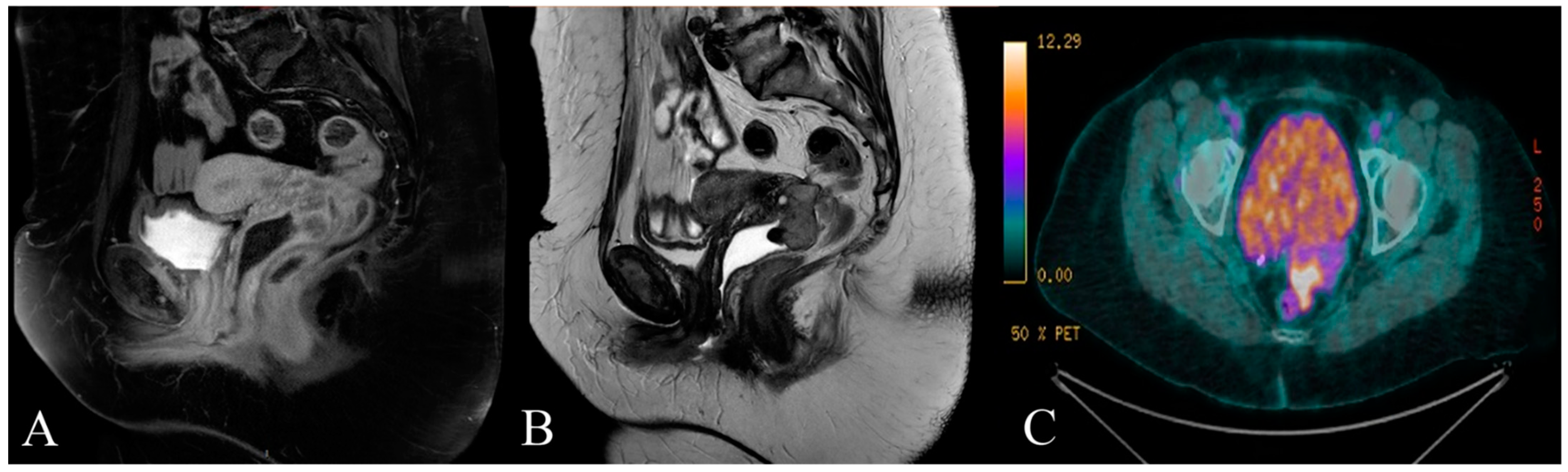
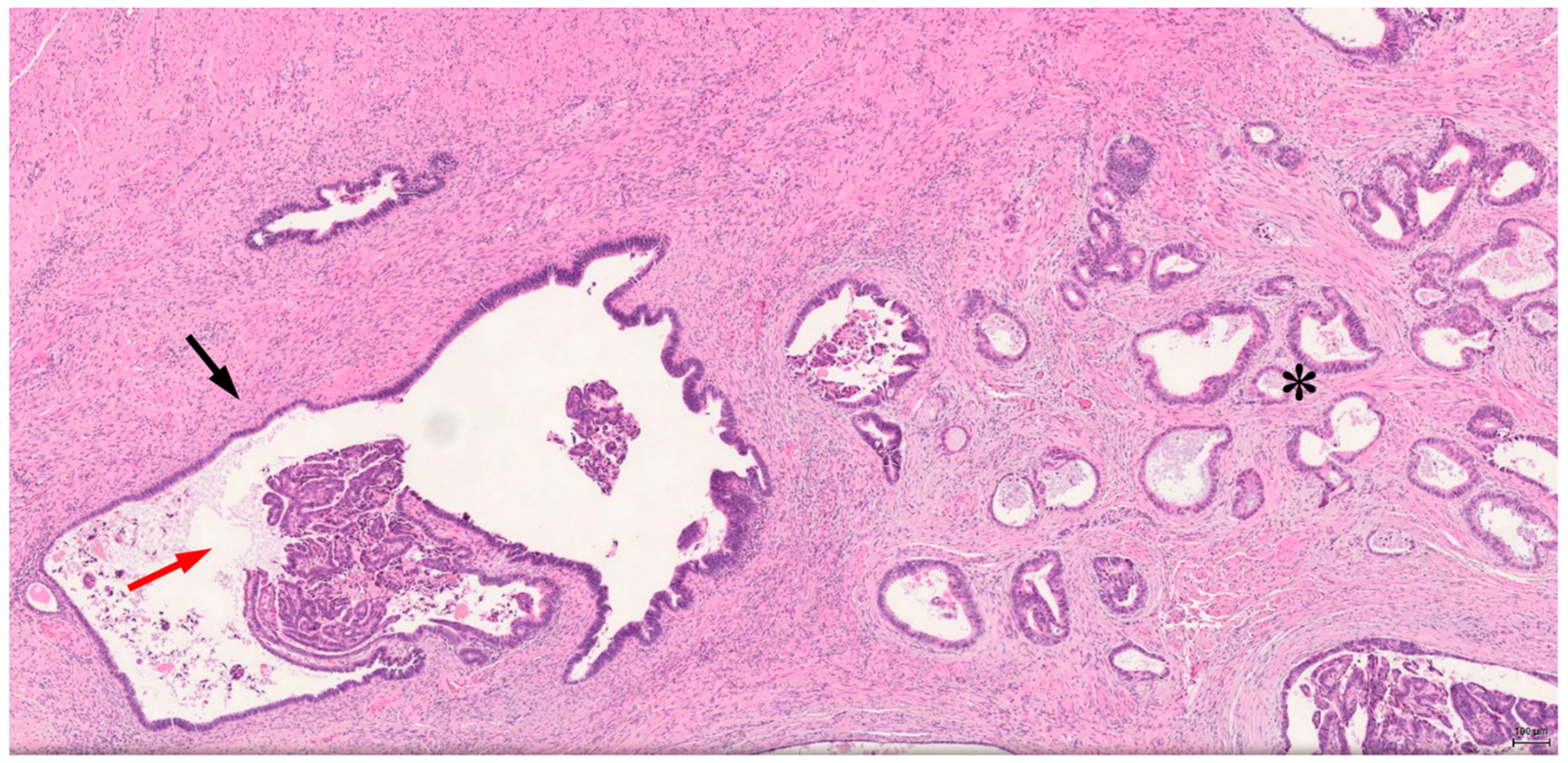
| Author | Grade | Location | Size (cm) | Age | Menopause | Nodes | Treatment | Evolution (No Recurrence) |
|---|---|---|---|---|---|---|---|---|
| Lim et al. 2009 [10] | 2 | 1/3 anterior vaginal wall, urethrovaginal septum | 4.3 × 4.2 | 61 | na | N0 | Anterior pelvic exenteration, BSO, pelvic lymph node dissection, urostomy with ileal conduct | 24 months |
| Fruscio et al. 2008 [11] | na | Upper third of the left vaginal wall | 3 × 3.5 | 40 | No | N0 | Neo-adjuvant chemotherapy, radical hysterectomy, BSO, bilateral pelvic and lomboaortic lymph node sampling, resection of left vaginal wall | 24 months |
| Lavery and Gillmer 2001 [12] | na | Vaginal vault | 5 × 3 | 50 | Yes | na | Upper vaginectomy, colon resection (8 cm), and reanastomosis | na |
| Lavery and Gillmer 2001 [12] | na | Left side of vaginal vault | na | 53 | Yes | na | None (non debulkable) | na |
| Fishman et al. 1996 [13] | na | na | na | 47 | Yes | na | Posterior exenteration and RT | 8 years |
| Fishman et al. 1996 [13] | na | na | na | 45 | Yes | na | Radical upper vaginectomy | 7 years |
| Haskel et al. 1988 [27] | na | Upper posterior vagina | na | 53 | Yes | N+ | TAH, vaginectomy, BSO, pelvic and para-aortic lymph node sampling, omental biopsy, partial colpectomy and RT | >24 months |
| Orr et al. 1989 [28] | na | Upper vagina | 6 × 4 × 4 | 60 | Yes | N+ | Resection with low rectal anastomosis, pelvic lymph node dissection, paraaortic node biopsy, and omentectomy | 6 months |
| Granai et al. 1984 [29] | na | Right vaginal apex | na | 47 | Yes | na | Laparotomy (enterolysis), RT, and hormonal therapy | 20 months |
| Kapp et al. 1982 [30] | nc | Posterior vaginal wall | 3 × 4 | 35 | Yes | na | RT | 7 years |
| Hyman 1977 [31] | na | na | na | 50 | na | na | None | na |
| Decelle 1969 [32] | na | na | na | 56 | na | N+ | TAH, BSO, nodal sampling, cystectomy | NA |
| Bamford 1967 [33] | na | Right posterolateral mid-third of vaginal wall | na | 42 | No | na | Hysterovaginectomy | 5 months |
| Author | Location | Size (cm) | Age | Menopause | Nodes | Surgery | Adjuvant Chemotherapy | Evolution (No Recurrence) |
|---|---|---|---|---|---|---|---|---|
| Okimura et al. 2018 [14] | Diaphragmatic | 2.4 × 1.6 | 59 | Yes | N+ | Partial resection of diaphragm and liver, BSO, partial omentectomy | 6 cy of carboplatin- paclitaxel | 6 months |
| Lee et al. 2017 [15] | Left paracolic area above the infundibulo-pelvic ligament | 10 × 10 × 8 | 53 | Yes | na | Hysterectomy, BSO, pelvic/para-aortic lymph node dissection, omentectomy | No | 26 months |
| Palla et al. 2017 [22] | Sigmoid colon | 6 | 73 | Yes | na | Sigmoidectomy | No | na |
| Ogi et al. 2016 [23] | Small intestine (ileum) | 6.5 × 4 | 55 | Yes | na | Partial small intestinal resection | No | 5 years |
| Jaiman et al. 2015 [24] | Recto-uterine pouch adherent to the right broad ligament and pelvic wall | 8.8 × 6.5 | 45 | na | na | Hysterectomy, BSO, and mass resection | 6 cy chemotherapy | na |
| Makihara et al. 2015 [25] | Small intestine (ileal mesentery) | 9.5 × 5.5 × 5 | 25 | No | N+ | Partial small intestinal resection | Yes: refusal | 10 months |
| Tarumi et al. 2015 [26] | Bladder | 2.3 | 45 | na | na | Total laparoscopic hysterectomy, partial bladder resection, insertion of bilateral ureteral stents; BO and omentectomy refused by patient | 6 cy docetaxel—carboplatin | 10 months |
| Bawazeer et al. 2014 [16] | Pelvic-abdominal | 16.7 × 10 × 14 | 53 | na | na | Mass excision | 6 cy carboplatin every 3 weeks | End of 6 cy |
| Caballero et al. 2013 [17] | na | 6.5 × 5.6 × 6.8 | 39 | na | na | Rectal anterior resection, hysterectomy, BO | chemotherapy | 1 year |
| Micha et al. 2011 [18] | Pelvic; sigmoid colon, left ureter | 9 cm tumor in the pelvis | 52 | Yes | N0 | Debulking with sigmoid colon resection with low rectal anastomosis, pelvic tumor debulking, bilateral pelvic ureterolysis, lymphadenectomy, appendicectomy, and omental biopsy | cisplatin, RT, and tamoxifen | 5 years |
| Agrawalet al. 2009 [19] | na | na | 50 | na | na | Resection of right anterior chest wall, partial resection of the diaphragm | 6 cy paclitaxel-carboplatin, megestrol acetate | 29 months |
| Park et al. 2009 [20] | Uterine cervix | 3 × 2.2 × 2.2 | 48 | No | na | TAH, BSO | 6 cy cyclophosphamide—cisplatin | 24 months |
| Susumu et al. 2005 [21] | Mesenterium of the sigmoid colon | 6 × 5 × 5 | 62 | Yes | N+ | Sigmoidectomy and lymph node resection | na | 28 months |
Publisher’s Note: MDPI stays neutral with regard to jurisdictional claims in published maps and institutional affiliations. |
© 2021 by the authors. Licensee MDPI, Basel, Switzerland. This article is an open access article distributed under the terms and conditions of the Creative Commons Attribution (CC BY) license (https://creativecommons.org/licenses/by/4.0/).
Share and Cite
Costanza, M.; Herrera, F.; Hastir, D.; Mathevet, P.; Sarivalasis, A. A Locally Advanced Endometrioid Adenocarcinoma Arising from Vaginal Endometriosis: Management and Review of the Literature. Reports 2021, 4, 29. https://doi.org/10.3390/reports4030029
Costanza M, Herrera F, Hastir D, Mathevet P, Sarivalasis A. A Locally Advanced Endometrioid Adenocarcinoma Arising from Vaginal Endometriosis: Management and Review of the Literature. Reports. 2021; 4(3):29. https://doi.org/10.3390/reports4030029
Chicago/Turabian StyleCostanza, Mariangela, Fernanda Herrera, Delfyne Hastir, Patrice Mathevet, and Apostolos Sarivalasis. 2021. "A Locally Advanced Endometrioid Adenocarcinoma Arising from Vaginal Endometriosis: Management and Review of the Literature" Reports 4, no. 3: 29. https://doi.org/10.3390/reports4030029
APA StyleCostanza, M., Herrera, F., Hastir, D., Mathevet, P., & Sarivalasis, A. (2021). A Locally Advanced Endometrioid Adenocarcinoma Arising from Vaginal Endometriosis: Management and Review of the Literature. Reports, 4(3), 29. https://doi.org/10.3390/reports4030029