Case Report

Chilaiditi’s Syndrome—What Every Endoscopist Should Know

1
General Surgical Department, Chesterfield Royal Hospital Foundation Trust, Chesterfield Road, Chesterfield S44 5BL, UK
2
Radiology Department, Chesterfield Royal Hospital Foundation Trust, Chesterfield Road, Chesterfield S44 5BL, UK
*
Author to whom correspondence should be addressed.
Reports 2020, 3(2), 11; https://doi.org/10.3390/reports3020011
Submission received: 27 March 2020 / Revised: 15 April 2020 / Accepted: 15 April 2020 / Published: 22 April 2020

Abstract

:
Chilaiditi’s syndrome is a rare and often asymptomatic anomaly, typically found as an incidental radiographic sign (gas under the diaphragm) due to hepato-diaphragmatic interposition of the transverse colon. We report a case of Chilaiditi’s syndrome following colonoscopy presenting with severe abdominal pain, dyspnoea and radiograph findings similar to the presence of bowel perforation (appearance of gas under the hemidiaphragm on erect chest radiograph). Computed tomography (CT) evidence of Chilaiditi’s sign prevented unnecessary laparotomy.

1. Introduction

Chilaiditi’s syndrome was first described by Demetrius Chilaiditi in 1910, it is a rare condition with acute abdominal symptoms resulting from entrapment of gas within or volvulus of an unusually mobile, anteriorly displaced transverse colon interposed between the liver and right hemidiaphragm (Chilaiditi’s sign) [1].
Chilaiditi’s syndrome has an incidence of 0.25% to 0.28% with a 4:1 male to female ratio [2,3,4]. This is normally diagnosed as an incidental finding on chest and abdominal radiographs [5].
We present a case of a healthy male who presented with severe abdominal pain following a colonoscopy which was thought to be due to a perforated viscus on erect radiograph; however, this was due to Chilaiditi’s syndrome.

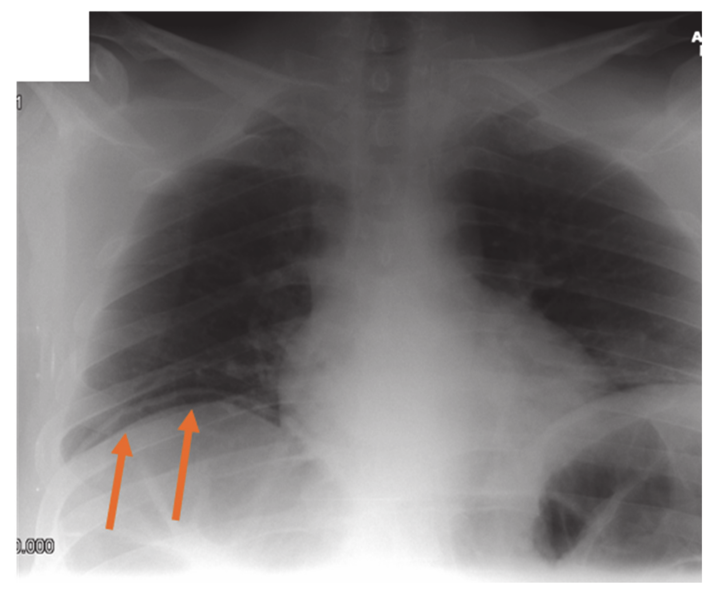
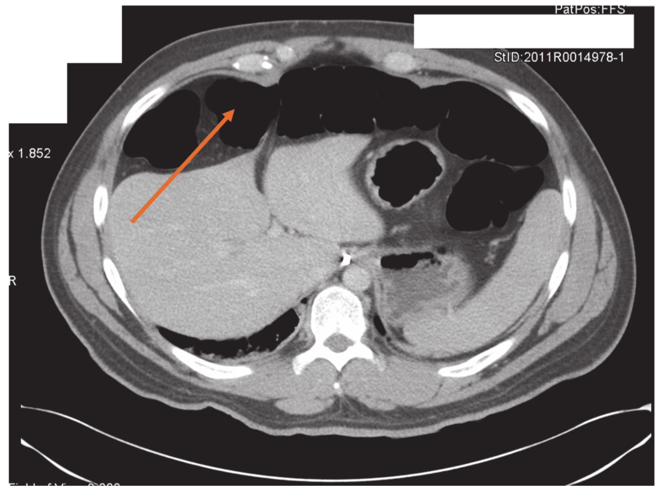
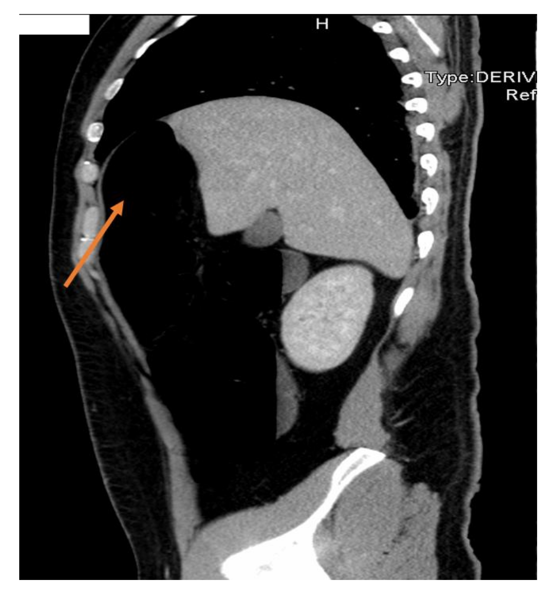
2. Case Presentation Section

A 31-year-old healthy male presented for elective colonoscopy and banding of haemorrhoids. Bowel preparation was performed with PicoPrep (Pharmatel Fresenius Kabi, Hornsby NSW). The procedure was performed in the left lateral position. Sedation and analgesia were achieved with Midazolam (2 mg), Alfentanyl (500 μg) and Propofol (cumulative total of 400 mg). The proceduralist was a colorectal surgeon and an accredited endoscopist (Joint Advisory Group on GI Endoscopy, UK). Regular air was used for the procedure and no abdominal pressure was required. No biopsy or polypectomy was performed. The procedure was completed uneventfully with little difficultly and took approximately thirty minutes. Banding of the haemorrhoids was performed at the end of the procedure via a proctoscope.
In the post-anaesthetic recovery room, the patient complained of severe abdominal pain, predominantly in the epigastric and right-upper-quadrant regions, colicky in character, reaching pain scores of 10/10 intermittently and worse on inspiration.
On examination, blood pressure was 135/80 mmHg, heart rate 72 beats per minute, respiratory rate was 24 respirations a minute with shallow breathing, pulse oximetry was 99% mmHg on 4 L of oxygen via nasal cannulae. The abdomen was soft but distended and tender in the epigastrium. Administration of 200 μg of Fentanyl, 100 mg of Tramadol, 1 g of Paracetamol and 10 mg of Buscopan did little to alleviate the pain. Given the severity of the pain, a chest radiograph was performed to exclude bowel perforation (Figure 1). This appeared to indicate free gas under the right hemidiaphragm and a large collection of gas in the left subdiaphragmatic region.
A subsequent CT scan revealed a segment of the transverse colon distended with gas and interposed between the liver and right hemidiaphragm anteriorly (Chilaiditi’s sign) (Figure 2 and Figure 3). There was no evidence of intraperitoneal gas. A large quantity of gas was also seen in the remaining segments of the colon.
Passage of a nasogastric tube was not successful in alleviating the pain. Subsequent management was conservative, requiring morphine patient-controlled analgesia and reassurance. A large quantity of flatus was passed in the ensuing few hours resulting in resolution of the pain.

3. Discussion

Multiple Chilaiditi’s case reports have been described in the literature [2]. One particular report identified a 58-year-old female presenting two days following her colonoscopy with right-upper-quadrant pain who failed conservative management and required a pexy of the ascending colon and caecum [6]. This case is different from ours as the patient’s symptoms resolved without the need for surgical intervention.
Other clinical scenarios when the syndrome may present include: cholecystitis, pancreatitis [7], blunt abdominal trauma [8], following upper GI endoscopic procedures, inexplicable shortness of breath, recurrent volvulus of the colon, extrinsic right bronchial compression on bronchoscopy [9] and in the presence of ascites [10]. Presentation with angina-like chest pains [11] and cardiac tamponade [12] have also been reported due to the proximity of the colon to the heart.
The condition can make a routine colonoscopy close to impossible [13]. This can predispose patients to perforation and trapped gas as occurred in this instance [3]. To our knowledge, there is only one case previously reporting the manifestation of the syndrome following successful colonoscopy; however, this case required operative intervention whereas our case settled with conservative management.
Pseudo-pneumoperitoneum is a term that has been used to describe radiograph and CT evidenced intra-peritoneal gas without clear evidence of perforated viscus. In these instances, it has been postulated that micro-perforations and trans-diaphragmatic movement of gas are possible mechanisms.
The cause of the anterior displacement of the transverse colon has been attributed to an abnormal long colon (dolichocolon) and/or laxity/absence of a falciform ligament, potentially coupled with liver cirrhosis/reduced hepatic volume or phrenic nerve palsy; resulting in an unusually mobile transverse colon [2].
An erect chest radiograph is extremely sensitive if performed correctly. CT scans (with or without contrast) have a higher sensitivity and specificity for detection of intra-peritoneal gas. Diagnosis has also been made with abdominal ultrasound [14].
Patients presenting with an acute surgical abdomen should be investigated with an erect chest radiograph, as well as the standard supine abdominal radiograph. The patient should be positioned sitting upright for 10–20 min before acquiring the erect chest radiograph image. This allows any free intra-abdominal gas to rise, forming a crescent beneath the diaphragm. It is said that as little as 1 mL of gas can be detected in this way. In our hospital, we use erect chest radiographs in the first instance before considering CT scans, but this is always correlated with the patients’ clinical need.
Increasing numbers of GI endoscopic procedures are being performed with anaesthetists in attendance. Knowledge of this unusual syndrome and its management is important in the differential diagnosis of severe abdominal pain and shortness of breath post-procedure. If, after a seemingly straightforward endoscopic procedure, perforation of a viscus is suspected, and plain abdominal radiograph indicates free air under the diaphragm, but clinical findings are not consistent with radiograph findings (i.e., a soft abdomen), we would consider it mandatory to confirm radiograph findings with a CT scan to avoid unnecessary surgical intervention.

4. Conclusions

Chilaiditi’s syndrome is a rare cause of abdominal symptoms due to entrapment of gas within or volvulus of an unusually mobile, anteriorly displaced transverse colon interposed between the liver and right hemidiaphragm. Chilaiditi’s syndrome can complicate endoscopic procedures. This can lead to apparent free air being seen on radiographs. Careful clinical correlation is warranted to negate surgical intervention.

Author Contributions

J.G.: Developing, proofing the manuscript and editing the images. A.V.G.: Proofing the manuscript and annotating the images. T.J.W.: Proofing the manuscript and concept development. All authors have read and agree to the published version of the manuscript.

Funding

This research received no external funding.

Acknowledgments

Consent was gained directly from the patient.

Conflicts of Interest

The authors declare no conflict of interest.

References

  1. Chilaiditi, D. Zur frage der hepatoptose und ptose im allgemeinen im anschluss an drei fälle von temporärer, partieller leberverlagerung. Fortschr. Geb. Rontgenstr. 1910, 16, 173–208. [Google Scholar]
  2. Saber, A.A.; Boros, M.J. Chilaiditi’s syndrome: What should every surgeon know? Am. Surg. 2005, 71, 261–263. Available online: http://www.ncbi.nlm.nih.gov/pubmed/15869145 (accessed on 22 March 2020). [PubMed]
  3. Gurvits, G.E.; Lau, N.; Gualtieri, N.; Robilotti, J.G. Air under the right diaphragm: Colonoscopy in the setting of Chilaiditi syndrome. Gastrointest. Endosc. 2009, 69, 758–759. [Google Scholar] [CrossRef] [PubMed]
  4. Nair, N.; Takieddine, Z.; Tariq, H. Colonic Interposition between the Liver and Diaphragm: “The Chilaiditi Sign”. Can. J. Gastroenterol. Hepatol. 2016, 2016. [Google Scholar] [CrossRef] [PubMed] [Green Version]
  5. Uygungul, E.; Uygungul, D.; Ayrik, C.; Narcı, H.; Babuş, S.B.; Köse, A. Chilaiditi sign: Why are clinical findings more important in ED? Am. J. Emerg. Med. 2015, 33, 733.e1–733.e2. [Google Scholar] [CrossRef] [PubMed]
  6. Yin, A.X.; Park, G.H.; Garnett, G.M.; Balfour, J.F. Chilaiditi syndrome precipitated by colonoscopy: A case report and review of the literature. Hawaii J. Med. Public Health 2012, 71, 158–162. Available online: http://www.ncbi.nlm.nih.gov/pubmed/22787564 (accessed on 26 March 2020). [PubMed]
  7. Gravante, G.; Wong, C.; Kelly, M. ‘Air under the diaphragm’ during acute pancreatitis/cholangitis. ANZ J. Surg. 2011, 81, 302–303. [Google Scholar] [CrossRef] [PubMed]
  8. Farkas, R.; Moalem, J.; Hammond, J. Chilaiditi’s sign in a blunt trauma patient: A case report and review of the literature. J. Trauma Inj. Infect. Crit. Care 2008, 65, 1540–1542. [Google Scholar] [CrossRef] [PubMed]
  9. Messina, M.; Paolucci, E.; Casoni, G.L.; Gurioli, C.; Poletti, V. A case of severe dyspnea and an unusual bronchoscopy: The Chilaiditi syndrome. Respiration 2008, 76, 216–217. [Google Scholar] [CrossRef] [PubMed]
  10. Bhattacharya, P.C.; Bhattacharya, A.K.; Dutta, S.; Mahanta, N.; Talukdar, R. Chilaiditi syndrome with ascites. J. Assoc. Physicians India 2002, 50, 860–861. Available online: http://www.ncbi.nlm.nih.gov/pubmed/12240875 (accessed on 22 March 2020). [PubMed]
  11. Sorrentino, D.; Bazzocchi, M.; Badano, L.P.; Toso, F.; Giagu, P. Heart-touching Chilaiditi’s syndrome. World J. Gastroenterol. 2005, 11, 4607–4609. [Google Scholar] [CrossRef] [PubMed]
  12. Makhija, Z.; Shaygi, B.; Deshpande, R.; Marrinan, M. Delayed cardiac tamponade following posttraumatic diaphragmatic hernia without an intrapericardial component. Interact. Cardiovasc. Thorac. Surg. 2009, 9, 132–133. [Google Scholar] [CrossRef] [PubMed]
  13. Bassan, M.; Thomson, A. Gastrointestinal: Chilaiditi syndrome. J. Gastroenterol. Hepatol. 2008, 23, 499. [Google Scholar] [CrossRef] [PubMed]
  14. Jones, R. Recognition of pneumoperitoneum using bedside ultrasound in critically ill patients presenting with acute abdominal pain. Am. J. Emerg. Med. 2007, 25, 838–841. [Google Scholar] [CrossRef] [PubMed]
Figure 1. Erect chest-radiograph showing the appearance of free gas under the diaphragm (orange arrows).
Reports 03 00011 g001
Figure 2. Axial computed tomography scan shows gas-filled large bowel between the liver and anterior abdominal wall (orange arrow).
Reports 03 00011 g002
Figure 3. Sagittal computed tomography scan showing distended large bowel anterior to the liver (orange arrow).
Reports 03 00011 g003

