Case Report

Mechanical Duodenal Perforation Due to Complications of Pancreatic Pseudocysts

1
General Surgical Department, Chesterfield Royal Hospital Foundation Trust, Chesterfield Road, Chesterfield S44 5BL, UK
2
Radiology Department, Chesterfield Royal Hospital Foundation Trust, Chesterfield Road, Chesterfield S44 5BL, UK
*
Author to whom correspondence should be addressed.
Reports 2020, 3(2), 10; https://doi.org/10.3390/reports3020010
Submission received: 25 March 2020 / Revised: 15 April 2020 / Accepted: 15 April 2020 / Published: 22 April 2020

Abstract

:
Pancreatic pseudocysts are a known complication of pancreatitis. There are a multitude of complications which have been described in relation to pancreatic pseudocysts. Perforation of a gastrointestinal hollow viscus is rare but has previously been reported. We report a case of a 72-year-old female with the development of three pancreatic pseudocysts four weeks post gallstone pancreatitis. The patient deteriorated and was found to have a duodenal perforation due to compression forces from the underlying pseudocysts. The patient required emergency surgical intervention and recovered fully following hospital admission.

1. Introduction

There are four types of pancreatic fluid collections as defined by the Atlanta guidance [1]. Pancreatic pseudocyst is one of these fluid collections. The pseudocysts are a maturing collection of pancreatic juice, encased by reactive granulation tissue, occurring in or around the pancreas because of pancreatitis (acute or chronic) or ductal leakage [2]. Pseudocysts are defined as pancreatic fluid collections, greater than four weeks old, surrounded by a defined non-epithelialised wall [3]. Several complications have been described relating to pancreatic pseudocysts [4]. We present a rare case of a large pancreatic pseudocyst leading to mechanical duodenal perforation. To our knowledge, this is the first case of this kind reported.

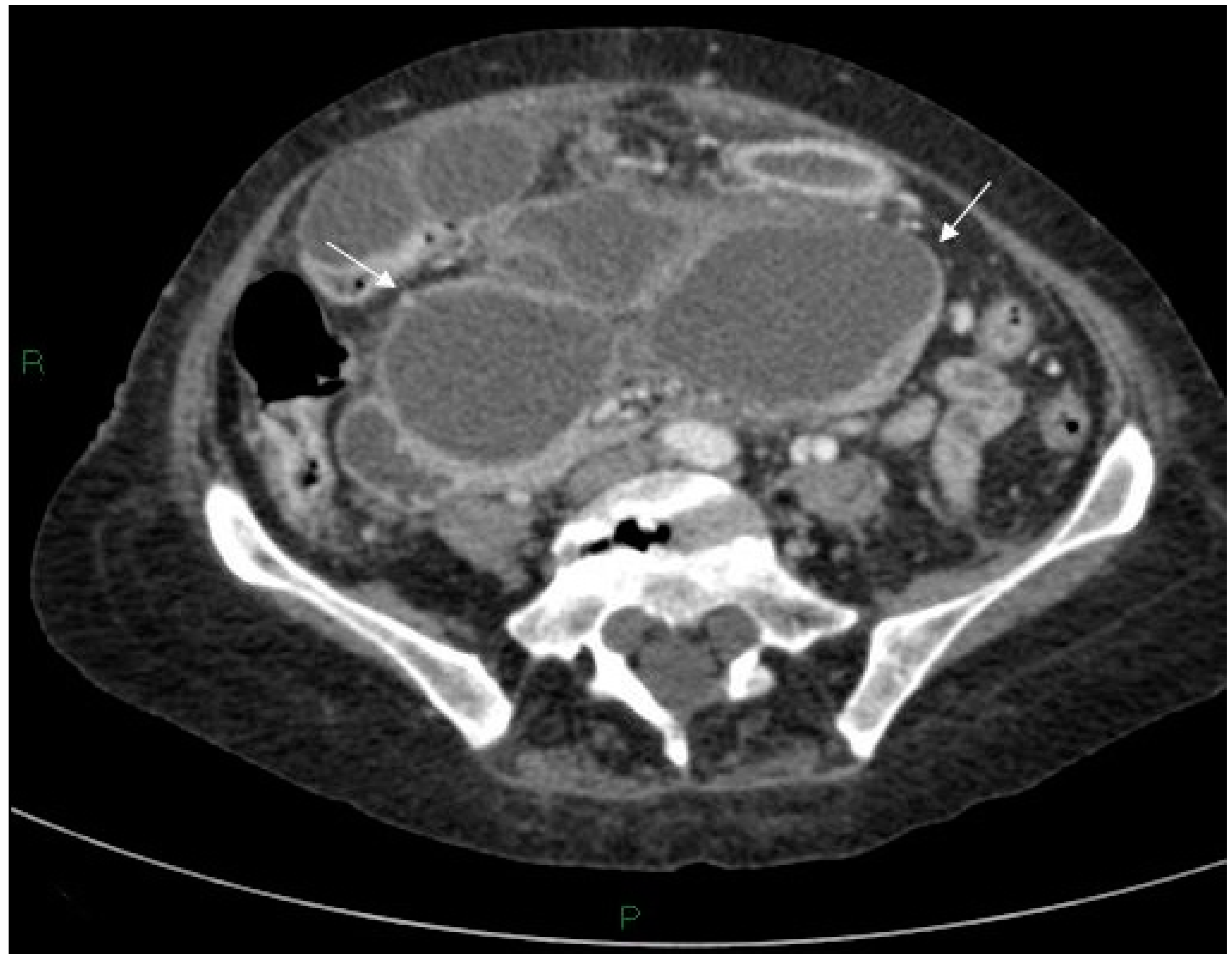
2. Case Presentation Section

A 72-year-old female attended for a laparoscopic cholecystectomy on the background of severe gallstone pancreatitis four weeks previously. Examination prior to her operation revealed that she had an upper abdominal mass. The procedure was cancelled, and computed tomography (CT) was performed. This revealed a pseudocyst with three components—a large 16 × 12 cm thin-walled cyst in relation to the head, body and proximal tail of the pancreas, a separate 6 cm cyst in the distal tail, and a 10 × 5 cm cyst in the area of the transverse mesocolon compressing the transverse colon (Figure 1 and Figure 2).
Advice from the regional hepatico–pancreatico–biliary (HPB) unit was for conservative management until the walls of the pseudocysts matured, when open drainage and cholecystectomy could be considered if symptoms did not resolve.
On day six her clinical condition deteriorated, she was pyrexial and haemodynamically unstable. The patient complained of increased abdominal pain and had a raised white cell count and a raised arterial lactate level. An erect chest x-ray revealed free air under the right hemidiaphragm. Following initial resuscitation and administration of antibiotics, a CT was performed revealing the known pseudocyst with anterior compression of the stomach, free fluid in the abdomen and considerable free intra-abdominal air (Figure 3, Figure 4 and Figure 5).
An emergency laparotomy was performed, and a large 1.5 cm split in the first part of the duodenum with free bile in the peritoneal cavity was noted. In the absence of a duodenal ulcer and ischaemic change, the cause of this mechanical perforation could only be attributed to excessive compression of the duodenum from the underlying pseudocyst. The gallbladder was densely adherent to the inflammatory phlegmon and was left untouched. Cystogastrostomy, pyloroplasty, omental patch and placement of nasojejunal feeding tube were performed. A drain was placed in Morrison’s pouch. The patient was then transferred to critical care postoperatively, where she remained for seventeen days. She was discharged home on postoperative day thirty. Consent was gained directly from the patient.

3. Discussion

Pancreatic pseudocysts can cause a multitude of complications which have previously been reported—including gastric outlet obstruction from compression of the stomach or duodenum, infection, pseudoaneurysm formation and bleeding, rupture of the cysts either intra-abdominally or into a viscus, stress-related gastrointestinal ulcers and fistulas [4,5]. Gastrointestinal perforation related to pseudocysts has previously been described, but it almost always followed a rupture of a cyst into the lumen of the bowel [6]. An example of this is an unusual case of colonic perforation reported in a 71-year-old male with a 10.5 cm pseudocyst which ruptured causing perforation at the splenic flexure [7].
Cases of gastric perforation have also been described in literature following focal gastric necrosis due to compression of the splenic artery [8], and gastric stress ulcers have also been recognised as a complication of pancreatitis and pancreatic pseudocysts [9]. These symptoms of compression are well noted and similar to our case, however in our case the perforation occurred in the duodenum. Pseudocyst formations post-traumatic injury have also been noted [10]. An example of this was a case of duodenal perforation was reported in a 23-year-old male with a large pancreatic pseudocyst which formed five days following minor blunt abdominal trauma [11]. This case is different from ours as there were no traumatic sequelae.
Most pseudocysts are singular [12]. Cases with multiple pseudocysts have been described mainly following episodes of acute alcoholic pancreatitis [13]. Our patient had three pancreatic pseudocysts following a single episode of severe acute gallstone pancreatitis four weeks earlier. She developed a duodenal perforation while being managed conservatively, with no precipitating factor such as trauma leading to this event. No duodenal ulcer or rupture of the cyst to the duodenum was identified intraoperatively. The duodenum was markedly stretched by this large pseudocyst leading to the perforation. To our knowledge this is the first case described in literature.
It has been suggested that the pseudocysts at highest risk of complications are those following an episode of acutely severe gallstone pancreatitis and particularly those involving the neck and the body of the pancreas [14]. This is in keeping with our case.
A Cochrane review has looked at the different treatment options for pancreatic pseudocysts [15]. Percutaneous drainage, endoscopic and endoscopic ultrasound drainage and open surgical management have all been described [15,16]. These procedures are not without serious adverse events but the highest was seen in open surgical drainage (15%)—however, the review did not reach statistical significance [15]. The decision to treat, which method to choose, and also the timing of the procedure have been debated throughout the literature [13,14]. Our patient acutely deteriorated and as such an urgent open intervention was required.
This patient required a cholecystectomy to prevent recurrent attacks of pancreatitis. There is some debate over the correct timing of cholecystectomy following an attack for gallstone pancreatitis. A Cochrane review in 2013 found that early cholecystectomy presents no evidence of increased complications, as well as suggesting that early cholecystectomy was favoured to delayed cholecystectomy in skilled hands for the treatment of mild gallstone pancreatitis [17]. Our case was a severe attack of gallstone pancreatitis and there needs further data to look at this for management. We would have ideally operated on this patient sooner, but this was not possible due to the delayed recovery of the patient and theatre availability. It is unclear as to whether an earlier operation would have prevented the outcome as the damage to the pancreas was done at the initial presentation.
This case demonstrates large pseudocysts are at a greater risk of complications and the mass effect caused by the pseudocysts may be directly responsible for the mechanical perforation of the duodenum or other hollow viscera and should be considered especially when there is clinical deterioration.

4. Conclusions

Pseudocysts have can cause a multitude of symptoms. To our knowledge, this is the first case to report a duodenal perforation following compression forces of pseudocysts due to gallstone pancreatitis. Pseudocyst management is challenging and should be taken on a case by case basis. Deterioration in the patient’s clinical status prompts early intervention.

Author Contributions

J.G., developing, proofing the manuscript and editing the images; C.F., developing the manuscript; H.J.H., editing and ascertaining images; K.R., proofing the manuscript; T.J.W., proofing the manuscript and concept development. All authors have read and agreed to the published version of the manuscript.

Funding

This research received no external funding.

Conflicts of Interest

The authors declare no conflict of interest.

References

  1. Banks, P.A.; Bollen, T.L.; Dervenis, C.; Gooszen, H.; Johnson, C.D.; Sarr, M.G.; Tsiotos, G.G.; Vege, S.S. Classification of acute pancreatitis—2012: Revision of the Atlanta classification and definitions by international consensus. Gut 2012, 62, 102–111. [Google Scholar] [CrossRef] [PubMed]
  2. D’Egidio, A.; Schein, M. Pancreatic pseudocysts: A proposed classification and its management implications. BJS 1991, 78, 981–984. [Google Scholar] [CrossRef] [PubMed]
  3. Bradley, E.L. A Clinically Based Classification System for Acute Pancreatitis. Arch. Surg. 1993, 128, 586–590. [Google Scholar] [CrossRef] [PubMed]
  4. Wang, Y.; Abu Omar, Y.; Agrawal, R.; Gong, Z. Comparison of treatment modalities in pancreatic pseudocyst: A population based study. World J. Gastrointest. Surg. 2019, 11, 365–372. [Google Scholar] [CrossRef] [PubMed]
  5. Varadarajulu, S.; Bang, J.Y.; Sutton, B.S.; Trevino, J.M.; Christein, J.D.; Wilcox, C.M. Equal Efficacy of Endoscopic and Surgical Cystogastrostomy for Pancreatic Pseudocyst Drainage in a Randomized Trial. Gastroenterology 2013, 145, 583–590. [Google Scholar] [CrossRef] [PubMed]
  6. King, B.; Speziale, A. Gastric perforation caused by a pancreatic pseudocyst. Gastrointest. Endosc. 2011, 74, 1403–1404. [Google Scholar] [CrossRef] [PubMed]
  7. Aghenta, A.A.; Kim, H.J. An Unusual Case of Colon Perforation Complicating Acute Pancreatitis. Case Rep. Gastroenterol. 2009, 3, 207–213. [Google Scholar] [CrossRef] [PubMed]
  8. Ertuğrul, I.; Yüksel, I.; Parlak, E.; Başar, Ö.; Uçar, E.; Şahin, B. Gastric Necrosis Due to Rapidly Growing Pancreatic Pseudocyst. Am. J. Gastroenterol. 2008, 103, 2949–2951. [Google Scholar] [CrossRef]
  9. Hsu, C.-Y.; Lee, K.-C.; Chan, C.-C.; Lee, F.-Y.; Lin, H.-C. Gastric Necrosis and Perforation as a Severe Complication of Pancreatic Pseudocyst. J. Chin. Med. Assoc. 2009, 72, 603–606. [Google Scholar] [CrossRef] [Green Version]
  10. Cannon, J.W.; Callery, M.P.; Vollmer, C.M. Diagnosis and Management of Pancreatic Pseudocysts: What is the Evidence? J. Am. Coll. Surg. 2009, 209, 385–393. [Google Scholar] [CrossRef] [PubMed]
  11. Tuboku-Metzger, V.R.; Seenath, M.M.; Tan, L.C. Peritonitis secondary to traumatic duodenal laceration in the presence of a large pancreatic pseudocyst: A case report. J. Med Case Rep. 2011, 5, 528. [Google Scholar] [CrossRef] [Green Version]
  12. Pitchumoni, C.; Agarwal, N. Pancreatic Pseudocysts. Gastroenterol. Clin. N. Am. 1999, 28, 615–639. [Google Scholar] [CrossRef]
  13. Andren-Sandberg, A.; Dervenis, C. Pancreatic pseudocysts in the 21st century. Part I: Classification, pathophysiology, anatomic considerations and treatment. JOP J. Pancreas 2004, 5, 8–24. [Google Scholar]
  14. Ocampo, C.; Oría, A.; Zandalazini, H.; Silva, W.; Kohan, G.; Chiapetta, L.; Alvarez, J. Treatment of Acute Pancreatic Pseudocysts After Severe Acute Pancreatitis. J. Gastrointest. Surg. 2007, 11, 357–363. [Google Scholar] [CrossRef]
  15. Gurusamy, K.S.; Pallari, E.; Hawkins, N.; Pereira, S.P.; Davidson, B.R. Management strategies for pancreatic pseudocysts. Cochrane Database Syst. Rev. 2016, 2016. [Google Scholar] [CrossRef]
  16. Aghdassi, A.A.; Mayerle, J.; Kraft, M.; Sielenkämper, A.W.; Heidecke, C.-D.; Lerch, M.M. Pancreatic pseudocysts—When and how to treat? HPB 2006, 8, 432–441. [Google Scholar] [CrossRef] [PubMed] [Green Version]
  17. Gurusamy, K.S.; Nagendran, M.; Davidson, B.R. Early versus delayed laparoscopic cholecystectomy for acute gallstone pancreatitis. Cochrane Database Syst. Rev. 2013, 2013, CD010326. [Google Scholar]
Figure 1. Axial post-contrast computed tomography (CT) scan of the upper abdomen showing a large pancreatic pseudocyst with the stomach (arrows) stretched anteriorly over it.
Reports 03 00010 g001
Figure 2. Axial post-contrast CT scan of the upper abdomen showing the inferior aspect of the cyst with bi-lobed appearance (arrows) in the pelvis.
Reports 03 00010 g002
Figure 3. Axial CT scan of the abdomen showing free air anterior to the liver (white arrows) and free fluid around the liver and spleen (black arrows).
Reports 03 00010 g003
Figure 4. Axial CT scan of abdomen showing free air anterior to the liver (white arrows) and free fluid around the liver and spleen (black arrows).
Reports 03 00010 g004
Figure 5. Initially, Axial CT scan showing the stomach containing a nasogastric tube anterior to the cyst (large white arrow), free air (small white arrows) and inflammatory changes in the soft tissues between the stomach/duodenum and the cyst (black arrow).
Reports 03 00010 g005

