Review

Nanomedicine-Based Advances in Brain Cancer Treatment—A Review

by
Borish Loushambam
1,
Mirinrinchuiphy M. K. Shimray
1,
Reema Khangembam
1,
Venkateswaran Krishnaswami
2 and
Sivakumar Vijayaraghavalu
1,*
1
Department of Life Sciences, Manipur University (A Central University), Imphal 795003, MN, India
2
Department of Pharmaceutics, SA Raja Pharmacy College, Vadakangulam, 3, Tirunelveli 627116, TN, India
*
Author to whom correspondence should be addressed.
Neuroglia 2025, 6(3), 28; https://doi.org/10.3390/neuroglia6030028
Submission received: 6 May 2025 / Revised: 15 July 2025 / Accepted: 15 July 2025 / Published: 18 July 2025

Abstract

Brain cancer is a heterogeneous collection of malignant neoplasms, such as glioblastoma multiforme (GBM), astrocytomas and medulloblastomas, with high morbidity and mortality. Its treatment is complicated by the tumor’s site, infiltrative growth mode and selective permeability of the blood–brain barrier (BBB). During tumor formation, the BBB dynamically remodels into the blood–brain tumor barrier (BBTB), disrupting homeostasis and preventing drug delivery. Furthermore, the TME (Tumor Micro Environment) supports drug resistance, immune evasion and treatment failure. This review points out the ways in which nanomedicine overcomes these obstacles with custom-designed delivery systems, sophisticated diagnostics and personalized therapies. Traditional treatments fail through a lack of BBB penetration, non-specific cytotoxicity and swift tumor adaptation. Nanomedicine provides greater drug solubility, protection against enzymatic degradation, target drug delivery and control over the release. Nanotheranostics’ confluence of therapeutic and diagnostic modalities allows for dynamic adjustment and real-time monitoring. Nanotechnology has paved the way for the initiation of a new era in precision neuro-oncology. Transcending the limitations of conventional therapy protocols, nanomedicine promises to deliver better outcomes by way of enhanced targeting, BBB penetration and real-time monitoring. Multidisciplinary collaboration, regulatory advancements and patient-centered therapy protocols customized to the individual patient’s tumor biology will be necessary to facilitate translation success in the future.

1. Introduction

1.1. Brain Cancer: Its Types and Prevalence

A brain tumor is a type of abnormal cell growth in the brain or its covering tissues, which results in a mass that can affect normal neurological function. Brain tumors are typically divided into primary and metastatic tumors [1]. Primary brain tumors develop in the brain itself or from tissues adjacent to the brain, including glial cells or other supportive structures like blood vessels and nerves [1]. In contrast, metastatic brain tumors result from cancers in other parts of the body, most commonly the lungs or breasts that spread to the brain via the bloodstream [1].
Brain tumors are classified by the World Health Organization (WHO) into four grades based on their histological characteristics: low-grade tumors, Grades I and II and high-grade, more virulent malignancies, Grades III and IV [2]. Table 1 presents a summary of primary brain tumors with information about their WHO grading, classification as benign or malignant tumors, their preferred anatomical sites, age distribution, associated clinical features and frequently employed treatment approaches. The WHO grading system shows how aggressive tumors become and how often they return through different categories from Grade I to Grade IV. Medical care for brain tumors depends on the type and size of the tumor as well as its placement and natural behavior patterns. So, surgical treatment, radiation, chemotherapy, hormone and seizure control therapies are employed.
Brain cancer risk factors include exposure to ionizing radiation, hormonal imbalances, poor nutrition, smoking, alcohol consumption and genetic predisposition. High radiation levels from power lines and electronic equipment may contribute to tumor development, while hormonal changes, especially in women, influence tumor incidence. Diets rich in processed meat and low in antioxidants increase risk, while smoking and alcohol consumption further elevate susceptibility [10].
The incidence of brain tumors has risen by over 40% in adults over the past two decades, with the highest rates observed in Northern Europe and the lowest in Africa. Differences in diagnosis, genetic backgrounds and medical advancements contribute to regional variations [10]. Mortality rates remain high, with an estimated 3.4 deaths per 100,000 globally. The increasing prevalence, especially in developed countries, is linked to aging populations, urbanization and lifestyle changes [10].
According to the GLOBOCAN 2020 report, brain and central nervous system (CNS) cancers represent the 19th most commonly diagnosed malignancy worldwide and rank 12th among the leading causes of cancer-related mortality, accounting for approximately 2.5% of all cancer deaths. In Iraq, this disease ranks 4th among all the cancer associated deaths. In the United States of America, between 2012 and 2018, 76.9% of cases were diagnosed at a local stage. However, the 5-year survival rate was low at 35.1%. Projections for the European region indicate a rise in new cases to 85,000 and deaths to 70,000 by 2030, highlighting the growing burden of this disease [11].
The incidence and mortality rates of brain and CNS cancers exhibit substantial global variation. In 2019, Denmark reported the highest age-standardized incidence rate (ASIR) at 17.1 per 100,000 population, followed by other Nordic countries such as Norway (15.5), Iceland (13.9), Finland (12.8) and Sweden (11.7). Conversely, São Tomé and Príncipe had the lowest ASIR at 0.4 per 100,000 population. Regarding mortality, Palestine recorded the highest age-standardized mortality rate (ASMR) at 7.2 per 100,000 population.
These disparities are largely attributed to differences in healthcare infrastructure, diagnostic capabilities and access to advanced treatment modalities. High-income countries often have better diagnostic tools and healthcare systems, leading to increased detection rates. In contrast, many low-income nations face challenges such as limited access to medical imaging and cancer treatment, resulting in underreporting and delayed diagnoses, which contribute to poorer survival outcomes [11].
Trends in brain cancer incidence have shown a 0.8% annual decline in the United States between 2008 and 2017, particularly among adults. However, the incidence among children and adolescents has been increasing. Many countries in Central and South America, as well as France, Spain and Canada, have also reported a rise in brain cancer cases over the past few decades. In contrast, some studies have reported decreasing trends in specific regions, such as Japan, where incidence rates declined between 1993 and 2007 [11].
Survival rates for brain cancer also show significant geographical disparities. The 5-year survival rate is highest in Japan (46%) and Croatia (42%), whereas Thailand reports the lowest survival rate at 15%. The mortality-to-incidence ratio, an indicator of survival outcomes, is lower in the Western Pacific and European regions (suggesting better survival rates) but higher in African and Southeast Asian regions (indicating poor survival). Factors contributing to these differences include variations in healthcare access, treatment options and genetic predispositions across populations. Despite advancements in brain cancer treatment, it remains challenging due to poor prognosis and resistance to therapy. These findings emphasize the need for improved public awareness, early detection strategies and innovative treatment approaches to address the growing global burden of brain cancer [11].

1.2. Nanomedicine for Overcoming the Limitations of Conventional Treatments

Brain cancer remains perhaps the most difficult malignancy to treat, largely due to the presence of the BBB, a highly selective and dynamic interface which regulates the permeation of molecules between the circulatory system and brain [12]. While essential for maintaining neural homeostasis, the BBB also acts as a significant barrier to the toxins, therapeutic agents and drug-delivery systems. Thus, larger or hydrophilic molecules, are unable to cross the BBB at therapeutic levels high enough to exert drug effects, rendering systemic administration approaches futile to a large extent. Traditional methods for circumventing this disadvantage, such as direct implantation of drug-eluting devices into brain tissue, intranasal delivery or temporary disruption of the BBB through focused ultrasound (FUS) are accompanied with significant drawbacks [12]. These include procedural invasiveness, possible damage to normal brain tissue, erratic drug distribution and poor patient compliance. Additionally, tumor vasculature heterogeneity, TME complexity and drug resistance mechanisms contribute further to the challenge of delivering effective treatment.
Given these persisting concerns, there is a critical requirement for the creation of novel, non-invasive and efficient drug delivery systems. For this purpose, nanomedicine has been acknowledged as an innovative approach, representing a significant progress from conventional modalities. Nanoparticles (NPs), with their nanoscale dimensions, surface functionalizability and ability to encapsulate varied therapeutic payload, have already demonstrated tremendous promise to overcome the BBB through mechanisms such as receptor-mediated and adsorption-mediated transcytosis. Modifications of the NP surface by ligands such as peptides, antibodies and aptamers enable site-specific interaction with the transport systems across the BBB, as well as with tumor-selective receptors, thereby enabling site-specific drug concentration along with reduced systemic toxicity. Nanocarriers can be engineered to provide controlled and extended release of the drug, allowing maximum therapeutic potency and reduced doses of administration [13,14].
It has been accounted for that the charge and particle size have an essential contribution toward impacting the capability of NPs in crossing the BBB and spreading into brain tissue. Smaller NPs (typically <100 nm) are quite prone to pass easily through the BBB and diffuse across brain parenchyma, but bigger ones have high loads of drugs encodable but are not very penetrative. Therefore, NP formulation optimization must be performed with caution such that both drug loading and brain uptake are in good balance [15]. Nanomedicine also has great potential for theranostics, where diagnostics and therapy can be achieved simultaneously by using diagnostic agents such as gadolinium (Gd)-based contrast agents in combination with therapeutic nanocarriers. The dual capability enables real-time monitoring of drug delivery and tumor response, which may be used to inform personalized treatment [16].
Nanomedicine also has great potential for theranostics, where diagnostics and therapy can be achieved simultaneously by using diagnostic agents such as gadolinium (Gd)-based contrast agents in combination with therapeutic nanocarriers. The dual capability enables real-time monitoring of drug delivery and tumor response, which may be used to inform personalized treatment [16].
In addition, artificial intelligence (AI) is refining the design of NPs by predicting what features are most optimal to target tumors. CRISPR-mediated delivery devices enable editing of specific genes in brain tumors, which presents a new prospect in genetic treatment. Similarly, RNA-loaded NPs that transport siRNA and RNA are being investigated in the context of regulating gene expression and sustaining protein replacement therapy in gliomas. These advancements aid in the development of individualized nanomedicine where therapy is customized according to individual molecular and immune profile. More recently, NPs are being applied to optimize immune-based therapies by targeting tumors with immune-stimulatory agents that may be more effective and tumor specific in halting immunosuppressive brain cancers [12].
A comparison of the conventional and nanomedicine strategies for brain cancer treatment is given in Table 2.

2. Pathophysiology of Brain Cancer and Challenges in Its Treatment

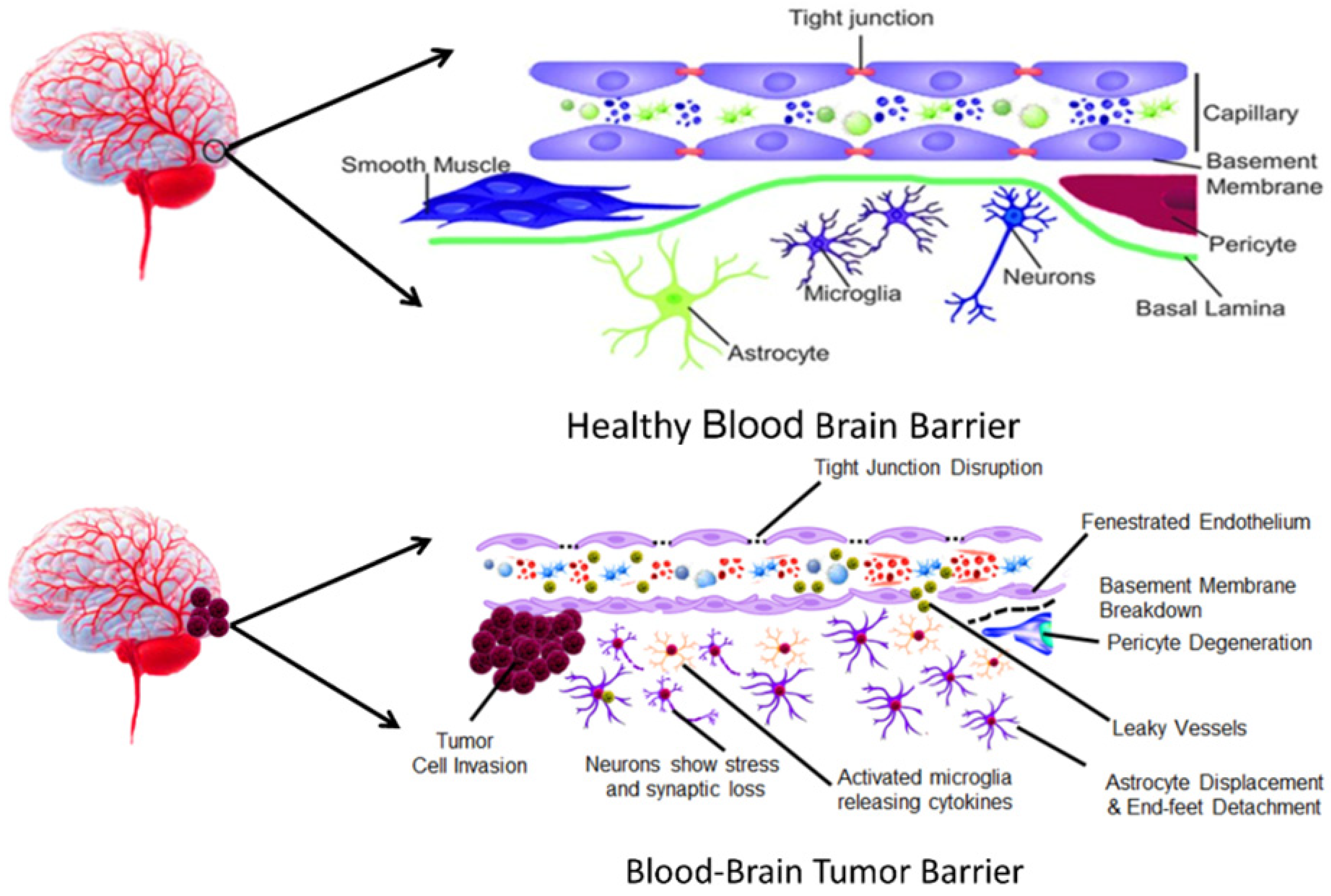
2.1. BBB and Its Role in Drug Delivery Challenges

BBB and blood–cerebrospinal fluid barrier is responsible for regulating the selective and vital entrance of important nutrients for the brain as well as the disposal of metabolic wastes, neurotoxins and other useful materials, thus preserving brain homeostasis. However, the same selective property that is beneficial in some ways becomes a huge hurdle in accessing therapeutic agents needed for treating various brain cancers [12].
The BBB is non-fenestrated and tightly joined endothelial cells that form the internal lining of capillary blood vessels. These non-fenestrated junctions have supporting pericytes, astrocyte end-feet, dual-basal membranes, as well as neurons which together make the neurovascular unit. Transportation through tight junctions and adherens junctions is obstructed. Parallel transport is suppressed and transcellular transport is carried out using designated systems. The unique nature of the BBB enables the structure to function as a physical barrier and biochemical barrier and selectively determine controlled entrances and exits [12].
Apart from its anatomical characteristics, the BBB also has different active efflux transporters like P-glycoprotein, breast cancer resistance protein and multidrug resistance-associated proteins, which actively remove xenobiotics and most drugs back into the bloodstream. Further, the reduced pinocytotic activity of brain endothelial cells decreases non-specific drug uptake. Therefore, over 98% of small-molecule medications and almost 100% of large-molecule biologics cannot pass the BBB in therapeutic concentrations. This greatly limits brain disease treatment and requires the identification of new delivery strategies for drugs [18].
Thus, while the BBB is essential for evading toxic substances from brain, its restrictive function presents a serious obstacle to drug delivery. The solution to these problems lies in novel strategies such as NP-based delivery systems, receptor-mediated transcytosis, FUS and intranasal delivery, all of which seek to improve penetration of drugs into the CNS without disrupting the barrier’s protective function [12].

2.2. Transformation of BBB During Tumor Progression

With the progression of brain tumors, specifically GBM and brain metastases, the BBB undergoes remodeling of its structure and function to form the BBTB. The shift from BBB to BBTB is neither consistent nor predictable. It differs notably based on the nature of the tumor, its location within the brain and its development stage [18]. Figure 1 presents a comparison between BBB and BBTB.
Within gliomas of higher grade such as GBM, BBTB becomes severely disordered, with fenestrated multilayered endothelial cells, disrupted basal laminae and displaced end-feet of astrocytes. These effects decrease the strength of tight junctions, mainly in the central part of tumors, where vessel leakage is maximally observed. Conversely, the invasive tumor border tends to have an intact or only partially disrupted BBB, further contributing to the survival of infiltrating cancer cells beyond therapeutic reach [19].
At the molecular level, the BBTB underlines dramatic changes due to the TME. Tumor cells release pro-angiogenic factors like Vascular endothelial growth factor (VEGF), sometimes overexpressed due to hypoxia-induced factor, HIF-1α. These mechanisms increase endothelial permeability by disrupting cell junctions and reorganizing the extracellular matrix [20]. Mislocalization and overexpression of aquaporin-4, a prominent water channel that is usually polarized in astrocytic end-feet, contribute to edema development and further impair barrier function. In parallel, Matrix metalloproteinases (MMPs; MMP-2 and MMP-9) break down structural elements of the BBB, promoting tumor invasion and with poor clinical outcome [21].
Although these structural and biochemical changes may imply enhanced drug penetration, clinical reality is not so simple. Permeability of the BBTB is extremely heterogeneous. While contrast-enhanced Magnetic Resonance Imaging (MRI) is able to delineate areas of leaky vasculature within the tumor center, the peritumoral infiltrative tumor cells tend to live in the areas where BBB is intact and impermeable to most of the therapeutic compounds. Thus, even in areas where the BBTB is deficient, drug concentrations often lie below therapeutic levels, leading to suboptimal treatment responses and the emergence of drug-resistant clones of cancer cells [22].
Additionally, a number of physiological elements exacerbate the challenge of drug delivery. After surgical removal, residual brain tissue commonly maintains an intact BBB, protecting remaining tumor cells from systemic therapy. Infiltrative cancer cells also have different metabolic patterns than the core of the tumor, depending more on fatty acid oxidation and showing greater resistance to chemotherapy [23]. Tumor-induced hypoxia, fueled by dysvascular and high interstitial pressure, is associated with necrosis, acidosis and radiation and chemotherapy resistance [24]. In addition, the acidic tumor environment, a result of enhanced glycolytic activity, changes drug ionization, transport and action, decreasing the efficacy of the drug and encouraging immune evasion [24].
Efflux transporters P-glycoprotein and breast cancer resistance protein, despite their expression at decreased levels in cancer, continue to restrict the intracellular level of chemotherapeutic drugs. These are frequently accompanied by upregulated drug-metabolizing enzymes like cytochrome P450 isoforms, which further breakdown drugs before they can exert their therapeutic effects. These factors: the structural heterogeneity of the BBTB, the metabolic and physical barriers of the TME and the efflux and metabolic activity synergize to form a hindrance to effective drug delivery in the treatment of brain tumors [25].
Deciphering the dynamic conversion of BBB into the BBTB as well as its resultant molecular and physiological adaptations holds the key for creating more effective therapeutic interventions. Targeted delivery of drugs through delivery systems, combination therapy modifying the BBTB, as well as diagnostic imaging differentiating permeable versus impermeable zones within a tumor, are some of what has been on the agenda towards surmounting such barriers. Overcoming the heterogeneity and complexity of the BBTB is still a major challenge in enhancing outcomes for patients with primary and metastatic brain tumors [26].

2.3. Tumor Microenvironment and Its Resistance Mechanisms

The brain TME, especially that of GBM, is the hallmark of resistance to standard therapies through the multi-modal interactions of cellular and molecular components. The BBB and its tumor-related, pathological counterpart the BBTB limit drug access through increased permeability induced by mechanisms such as VEGF and MMPs [27]. Glioma stem cells are especially challenging because they possess very efficient DNA (Deoxyribonucleic Acid) repair capabilities, are resistant to cell death and, in general, are protected by their microenvironment and hence lead to tumor recurrence [28]. Resistance is also facilitated by high levels of DNA repair enzyme MGMT (O6-Methylguanine-DNA Methyltransferase), which counteracts the action of alkylating agents like TMZ (Temozolomide) [28] The other repair processes such as mismatch repair and base excision repair reduce drug-caused DNA damage. GBM cells also adapt through altered metabolism and increased autophagy so that they are able to live under stress conditions [28]. Extracellular vesicles also enable cell-to-cell signaling between stromal and cancer cells, passing resistance traits [27]. Addressing these obstacles requires multi-modal approaches, including approaches to enhance drug delivery, inhibit primary resistance mechanisms, target glioblastoma stem-like cells and reprogram the immune microenvironment to enable better therapies. Table 3 presents different nanomedicine strategies targeting the TME.

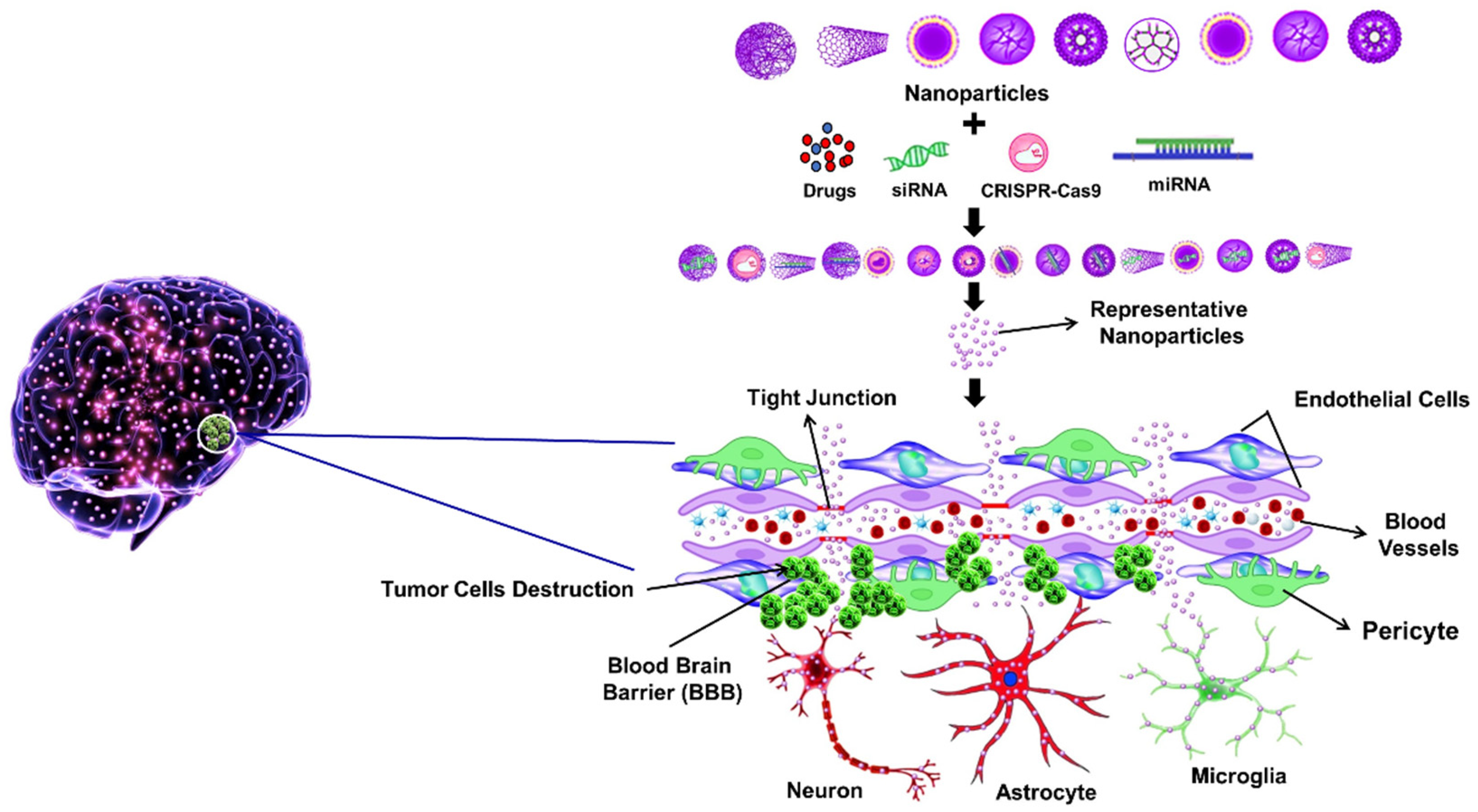
3. BBB Penetration Strategies Using Nanotechnology

Nanotechnology offers innovative solutions to enhance drug delivery across the BBB, using different mechanisms. Some of these strategies used by NPs in penetrating BBB to improve treatment outcomes for brain cancers include the following mechanisms.

3.1. Passive Diffusion Mechanism

Liu et al. [12] reported DOx@PNIPAM-PEI-CPP (Doxorubicin poly(N-isopropylacrylamide polyethylenimine cell-penetrating peptide or DPPC) which functions as a thermosensitive and CPP-modified nanogel delivery system to enable double-targeted BBB penetration. The nanogel incorporates both passive targeting with PNIPAM (Poly(N-Isopropylacrylamide)) but also utilizes active targeting through CPPs to break down astrocyte membranes. The elevated temperature inside glioma tissues (~42 °C) activates drug release from this system which enhances delivery of DOx specifically to glioma tissues. BBB models in vitro demonstrated that DPPC had considerable tissue penetration by showing stronger fluorescence signals coupled with higher tumor death from cytotoxic treatments when temperature rose. Experimental results indicated that passive and active targeting in combination resulted in greater drug loading that suppressed the viability of tumor cells more compared to utilizing each method individually [31].
Most of CNS drug entry is reported to be through passive diffusion based on computational BBB permeability assessment [32,33]. The study determined that passive transport is largely based on four physicochemical properties such as LogD lipophilicity values, molecule weight, topological polar surface area values and hydrogen bond donor interactions. The study applied parallel artificial membrane permeability assay to prove that compounds with low topological polar surface area values and balanced lipophilicity characteristics permeate through the BBB. The data from parallel artificial membrane permeability assays is strongly linked with drugs that process through the BBB successfully mainly because passive diffusion works as a dominant factor in brain drug delivery. The passive diffusion property in XGBoost-based models showed precise correspondence with the measured properties among CNS-active approved compounds. The model comparisons revealed that passive diffusion (parallel artificial membrane permeability assays and BBB endothelial assays) delivered superior performance to active influx or efflux mechanisms for predicting brain drug permeability [32].

3.2. Receptor-Mediated Transport

Receptor-mediated transport provides a top choice for non-invasive nanotherapeutic delivery across BBB and BBTB which enables precise brain cancer treatments. The transcytosis process through the BBB becomes enhanced when nanocarriers use targeted ligands that recognize the elevated receptors on brain endothelial cells. The most commonly targeted receptors in receptor-mediated transport are the transferrin receptor and the insulin receptor, low-density lipoprotein receptor together with lipoprotein receptor-related protein 1. The exploitation of these receptors happens often because they display elevated expression levels on both brain endothelial cells and glioma cancer cells [32]. Drug-loaded NPs with receptor-targeting ligands or antibodies attached to their surface are able to collaborate in order to provide selective tumor tissue targeting by binding endothelial receptors and subsequent endocytosis with reduced unwanted side effects. The study of Wu et al. 2024, revealed that TfR-targeting liposomes exhibited significant brain penetration and anti-tumor efficacy in glioma model studies [34]. The functionality of nanocarriers with targeted receptors allows them to identify dual markers of BBB alongside glioma-specific targets to enhance delivery accuracy. BBB translocation through receptor-mediated transport pathways becomes more efficient when NPs receive transferrin modifications and endogenous ligand peptide coatings because this approach leads to better therapeutic response and reduces systemic drug toxicity [35]. Nanocarrier systems become more effective in penetrating tumors and distributing drugs throughout the body by adding nanobodies and cell-penetrating peptides. The NP’s small structure together with strong molecular binding capabilities increases their ability to penetrate both the BBB and reach glioma. Nanotechnological interventions based on receptor-mediated transport demonstrate a major clinical potential for direct delivery of drugs to brain cancer while providing receptor-specific therapy through non-invasive pathways.

3.3. Adsorptive-Mediated Transport

Adsorptive-mediated transport has been a highly promising route to the delivery of nanomedicine across the BBB in brain cancer treatment. The mechanism exploits electrostatic forces between positively charged ligands on NPs and the negatively charged luminal surface of BBB endothelial cells. Unlike receptor-mediated transport, adsorptive-mediated transport does not need receptor-specific interaction and offers a more general and perhaps more adaptable avenue for penetration of the BBB [33]. Among enhancing constituents of adsorptive-mediated transport, cell-penetrating peptides such as the Human Immunodeficiency Virus-1-derived Trans-Activator of Transcription peptide and arginine-rich peptides such as R9 are often utilized to mediate uptake through non-specific electrostatic binding. These x peptides significantly enhance cellular internalization and have been successfully conjugated onto NPs for better CNS delivery [35]. Despite the comparatively lesser selectivity of adsorptive-mediated transport relative to receptor-based strategies, its high binding ability facilitates considerable uptake, particularly when functionalized nanocarriers are used. NPs with CPPs or cationic dendrimers have exhibited excellent BBB penetration as well as effective drug delivery to GBM tissue. However, toxicity from overcharging with cationic charge demands rigorous design and surface modification methods, such as PEGylation, to provide efficacy over safety. Nanocarriers based on adsorptive-mediated transport thus offer a promising pathway to brain-directed therapy, especially for macromolecular and gene-based therapeutics needing effective BBB translocation independent of receptor specificity [33,35].

3.4. Cell-Mediated Transport

Macrophages, particularly M1 polarized macrophages, not only deliver drug-loaded NPs through the BBB but also stimulate anti-tumor activities in the TME. Neutrophils and other immune cells have been utilized as Trojan horses to target NPs to inflamed glioma tissues by taking advantage of their inherent infiltration properties. Cell-based carriers enhance specificity in targeting, reduce systemic toxicity and allow for controlled release of drugs within the TME. When designed with biocompatible and biodegradable NPs, these systems have demonstrated improved therapeutic effects in preclinical GBM models. In addition, surface modifications of NPs, including peptide or antibody conjugation, can be integrated with cell-based carriers to further optimize specificity and drug delivery efficacy [36].

3.5. Intranasal Delivery

The nasal mucosa offers high surface area and excellent vascularization, allowing for rapid drug absorption and minimizing systemic toxicity. Intranasal delivery has already been proven to deliver therapeutic agents across the BBB via extracellular and intracellular pathway routes. Olfactory epithelium and the trigeminal nerve serve as main conduits, promoting CNS targeting with patient compliance maintained through ease of administration [34]. Delivery systems of nanocarriers like liposomes, micelles and polymeric NPs can be designed for improved mucosal adhesion, extended nasal retention and controlled drug release. The delivery platforms can be surface-engineered using peptides or ligands to increase targeting specificity and penetration. Preclinical studies have shown that NPs, intranasally administered, improve therapeutic index and restrict off-targeted effects and therefore, their potential for glioma treatment.

3.6. Focused Ultrasound (FUS)

FUS is an effective non-invasive method to transiently and safely disrupt the BBB and boost drug delivery to treat brain cancer. When fused with nanotechnology, FUS provides a site-specific modality for enhancing permeability of BBB, particularly in combination with microbubbles. These gas bubbles in MBs vibrate on exposure to US waves, inducing mechanical forces responsible for breaking down tight junctions in endothelial cells of the BBB. This transient window allows therapeutic NPs to enter the brain parenchyma more efficiently [36]. Two primary mechanisms are accountable for this effect: stable cavitation, where rhythmic contraction and expansion of microbubbles generate shear forces and inertial cavitation, where microbubble collapse and shock wave generation take place, both of which facilitate BBB permeabilization. Among nanocarriers, liposomes have shown considerable promise because they are biocompatible and can encapsulate a wide range of drugs. For example, liposome-encapsulated MGMT inactivators with MRI-guided low-intensity pulsed FUS effectively improved delivery to TMZ resistant gliomas. Likewise, polymeric NPs like PLGA or PEG-coated systems were shown to improve delivery and therapeutic outcomes when delivered along with US. The combination of FUS and NPs, therefore, facilitates localized, amplified and tunable delivery of chemotherapeutics to glioma tissue, overcoming one of the most recalcitrant challenges in neuro-oncology. However, long-term consequences must be continuously optimized and tracked to facilitate wider clinical translation [36]. Table 4 presents different nanotechnology-based delivery strategies for BBB penetration.

4. Various Nanoparticles Used in the Treatment of Brain Cancer

NPs offer promising solutions to the key issues in brain cancer therapy with their targeted drug delivery, better imaging and higher efficacy, thereby overcoming the limitations of conventional treatments. This section discusses the various NPs used in brain cancer treatment.

4.1. Liposomes

Nanotechnology research features liposomes as one of its most well-studied nanomaterials because these vesicles exist in spherical forms of 100 to 200–800 nm with aqueous cores constructed from natural or synthetic phospholipid bilayers [37].
The brain tissue accepts liposome delivery by means of intranasal, intracarotid, intracranial and intraperitoneal injections together with convection-enhanced delivery. Through intracarotid delivery, drugs enter the carotid artery directly and through intranasal methods they circumnavigate the BBB by utilizing the nasal passages. Intracranial administration delivers drugs directly to targeted brain areas while intraperitoneal mode provides drug distribution using the peritoneal pathway. Pharmaceutical professionals use the convection-enhanced delivery method to establish a pressure differential through the catheter tip positioned within brain tissue that enables drug delivery directly into brain tissue interstitial compartments [38].
Liposomes prove effective for tumor drug delivery to the brain through their ability to release drugs precisely in tumor vascular and extravascular areas. The targeted delivery systems enable liposomes to transport anticancer medications efficiently to both BBB regions and GBM tumor areas. The mechanisms utilizing receptor-mediated endocytosis and transmembrane lipidation combined with certain drug formulations except CPPs (Cell-Penetrating Peptides) and transferrin achieve high levels of success in enhancing in vivo survival rates [38].
Scientific research has included studies on both targeted and non-targeted liposomes meant for brain drug delivery applications. The pharmaceutical agent Myocet which contains liposomal doxorubicin (DOx), proved beneficial in treating recurrent gliomas of pediatric patients based on study NCT02861222 while liposomal irinotecan demonstrated promise in experimental trials for high-grade recurrent GBM according to NCT02022644 [39]. Clinical evaluations of liposomal irinotecan (NL CPT-11) in 2008 determined inadequate Phase II GBM research potential [40]. However, another liposomal drug (MM-398) achieved promising anti-tumor activity during treatment of brain metastasized advanced breast cancer (NCT01770353) [41]. Liposomes demonstrate outstanding potential to enhance brain tumor drug delivery and targeted drug release which results in improved therapeutic results.
Immunoliposomes combined with antibody molecules emerged as a powerful method to treat cancer. The FDA (U.S. Food and Drug Administration) has approved several monoclonal antibodies for cancer treatment and their utilization has become possible in these systems [42]. Immunoliposomes use antibody-linked targeting to effectively reach angiogenesis and metastatic processes in brain tumors because these tumors show elevated VEGF and VEGFR2 expression [43]. Gliomas resist the microtubule-targeting agent Paclitaxel despite its use in treating various cancers because this drug cannot penetrate the BBB properly [44]. Pharmaceutical scientists have tried to improve paclitaxel delivery by encapsulating it inside PLGA (poly(lactic-co-glycolic acid)) microspheres. Research indicates that when polyvinyl alcohol concentration rises during manufacturing it leads to smaller microsphere diameters which enhance drug release because the diffusion distance shortens independently of drug content [45]. Scientists developed new liposomal paclitaxel and artemether formulations which show powerful effects in treating tumors and triggering apoptosis [44].
The application of liposomes extends beyond medication delivery because researchers use them in gene therapy by combining DNA with cations like dioleoylphosphatidylethanolamine (DOPE) or N-[1-(2,3-Dioleoyloxy)propyl]-N,N,N-trimethylammonium chloride (DOTMA) [14]. The use of liposomes enables their implementation as sophisticated imaging devices during diagnostic procedures. Iodine-filled liposomes function as computed tomography (CT) imaging agents to detect tumors together with blood disorders. MRI technology benefits from Gd-containing liposomes because they provide better contrast imaging capabilities together with extended bloodstream survival duration [14]. Liposomes containing air bubbles also serve as imaging tools for ultrasound (US) practice [14].

4.2. Dendrimers

The nano-sized hyper-branched structure of dendrimers provides a well-structured multilevel architecture together with broad surface areas which enables optimal drug delivery work. The series of organic compounds named dendrimers provide several advantages thanks to their nanoscopic size and stability properties as well as their quick uptake rate into cells and specific drug delivery functionality. The presence of functional groups on their surface facilitates the attachment of various therapeutic agents. Researchers have developed surface modification techniques called pegylation and glycosylation to improve both safety performance and biological system compatibility of such materials. Through the inclusion of targeting ligands on dendrimer surfaces researchers can accomplish targeted brain tumor cell drug delivery which protects surrounding healthy tissue. Researchers today concentrate on building new dendrimers and boosting drug-loading capacities and biological tolerability to expand clinical applications [46].
Research on brain tumor treatment includes studies focusing on poly(amidoamine), polypropylenimine, poly-l-lysine, carbosilane, phosphorus-based peptide, glycodendrimers, triazine, polyglycerol, citric acid-based, polyether and surface-modified dendrimers [47].
The precise nanoscale structure of poly(amidoamine) dendrimers makes them highly important for oncology through their capability to deliver targeted drugs and genes precisely. Researchers have developed numerous nanotheranostic systems containing poly(amidoamine) dendrimers during recent years for improving brain drug delivery [48]. The group of Sharma et al. [49] developed a nanoformulation of TMZ by using poly(amidoamine)-chitosan conjugates for GBM treatment. The nanoformulation established better therapeutic effects against glioma cell lines U-251 and T-98G than pure TMZ administered separately. The formulation showed adequate characteristics through Nuclear Magnetic Resonance measurements, Fourier Transform Infrared spectroscopy, surface and morphological tests. Scientific studies on live animals proved the safety of this formulation and pharmacokinetic testing demonstrated that it released the drug over an extended period relative to TMZ alone. Bio-distribution analyses demonstrated that heart tissue contained more drug substance than brain tissues yet overall findings showed promise for dendrimer-based chitosan formulations to improve GBM therapy [49]. The branched structure combined with internal compartments of dendrimers enables these structures to serve as ideal drug carriers that deliver therapeutic agents to cells of both brain and cancer types while bypassing viral transduction methods [47].

4.3. Nano-Micelles

The ability of micelles to transport therapeutic agents through physiological barriers has made them highly considered nanocarriers for targeting brain cancer treatments specifically because they penetrate the BBB successfully [50]. The nanoscale carriers consist mainly of amphiphilic copolymers, which form core–shell structures naturally above critical micelle concentration when placed in aqueous solutions. The inner hydrophobic portion of the carrier contains poorly dissolved lipophilic drugs, while the exterior hydrophilic parts maintain structural integrity and minimize drug elimination before therapeutic action. With its dual properties, the micelle structure delivers efficient drug encapsulation to both water-hating compounds and hydrophilic drugs, leading to better drug dissolution, extended stability and improved absorption rates [51]. The small size of these NPs allows for enhanced permeability and retention-mediated passive targeting through tumor-impaired blood vessel barriers. However, active targeting can be achieved by modifying them with specific receptor-binding ligands for endocytosis [50]. Micelles exhibit better drug-loading capacity, enhanced biodistribution and reduced systemic toxicity than other conventional nanomedicine platforms like liposomes, niosomes, dendrimers and polymeric NPs [50].
Research findings have shown that micelle-based delivery systems demonstrate effectiveness in preclinical experiments of GBM tumors alongside dynamic brain tumor models. Gao et al. [52] established methoxy poly(ethylene glycol)-poly(ε-caprolactone) (MPEG-PCL) micelles with honokiol (HK) and DOx through their method resulting in micelles with 34 nm average size. The formulation demonstrated an extended drug release mechanism and achieved high levels of apoptosis together with glioma cell growth suppression while maintaining its efficiency against glioma cells in test tube experiments. In vivo data demonstrated that HK-DOx-MPEG-PCL [HK peptide doxorubicin methoxy poly(ethylene glycol)-block-poly(ε-caprolactone)] micelles achieved better therapeutic results than single-drug treatments because they demonstrated superior tumor growth suppression and enhanced treatment ability making them promising for glioma therapeutic applications [52]. Prevailing studies utilized transferrin-conjugated vitamin E D-α-tocopheryl polyethylene glycol succinate micelles to enhance specific brain drug delivery. The micelles delivered docetaxel while achieving enhanced drug loading efficiency together with controlled release characteristics and enhanced brain delivery when compared to standard delivery systems [53].
Huang and his team developed a new treatment system that presented a therapeutic solution for GBM patients who suffer from this aggressive brain cancer. The researchers combined amphiphilic CB-poly(ethylene glycol)-Chlorin e6 polymers for CPC (Cationic Peptide Carrier) micelle formation to encapsulate 5-(3-methyltriazen-1yl)-imidazole-4-carboxamide (MTIC) chemotherapy drug and layer-dressed CPC micelles with a hybrid membrane consisting of macrophage (HMC3) and glioma cell membrane components (U87MG) at a ratio of 1:2 to create mUMH. The dual membrane structure on micelles enabled targeted tumor cell attachment and managed drug release responses to the tumor tissue conditions. The combination of mUMH@CPC@MTIC(Modified ultrasmall micelle hybrid @ cetylpyridinium chloride @ Monomethyl triazeno imidazole carboxamide) micelles showed enhanced tumor-seeking properties and better anti-tumor effects when tested with mice compared to single-component or uncoated systems, thus indicating potential progress for GBM chemotherapy [53]. The research findings showcase how micelle-based delivery systems effectively deliver drugs for more effective brain cancer treatment by surpassing traditional therapeutic boundaries.

4.4. Carbon Nanotubes (CNTs)

CNTs possess excellent mechanical properties and broad surface area while offering rare electrical abilities. The drug delivery capabilities of CNTs work well for brain cancer cell targeting because of their advantageous properties. CNTs possess an exceptional quality since their surfaces become processable after functionalization with biological compounds that include polymers, peptides, carbohydrates and other organic substances. Enhanced functionality of CNTs enables better solution dissolution as well as targeted biological interactions combined with high biocompatibility suitability for cancer therapies and drug delivery systems [54]. The notable progress in pharmaceutical science came from Ren et al. [55] as they created dual-targeting delivery through their combination of PEGylated and Angiopep-2 (ANG-2)-conjugated oxidized multi-walled CNTs (O-MWNTs-PEG-ANG). Through efficient BBB penetration the system proved to exhibit selectivity towards glioma cells, DOx drug demonstrated better efficacy toward glioma control combined with reduced toxicity in the formulated delivery system versus unbound drug [55].
Present studies focus on assessing the cytotoxic impacts linked to CNTs because of growing safety issues. The research by Han et al. [56] tested the toxicological features of multi-walled CNTs within C6 glioma cells using tests that showed reduced cell viability, enhanced oxidative stress effects and increased apoptosis mechanisms and G1 phase cell cycle arrest in relation to the dose amount and time duration. The research identified that shorter CNTs show greater toxicity because oxidative stress leads to main tissue damage [56]. The production of CNT-based drug delivery systems for brain cancer therapy requires careful safety assessments according to studies that show their therapeutic potential.

4.5. Silver NPs (AgNPs) and Gold NPs (AuNPs)

AgNPs and AuNPs exhibit multiple benefits which include superior biocompatibility, adaptable dimensions, morphologies and biomolecule conjugation capabilities to improve their specificity and functionality [57]. The major cytotoxic power of AgNPs results from ROS (reactive oxygen species) production that leads to oxidative stress which destroys DNA, lipids and proteins. Depending on exposure duration and dosage, these oxidative interactions damage essential cellular functions which leads to either apoptosis or necrosis of damaged cells [58]. Multiple scientific investigations have shown the clinical effectiveness of AgNPs when treating brain cancers. Human GBM (U-87) cells grown on chicken embryo chorioallantoic membranes applied with colloidal AgNPs (40 μg/mL) exhibited suppressed tumor growth combined with modified cellular appearance and apoptotic response indicators [59]. Research indicated that AgNPs enhanced apoptotic pathways by raising the expression of activated caspase-9 and caspase-3 which leads to programmed cellular death but their main effect was observed on cell proliferation rather than on death rates alone [60]. The C6 rat glioma cell lines exposed to AgNPs for 24 h demonstrated a 21% decline in viability while exhibiting coexistence of G0/G1 phase arrest and reduced G2/M phase entry indicating DNA damage and delayed mitotic entry [61]. Researchers used AgNPs in multifunctional nanoplatforms with alisertib (Aurora kinase A inhibitor) and chlorotoxin (a peptide that binds specifically to MMP-2) to deliver AgNPs only to tumor cells. The AgNP–chlorotoxin conjugate displayed targeted tumor tissue accumulation which researchers validated using technetium-99m (99ᵐTc) radiolabeling before showing significant tumor volume decrease in animal models [61].
AuNPs exhibit superb photothermal properties and diagnostic ability. The large surface area of AuNPs makes therapeutic drug binding possible through chemical or electrostatic interactions that enhance drug delivery and sustain release at better pharmacokinetic levels [57]. Two years after Yu et al. [62] anti-EphA3 (Antibody against Ephrin type-A receptor 3) modified AuNPs delivery system reached the market, it managed to transfer TMZ to treat GBM T98G cells while fighting against TMZ resistance. Radiation with laser power caused TMZ@GNPs (temozolomide loaded gold nanoparticles) to increase cellular drug absorption which started cell death processes and produced longer survival outcomes than free TMZ in rats with GBM tumors [62]. The eco-friendly synthesis of CMXG@AuNPs (Carboxymethyl xyloglucan@Gold Nanoparticles) depended on CMXG (carboxymethyl xanthan gum) as both green reducing and capping agent. The synthesis of these NPs happened rapidly with uniform results by using microwave-assisted technology. The CMXG@AuNPs loaded with DOx displayed pH-sensitive drug release features where the drug released at high levels in acidic tumor conditions compared to normal pH which boosted both drug accessibility inside cells and overall toxicity. The cytotoxic properties of DOx@CMXG@AuNPs (gold nanoparticle system coated with carboxymethyl xyloglucan and loaded with doxorubicin) surpassed those of unbound DOx by 4.6 times when tested through laboratory experiments [63].
Engineers produced dual-ligand targeted AuNP system through PEGylating AuNPs and then combining them with both folate and transferrin antibodies for glioma-specific delivery. The system contained curcin as its main ingredient while being designed to express this potent type I ribosome-inactivating protein under tumor environments with acidic pH levels. Studies have shown that Curcin-loaded AuNPs stopped glioma cell proliferation and migration by enabling cellular apoptosis and causing mitochondrial depolarization together with elevated ROS production and cytoskeletal destruction [64]. Selective tumor cell toxicity emerges from AuNPs treatment which preserves healthy tissues in the process. Derived from the photothermal ablation, the ability of AuNPs, when exposed to light, makes them effective for precise tumor colony destruction without invasive methods. It also demonstrates strong treatment potential of AgNPs and AuNPs for GBM patients who benefit from targeted delivery systems and precise drug release combined with ROS toxicity and photothermal capabilities which presents an integrated therapeutic solution to the issues of drug resistance and poor permeability along with non-specific toxicity in brain cancer therapies [62,63].

4.6. Zinc Oxide NPs (ZnONPs)

The anticancer technology and cosmetic industry alongside agriculture and energy industries benefit from the applications of multifunctional low-cost inorganic nanomaterials named ZnONPs. ZnONPs serve as highly effective experimental materials in multiple biomedical and industrial settings because of their attractive combination of extensive catalytic activity along with high biocompatibility and strong adsorption performance and rapid electron-transfer speeds. Different synthesis techniques such as hydrothermal methods, ultrasonication and thermal evaporation have produced various forms of ZnO nanomaterials including nanorods, nanotubes, nanowires, nanobelts, nanobridges, nanoribbons and nano-nails through which researchers can customize their operational characteristics for specific applications [65].
The penetration capability of ZnONPs through the human CNS post oral consumption happens through two pathways: neural transport mechanisms and BBB disruption. Apolipoprotein E exists within protein corona structures on ZnONPs thus potentially directing their BBB translocation and neurological disorder targeting capability [58]. Brain tissue cells died after they internalized ZnONPs through oxidative stress mechanisms that produced toxic cell death in microglial cells [66].
Wahab et al. [67], tested the cytotoxic properties of different ZnO nanostructures which included magnetic fluids, NPs, microspheres and nanospheres on U87 human GBM, HeLa cervical cancer as well as normal human embryonic kidney cell lines. His research showed that ZnO nanostructures reduced cancer cell growth while causing cell death by apoptosis at increasing concentrations although nanosheets along with NPs displayed the most powerful anti-tumor effects. The treatment of U87 cells with ZnO nanostructures resulted in heightened micronuclei formation because DNA damage appeared to stem from repair process strand rejoining interruption [67].
Research conducted on living organisms has revealed that ZnONPs possess toxicity properties toward neural tissue. Rats receiving 40 or 100 mg/kg of oral treatment through their mouth for a duration of 24 h manifested no observable signs of neurotoxicity. The combined stress of seven days of exposure resulted in brain tissue damage which showed itself through higher malondialdehyde levels coupled with weakened antioxidant protection mechanisms. The extended exposure to ZnONPs both fragmented DNA molecules and dramatically elevated the levels of pro-apoptotic markers including caspase-3, Fas proteins, heat shock protein-70 (Hsp 70) and cytokines thus establishing their role in causing neuronal stress together with apoptosis during continuous dosing [68].

4.7. Nucleic Acid-Based NPs

There has been recent advancement in understanding the molecular processes of brain tumors that has identified several genetic targets and therefore paved the way for gene therapy approaches in neuro-oncology. Even though traditional DNA delivery frameworks utilize viral vectors, the potential of smart nanoplatforms brings unparalleled advantages, including improved penetration across the BBB, greater versatility of cargo and superior biocompatibility. These nanoengineered platforms provide the promise of the delivery of DNA or RNA into treating multiple fronts of tumor biology concurrently. Such methods can include reactivating tumor suppressor genes, inactivating oncogenes, enhancing programmed cell death, improving chemosensitivity, inducing tumor cell differentiation of cancer-initiating cells and manipulating tumor cell proliferation and motility [69].
In addition, nucleic acid-based nanocarriers are also being researched more and more for immunotherapeutic purposes by controlling the immune response of the body to detect and destroy tumor cells [69]. High-end genetic engineering technologies like the CRISPR/Cas9 (Clustered Regularly Interspaced Short Palindromic Repeats-associated protein 9) system are also being incorporated into nanoplatforms to provide targeted precision interventions in CNS cancers [69].
These NPs are widely classified into DNA- and RNA-based types. DNA nanocarriers tend to be engineered to deliver therapeutic genes that either cause the apoptosis of cancer cells or make them sensitive to cytotoxic drugs. RNA nanocarriers have a broader scope of therapy, as they may deliver different types of RNA, such as mRNA, miRNA, siRNA and ribozymes. Although miRNAs and siRNAs are frequently utilized to control gene expression by silencing certain mRNA targets, NPs based on mRNA are becoming increasingly popular as vaccine platforms for cancer immunotherapy [69].

4.8. Viromimetic NPs

Viruses, with their built-in capacity for immune evasion and cell-specific tropism, present an attractive model for NP design. Consequently, there is increasing interest in viromimetic NPs as tools for brain cancer treatments. For example, the rabies virus, with its capacity for BBB penetration, has been a source of inspiration for the development of NPs both for chemotherapeutic [69] and photothermal [69] applications in glioma models. Likewise, the Trans-Activator of Transcription peptide from the Human Immunodeficiency Virus has been extensively employed to improve BBB penetration in drug delivery for glioma therapy [69].
Besides animal viruses, plant viruses like cowpea mosaic virus and tomato bushy stunt virus are also investigated for their high cytotoxicity against brain tumor cells as NP scaffolds. The in vivo potential of these plant virus-derived NPs to traverse the BBB, however, still needs to be confirmed [69].

4.9. Upconversion NPs

Upconversion NPs, mostly made of lanthanides, have the ability to convert NIR (near-infrared) light, which is deeply absorbed by tissues, into visible or ultraviolet emission. Due to their characteristics narrow emission bands, high photostability and low background noise they are of great utility in diagnostics and therapy for brain tumors. Their shape and surface can be easily tuned for enhancing BBB crossing [69].
In in vitro experiments, photothermal agent-loaded hybrid upconversion NP formulations have displayed encouraging cytotoxicity against astrocytoma cells [69]. Transferrin-conjugated liposome-mediated upconversion NP systems have also shown effective drug delivery and tumor cell killing in glioma models. While most of the in vivo experiments with upconversion NPs have focused on tumor imaging, recent advances suggest they are promising for therapy. One such example is a PEG (Polyethylene Glycol) diacrylate-based upconversion NP implant applied for wireless photodynamic GBM treatment in mouse models [69].

4.10. Albumin-Based NPs

Proteins, like polysaccharides, provide a cheap, biodegradable and biocompatible platform for drug targeting in the brain. Their topographies facilitate facile modification with ligands or imaging agents for enhanced targeting. Transferrin NP-based particles are a classic case that takes advantage of transferrin receptor-mediated transfer across the BBB/BBTB for glioma treatment [69]. Casein, a milk protein, has been shown to exhibit inherent brain-targeting capability, as seen in glioma mouse models. It is therefore a promising candidate for oral nanodelivery systems based on milk exosomes that are not only BBB-permeating but also gastrointestinal-stable [69].
Among protein-based nanocarriers, albumin NPs are especially predominant owing to the high energy expenditure of tumor cells that overexpress albumin receptor binding molecules such as gp60. Cationized albumin resulted in increased uptake by brain endothelial cells with little toxicity [69]. These NPs also increase the circulation time and bioavailability of the drug. Albumin carriers engineered in conjunction with herbal brain target agents exhibited excellent BBB penetration and glioma-specific accumulation [69]. Moreover, albumin-coated magnetic NPs functionalized with antibodies against VEGF have proved to be useful in both diagnosis and treatment of brain tumors. Hybrid magnetic NPs, combining substances such as Gd or iron oxide with bovine serum albumin, provide a double advantage of low toxicity and increased MRI visibility for glioma theranostics [69].

4.11. Amorphous Carbon-Based Nanomaterials

Amorphous carbon-based nanomaterials graphene oxide (GO) and reduced graphene oxide (rGO) have been popular as advanced nanocarriers in the therapy of brain cancer owing to their physicochemical properties, such as large surface area, undulation and ease to functionalization [70]. Functionalizing with biocompatible substances like hyaluronic acid, proteins or even specific targeting ligands can enhance their solubility, stability and selective uptake by cancer cells with surface receptors [71].
GO and rGO can encapsulate different anticancerous drugs like doxorubicin, curcumin and rutin and subsequently, release them in controlled, pH-sensitive even distribution, which increases drug localization in tumor cells and decreases their negative effects on healthy cells [70]. Due to their capacity of overcoming biological barriers and reacting to the acidic atmosphere present in tumors, they are specifically adapted towards attacking brain tumors [71].
In addition, these nanomaterials display high absorption in near-infrared (NIR) wavelengths and can be used in photothermal therapy. They transform light energy into heat in the process of hyperthermia (localized), thus effectively destroying tumor cells when irradiated with NIR [71]. Such graphene systems may be designed to pursue integrative therapeutic plans, by incorporating chemotherapy, photothermal therapy and photodynamic therapy to maximize therapeutic effects through the induction of increased cancer cell apoptosis and marked tumor reduction. They can be tailored for target-specific, pH-responsive and NIR-induced drug release to maximize efficiency and reduce side effects [71].
Functionalized GO and rGO are typically biocompatible and show little to no toxicity in healthy cells [72]. It has been shown in animal models that they are effective tumor-targeted agents with minimal systemic toxicity [71]. Additionally, such nanocarriers have the potential to affect the cellular signaling pathways that regulate apoptosis and autophagy, sensitize tumor cells to chemotherapeutics and induce cellular DNA damage through ROS production [72]. On the whole, GO and rGO-based nanoplastic platforms present multi-forward, safe and effective approach to treating brain cancer.

4.12. Carbon Dots

Carbon dots are ultrasmall (<10 nm), carbon-based nanomaterials, which have gained increasing interest in brain cancer nanomedicine by showing a range of optical, chemical and biological properties. This is in part enabled by their tiny size due to which they penetrate efficiently across the BBB which presents a major challenge to drug delivery in neuro-oncology. Besides being water soluble and good chemical stability, the carbon dot is relatively nontoxic and biodegradable, thus reasonably compatible with biomedical usage [73,74]. The most striking property is the tunable fluorescence of the carbon dots which enable non-invasive real-time imaging and tracking of the tumors favoring theranostic applications [73].
In addition, carbon dots could be easily modified with targeting ligands including transferrin or other therapies such as temozolomide and curcumin, as well as imaging probes, enabling multimodal and targeted delivery to GBM and other brain malignancies [75]. A number of studies have shown that conjugation of carbon dots with chemotherapeutic agents (e.g., epirubicin, temozolomide, gemcitabine) increases their selectivity and cytotoxicity to brain tumor cells and may be further enhanced by targeting ligands, such as transferrin. At low in vitro concentrations, a triple-conjugated carbon dot system that delivered transferrin, epirubicin and temozolomide induced between 86 percent GBM cell death compared to non-targeted or free drug controls [75]. Moreover, the targeted drug delivery through carbon dot-based nanocarriers has been designed to be pH-sensitive, prolonged and controlled drug release, thus increasing the concentration of drugs in the tumor region with less-to-no side effects on the overall system [76].
They are also used in the treatment of GBM in children where the combination of carbon dots and gemcitabine showed effective performance and strong antitumor effects [77,78].
The preclinical studies involving carbon dots are encouraging; however, the issues of scalable production, prolonged safety study and regulatory acceptance must still be resolved prior to translation into the clinical practice. However, it also leaves a possibility of individualized nanomedicine, where carbon dot formulations may be designed to suit a particular tumor profile of the patient [73,78].

4.13. Iron-Oxide–Graphene-Based Hybrid NPs

Hybrids that contain iron-oxides and graphene are outstanding candidates of multimodal cancer therapy, in which magnetic hyperthermia, photothermal therapy and chemotherapy are combined on a single therapeutic platform. The GO or rGO sheets provide a large surface-to-volume ratio with many functional groups (hydroxyl, carboxyl, epoxide) that allow the loading as well as the covalent or non-covalent conjugation of a great number of chemotherapeutic agents, including doxorubicin or artesunate [79]. The release of such drugs is pH-sensitive and they are preferentially delivered in and around the tumor due to the acidic microenvironment and having lower side effects systemically. Investigations into drug release capabilities have demonstrated that this hybrid system enables controlled and lasting delivery, enhancing therapeutic window and reducing the dosing interval [80].
The iron oxide content enables magnetic field-mediated targeting, where the interaction of NPs with external magnetic fields helps it target tumors, increasing localization and tumor-selective accumulation [79]. Once it is localized, an alternating magnetic field can be passed through it to result in magnetic hyperthermia, the process in which magnetic energy is transformed into local heat by the superparamagnetic particles and the local temperature in the tumor is raised to 42–45 °C. This condition induces apoptosis to the cancer tissue but not the normal tissue. It is worth mentioning that hybrid design enhances the specific absorption rate because of an elevated magnetic dipole alignment and consequently reduces the NP dose needed to afford efficient hyperthermia [79].
The photothermal conversion capability of the hybrids is due to their ability to absorb near-infrared (NIR) in the graphene structure. The method allows distant field-independent killing of brain tumor cells and is especially useful in treatment of the infiltrative gliomas that do not respond to known therapies. Used in combination, magnetic hyperthermia and photothermal therapy have synergistic antitumor effects and are more effective than either one of them used alone [81].
Hybrid systems with AuNPs have also been developed, which allows a new thermo-radiotherapy strategy. These hybrids are a good choice of radiosensitizers since the atomic number of gold is high and therefore it enhances radiotherapy treatment by maximizing local delivery of radiation [81]. Results of in vivo experiments demonstrate that these multi-stimuli-responsive platforms are successfully used to suppress tumors, greatly reduce tumor size and increase survival time of brain cancer models compared to free drugs or monotherapies [81].
Lastly, use of biocompatable coatings, including polyethylene glycol (PEG) and natural polysaccharides, increases biostability and minimizes cytotoxicity. This is confirmed through low rates of hemolysis and high viability rates of healthy cellular cultures in toxicity tests indicating the potential of the platform to translate [79].

4.14. Sillica-Based NPs

Nanocarriers that are silica-based, especially mesoporous silica nanoparticles (MSNs), are commonly investigated for use with chemotherapy, gene therapy and stimuli-sensitive therapy against brain tumors, especially GBM. Their mesoporous framework, high drug-loading efficiency and controlled releases are vital in breaching the therapeutic barriers that the BBB and tumor microenvironment offer [82]. Most often, doxorubicin, temozolomide, methotrexate and paclitaxel agents are used, which exhibit an increased cellular uptake, higher cytotoxicity and decreased off-target effects under MSNs [83,84]. Functionalization of MSNs with targeting ligands (e.g., transferrin, cyclic Arg-Gly-Asp (RGD) peptide, Trans-Activator of Transcription peptide) or PEGylation to achieve stealth performance results in significant increase in accumulation in glioma tissues and enhanced therapeutic index. Strategies of ligand-free receptor-mediated endocytosis were also utilized successfully. Interestingly, co-delivery of chemotherapeutics and gene-silencing compounds (e.g., siRNA against drug resistance genes such as Hepatoma-Derived Growth Factor) using MSN platforms has produced synergistic effects that suppress both forms of malignancy (proliferation and resistance) in GBM [84]. Additionally, one can design MSNs to become responsive to a particular endogenous trigger (e.g., acidic pH, glutathione, enzymes) or an external stimulation (e.g., magnetic field, NIR light), which will make the delivery of the therapeutic payload specific and on-demand [85]. MSN-based therapy has shown to cause considerable regression of tumors, extended life duration and low systemic treatment in mice models which shows their potential in translation [84]. Nonetheless, long-term biocompatibility, high-level reproducibility and regulatory processes should also be considered to make it applicable to clinics [86].
Figure 2 presents an illustration of NP-mediated delivery systems for brain cancer therapy.

5. Various Nanoparticles Used in the Diagnosis and Biosensing of Brain Cancer

The field of cancer diagnosis benefits significantly from nanotechnology innovations which particularly benefit brain tumor detection. The precise physical and chemical properties of NPs enable them to function as strong contrast agents which enhance tissue imaging procedures in both laboratory and living samples.
Nanomaterials with optimized dispersion properties and exact engineering on their surfaces can collect at tumors while clearing away from external areas effectively. The NPs enable tumor cells to engulf them through phagocytosis which provides detailed images along with distinct separation between cancerous and normal brain structures. The development of tumor-targeted strategies and targeted surface coatings has improved the precision of tumor tissue localization [87].

5.1. Magnetic NPs

The properties of magnetic NPs, particularly superparamagnetic iron oxide NPs (SPIONs) and ultra-small SPIONs make them ideal for cancer imaging and diagnostic applications [88]. The particles demonstrate both magnetic sensitivity and structural stability that enables accurate cell observation under the microscope. Experimental tests show that SPIONs coated by folic acid and bovine serum albumin demonstrate excellent biocompatibility and cellular uptake for GBM detection via MRI in U251 glioma cell models [89].
Magnetic NPs integrated with polymer hybrids combined with fluorescent tagging substances have demonstrated high potential for detecting human GBM. PLGA-coated SPIONs tagged with fluorescein isothiocyanate-tagged polyethyleneimine showed increased uptake by U251 GBM cells when compared to untreated particles. The system demonstrated dual capabilities to detect cancer cells while delivering medicines in a protective manner for healthy cells [90].
Scientists created PEGylated ultra-small SPIONs for GBM vasculature targeting by incorporating ANG-2 functionalization that binds to the GBM-overexpressed low-density lipoprotein receptor-related protein. The nanoprobes managed to cross through the BBB while imaging cerebral tumors in addition to showing excellent biocompatibility which positions them as promising tools for brain cancer imaging diagnostics [91].

5.2. Extracellular Vesicles and Exosomes

The detection and monitoring of gliomas call for reliable non-invasive diagnostic approaches. Extracellular vesicles constitute nano-scale membrane-bound particles which emerge from cells to transmit DNA and RNA together with lipids and proteins. These markers demonstrate suitable characteristics for brain tumor biomarker study because they cross the BBB utilizing transcytosis [92]. Adult glioma extracellular vesicles contain epidermal growth factor receptor (EGFR) proteins that serve as precise tumor markers for targeted detection [93].
Among different kinds of extracellular vesicles, the exosome subtype consists of lipid bilayers and cells produce them through secretion. Within exosomes lie both coding and non-coding RNAs together with lipids which enable BBB penetration and their use as promising non-invasive diagnostic elements. Research scientists embedded SPIONs into exosomes taken from glioma cells for optimized imaging purposes. The exosomes received specific design modifications to detect neuropeptide-1 which resides on cell membranes. The exosomes delivered curcumin as a therapeutic payload for its exploitation. Application of magnetic fields enabled the efficient concentration of SPIONs in tumor tissue so medical providers could undertake two simultaneous modes of therapy and imaging [94].

5.3. Metallic NPs for Brain Cancer Imaging and Diagnosis

Metallic NPs, AuNPs in particular, have been studied intensively for their therapeutic as well as their diagnostic potential. Initially employed in infections and rheumatological diseases, AuNPs were repurposed as powerful tools for cancer detection owing to their distinctive optical and chemical properties. Enzyme and ligand functionalization of the surface has made them candidate materials for immunoassays and cancer diagnosis. One of the most vigorous features of AuNPs is plasmonic resonance, which increases imaging capability within biological systems. AuNPs are readily detectable with imaging modes such as CT, MRI and FUS [95].
AuNPs have shown promise in MRI-based glioma imaging. Intravenous injection of AuNPs significantly enhances fluorescence imaging, with enhanced visualization in glioma models compared to direct injections into the tumor site [96]. The use of biocompatible scaffolds and AuNPs has also advanced glioma imaging technologies. An example is the use of AuNPs that are conjugated with chlorotoxin peptide and radiolabeled with iodine-131. These NPs not only displayed X-ray attenuation and cytocompatibility but also displayed crossing capability of the BBB in rat glioma models, which makes them worthy of use in CT-guided radionuclide therapy [97].
Surface functionalization of AuNPs with ligands like transferrin, fibroblast growth factors, antibodies and low-density lipoproteins increases their permeability through the BBB and subcellular resolution imaging of the brain tumor by multiphoton and scanning electron microscopy. For instance, peptide-conjugated AuNPs that target CD133, a marker for GBM, showed efficacious cell targeting with biocompatibility and fluorescence sensitivity, which makes them worthy of use in tumor visualization [98].
Silica-coated iron oxide nanocomposites are also a valuable imaging agent. NIR fluorescent NPs are highly fluorescent for intraoperative imaging and have proved effective in targeting tumor-associated macrophages, both in vivo and in vitro. Their advantage is water dispersibility and multimodal imaging agent potential. In a second strategy, fluorescent silica NPs functionalized with glucose and glucose-PEG derivatives revealed the capability of crossing the BBB, as revealed by confocal laser scanning microscopy, thereby attesting to their diagnostic utility [99].
New theranostic systems have also been developed, including NIR nanoprobes that integrate organoplatinum (II) metallocycles and Pluronic F127, a biocompatible polymer. They were photostable and exhibited real-time imaging of therapeutic interventions with selective internalization in glioma U87MG cells compared to non-cancerous cells [100].

5.4. Quantum Dots (QDs)

QDs offer significant potential for the imaging of brain tumors due to their fluorescent features of narrow emission spectra, broad Stokes shifts and photoluminescence that can be tuned. These characteristics allow for high-resolution imaging in addition to dual-modality utilization during neurosurgery. PEG-coated QDs, for example, Cadmium Selenide/Zinc Sulfide-based, have shown to be functionalized with asparagine–glycine–arginine peptides targeting CD13, a cell surface glycoprotein overexpressed in glioma cells. These QDs showed in vivo fluorescence imaging and can potentially aid surgical resection [101].
Indium phosphide QD-encapsulated lipid-phase nanobubbles have also been prepared as a non-toxic imaging strategy. The nanobubbles accumulated in brain tumors permitted both US and optical imaging, enhancing their diagnostic use [102]. A surgery device has also been designed from QD-conjugated aptamers to target the EGFRvIII mutation in glioma. Such probes reached across the BBB and imaged tumor tissues effectively in animal models [101].
Another strategy was IL-13-associated PEG-modified cadmium-based QDs targeting the IL-13Rα2 receptor found on glioma stem cells. These QDs were more specific to glioma tissues, as verified by transmission electron microscopy and flow cytometry [103]. Additionally, biocompatible polysaccharide-based QDs functionalized with L-cysteine and poly-L-arginine were synthesized using carboxymethylcellulose. These nanoconjugates showed effective cellular uptake and fluorescence, which makes them appropriate for brain cancer bioimaging [104].
Graphene QDs have become prominent due to the fact that they show lower cytotoxicity compared to traditional QDs and are conductivity tunable, which is beneficial for biosensing. Their fluorescence is structural in nature such as chirality and sp2 hybridization, which influence their fluorescence. Fluorescence was improved in graphene QDs when gamma irradiation was applied to them, leading to higher quantum yields. This method also enabled effective photodynamic therapy by generating reactive singlet oxygen species in cancer cells [105].
Gamma-irradiated graphene QDs (e.g., 50 kGy) greatly enhanced imaging clarity under UV light, indicating potential in combined diagnostic and treatment applications [106].
In addition, new 2D materials like MXenes are being investigated in combination with QDs for their applications in photothermal therapy and imaging of glioma. These developments expand the imaging range of QDs over near-infrared bio-windows (NIR-I and NIR-II), making QDs as general, biodegradable and effective agents for in vivo imaging of glioma and guided therapy [107]. Table 5 presents various types of quantum dots (QDs), highlighting their associated toxicities and potential for clinical translation.

5.5. Polymeric Nano-Vehicles for Brain Cancer Imaging

Conventional contrast-enhanced MRI techniques are found to have constraints in brain tumor imaging due to factors such as low circulation time, poor BBB permeability and toxicity constraints. As a substitute for the above, nanostructures based on polymers have gained importance with their biodegradability and biocompatibility, particularly concerning glioma-based enhancement in imaging. These polymeric systems evolved for better diagnosis through the practice of principles derived from nanotechnology [110].
One of the innovative approaches involved the synthesis of red fluorescent carbonized polymer dots with superior internalization into glioma cells. The nanoprobes were nontoxic, photostable and featured long excitation/emission wavelengths, thereby enabling successful real-time, image-guided surgery. Another group designed polymeric NPs with cyclic peptide cyclo (Arg-Gly-Asp-D-Phe-Lys(mpa)) targeting integrins expressed on tumor cells. These NPs had superior NIR absorbance, which allowed for precise photoacoustic imaging and site-specific photothermal therapy. The technique was used to image glioma tissues to a depth of 3 mm with a high signal-to-noise ratio [110].
Fluorinated semiconducting polymer dots were also engineered to enhance fluorescence for accurate brain tumor imaging. NIR-emitting fluorinated polymers generated images that were three times brighter than their non-fluorinated counterparts, facilitating enhanced visualization and detection [111]. Further, a dual-mode imaging technique was employed in order to bypass the false positive issue of tumor imaging. A polymeric nanostructure bearing paramagnetic iron ions offered both T1 and T2 imaging modalities. In murine GBM models, these coordination polymer-based NPs showed sustained biodistribution, unambiguous imaging resolution and high biosafety [112].

5.6. Biomimetic Nanocomposites for Multimodal Imaging of Glioblastoma

The heterogeneous TME and structural barriers in GBM, such as the intact BBB in infiltrative zones, pose a significant challenge to efficient imaging and diagnosis. Emerging developments have sought biomimetic approaches through surface-engineered nanocomposites that imitate tumor biology for increased targeting and BBB penetration.
A new method utilized cancer cell membrane coatings to fabricate multifunctional nanocomposites for navigating and accumulating in GBM tissues. In a report, nanocomposites were bio-orthogonally tagged and membrane-coated from brain tumor cells. These entities were subsequently modified with a cyclic peptide containing the RGD motif, which specifically binds to integrin receptors on tumor-associated endothelial cells. This approach allowed specific tumor targeting and enabled real-time imaging during resection surgery [113].
Equally, scientists fabricated a nanocarrier system from brain metastatic tumor cell membranes. These were filled with indocyanine green, a fluorescent dye, to allow for imaging as well as phototherapeutic uses. The biomimetic nanocarriers showed efficient BBB penetration and tumor targeting, without any toxicity seen. In another similar study, proteolipid-based biomimetic NPs with indocyanine green grafted were developed for glioma imaging. These NPs effectively penetrated the BBB and outlined tumor margins under NIR fluorescence imaging, both for diagnosis and therapy with minimal cytotoxicity [114].
Furthermore, a biomimetic strategy based on fluoride-modified nanocrystals conjugated with bovine serum albumin was employed for enhancing MRI contrast in glioma imaging. Such nanocrystals showed improved T1 and T2 contrast effects than non-biomimetic counterparts, providing tissue resolution in detail and enhanced tumor detection [115].

5.7. Iron-Oxide–Graphene-Based Hybrid NPs as Diagnostic Agents

The hybrids of iron-oxide and graphene are one of the highest developed diagnostic platforms, due to built-in magnetic and optical capabilities. Iron oxide nanoparticles (Fe3O4 or y-Fe2O3) are superparamagnetic and are very strong contrast enhancers of T2-weighted MRI, a non-invasive imaging technique that has been explored in deep tissue in the brain via such hybrids [80]. Immobilization of iron oxide NPs on GO or rGO nanosheets enhances the dispersion and colloidal stability of iron oxide NP that can reduce NP aggregation in biological environments and guarantee the uniformity of imaging appearance [80]. This structural synergy enhances magnetic sensitivities and increases signal contrast in MRI scans of tumors in the brains.
Besides MRI, multimodal imaging using these hybrids is based on the optical properties of GO. GO may be conjugated to fluorescent labels or have intrinsic fluorescence allowing real-time monitoring of biodistribution and cellular delivery by simultaneous fluorescence imaging [79]. This enables active tracking of NP penetration through the systemic circulation to the tumor and it visualizes kinetics of the drug delivery. Moreover, such hybrids are capable of long-term and highly sensitive optical imaging due to the high dye loading and stabilization that could be achieved because of the large π-conjugated surface of GO [116].
Furthermore, these hybrid platforms that hold AuNPs on rGO add more functionality to the benefit of such a diagnostic tool because of the new imaging with additional options. Photoacoustic imaging and CT can be performed in high resolution using AuNP coated hybrids due to their high atomic number, high X-ray attenuation and great photothermal conversion efficiency [98,116]. The triplet of imaging modalities, including MRI, photoacoustic and fluorescent, on the same hybrid platform functionalities will allow full imaging of the tumor and subsequent navigation throughout treatment. The strategies are real-time and imaging-guided, which maintains higher localization accuracy of brain tumors, assessment of the efficacy of treatments and gives greater individual planning of therapy.

5.8. Silicate Nanocarriers

There is also a significant potential of silicate nanocarriers, especially MSNs, as brain cancer imaging and theranostic agents. They have high surface areas that are permeable, which makes them interchangeable with imaging agents like fluorescent dyes, magnetic substances and radionuclides to accommodate multimodal imaging like MRI, fluorescence imaging and photoacoustic imaging [99,103]. These properties enable real time observation in NP biodistribution, accumulation in tumors and therapeutic activity. Notably, ultrasmall MSNs (~2540 nm) have shown better penetration of brain parenchyma and longer circulation due to which enhanced image resolution and localization of glioma tissues are possible [99]. Theranostic MSNs are also used with the combined diagnostic and therapeutic ability to provide information of drug release and in vivo treatment response. These functionalities qualify MSNs as prime participants of personalized medicine based on the fact that they can perform accurate spatiotemporal control on diagnosis and treatment [103].

6. Nanotheranostics: Integrating Diagnosis and Therapy

6.1. NPs in Imaging-Guided Therapy

Theranostics places diagnostics and therapeutics on a common integrated platform. Theranostic approach is the simultaneous disease diagnosis and administration of the therapy, maximizing the efficacy of the therapy without side effects. NPs are a crucial part of the system due to the feasibility of combining therapeutic and imaging functionality [117].
Imaging-guided therapy utilizes real-time imaging to monitor and maximize drug delivery and response to therapy. Nanomaterials have been utilized in imaging-guided therapy with enhanced diagnostic performance and enhanced treatment performance [118].

6.1.1. MRI

MRI is a major diagnostic imaging procedure yielding anatomical as well as functional information and permitting the detection and characterization of numerous diseases with better soft tissue contrast, high resolution and non-ionizing radiation characteristics. For optimum sensitivity and specificity of MRI in some disease states, the application of contrast agents is required. Contrast agents are used to enhance the appearance of some tissue or structures such that the diagnosis will be more precise [119].
Nanomaterials as MRI Contrast Agents
  • SPIONs
SPIONs are commonly utilized as MRI contrast agents owing to their superior magnetic characteristics and inborn biocompatibility. As they are biocompatible, providing minimal side effects and toxicity in vivo, SPIONs are suitable for the clinical scenario [120]. SPIONs augment MRI contrast by modifying the relaxation times of adjacent water molecules, thus producing images with darker or brighter signals. Relaxation time difference enables improved imaging of certain tissues or structures and useful information regarding SPION distribution and presence in the body [121].
Dextran-coated iron oxide NPs provide a useful platform for molecular diagnostics and targeted imaging. Dextran coating enhances the stability and biocompatibility of the NPs, as well as offers a surface for the attachment of targeted ligands. These target-specific ligands enhance MRI sensitivity and specificity by selective localization within target tissues [121]. Exceedingly small magnetic iron oxide NPs (<5 nm) can be potential T1 contrast agents for MRI because they do not have the shortcomings of magnetic iron oxide NPs or Gd-chelates. They are more contrast-enhancing and less toxic compared to conventional MRI contrast agents [122].
  • Gd-Based NPs
Gd-doped iron oxide NPs harness the advantages of SPIONs and Gd to provide enhanced contrast and lower toxicity profiles. Multimodal imaging is facilitated by the Gd moiety, which provides T1 contrast and the iron oxide moiety provides superior T2 contrast. Gd-based MRI agents also may be covalently attached to fluorescent probes in the same compartment of nanomaterials, creating multifunctional agents for the simultaneous MRI and fluorescence imaging. Such multi-functioning imaging enhances the diagnostic capability of the NPs [123].
Inorganic NPs for MRI
Inorganic NP engineering targets their optimal size, shape, composition and crystallinity. Their size and shape will determine the biodistribution, circulation half-life and target concentration of the NPs. Functionalization of surfaces using polymers, zwitterions and functional proteins is more biocompatible and has increased targeting efficacy. Surface functionalization enhances the stability of the NPs, immune system clearance is decreased and targeted distribution is increased to a target cell or tissue [122].
Core–shell NPs stabilize the NPs and enable good-quality MRI contrast. Properties of the NPs can be tuned by encapsulating a core material within a variable-composition shell [120]. They are prevented from aggregating and they work better in water-based systems through encapsulating NPs with hydrophilic groups. Hydrophilic coating makes the NPs water-loving, thus their dispersibility is boosted to allow easy distribution and better contrast enhancement [124].

6.1.2. Positron Emission Tomography (PET)

PET imaging is an important technique of tracking drug delivery and biodistribution. In PET imaging, an imaging agent containing a radioactive tracer is used that releases positrons which can be detected by the PET scanner, making it possible to visualize and measure the metabolic processes and molecular targets [125]. The resolution and sensitivity of PET scans are influenced by the radioactive decay properties of the used radioisotope. The size of the imaging window is dictated by the half-life of the used radioisotope [125]. Because of its ideal half-life and radioactive decay properties, Copper (Cu) mainly 64Cu is extensively utilized in PET scanning. The 64Cu, positron emission provides an excellent sensitivity and acceptable resolution, while its 12.7 h half-life offers ample time for imaging studies. 64Cu-functionalized NPs have already demonstrated effectiveness in PET imaging for the detection and discrimination of different cancers in vivo [125]. 64Cu enables good and stable PET imaging because it can adsorb strongly without chelating molecules. Chelator-free radiolabeling techniques end the necessity for sophisticated chelating molecules and thus minimize the risk of toxicity and overall inefficiency in radiolabeling [126].

6.1.3. Fluorescence Imaging

While fluorescence imaging is available to supply real-time information regarding NP distribution and therapeutic outcomes, this can also be achieved in a complementary way with MRI and PET [123]. Fluorescent tags may be incorporated into NPs to permit the visualization of the NPs directly as a function of location and activity in cells and tissues.
QDs as Fluorescent Probes
QDs are extremely fluorescent NPs with emission wavelengths that can be tuned and hence they are excellent candidates for bioimaging applications [123]. Their distinctive optical properties enable the generation of bright and stable fluorescent probes that can be employed to visualize biological processes in real-time. QDs provide brighter, more stable and photobleaching-resistant performance than conventional dyes, offering better performance in long-term imaging studies [123]. The increased stability and brightness enable more precise and credible detection of molecular targets.
Surface functionalization of QDs enables targeted imaging and drug delivery and allows for the selective deposition of QDs in target sites of diseases. By conjugating targeting ligands to the surface of QDs, they may be targeted towards certain cells or tissues, enhancing sensitivity and specificity of imaging. QDs can be employed for imaging protein localization at the cellular surface or at the surface of intracellular organelles, for cancer marker screening in biofluids and for diagnosing primary and metastatic tumors in vivo. This flexibility renders QDs a useful tool for many applications in biomedicine [123].
AuNPs for Fluorescence Enhancement
Development of fluorophore functionalised AuNPs can create advanced multimodal imaging probes incorporating the advantages of both fluor due dyes and the optical properties of Au NPs that are unique. Such integration enables development of versatile and highly efficient imaging platforms that can be used in a large number of biomedical fields. The intense light scattering, surface plasmon-enhanced luminescence, and high sensitivity and contrast make Au NPs especially valuable in bio-imaging. These characteristics render them very useful in visualizing in vitro and in vivo cells as well as tissues. Light scattering under dark-field microscopy is typically used to visualize light scattering by Au NPs whereas two-photon luminescence microscopy is used to monitor surface plasmon-enhanced luminescence, which can be used to achieve high spatial resolution in terms of nanoparticle analysis and characterization [123].
AuNPs can be functionalized with fluorophores to develop multimodal imaging probes, which integrate the benefits of fluorescent dyes and AuNPs [123]. This makes it possible to develop flexible imaging probes for a range of applications. AuNPs are utilized for bio-imaging because they exhibit intense light scattering and surface plasmon enhanced luminescence and are highly sensitive with high contrast [123]. Their distinctive optical properties render them suitable for visualizing cells and tissues in vitro and in vivo. Light scattering by AuNPs is typically visualized using dark-field microscopy, while surface plasmon enhanced luminescence is typically tracked by two-photon luminescence microscopy [123]. Such methods enable detection and characterization of AuNPs with high spatial resolution.
Polymer-Encapsulated Organic NPs
Polymer-encapsulated organic NPs provide tunable brightness, excellent photo- and physical stability and good biocompatibility and are thus good candidates for bioimaging and drug delivery [127]. The polymer encapsulation stabilizes the organic NPs against degradation and improves their stability in biological systems. Conjugated polymers or aggregation-induced emission fluorogens serve as the core, which exhibit excellent fluorescence performance and biocompatibility [127]. Aggregation-induced emission fluorogens have increased fluorescence emission upon aggregation, which makes them suitable for designing bright and stable fluorescent probes.
These NPs find applications in cell marking, targeted in vivo and in vitro imaging, imaging of blood vessels, tracing of cells, monitoring of inflammation and molecular imaging [127]. They are versatile and biocompatible and thus find applications in a variety of biomedical applications. Methods of optimizing the NP property (e.g., size and quantum yield of fluorescence) by accurate organic core engineering and judicious polymer matrix selection are essential for the optimization of their performance [127]. This enables the synthesis of NPs with desired properties for targeted applications.

6.1.4. Integration of Imaging Modalities

The integration of PET and MRI provides complementary information, allowing for improved diagnostic accuracy and a more complete understanding of disease processes. PET offers excellent sensitivity for identifying molecular targets, while MRI provides detailed, high-resolution anatomical visualization. SPIONs may be surface modified for 64Cu radiolabeling, allowing for PET/MRI bimodal imaging and the leveraging of the strengths of both modalities [128]. This permits the simultaneous visualization of molecular targets and anatomical structures and enhances diagnostic accuracy and treatment planning.
Bimodal imaging capability with high r2 and r2* values along with good stability can be obtained through these NPs and therefore, they are good candidates for clinical translation [128]. High r2 and r2* values reflect good MRI contrast enhancement and the good stability ensures that the NPs are intact and functional in vivo. PET/MRI strategy is among the T1-MRI visual strategies for exceedingly small magnetic iron oxide NPs, providing a versatile imaging-guided therapy approach [122]. This strategy enables real-time tracking of drug delivery and treatment response to maximize therapeutic benefits.
Integration of MRI, PET and fluorescence imaging offers complete diagnostic information, enhancing the sensitivity and accuracy of disease detection [129]. Multimodal imaging increases sensitivity, resolution and specificity, enhancing diagnostic accuracy and treatment planning. Multimodal imaging modalities are more sensitive, have higher resolution and specificity with reduced cost and toxicity although they possess a number of disadvantages. Molecular imaging techniques are increasingly being used in clinical and preclinical applications [129].

6.2. Therapy and Diagnosis Using Nanoplatforms: Real-Time Monitoring of Treatment Response

Smart nanotechnology has revolutionized procedures for brain tumor diagnosis and treatment of GBM because this aggressive cancer shows both high invasion and multiple recurrences together with treatment resistance. Nanotheranostic systems undergo continuous engineering developments to address three main issues such as low drug penetration through the BBB and systemic toxicity together with insufficient real-time monitoring capabilities. Next-generation nanoformulations according to Ahmad et al. [130] serve as platforms for managed drug delivery to target sites and they enable both advanced imaging techniques and improved therapeutic outcomes. Nanocarriers have been bioengineered to react not only to endogenous purposes such as pH, bodily responses, oxidative stress and enzymatic activity but also external cues that involve magnetic fields and sonication. The BBB penetration of therapeutic agents is boosted by three main mechanisms including receptors used for transcytosis such as low density lipoprotein, transferrin receptors as well as BBB disruption through FUS and delivery using stem or immune cells [130]. Single nanosystems with integrated targeting ability, imaging and therapeutic functions help both purposes: real-time treatment monitoring and personalized therapeutic approaches.
Sonali et al. [131] presents extensive information about nanotheranostics for brain cancer which includes fundamental discussions of AuNPs, magnetic NPs, mesoporous silica NPs, QDs and dendrimers. As a clear example, AuNPs function successfully as carriers of targeting ligands and therapeutic loads including paclitaxel or photodynamic agents which provide dual imaging capabilities with therapeutic benefits. Their special optical characteristics enable them to work effectively in defining tumor margins during intraoperative procedures [131].
In parallel, magnetic NPs and iron oxide-based systems, have the benefits of magnetic targeting and MRI tracking while allowing local hyperthermia or concurrent therapy. Particularly, Sonali et al. [131] point to 3D in vitro BBB models and cancer stem cell-targeted approaches to enhance NP design optimization and assess translational applicability. Cancer stem cell markers like CD44 and CD133 are being targeted ever more frequently with ligand-functionalized nanocarriers to increase tumor selectivity and therapeutic efficacy. These dual- or multi-targeted nanoplatforms not only enhance drug accumulation in the tumor but also enable real-time diagnostic feedback and adaptability of therapeutic regimens [131].

Monitoring and Evaluation Techniques

Yang et al. [132] designed a ratiometric NIR-II fluorescent nanoplatform, which can perform real-time observation and evaluation of sonodynamic therapy. The nanoplatform consists of a TiO2 nanoshell, a sonosensitizer and an anchored NIR-II fluorescence dye (IR-1061) on a down conversion NP (NaYF4:Yb,Er,Ce). When irradiated with US, the TiO2 shell produces singlet oxygen (1O2), a primary cytotoxic species in sonodynamic therapy, which also induces an equal, measurable decline in IR-1061 fluorescence at 1100 nm while the control signal at 1550 nm is unchanged. This ratiometric fluorescence shift in FL1550/FL1100 is a measurable and selective probe of therapeutic response that enables non-invasive real-time monitoring of the efficacy of sonodynamic therapy within tumor tissue. Notably, the NIR-II fluorescent nanoplatform demonstrated excellent biocompatibility, tumor accumulation and high tumoricidal efficacy, with optimal correlation of 1O2 generation, apoptosis induction and variation in fluorescence signal [132].
Xie et al. [133] presented a dual-responsive NIR-II fluorescent drug delivery nanoplatform that synergistically integrates photodynamic therapy and chemotherapy. Synthesized from hyaluronic acid-coated vesicles containing DOx, chlorin e6, disulfide crosslinks and RGD peptides, this platform is inert (“off”) within normal tissues but turns on within hyaluronidase-abundant TME replete with glutathione. When turned on, the vesicles break, releasing their payload of therapy and restoring NIR-II fluorescence of quenched IR-1061. This hyaluronidase/glutathione dual-stimulus response enables spatiotemporal control over drug release with accuracy and a linear fluorescence intensity at 1100 nm vs. cumulative drug release relationship, such that therapeutic outcome can be predicted in real time. Moreover, this nanoplatform also exhibited targeted cell uptake in glioma cells through CD44 and alpha-v beta-3 integrin receptors, effective ROS production under light illumination and strong tumor regression upon combining photodynamic therapy with chemotherapy [133].
The nanoplatform developed by Wang et al. [134] named PFO@CPPO@hemin-GOD (Poly(9,9-dioctylfluorene) @ Bis(2-carbopentyloxy-3,5,6-trichlorophenyl) oxalate @ Hemin conjugated with Glucose Oxidase) combines chemiluminescence with ROS generation to achieve treatment along with real-time imaging of therapeutic outcomes. A single NP platform combines four elements that consist of the semiconducting polymer Poly(9,9-dioctylfluorene-alt-benzothiadiazole) (PFODBT), the chemiluminescence substrate CPPO (Bis(2,4,5-trichloro-6-carbopentoxyphenyl)oxalate), the iron-based catalyst hemin and glucose oxidase. The activation of glucose oxidase through the cascade reaction produces hydrogen peroxide that leads to the hemin-mediated conversion into hydroxyl radicals (•OH). The NIR chemiluminescence from CPPO occurs when ROS radicals activate the substrate while also enabling real-time noninvasive imaging of ROS production as per the chemiluminescent resonance energy transfer mechanism [134].
A chemiluminescence signal shows quantitative associations to the tumor inhibitory effects and ROS production levels so that chemiluminescence intensity provides surrogate measurements about therapeutic performance. Research under controlled laboratory settings revealed a direct linear connection between measured chemiluminescence intensity and cancer cell inhibition rates (R2 = 0.986). When applied to living mice with 4T1 tumors the setup produced equally strong correlations of tumor suppression against chemiluminescence signal output (R2 = 0.991). The nanoplatform exhibited prolonged luminous behavior lasting for longer than 60 h together with excellent compatibility in biological environments and strong discriminatory power for detecting hydrogen peroxide compared to other species present in biological systems. The adjustable ratios of CPPO to hemin to glucose oxidase enabled researchers to optimize therapeutic output and imaging contrast capabilities of the nanoplatform [134].
This dual-functionality approach is a significant step forward in personalized cancer treatment. It not only improves the accuracy of chemodynamic therapy but also offers “a real-time feedback loop” for modifying treatment parameters like drug dose or injection timing. The strong association between chemiluminescence signals and therapeutic efficacy renders this system especially promising in the context of early response evaluation in brain cancer, where detection of tumor dynamics in a non-invasive form is essential. By combining both diagnostic and therapeutic elements into an integrated “all-in-one” nanoplatform, this technology represents the evolving frontier of intelligent theranostic systems for oncology [134]. Table 6 presents different types of nanoplatforms and their mechanisms in brain cancer theranostics.

6.3. NPs on Targeted Radiotherapy of Brain Tumors

Understanding the risk of brain tumors, high resistance and the promising strategy to manage the limitation of conventional radiotherapy through nanoparticle-enhanced radiotherapy approach was proposed as an effective methodology in addressing the complications of brain tumors [135].
They enhance the production of secondary electrons and ROS to enhance DNA damage and apoptosis. Nanowires can be used to coat the NP surfaces to deliver them specifically to the tumor cells, boosting the specificity of therapy with the consequent minimization of the systemic side effects. Gadolinium NPs like AGuIX (Activation and Guidance of Irradiation by X-ray) have been notable among the different NPs that are used in brain tumor radiotherapy. AGuIX is a T1-weighted MRI contrast agent and a radiosensitizer, enhancing the in vivo precision of imaging and therapy. In preclinical models, AGuIX in combination with whole-brain radiotherapy has been shown to increase survival in rodent glioma models two-fold compared with whole-brain radiotherapy alone. To a certain extent, these ~3 nm particles are designed to undergo renal clearance while maintaining circulation long enough to take advantage of the enhanced permeability and retention effect for tumor accummulation [136].
Metallic NsP that are considered potent radiosensitizers include gold, silver and iron oxide, whereas polymeric and lipid-based NPs ensure controlled drug release and BBB permeation. The NPs made of carbon also offer photothermal properties whereas hyperthermia-induced ablation of the tumor can be achieved by using magnetic NPs [135].
Clinical relevance of these NPs is shown by NANO-RAD phase I trial (NCT02820454) that investigated intravenous AGuIX with whole-brain radiotherapy in patients with several brain metastases. Safety, maximum tolerated dose were primary endpoints, whereas pharmacokinetics, MRI contrast and survival outcomes were secondary objectives. AGuIX demonstrated positive radiosensitizing effects with promising biodistribution and low toxicity [136].
Nevertheless, challenges such as toxicity, heterogeneity of tumor accumulation and translational gaps are still present. The main challenges to the clinical adoption are standardization and regulatory [135,136]. AGuIX nanoparticles composed of Gd chelates by a 5 nm polysiloxane core have been tested in a phase I trial, NANO-RAD in patients with brain metastases who are unsuitable for stereotactic radiotherapy. Doses of 15–100 mg/kg administered prior to whole-brain radiotherapy demonstrated no dose-limiting toxicity. Tumor-specific AGuIX accumulation resulted in prolonged (up to a week) high MRI contrast and 13 out of 14 patients, maintained tumor volume stability or reduction. The reported trial showed the median progression-free survival and overall survival of 5.5 months, with five patients still alive at the 12-month follow-up [136].
At a mechanistic level, the action of NP radiosensitizers exhibit three synergistic mechanisms: at the physical level, elements such as gold, gadolinium and bismuth can elevate local radiation dose through photoelectric and Auger electron emissions; at the chemical level, contribute to ROS formation to enhance DNA damage; at the biological level, they alter tumor microenvironment and apoptotic signaling pathways [137].
AGuIX NPs have completed the first in human trials assessing their MRI-based imaging and safety. The use of the compounds as diagnostic and radiotherapeutic agents was validated in these studies in the case of brain metastases. AGuIX generated the best MRI contrast with tumors (e.g., Non-small cell lung, breast, melanoma, colon) and doses of up to 100 mg/kg showed an MRI signal enhancement of up to 120%, with the maximum persistence up to one week. The levels of the tumor were up to 63 mg/L, which corresponds to the preclinical results [138]. On the pharmacokinetic level, these ~4 nm particles of r1 relaxivity of 8.9 mM-1s-1 showed selective uptake in a tumor without significantly exposing the remaining healthy tissue of the brain tissue to the drug despite the short plasma half-life of approximately 1 h. The radiosensitizing activity was attributed to clustered Gd atoms promoting Auger electron emission. This gave the option of either intensified therapy or lesser dose of radiation for better tissue sparing. Remarkably, AGuIX showed consistent tumor accumulation irrespective of their size or of origin [138].
No grade 3–4 adverse effects were observed in the clinical trial, where 15 patients were given 15–100 mg/kg AGuIX and subsequent whole-brain radiotherapy treatment. The trial was continued in Phase II NANORAD2 trial (NCT03818386) based on its positive results [138]. The distribution of AGuIX can be visualized by use of MRI, which enables adaptive radiotherapy, tailored therapy planning with consideration of MR-Linac integrated platforms which combine both imaging and linear acceleration [138].
Table 6. Nanoparticle-based strategies for BBB penetration and theranostic applications in treatment of brain cancer.
Nanoparticle StrategyMechanism of ActionTheranostic FunctionAdvantagesReferences
pH-Responsive NPs
(e.g., CaCO3)
Stable at physiological pH; degrade in acidic TME to release drugsEnables real-time drug release monitoring via enhanced MR imagingSite-specific drug release, improved imaging contrast, low systemic toxicity[139]
Quantum Dot-Based Platforms
(e.g., Ag2S QDs)
Emit fluorescence in the NIR-II window, allowing deep tissue imagingReal-time in vivo tracking of drug targeting and efficacyHigh spatial resolution, deep penetration, non-invasive monitoring[140]
Biomimetic Nanoplatforms (e.g., macrophage membrane-coated)Mimic natural cells to traverse the BBB and target tumorsEnhanced drug delivery and synergistic chemo-photothermal therapyImmune evasion, BBB penetration, tumor-specific targeting[141]
Self-Adaptive NanoplatformsDynamically adjust size, charge or surface features based on tumor microenvironmentImproved targeting and controlled drug release in glioblastomaResponsive delivery, deep tumor penetration, personalized intervention[142]
Upconverting covalent organic frameworksGenerate and monitor ROS during photodynamic therapyReal-time tracking of therapeutic response and oxidative stressPhotostable, tunable, dual imaging and therapeutic monitoring capability[143]
NPs: Nanoparticles, CaCO3: Calcium carbonate, Ag2S QDs: Silver sulfide quantum dots, TME: Tumor Microenvironment, MR: Magnetic Resonance, NIR-II: Near-Infrared Window II, BBB: Blood–Brain Barrier.

7. Immuno-Nanomedicine for Brain Cancer

7.1. Nanovaccines for Immunotherapy of GBM

7.1.1. Autologous Nanovaccines

The autologous nanovaccine platform known as low-profile land grid array gel demonstrates excellent potential for GBM immunotherapies through its ability to transform the post-operative TME from an immunosuppressive state to an immunostimulatory state. The system uses glioblastoma cell lysates in combination with TLR4 (Toll-like Receptor 4) agonist lipopolysaccharide that is present within layered double hydroxide nanosheets prior to the delivery of these components, receiving encapsulation within an alginate hydrogel for local injection. Combination delivery of tumor-specific antigens and immune adjuvants enhanced the induction of pyroptosis among glioma cells as well as the immune cascade, which encompassed improved DC maturation and polarization of macrophages to an M1 phenotype. Such consequences resulted in increased CD8+ T cell infiltration while reducing the regulatory T cell population within the tumor, thus potentiating anti-tumor immunity [144].
Orthotopic GBM mouse studies demonstrated that post-resection patients treated with low-profile land grid array gel presented reduced tumor relapse and patients survived for 103 days while maintaining their body weight thus showing minimal systemic side effects. Hospital stays were prolonged up to 103 days by using the hydrogel matrix which delivered immunotherapeutics in a sustained manner directly into the tumor cavity thereby optimizing immune activation duration and bioavailability. Immune signals from immunogenic cell death became more apparent due to increased calreticulin expression as well as the release of Adenosine Triphosphate (ATP) and HMGB1 (High Mobility Group Box 1) molecules. Studies have verified low-profile land grid array gel as an effective postoperative nanovaccine treatment against GBM that provides new prospects for using it with checkpoint blockers and chimeric antigen receptor T cells to improve therapeutic results [144].

7.1.2. Dendritic Cell-Based Nanovaccines

Wang et al. [145] proposed a novel approach to improve DC based nanovaccines for GBM therapy. The study explored nanovaccines (DCNV(CSD)) (Dendritic Cell-targeted Nanovaccine) by loading matured DC membranes with copper selenide (Cu2−xSe) NPs to create a single platform that delivers antigens and triggers immunity while targeting lymph nodes. The Cu2−xSe NPs play a multifunctional role: inducing immunogenic cell death in tumor cells, facilitating the lysosomal escape of tumor-associated antigens for major histocompatibility complex I (MHC I) presentation and sustainably releasing Cu ions to promote T cell proliferation. Through these characteristics the system provides stronger maturation pathways for DCs in addition to enhanced antigen presentation and targeted lymph node migration [145].
Both in vitro and animal studies showed that CD8+ T-lymphocytes were strongly activated and MHC I, CD80 and CD86 costimulatory molecules amplified in expression levels as tumor cell apoptosis reached significant numbers. In orthotopic GBM mouse models, DCNV(CSD) nanovaccines showed superior lymph node accumulation, effective CD8+ T cell infiltration into tumor tissue and significantly extended survival times compared to controls. Additionally, memory T cells were induced, implying long-term immunity and recurrence prevention capability. The nanovaccine also surpassed conventional DC vaccines in homing efficiency, immune cell stimulation and therapeutic efficacy, revealing its potential as a personalized and highly effective platform for glioblastoma immunotherapy [145].

7.1.3. Hydrogel-Based Nanovaccines

Cheng et al. [146] created a nanovaccine-in-hydrogel platform to target the immunosuppressive TME of GBM and other weakly immunogenic tumors. This platform is made up of polymeric NPs bearing three synergy-producing immunostimulants [TLR7/8/9 and STING (Stimulator of Interferon Genes) agonists] in concert with immune checkpoint blockade antibodies, all embedded in a thermoresponsive injectable hydrogel. Such a formulation allows extended tumor retention, targeted release of immunotherapeutics and lowered systemic inflammation. Local and systemic antitumor immune reactions became robust after nanovaccine-in-hydrogel delivery at a single dose through intratumoral administration which activated CD8+ T cells and promoted DC maturation as well as macrophage repolarization. Remarkably, nanovaccine-in-hydrogel elicited an abscopal effect regressing not only primary tumors but also distant, untreated orthotopic GBM by inducing systemic antitumor immune memory. The platform demonstrated potent efficacy in multiple tumor models, including GL261 GBM and was well tolerated without causing systemic toxicity. These results identify nanovaccine-in-hydrogel as a highly effective and minimally invasive approach to GBM immunotherapy [146].

7.2. Checkpoint Inhibitors Combined with Nanocarriers

Advancements in nanocarrier technologies allow scientists to introduce novel strategies for immune therapy treatment against highly aggressive GBM tumors with their immunosuppressive nature. The Sun et al. [147] study offers a biomimetic nanocarrier platform that exhibits concomitant silencing ability towards immunosuppressive PDL1 and immuno-sensitizing Pbrm1 (Polybromo 1) genes using the siRNA-CaP@PD1-NVs (small interfering RNA-calcium phosphate PD-1 engineered Nanovesicles) platform. Researchers combined three elements so the virus-like carrier system utilizes a calcium phosphate core to deliver siRNA at specific pH conditions and provides PD-1 (Programmed Cell Death Protein 1)—labeled cell membranes for immune checkpoint blocking and utilizes calcium ions to activate DCs through immunoadjuvant functions. Engineered CD8+ T cell infiltration along with activation exists within the system for better immune memory while tumor recurrences and growth take advantage of simultaneous double targeting as reported in preclinical investigations for its future development [147].
Allen et al. [148] designed PD-1-blocking nanocarriers with embedded Glycogen Synthase Kinase 3 inhibitor AZD1080 by silicasome encapsulation to inhibit T-cells from expressing PD-1 receptors. A small molecule with tumor-targeting properties offers a potential new alternative to employing antibodies for checkpoint blockade therapy. The silicasome, composed of lipid-coated mesoporous silica NPs, enables enhanced drug loading, controlled release and improved biodistribution. In some murine cancer models like colorectal, pancreatic and lung cancers, treatment with silicasome-AZD1080 caused significant tumor regression, increased infiltration of perforin-secreting CD8+ T-cells and reduced PD-1 expression without observable systemic toxicity. These findings place the silicasome in the position of a potent and nontoxic delivery vehicle for small molecule immunomodulators [148].
Jia et al. [149] built a nanocarrier-mediated co-delivery platform, BPEI-SS-Pt/HAase@CaP (BSP/H@CaP) (branched polyethylenimine disulfide linker platinum-based hyaluronidase), to overcome the tight stromal barrier of pancreatic cancer and enhanced the combinatorial efficacy of chemo- and immuno-therapy. The platform activates hyaluronidase to break the extracellular matrix barriers which creates effective penetration routes for both cisplatin and anti-PD-L1 antibodies. Research with mice showed BSP/H@CaP’s therapeutic combination with anti-PD-L1 therapy provided superior effectiveness than individual treatments by increasing tumor-connected benefits including apoptosis, decreased cell proliferation and enhanced CD8+ T cell infiltration together with IFN-γ. The system shows its ability to modify TME structure while activating combined immune stimulation and tumor reduction effects [149].
Zhang et al. [150] reviewed the development of nanocarriers with stimulus-responsive features that combine immunogenic cell death inducers and immune checkpoint inhibitors including PD-1/PD-L1 antibodies. These nanocarriers possess the capability of responding to interior stimuli (e.g., pH, redox, enzymes) or exterior stimuli (e.g., light, heat, magnetic fields) and this enables regulated and site-specific drug release. Their ability to improve co-delivery, targeting and bioavailability of immunogenic cell death inducers and immune checkpoint inhibitors increases antitumor immunity with lowered systemic toxicity. Preclinical studies demonstrated the potential of such nanocarriers to reprogram immunologically “cold” tumors to “hot” tumors, induce immune memory and cause tumor regression and metastasis suppression. Integration of these nanocarriers into cancer immunotherapy platforms is effective in the direction of precision oncology [150].
NP-based immunotherapy platforms have also shown promise in GBM treatment by modulating the TME and augmenting immune responses. Lee et al. [151] developed a chemo-induced apoptotic tumor cell-selective nanocarrier system based on annexin A5-functionalized PLGA NPs (AnnV-PLGA-NPs) that selectively targeted chemo-induced apoptotic tumor cells with exposed phosphatidylserine, a critical marker of immunosuppression following chemotherapy. These NPs have tumor-specific or neoantigens and function by inhibiting the interaction of phosphatidylserine with DC phosphatidylserine receptors, promoting DC maturation and activation. Research using live samples showed that combined delivery of AnnV-PLGA-NPs with chemotherapy drugs alongside PD-1/PD-L1 checkpoint inhibitors successfully increased CD8+ T cell activity while stimulating IFN-γ production along with reducing cancer tissue mass and improving patient outcomes. This approach successfully repurposes an immunosuppressive TME to an immunostimulatory one, implying a potent potential of combination immunotherapy approaches [151].
Xie et al. [152] engineered Hsp70 targeting and acid-sensitive AuNPs functionalized with Donor-Acceptor-Donor-Acceptor structures/Triphenylphosphonium, for acid-triggered release of DOx within glioma cells. The particles self-assemble in acidic environments, facilitating greater tumor retention and better receptor binding by the Hsp70-targeting peptide. This delivery method enhances intracellular drug levels and induces immunogenic cell death, as shown by the exposure of calreticulin, release of ATP and exposure of HMGB1, to initiate dendritic activation and cytotoxic T cell priming. Combined with PD-1 checkpoint blockade, Donor-Acceptor-Donor-Acceptor structures/Triphenylphosphonium strongly suppressed tumor growth, enhanced drug biodistribution to the brain and enhanced median survival in glioma-bearing mice. This two-pronged approach illustrates the merit of combining immune checkpoint blockade with targeted drug delivery to achieve maximum therapeutic benefit in glioma [152].

7.3. Engineered NPs for Tumor-Specific Immune Activation

The study by Cheng et al. [153] demonstrated that STING agonist NPs enhanced antitumor immunity via PD-L1–refractory triple-negative breast cancer models. The researchers employed liposomal NPs to carry STING agonist cyclic GMP-AMP (cGAMP) into cells which minimized the disadvantages associated with soluble cGAMP. NP delivered STING agonist cGAMP-NPs successfully transformed M2-like immunosuppressive tumor-associated macrophages into the pro-inflammatory M1-like phenotype which activated extensive CD8+ T cell response and raised tumor cell death and improved tumor reduction. Different preclinical models of triple-negative breast cancer and melanoma skin cancer showed that cGAMP-NP medication activated powerful type I interferon responses while boosting MHC and costimulating molecule displays and increasing cytotoxic T cell migration. Notably, cGAMP-NPs alone were able to inhibit tumor growth and prolong survival, even in resistant models to PD-L1 checkpoint blockade, demonstrating their strong standalone therapeutic potential [153].
Huang et al. [154] constructed tumor-targeted lipid-dendrimer-calcium-phosphate NPs for dual delivery of PD-L1 siRNA and IL-2 plasmid DNA in hepatocellular carcinoma. These NPs were engineered with thymine-functionalized dendrimers and calcium phosphate cores for pH-sensitive endosomal release and efficient gene delivery. The system improved gene transfection, STING pathway activation and served as an immune adjuvant. Tumor-targeted lipid-dendrimer-calcium-phosphate NPs strongly suppressed PD-L1 expression and stimulated IL-2 secretion by tumor cells and thus improved CD8+ T cell activation and infiltration. In an orthotopic hepatocellular carcinoma mouse model, these NPs not only inhibited primary tumor growth but also blocked lung metastasis. Their delivery was safe, with no apparent systemic toxicity, showing the feasibility of employing multifunctional nanocarriers for effective and safe immunogene therapy [154].
Zhang et al. [155] designed a multifunctional redox-responsive nanoplatform that enabled tumor-associated macrophages and T cells to improve antitumor immune responses. The nanoplatform encapsulates a TLR agonist (motolimod) that regulate ROS and siRNA against PD-L1. The planned combination enables the platform to repolarize tumor-associated macrophages to pro-inflammatory M1-like macrophages and mitigate the inhibitory action of ROS on T cells. The delivery system also effectively silenced PD-L1 gene expression in tumor cells and demonstrated high levels of tumor accumulation and biocompatibility in vivo. The study revealed that the system demonstrated sturdy antitumor effects in Hepa1–6 and 4T1 mouse models which did not result in systemic toxicity while providing a robust and protected way to control tumor inflammatory environments [155].
In a supplementary study, Sun et al. [156] prepared pH-responsive, hollow mesoporous silica-coated manganese oxide NPs internalizing RGD peptide NPs for MRI-guided tumor immune-chemodynamic therapy. These NPs integrate the tumor-targeting capacity of internalizing RGD peptides with the chemodynamic and immunostimulatory properties of Mn2+. At acidic TMEs, the MnO cores degrade to release Mn2+ ions, which catalyze Fenton-like reactions to produce ROS and activate the cGAS-STING pathway, thus boosting immune responses. The platform was used as a complementary agent with α-PD-1 checkpoint blockade to augment cytotoxic T-lymphocyte infiltration which resulted in melanoma inhibition and validated its tumor-specific nature and biological compatibility. The two-fold nature of this platform illustrates potential as a forefront therapy for site-specific cancer therapy and diagnostic uses [156].
Visible-light-triggered prodrug NPs have been synthesized by Choi et al. [157] which integrate photodynamic therapy and chemotherapy to potentiate immune checkpoint blockade for cancer immunotherapy. These findings report the development of visible-light-triggered prodrug NPs through the conjugation of verteporfin and DOx onto cathepsin B-cleavable peptide for tumor-targeted release. Upon accumulation within tumor tissues based on the enhanced permeability and retention effect, the NPs are cleaved in cathepsin B-overexpressing cancer cells, leading to immunogenic cell death upon visible light exposure. It results in DC maturation, activation of cytotoxic T cells and immunogenic ‘hot’ tumor formation from immunosuppressive ‘cold’ tumors. When it is used in combination with anti-PD-L1 therapy, these NPs result in complete tumor regression and suppressed lung metastases in mouse models, with eminent systemic immunity and memory effects [157].
Lee et al. [151] suggested a novel nanocarrier system using AnnV-PLGA-NPs to immunomodulate the TME after chemotherapy. Phosphatidylserine, which is exposed on apoptotic tumor cells, binds with DCs to inhibit immune activation. Annexin A5, which has high phosphatidylserine affinity, serves as an intrinsic immune checkpoint inhibitor by inhibiting the interaction of phosphatidylserine with DC, facilitating the maturation of DC and increasing tumor-specific CD8+ T cell response. The AnnV-PLGA-NPs were loaded with neoantigenic or tumor-specific antigens and co-injected with cisplatin and PD-1/PD-L1 antibodies. This combination greatly decreased tumor burden, elevated pro-inflammatory cytokine expression (TNF-α, IL-12) and dampened immunosuppressive cytokines (IL-10, TGF-β), leading to improved systemic and tumor-localized immunity and extended life span in mouse models [151].
Tang et al. [158] created a multifunctional nanoplatform, M@BTO (metal/molecule encapsulated with barium titanate) NPs, which integrates genetically modified cell membranes and ultrasmall barium titanate NPs for target-specific piezocatalysis-immunotherapy in melanoma. The NPs combine ROS and oxygen (O2) generation via US-stimulated piezocatalysis with matrix metalloproteinase-2 (MMP2)-mediated PD-L1 blockade to amplify immune checkpoint inhibition locally in TMEs. Tumor-specific activation via use of cell membranes displaying MMP2-sensitive PD-L1 antibodies avoids off-target immune activation to circumvent several important limitations inherent to conventional immune checkpoint blockade therapeutics. In vitro and in vivo experiments showed extensive tumor cell apoptosis, enhanced oxygenation in hypoxic TMEs and strong induction of immunogenic cell death, such as release of calreticulin, HMGB1 and Hsp70. Laboratory tests using B16F10 melanoma mouse models revealed that M@BTO NPs when combined with US produced beneficial results which included suppressing tumor growth and lung metastases alongside increased cytotoxic T cell infiltration and enhanced DC activation. It also exhibited prolonged blood circulation, enhanced targeted retention and reduced systemic toxicity, performing better than free PD-L1 antibodies as well as regular NPs. The study demonstrates how M@BTO NPs represent a promising approach for uniting mechanical modality-enhanced immunological cancer therapy based on safety and efficiency criteria [158].

8. Pre-Clinical and Clinical Studies

Preclinical and clinical studies show that intranasal delivery operates effectively as a primary approach to bypass the BBB in the treatment of GBM. In vitro research illustrated that intranasal delivery of curcumin conjugated with CD68 antibody resulted in the significant reduction in GL261 glioma tumors in mice with enhanced survival outcomes in mice harboring this particular form of glioma [159]. Similar findings showed that intranasal rhein administration blocked CD38 activities in microglia cells which led to significant 74% reduction in wild-type mouse glioma volumes [160]. The combined use of intranasal 5-fluorouracil with acetazolamide produced enhancements of 200 to 300% in central spinal fluid drug concentrations [161]. Methotrexate delivered intranasally with chitosan microspheres improved brain penetration strength and exceeded drug levels of intravenous or solution forms [162]. Rats with C6 glioma tumors who received intranasal TMZ experienced both diminished tumor volume and improved survival duration to 31 days in comparison to 20–21.5 days survival following other administration routes [163]. TMZ administered through intranasal delivery led to delayed tumor growth along with enhanced survival in human glioma xenograft models specifically affecting TMZ-sensitive tumor lines [164].
Perillyl alcohol succeeded in medical trials which Brazil and the United States jointly conducted. Perillyl alcohol administered intranasally produced disease stabilization along with partial responses within patients who experienced GBM recurrence. The clinical remission status of 19 percent patients persisted throughout four years of continuous perillyl alcohol treatment [165]. Patients experienced better outcomes when they received perillyl alcohol therapy under a combined treatment protocol with a ketogenic diet. NEO100 represents a synthetic version of perillyl alcohol which is currently undergoing Phase 1/2A clinical trials in the United States, where its safety profile is established and efforts are underway to determinine an effective dosage strategy (NCT02704858) [165].
TMZ, the conventional chemotherapeutic for GBM, encapsulated within NPs, was shown to induce enhanced survival of glioma models. ANG-2 and anti-CD133 antibody-modified dual-targeting liposomes greatly enhanced survival rates in tumor-bearing mice to almost double that of free TMZ [166]. Transferrin-conjugated liposomes co-delivering TMZ and the Bromodomain-Containing Protein 4 inhibitor JQ1 (A BET bromodomain inhibitor) also remarkably extended survival in U87MG and GL261 mouse models [167]. Equally, albumin- and lactoferrin-derived NPs improved TMZ brain targeting and minimized systemic toxicity [168]. Nanocarriers co-delivering TMZ and siRNA to TGF-β showed a significant survival advantage in preclinical models [169]. Further targeting by T7 peptide and Pep-1 modifications improved efficacy and tumor selectivity [170]. Lomustine-filled nanocapsules not only improved survival but also enabled more tolerated dosing [171]. In platinum-based treatments, liposomal formulations of cisplatin and oxaliplatin greatly surpassed free drugs for survival [172]. PEGylated nanogels with brain-specific anion transporter 1 and Connexin 43 antibodies conjugated extended survival and had improved targeting [173]. Convection-enhanced delivery of cisplatin with poly(aspartic acid) NPs produced over 100 days of survival in glioma-bearing rats, whereas the free drug yielded only 12 days [174]. FUS further increased NP delivery, with polymeric cisplatin NPs showing a median survival of 35 days in aggressive glioma models [175]. These studies cumulatively bring out the contribution of nanomedicine to enhancing drug bioavailability, tumor selectivity and overall therapeutic response in glioblastoma therapy.
Immune cell treatments show promise for GBM management although clinical success remains confined at present. During the CheckMate 143 phase III clinical trial, researchers examined how nivolumab as a PD-1 inhibitor compared to bevacizumab without determining any improvements in total patient survival outcome [176]. Research conducted in the phase II trial demonstrated that G47Δ herpes simplex virus combined with TMZ pushed recurrent GBM survival to 92.3% for one year [177]. Recent data shows that clinically safe chimeric antigen receptor T-cell therapies face challenges because of tumor antigen differences and immunosuppressive tumor environments [178]. The median survival rates for GBM patients without prior treatment were improved through DC vaccines according to phase II trial data [179].
Immunotherapy employs EGFRvIII-targeted vaccines under the name rindopepimut. The ACT IV phase III trial discarded previous hopes after showing that rindopepimut with TMZ did not lead to any survival benefit among treated patients [180]. Tumor lysate vaccines have been tested in rat models, wherein they induced elevated survival, decreased tumor volume and improved immune cell infiltration [181]. Immunotherapies based on nanomedicine, including Cytosine-phosphate-Guanine Oligodeoxynucleotide loaded high-density lipoprotein nanodiscs and EGFRvIII antigen-loaded nanoclusters, have exhibited better CD4+ and CD8+ T-cell activation and longer survival in preclinical models [182,183]. Future directions in the area of GBM treatment focus on combination therapies, involving delivery via CRISPR/Cas9 for gene editing and CNS lymph vessel modulation for enhanced T-cell infiltration [184]. Despite many challenges, the combination of immunotherapeutic approaches with other modalities of treatment can lead to more effective control of GBM. Table 7 presents a summary of different pre-clinical and clinical trial studies on nanomedicine-based therapeutics in brain cancer.

9. Emerging Nanotechnologies for Brain Cancer Treatment

9.1. CRISPR(Clustered Regularly Interspaced Short Palindromic Repeats) Based Nanomedicine for Gene Editing

Recent progress in CRISPR/Cas9 gene editing provides promising routes for targeted therapy for the treatment of GBM. Different nanomedicine approaches established to improve delivery and efficiency of CRISPR-mediated gene editing in GBM are discussed in Table 8.

9.2. RNA-Based Nano-Delivery for Tumor Suppression

Advances in nanotechnology have provided an added boost to the viability of siRNA, miRNA and mRNA-based treatments for the therapy of GBM. RNAi, especially with the utilization of siRNA, has been found to be a potential approach for silencing oncogenes and intervening in tumor behavior. The poor stability, fast degradation and reduced cellular uptake of naked siRNA, however, prevent its efficient delivery. To counter this, nanocarrier systems like liposomes, polymeric NPs, dendrimers and lipid-polymer hybrids have been utilized to encapsulate siRNA and facilitate its delivery through the BBB [191].
Studies have revealed that siRNA targeting via NPs can successfully knock down genes participating in glioma growth, metastasis, chemotherapy resistance and immune evasion. For example, ANG-2-targeted nanocapsules carrying Polo-Like Kinase 1 siRNA exhibited successful BBB penetration and cancer targeting, resulting in inhibited cancer growth in GBM models. Multifunctional nanocarriers co-loaded with siRNA and chemotherapy drugs, like DOx, have also demonstrated synergistic effects in decreasing systemic toxicity alongside improved therapeutic efficacy [191].
mRNA therapies also use NPs to deliver mRNA to tumor cells for gene replacement or expression of therapeutic proteins. Incorporation of imaging agents, targeting ligands and responsive release systems into nanocarriers has made it possible to track in real time, release at the site of interest and enhance bioavailability of nucleic acid drugs. These architectures have shown improved selectivity, lower off-target effects and improved therapeutics in GBM models [191].
Guo et al. [192] illustrated an innovative use of cationic lipid-polymer hybrid NPs combined with microbubble-enhanced FUS for effective siRNA delivery in preclinical medulloblastoma and glioma models. The system increased siRNA penetration and uptake by greater than 10-fold, allowing for successful silencing of smoothened signaling within the sonic hedgehog subgroup of medulloblastoma and inducing apoptosis [192].
Tang et al. [193] combined three strategies into their concurrent method to develop cholesterol-modified T7 peptide-based micelles (T7-C) as an intranasal delivery system targeting glioma through siRNA. The T7 peptide inside the system allowed BBB transportation after binding to transferrin receptors which are highly prominent on glioma cells. The delivery of siRNA through T7-C triggered immune responses because it repolarized the TME to transform M2 macrophages into M1 subtypes and activated CD8+ T cells. T7-C nanomicelles demonstrated both highly potent gene silencing effects along with targeted cancer properties which resulted in tumor inhibition and improved survival for mice with gliomas [193].
Multiple GBM models demonstrated strong gene silencing by using siRNA-based platforms. Researchers adopt Polo-Like Kinase 1 and STAT3 (Signal Transducer and Activator of Transcription 3) suppressing micelles as a promising strategy to simultaneously slow down tumor expansion and enhance the efficacy of chemotherapy. MGMT-siRNA delivered via iron oxide NPs increased the responsiveness of tumors to TMZ chemotherapy and X-ray responsive systems used for targeting Cofilin-1 improved resistance to radiation treatment. SiRNA targeting Metastasis-Associated Lung Adenocarcinoma Transcript 1 was delivered and it enhanced the sensitivity to chemotherapy while inhibiting the progression of tumor cells [193].
The transfection of immunomodulatory mRNAs via nanotechnology platforms is an approach to stimulate immune responses and rebuild genetic proles. The use of exosomes and NPs containing Phosphatase and Tensin Homolog-mRNA has restored tumor suppressor functions which led to improved patient survival rates. Researchers developed exosomes containing IFN-γ (Interferon Gamma) mRNA that expressed CD64 together with IRF5/IKKβ (Interferon Regulatory Factor 5/Inhibitor of Nuclear Factor Kappa-B Kinase Subunit Beta) mRNA nanocarriers as they corrected the immune microenvironment to produce antitumor immune responses. One prominent clinical case included dendritic cell (DC) vaccines filled with mRNA-encoded tumor antigens, which triggered powerful immune responses and long-term survival without toxicity [194].
Several approaches have been made to restore tumor suppressor miRNAs or target oncomiRs. Restoration of suppressed miRNA needs miRNA mimics while antisense oligonucleotides together with antagomirs and locked nucleic acid anti-miRNAs facilitate targeting of pathological miRNAs that become hyperactive. Clinical trials illustrates the ability of miRNA mimics miR-138 and miR-370-3p delivered intratumorally or systemically to induce GBM tumor regression and enhanced sensitivity to the chemotherapeutic drug TMZ [195].
NPs are effective miRNA carriers that impart stability, extend circulation half-life and allow for targeted delivery. Synthetic carrier delivery vehicles such as liposomes, AuNPs and polymeric NPs have been used to deliver miRNAs such as anti-miR-21, miR-124 and miR-34a. The vehicles have been shown to exert increased gene silencing, tumor inhibition and immunostimulation in in vitro and in vivo models [196].
Moreover, viral vectors, particularly lentiviral systems, have also been employed to deliver miRNA into glioma cells. Delivery through lentivirus of miR-519a and miR-524-5p enhanced chemosensitivity and reduced tumor burden by targeting STAT3/Bcl-2 and other cancerous pathways [197]. Non-viral approaches like liposomes loaded with miRNA and polymeric micelles are also favored increasingly because of their safety profiles and reproducibility. These nanocarriers have also been blended with ligands for targeted delivery, such as transferrin and folate receptors, which are overexpressed in GBM cells [197]. Table 9 presents NP-based siRNA delivery systems for brain cancer therapy.

9.3. Smart NPs with Stimuli-Responsive Drug Release

9.3.1. Types of Stimuli

The new drug delivery system called stimuli-responsive NPs (srNPs) uses designed NPs to activate predefined responses within both internal TME conditions and external stimuli. This enables targeted treatment delivery with lower systemic toxicity levels. The BBB and tumor heterogeneity pose treatment challenges to therapy of GBM. Therefore, srNPs demonstrate promise as targeted drug release systems for site-specific delivery in these brain cancers.
One of the important strategies includes the targeting of endogenous stimuli found in the TME, (including acidic pH, increased glutathione concentration, overexpressed enzymes, hypoxia and ROS). Acid-responsive nanocarriers exploit acid-cleavable linkages like hydrazone and imine bonds, which are physiologically stable but cleave in acidic TME, leading to target-specific drug release [202]. The glutathione concentrations which are highly concentrated within tumor cells initiate the breakdown of disulfide bonds in redox-responsive delivery systems resulting in drug release in targeted sites. These delivery systems specifically strengthen therapeutic effects inside the lysosomes and endosomes because these cellular compartments maintain dominant reductive and acidic environments [202].
Ismail et al. [203] investigated multi-trigger based polymeric nanocarriers which improved tumor-specific drug distribution. The consecutive use of polymer-drug conjugates responds to pH, redox and enzyme signals to deliver drugs based on complex TME dynamics. Their research led to the development of dual drug-loaded hydrogel systems that use hydrazone linkages which create an efficient delivery system for GBM cells of chemotherapy drugs TMZ and carmustine through lysing at acidic TME conditions. The combination strategy boosts therapy performance by activating the immune system through two mechanisms that include the triggering of immunogenic cellular death and adjustments to tumor-related immune-suppressive cells [203].
Karimi et al. [204] studied thermoresponsive polymers especially Poly(N-isopropylacrylamide) because these materials display a lower critical solution temperature characteristic at physiological temperature. The therapeutic carriers experience structural modifications after local hyperthermia and magnetic stimulation which leads to drug release. The platform of graphene-based carriers operates as efficient NIR-induced delivery systems because they absorb near-infrared wavelengths and possess large surface areas while remaining safe at low doses. Their ability to respond both to NIR as well as to pH conditions additionally facilitates dual-triggered release techniques [204].
Micelle-based NPs have proven to be a leading category of srNPs based on their simple synthesis, biocompatibility and capacity to encapsulate hydrophobic agents. These self-assembled systems have a hydrophobic core and hydrophilic shell, allowing for extended circulation time, reduced protein adsorption and enhanced drug accumulation at the tumor site via the enhanced permeability and retention effect [205]. In addition, polymeric micelles can be controlled in size, charge and composition for optimal function under physiological and intracellular conditions.
The rational design of smart NPs with pH, redox and photo-sensitivity has led to enormous advancement in GBM therapy. An et al. [206] created a three-arm miktoarm star quaterpolymer containing photothermal agents and chemotherapeutics, which allows for drug release when exposed to acidic pH, high glutathione content and NIR light. This architecture enables irreversible phase transition and localized hyperthermia, leading to increased intracellular drug delivery and synergistic anticancer efficacy without tumor regrowth. These multifunctional nanostructures also avoid off-target effects and improve therapeutic indices [206].
Wu et al. [207] described the significance of dual-stimuli responsive nanocarriers and particularly pH- and redox-responsive nanocarriers, as they can conquer multidrug resistance and exhibit site-specific, controlled drug delivery. These platforms are typically developed on the basis of hydrazone and disulfide linkages, which preferentially break down in the TME to trigger therapeutic payloads [166]. In addition, double-drug delivery systems with drugs like DOx and curcumin have yielded better therapeutic outcomes in multidrug-resistant cancer models with reduced systemic toxicity [207].

9.3.2. Applications and Efficacy

pH-sensitive srNPs, especially the pHLIP (pH Low Insertion Peptide) modified srNPs, have shown higher effectiveness in the acidic TMEs by promoting deeper penetration in tumor cells and higher intracellular drug accumulation. The NPs under acidic conditions release their payload straight into glioma cells, thus leading to elevated therapeutic indices as well as little systemic toxicity. Such systems have produced better survival results in mouse GBM models [202].
Redox-sensitive srNPs exploit the high concentration of glutathione within cancer cells to initiate drug release. Disulfide nanocarriers have also been specifically demonstrated to deliver siRNA and chemotherapeutics at the same time targeting resistant mechanisms such as survivin and P-glycoprotein. Employing the two-in-one strategy led to fewer tumor recurrences and enhanced apoptosis, demonstrating therapeutic promise for redox-triggered platforms [208]. The enzyme-responsive NP technology works well in combination with therapeutic efficiency through detection of TME specific enzymes such as MMPs. Drug release from these nanocarriers can happen only when particular enzymes exist in the system thus reducing the impact on healthy tissue. Incorporating MMP-cleavable linkers in NP construction facilitates improved drug delivery and inhibition of P-glycoprotein in glioma models that enhanced therapeutic outcomes and patient survival [208].
Photothermal therapy and photodynamic therapy with light-responsive NPs have resulted in significant tumor reduction and enhanced drug delivery. US-triggered srNPs, when combined with microbubbles, have facilitated siRNA and chemotherapeutic delivery to deep brain tumors, showing a 10-fold enhancement in delivery efficacy and better therapeutic effects in vivo [201].
Intelligent drug delivery systems are effective approaches to surmount limitations of traditional treatments for brain cancers. Intelligent NPs utilize changes in the tumor environment or external stimulants for the release of targeted drugs to improve therapeutic responses. One illustration is the formation of multi-stimuli-sensitive NPs that are capable of delivering chemotherapeutic drugs in response to pH, reducing conditions and NIR illumination. These mechanisms enable deep tumor penetration and site-specific cytotoxicity while minimizing injury to adjacent normal tissues [209].
An et al. [206] developed a miktoarm star copolymer that conjugated with photothermal dye and paclitaxel to make NPs which exhibited synergistic behavior to intracellular pH and glutathione content as well as NIR illumination [206]. These NPs exhibited a remarkable time-and-space-dependent drug release capacity since they penetrated deeply in tumors and were responsive to heat-activated cell death. Rapid and irreversible drug release by NIR-induced photothermal effects increased cellular uptake and apoptotic injury leading to complete tumor eradication in preclinical models.
Wang et al. [209] described how endogenous and exogenous stimuli regulate the process of drug release. For example, enzyme-sensitive NPs have been developed to be circulation-stable yet selectively release the payload within the tumor tissue by increasing drug bioavailability with decreased systemic toxicity. In brain tumor models, similar approaches have helped achieve intracellular drug release in the form of DOx and siRNA through enzymatic cleavage of enzyme-sensitive linkers and facilitated high therapeutic efficacy. Tumor cell intracellular targets can be effectively reached by utilizing redox-sensitive NPs. Excessive concentrations of glutathione within GBM environments induce NP disulfide bond reduction through the mechanism of releasing drugs into cells. The delivery systems of drugs operate by increasing entry of drugs such as chemotherapeutics and gene modifiers into cells in order to circumvent drug resistance and enhance therapeutic outcomes. Exogenous stimulus methods that utilize NIR light and US serve as non-invasive control-initiating mechanisms for therapeutic delivery. NIR-sensitive NPs create powerful tumor-ablating and photodynamic effects that become more effective when combining with immunomodulatory functions which block PD-L1 (Programmed Death-Ligand 1) signaling [209]. Such therapeutic approaches blend independent site-specific therapeutic activity with broad immune activation functions thus extending their value as comprehensive cancer therapy methods.

10. Future Perspectives and Challenges

10.1. Personalized Nanomedicine Approaches for Brain Cancer

Personalized nanomedicine is rapidly emerging as a revolutionary therapeutic agent in brain cancer treatment by merging the concepts of precision medicine with the multi-modal benefits of nanotechnology to design personalized therapeutic regimens according to the unique genetic, epigenetic and proteomic signature of each patient. Compared to conventional therapies, which are not highly selective and are plagued with major hurdles such as poor BBB permeation and systemic toxicity, personalized nanomedicine employs judiciously engineered nanocarriers that can freely permeate through the BBB and concentrate specifically in the tumor tissues. These nanocarriers are engineered to be tunable to possess specific physicochemical properties, i.e., size, charge, shape and ligand functionalization to allow for specific interactions with overexpressed receptors or abnormally occurring molecular pathways of brain tumors such as GBM [130].
Moreover, stimuli-responsive NPs can also be engineered to respond to tumor-specific microenvironmental cues such as low pH, elevated glutathione levels, hypoxia or enzyme overexpression for delivering therapeutic payloads. This site-specific delivery not only optimizes drug efficacy but also minimizes damage to healthy brain tissue. Surface-functionalized NPs, such as those that are transferrin, folate or monoclonal antibody conjugated, offer active targeting functionality that adds further specificity to drug delivery and intracellular transport. Nanomedicine that is personalized also makes it possible for co-delivery of two or more drugs of therapy, i.e., chemotherapeutic drugs, siRNA, miRNA or the components of CRISPR/Cas9 for synergistic multi-targeted therapeutic interventions to retard tumor development, induce apoptosis, modulate gene expression and overcome drug resistance [130].
Another important innovation here is the design and engineering of theranostic nanoplatforms that encompass the union of therapeutic and imaging modalities into a unified nanosystem. Such platforms allow for real-time, non-invasive imaging (e.g., MRI, PET, fluorescence or photoacoustic imaging) to dynamically track drug delivery, tumor progression and therapeutic outcome and permit continuous adjustment of the treatment regimen based on patient-specific feedback. For instance, SPIONs or QD-conjugated nanocarriers may be used to image tumor sites while simultaneously delivering cytotoxic agents [69].
Moreover, nano-immunotherapy and nano-gene therapy are two advanced personalized nanomedicine technologies of neuro-oncology. Immune-modulating agents or tumor antigens in nano-immunotherapy are administered via NPs to stimulate the patient’s own immune system to detect and attack cancer cells. Nano-gene therapy involves NPs used to deliver genetic materials like siRNAs, miRNAs or CRISPR/Cas9 elements to silence oncogenes or restore tumor suppressor genes. These gene-modulating strategies can be engineered to the patient’s precise mutational fingerprint, so they can make very specific and long-lasting therapeutic effects [69].
The intersection of high-throughput genomic, transcriptomic and proteomic profiling with nanotechnology has allowed the discovery of actionable molecular targets and biomarkers that permit the rational development of personalized nanomedicines. This intersection allows the transition from a “one-size-fits-all” therapy to precision medicine for consideration of individual heterogeneity in disease biology, treatment response and side effects [69].

10.2. AI and Machine Learning (ML) in Nano Drug Design

10.2.1. Integration of AI and ML

Integration of the AI and ML in the process of the nanodrug design is a strong candidate for advancement of the field. With AI and ML, nanodrug discovery can become more precise and efficient by in silico designing and screening of nanomaterials with desired biomedical functions. Personalized precision nanodrugs need optimized structures and surface functionalities and composition optimization because this is crucial for their development. This approach may overcome the limitation of conventional drug design, for instance, low targeting efficacy and high toxicities [210].
AI systems that integrate physical rules will transform molecular research by enabling automatic therapeutic development and will enhance the search of chemical compounds throughout the space [211]. AI dedicates itself to examining extensive biological datasets which creates opportunities for customized medical approaches resulting in better clinical results and better patient commitment to their own treatment. The examination of bio-nano interactions becomes faster through this approach which leads to better nanodrug therapeutic performance [210].

10.2.2. Drug Formulation Design

AI is set to revolutionize the design of drug formulations to a great extent through improved efficiency, accuracy and personalization. AI technologies like ML and deep learning are capable of rationalizing the process of drug formulation by forecasting pharmacokinetics, optimizing dosage form and enhancing bioavailability and targeted delivery [212,213]. Platforms based on AI like FormulationDT are already demonstrating the potential of developing rational formulation strategies by the avoidance of trial-and-error approaches and an increase in the drug development efficiency [192]. AI’s ability to handle large biological data allows for the detection of disease-related targets and prediction of drug interactions, allowing for a more targeted approach to drug discovery and development. AI can also contribute to the creation of personalized medicine by analyzing real-world patient data to maximize treatment outcomes and compliance [213].

10.2.3. Autonomous Molecular Design

Developments in domain-sensitive AI and physics-guided ML are laying the groundwork for autonomous molecular design systems. These systems will be able to perform improved design hypotheses by using feedback analysis and data integration, which can result in end-to-end automation of compound discovery and optimization. This would significantly shorten the time for therapeutic discovery and optimization, particularly in reaction to new zoonotic transmission events [211].

10.2.4. Improved Predictive Models

ML and AI are utilized in the creation of predictive models that have the potential to enhance the design of cancer nanomedicine with increased tumor delivery efficiency. The models are capable of examining the contributions of NP physicochemical properties and cancer types, making suggestions on enhancing delivery efficiency. Models show the viability of coupling AI with physiologically based pharmacokinetic modeling strategies [210].

10.3. Challenges in Personalized Nanomedicine for Brain Cancer

10.3.1. BBB Crossing Challenges

The primary obstacle in brain cancer nanomedicine therapy is the barrier effectiveness of the BBB which prevents therapeutic agents from reaching the target area. Nanomedicine faces difficulties delivering therapeutic agents because the BBB provides brain protection from harmful substances. Nanocarriers demonstrate potential for brain drug delivery enhancement yet the BBB poses a substantial impediment according to research findings [12]. Better BBB permeability together with improved drug delivery methods are critical for nanomedicine success when treating brain cancer. An additional major difficulty exists in comprehending how NPs engage with the protein-influenced biological conditions found within the brain. Nanomedicine-based therapy effectiveness requires additional research to optimize the interactions between them and maximize their treatment benefits [12,214].

10.3.2. Tumor Heterogeneity

Brain cancers present high heterogeneity through different origins and biochemical characteristics that make treatment more difficult. Personalized nanomedicine requires effectiveness through adapting versatile nanocarriers because brain tumors show heterogeneous characteristics [215]. Gliomas together with other brain tumors demonstrate distinctive resistant behavior against standard treatments and display heterogeneous characteristics. Tailored nanomedicine treatments have emerged to resolve these difficulties through creating customized therapies based on specific tumor genetic and molecular characteristics [216]. Researchers work to develop NPs which both recognize tumor-initiating cells and modify TMEs with the purpose of improving therapy results in the face of resistance mechanisms. A thorough understanding of tumor biology together with advanced nanocarrier development becomes essential for this strategy to work effectively with brain tumors that possess dynamic characteristics [216].

10.3.3. Clinical Translation and Safety

The clinical adoption of nanomedicine faces substantial barriers when trying to convert research into practical applications. Successful implementation of nanomedicines demands evidence of their safety and efficacy especially regarding their long-term effects and toxic properties. Implementing these innovations to patients requires overcoming the regulatory challenges together with completing lengthy clinical trials [129]

10.4. Challenges of Using AI and ML in Nano Drug Design

10.4.1. Data Limitations

One of the major challenges in using AI and ML for nanodrug design is the scarcity of large datasets. The small population of patients that can be treated with sophisticated nanomedicine and the restricted data sources limit “big data” strategies [217]. This calls for intelligent combinations of human and AI to overcome data limitations [210].

10.4.2. Scalability and Error Quantification

Although AI and ML offer great capabilities for drug design, they pose scalability and end-to-end error quantification challenges. The hype surrounding current AI is questionable and the requirement for sound quantum physics-based molecular representation and data generation tools is imperative for larger-scale application [211].

10.4.3. Physical Explanation of AI Models

Giving a tangible explanation of AI-based models still proves difficult. Although successful throughout different phases of drug discovery, AI models generally lack transparency that can slow down their adoption and use in the field [210,211].

10.4.4. Integration with Existing Processes

The incorporation of AI and ML into current biopharmaceutical production processes is not without its challenges, specifically in applying these methods at the manufacturing scale. Industry 4.0 and the demands of smart, autonomous monitoring necessitate substantial adjustment and investment [210,211].

10.4.5. Model Accuracy and Ethical Considerations

Model interpretability is also a concern since the process of decision-making of AI models is essential for regulatory approval as well as clinical acceptance. Ethical aspects such as privacy, bias and accountability must also be taken care of to provide responsible AI adoption in drug development [213]. AI holds vast potential to revolutionize drug formulation design by making it more efficient, precise and personalized. However, challenges will have to be addressed to realize its full potential. Continued innovation in AI technologies and interdisciplinary collaboration will play a key role in driving innovation in this field.

10.5. Ethical Concerns with Nanomedicine for Neuro-Oncology

10.5.1. Biocompatibility and Toxicity

There are concerns regarding the biocompatibility and long-term potential toxicity of NP. Even though nanomedicine is designed to exclusively target brain tumor, they can transit through other biological barriers, either the BBB or the placental barrier and become concentrated in other organs. This can give rise to undesired toxic side effects on the secondary target organs. Long-term effects are currently unclear, but there are questions as to how likely these adverse effects will arise. Long-term rigorous research is required to confirm the efficacy and safety of these treatments [218].

10.5.2. Regulatory and Translational Barriers

Since nanomedicine evolves much more rapidly than the current regulatory framework, it is challenging to ascertain these treatments to be safe and effective. Creation of proper regulations is one solution to these challenges [219]. The complex nature of nanomedicine formulations and the need for thorough safety and efficacy testing pose significant challenges to their approval and clinical use. The overcoming of these challenges is crucial to the successful translation of nanomedicine innovation from the bench to the clinic.

10.5.3. Informed Consent and Patient Autonomy

Nanomedicine involves complex scientific concepts and technicalities that may be difficult for patients to understand. The technological nature of nanomedicine technology can complicate the ability of patients to understand the treatment options, potentially impairing informed consent. Making certain that patients are well-informed regarding the benefits and risks associated with nanomedicine is critical to maintain autonomy. This can help ensure patients can provide appropriately informed consent prior to participating in clinical trials or receiving nanomedicine treatments.

10.5.4. Public Acceptance and Risk

Assessment Public acceptability of nanomedicine is essential to its successful adoption. The risk and benefits of nanomedicine need to be communicated openly so that patients and health professionals are well informed in making decisions. The willingness of society to embrace the risks of novel technologies prior to being conclusively proved effective is a crucial challenge [220].

10.5.5. Equity and Access

The development and production of nanomedicine could require huge investment in research, development and production facilities. This could lead to high costs for nanomedicine treatment, rendering it unaffordable to most patients. Limited access to nanomedicine could create unequal healthcare access, with only affluent individuals or patients having access to specialty healthcare centers that can take advantage of such advanced treatments.
Therefore, the extremely expensive and advanced nature of nanomedicine may limit access to these treatments, further increasing current health inequities. Ensuring equitable access to such new therapies is an actual ethical concern [220].

10.5.6. Privacy and Data Security

Application of nanotechnology in personal medicine involves dealing with sensitive genetic and health information, thus resulting in privacy as well as data security issues. Patient data protection is required to maintain trust on such technology. Nanomedicine raises a variety of ethical and societal issues. Technology development tends to overlook ethical issues primarily during early stages. But societal and ethical issues need to be integrated into nanomedicine technology design and development right from the start. This can help to ensure that these technologies are developed in a responsible manner and harm is minimized. It is also important to address these problems proactively and openly, so that nanomedicine can be developed and used in a way that provides benefits to society as a whole [220].

11. Conclusions

The field of nanomedicine now drives major improvements in brain cancer treatment through advanced methods for diagnosing and treating traditional problems. GBM along with many other invasive brain cancers continues to be fatal because their cells are complex to track and they originate from multiple sources in the brain and need to cross the difficult-to-penetrate BBB to receive adequate drug treatments. Platforms based on nanotechnology have arrived as powerful tools in overcoming these obstacles, offering site-specific delivery, drug solubilization and multi-component functions integrating therapy and diagnostics.
NPs such as liposomes, dendrimers, micelles, CNTs, metal NPs (AgNPs, AuNPs, ZnONPs) and bio-inspired NPs like albumin-based and nucleic acid-based nanocarriers are widely studied, from a large number of candidates to very few effective ones. The latest imaging technology employs systems based on viromimetic NPs and upconversion NPs to surpass previous boundaries in real-time imaging and biosensing application for tumor localization. The development of intelligent NPs adds precision to drug delivery by directing treatment application to specific sites and controlling therapeutic release in order to enhance drug efficacy while reducing toxicity.
Despite such promising advancements, more extensive investigation and longer research studies are urgently needed to understand the pharmacokinetics, biodistribution, immunological responses and off-target activities of such nanomaterials. How the BBB reorganizes itself with tumor growth, how tumor microenvironment interacts intricately and how mechanisms of drug resistance emerge is not well understood and requires more mechanistic studies. Moreover, the behavior of the NPs in heterogeneous tumor populations, their biocompatibility with non-cancerous neural tissues and their long-term biocompatibility must be understood by extensive preclinical and clinical studies.
While more nanomedicine preparations have entered into clinical trials, scalability, reproducibility, regulatory approval and cost-effectiveness are challenges yet to be overcome for bench-to-clinic translation. Creation of standardized NP synthesis and quality control protocols is urgently needed to facilitate clinical translation. In addition, further investigations are needed for optimization of the delivery system toward personalized therapy and considering patient-specific tumor biology as well as genomic backgrounds.
Emerging technologies such as CRISPR-based nanotherapeutics, AI-aided nanodrug design and immuno-nanomedicine (such as nanovaccines and nanoadjuvants for glioma immunotherapy) promise new directions, but they must also have rigorous experimental evidence and interdisciplinary support. Ethical considerations, particularly in neuro-oncology and social acceptance of nanomedicine applications also require extensive studies and public engagement.
In short, nanomedicine holds revolutionary promise for the treatment and diagnosis of brain cancers. However, to realize this full potential, there must be sustained and multidisciplinary research endeavors. Interdisciplinary collaboration across disciplines such as neuroscience, oncology, nanotechnology, bioengineering, pharmacology, computational biology and regulatory science is necessary to stimulate innovation and ensure safety, effectiveness and availability. The future of brain cancer therapy lies in collaborative scientific advances, supported by sustained discovery, rigorous evaluation and prudent use of nanotechnological innovations.

Author Contributions

B.L., as the first author, was responsible for data collection and drafting the manuscript. M.M.K.S. and R.K., contributed to data collection and manuscript drafting. V.K., provided data analysis and guidance during manuscript preparation. S.V. supervised the study, contributed to data analysis, conceptualized the review, supervised the study and finalized the manuscript. S.V. is the corresponding author, reflecting his leadership in supervision, conceptualization and manuscript finalization. All authors have read and agreed to the published version of the manuscript.

Funding

This research received no external funding.

Data Availability Statement

This narrative review synthesizes information from previously published studies, which are appropriately cited within the manuscript. No new data were generated or analyzed in this study. Therefore, data sharing is not applicable.

Conflicts of Interest

The authors declare no conflicts of interest.

Abbreviations

The following abbreviations are used in this manuscript:
AbbreviationFull Form
AgNpSilver Nanoparticle
AGuIXActivation and Guidance of Irradiation by X-ray
AIArtificial Intelligence
ANG-2Angiopep-2
ATPAdenosine Triphosphate
AnnV-PLGA-NPsAnnexin A5-functionalized PLGA nanoparticles
AuNpGold Nanoparticle
BBBBlood–Brain Barrier
BBTBBlood-Brain Tumor Barrier
Bcl-2B-cell Lymphoma 2
Cas9CRISPR-associated protein 9
CDCluster of Differentiation
cGAMPCyclic GMP-AMP
CNSCentral Nervous System
CNTsCarbon Nanotubes
CPCCationic Peptide Carrier
CPPCell Penetrating Peptide
CPPOBis(2,4,5-trichloro-6-carbopentoxyphenyl)oxalate
CRISPRClustered Regularly Interspaced Short Palindromic Repeats
CTComputed Tomography
CuCopper
DCDendritic cell
DNADeoxyribonucleic Acid
DOxDoxorubicin
DPPCDOx@PNIPAM-PEI-CPP
EGFREpidermal Growth Factor Receptor
FUSFocused Ultrasound
GBMGlioblastoma Multiforme
GdGadolinium
GOGraphene Oxide
HKHonokiol
HMGB1High Mobility Group Box 1
HSP70Heat Shock Protein 70
IFN-γInterferon Gamma
JQ1A BET Bromodomain Inhibitor
MGMTO6-Methylguanine-DNA Methyltransferase
MHC IMajor Histocompatibility Complex I
miRNAMicroRNA
MLMachine Learning
MMPMatrix Metalloproteinases
mpaMercaptopropionic Acid
MPEG-PCLMethoxy Poly(Ethylene Glycol)-Poly(Ε-Caprolactone)
MRIMagnetic Resonance Imaging
mRNAMessenger RNA
MSNMesoporous Silica Nanoparticle
MTIC5-(3-Methyltriazen-1yl)-Imidazole-4-Carboxamide
nmNanometer
NIRNear-infrared
NPNanoparticle
Pbrm1Polybromo 1
PD-1Programmed Cell Death Protein 1
PD-L1Programmed Death-Ligand 1
PEGPolyethylene Glycol
PETPositron Emission Tomography
pHLIPpH (Low) Insertion Peptide
PLGAPoly(Lactic-Co-Glycolic Acid)
QDQuantum Dot
RGDArg-Gly-Asp
rGOReduced Graphene Oxide
RNARibonucleic Acid
RNAiRNA interference
ROSReactive Oxygen Species
siRNASmall Interfering RNA
SPIONSuperparamagnetic Iron Oxide Nanoparticle
SrNPsStimuli-Responsive Nanoparticles
STAT3Signal Transducer and Activator of Transcription 3
STINGStimulator of Interferon Genes
TGF-βTransforming Growth Factor Beta
TLRToll-like Receptor
TMETumour Microenvironment
TMZTemozolomide
TNF-αTumor Necrosis Factor alpha.
TPPTriphenylphosphonium
USUltrasound
VEGFVascular Endothelial Growth Factor
WHOWorld Health Organization
ZnONPZinc Oxide Nanoparticle

References

  1. Biratu, E.S.; Schwenker, F.; Ayano, Y.M.; Debelee, T.G. A Survey of Brain Tumor Segmentation and Classification Algorithms. J. Imaging 2021, 7, 179. [Google Scholar] [CrossRef] [PubMed]
  2. van den Bent, M.J.; Weller, M.; Wen, P.Y.; Kros, J.M.; Aldape, K.; Chang, S. A clinical perspective on the 2016 WHO brain tumor classification and routine molecular diagnostics. Neuro-Oncol. 2017, 19, 614–624. [Google Scholar] [CrossRef] [PubMed]
  3. Robles, L.A.; Mundis, G.M. Chondromas of the Lumbar Spine: A Systematic Review. Glob. Spine J. 2021, 11, 232–239. [Google Scholar] [CrossRef] [PubMed]
  4. Pekmezci, M.; Villanueva-Meyer, J.E.; Goode, B.; Van Ziffle, J.; Onodera, C.; Grenert, J.P.; Bastian, B.C.; Chamyan, G.; Maher, O.M.; Khatib, Z.; et al. The genetic landscape of ganglioglioma. Acta Neuropathol. Commun. 2018, 6, 47. [Google Scholar] [CrossRef]
  5. Younes, E.; Montava, M.; Bachelard-Serra, M.; Jaloux, L.; Salburgo, F.; Lavieille, J.P. Intracanalicular Vestibular Schwannomas: Initial Clinical Manifestation, Imaging Classification and Risk Stratification for Management Proposal. Otol. Neurotol. 2017, 38, 1345–1350. [Google Scholar] [CrossRef]
  6. Dehdashti, A.R.; Chakraborty, S. Aggressive pituitary adenomas: Is pathology the only feature of aggressiveness? Acta Neurochir. 2018, 160, 57–58. [Google Scholar] [CrossRef]
  7. Wilson, D.A.; Awad, A.W.; Brachman, D.; Coons, S.W.; McBride, H.; Youssef, E.; Nakaji, P.; Shetter, A.G.; Smith, K.A.; Spetzler, R.F.; et al. Long-term radiosurgical control of subtotally resected adult pineocytomas. J. Neurosurg. 2012, 117, 212–217. [Google Scholar] [CrossRef]
  8. van den Bent, M.J.; Chang, S.M. Grade II and III Oligodendroglioma and Astrocytoma. Neurol. Clin. 2018, 36, 467–484. [Google Scholar] [CrossRef]
  9. Tan, A.C.; Ashley, D.M.; Lopez, G.Y.; Malinzak, M.; Friedman, H.S.; Khasraw, M. Management of glioblastoma: State of the art and future directions. CA Cancer J. Clin. 2020, 70, 299–312. [Google Scholar] [CrossRef]
  10. Farmanfarma, K.K.; Mohammadian, M.; Shahabinia, Z.; Hassanipour, S.; Salehiniya, H. Brain cancer in the world: An epidemiological review. World Cancer Res. J. 2019, 6, e1356. [Google Scholar]
  11. Ilic, I.; Ilic, M. International patterns and trends in the brain cancer incidence and mortality: An observational study based on the global burden of disease. Heliyon 2023, 9, e18222. [Google Scholar] [CrossRef]
  12. Loushambam, B.; Yanglem, S.; Krishnaswami, V.; Kumar, M.; Vijayaraghavalu, S. Nanomedicine: Pioneering Advances in Neural Disease, Stroke and Spinal Cord Injury Treatment. Neuroglia 2025, 6, 9. [Google Scholar] [CrossRef]
  13. Zhou, Y.; Peng, Z.; Seven, E.S.; Leblanc, R.M. Crossing the blood-brain barrier with nanoparticles. J. Control. Release 2018, 270, 290–303. [Google Scholar] [CrossRef] [PubMed]
  14. Zottel, A.; Videtič Paska, A.; Jovčevska, I. Nanotechnology meets oncology: Nanomaterials in brain cancer research, diagnosis and therapy. Materials 2019, 12, 1588. [Google Scholar] [CrossRef] [PubMed]
  15. Jo, D.H.; Kim, J.H.; Lee, T.G.; Kim, J.H. Size, surface charge and shape determine therapeutic effects of nanoparticles on brain and retinal diseases. Nanomedicine 2015, 11, 1603–1611. [Google Scholar] [CrossRef]
  16. Wu, X.; Yang, H.; Yang, W.; Chen, X.; Gao, J.; Gong, X.; Wang, H.; Duan, Y.; Wei, D.; Chang, J. Nanoparticle-based diagnostic and therapeutic systems for brain tumors. J. Mater. Chem. B 2019, 7, 4734–4750. [Google Scholar] [CrossRef]
  17. Bai, F.; Deng, Y.; Li, L.; Lv, M.; Razzokov, J.; Xu, Q.; Xu, Z.; Chen, Z.; Chen, G.; Chen, Z. Advancements and challenges in brain cancer therapeutics. Exploration 2024, 4, 20230177. [Google Scholar] [CrossRef]
  18. Haumann, R.; Videira, J.C.; Kaspers, G.J.L.; van Vuurden, D.G.; Hulleman, E. Overview of Current Drug Delivery Methods Across the Blood-Brain Barrier for the Treatment of Primary Brain Tumors. CNS Drugs 2020, 34, 1121–1131. [Google Scholar] [CrossRef]
  19. Watkins, S.; Robel, S.; Kimbrough, I.F.; Robert, S.M.; Ellis-Davies, G.; Sontheimer, H. Disruption of astrocyte-vascular coupling and the blood-brain barrier by invading glioma cells. Nat. Commun. 2014, 5, 4196. [Google Scholar] [CrossRef]
  20. Treps, L.; Perret, R.; Edmond, S.; Ricard, D.; Gavard, J. Glioblastoma stem-like cells secrete the pro-angiogenic VEGF-A factor in extracellular vesicles. J. Extracell. Vesicles 2017, 6, 1359479. [Google Scholar] [CrossRef]
  21. Li, S.; Li, Y.; Huang, S.; Fan, B.; Wei, J.; Ouyang, L.; Chen, Z.; Jiang, B. Silencing matrix metalloproteinase 9 exerts a protective effect on astrocytes after oxygen-glucose deprivation and is correlated with suppression of aquaporin-4. Neurosci. Lett. 2020, 731, 135047. [Google Scholar] [CrossRef]
  22. Sarkaria, J.N.; Hu, L.S.; Parney, I.F.; Pafundi, D.H.; Brinkmann, D.H.; Laack, N.N.; Giannini, C.; Burns, T.C.; Kizilbash, S.H.; Laramy, J.K.; et al. Is the blood-brain barrier really disrupted in all glioblastomas? A critical assessment of existing clinical data. Neuro-Oncology 2018, 20, 184–191. [Google Scholar] [CrossRef]
  23. Badr, C.E.; Silver, D.J.; Siebzehnrubl, F.A.; Deleyrolle, L.P. Metabolic heterogeneity and adaptability in brain tumors. Cell. Mol. Life Sci. 2020, 77, 5101–5119. [Google Scholar] [CrossRef] [PubMed]
  24. Harrison, D.K.; Vaupel, P. Heterogeneity in tissue oxygenation: From physiological variability in normal tissues to pathophysiological chaos in malignant tumours. Adv. Exp. Med. Biol. 2014, 812, 25–31. [Google Scholar] [CrossRef] [PubMed]
  25. Talib, W.H.; Alsayed, A.R.; Barakat, M.; Abu-Taha, M.I.; Mahmod, A.I. Targeting Drug Chemo-Resistance in Cancer Using Natural Products. Biomedicines 2021, 9, 1353. [Google Scholar] [CrossRef] [PubMed]
  26. Fortin, D. Drug Delivery Technology to the CNS in the Treatment of Brain Tumors: The Sherbrooke Experience. Pharmaceutics 2019, 11, 248. [Google Scholar] [CrossRef]
  27. Tripathy, D.K.; Panda, L.P.; Biswal, S.; Barhwal, K. Insights into the glioblastoma tumor microenvironment: Current and emerging therapeutic approaches. Front. Pharmacol. 2024, 15, 1355242. [Google Scholar] [CrossRef]
  28. Yalamarty, S.S.K.; Filipczak, N.; Li, X.; Subhan, M.A.; Parveen, F.; Ataide, J.A.; Rajmalani, B.A.; Torchilin, V.P. Mechanisms of Resistance and Current Treatment Options for Glioblastoma Multiforme (GBM). Cancers 2023, 15, 2116. [Google Scholar] [CrossRef]
  29. Meng, W.; Huang, L.; Guo, J.; Xin, Q.; Liu, J.; Hu, Y. Innovative Nanomedicine Delivery: Targeting Tumor Microenvironment to Defeat Drug Resistance. Pharmaceutics 2024, 16, 1549. [Google Scholar] [CrossRef]
  30. Fakhri, S.; Moradi, S.; Faraji, F.; Farhadi, T.; Hesami, O.; Iranpanah, A.; Webber, K.; Bishayee, A. Current Advances in Nanoformulations of Therapeutic Agents Targeting Tumor Microenvironment to Overcome Drug Resistance. Cancer Metastasis Rev. 2023, 42, 959–1020. [Google Scholar] [CrossRef]
  31. Liu, J.; Li, M.; Huang, Y.; Zhang, L.; Li, W.; Cao, P.; Min, W.; Li, J.; Jing, W. A Nanogel with effective blood-brain barrier penetration ability through passive and active dual-targeting function. J. Nanomater. 2021, 6623031. [Google Scholar] [CrossRef]
  32. Cornelissen, F.; Markert, G.; Deutsch, G.; Antonara, M.; Faaij, N.; Bartelink, I.; Noske, D.; Vandertop, W.; Bender, A.; Westerman, B. Explaining blood–brain barrier permeability of small molecules by integrated analysis of different transport mechanisms. J. Med. Chem. 2023, 66, 7253–7267. [Google Scholar] [CrossRef]
  33. Shi, S.; Ren, H.; Xie, Y.; Yu, M.; Chen, Y.; Yang, L. Engineering advanced nanomedicines against central nervous system diseases. Mater. Today 2023, 69, 355–392. [Google Scholar] [CrossRef]
  34. Wu, H.; Wang, Y.; Ren, Z.; Cong, H.; Shen, Y.; Yu, B. The nanocarrier strategy for crossing the blood-brain barrier in glioma therapy. Chin. Chem. Lett. 2025, 36, 109996. [Google Scholar] [CrossRef]
  35. Culkins, C.; Adomanis, R.; Phan, N.; Robinson, B.; Slaton, E.; Lothrop, E.; Chen, Y.; Kimmel, B. Unlocking the Gates: Therapeutic Agents for Noninvasive Drug Delivery Across the Blood-Brain Barrier. Mol. Pharm. 2024, 21, 5430–5454. [Google Scholar] [CrossRef] [PubMed]
  36. Lim, S.; Yee, G.; Khang, D. Nanoparticle-Based Combinational Strategies for Overcoming the Blood-Brain Barrier and Blood-Tumor Barrier. Int. J. Nanomed. 2024, 19, 2529–2552. [Google Scholar] [CrossRef] [PubMed]
  37. Nsairat, H.; Khater, D.; Sayed, U.; Odeh, F.; Al Bawab, A.; Alshaer, W. Liposomes: Structure, composition, types and clinical applications. Heliyon 2022, 8, e09394. [Google Scholar] [CrossRef] [PubMed]
  38. Raju, R.; Abuwatfa, W.H.; Pitt, W.G.; Husseini, G.A. Liposomes for the treatment of brain cancer. A review. Pharmaceuticals 2023, 16, 1056. [Google Scholar] [CrossRef]
  39. Ruiz-Molina, D.; Mao, X.; Alfonso-Triguero, P.; Lorenzo, J.; Bruna, J.; Yuste, V.J.; Candiota, A.P.; Novio, F. Advances in preclinical/clinical glioblastoma treatment: Can nanoparticles be of help? Cancers 2022, 14, 4960. [Google Scholar] [CrossRef]
  40. Hare, J.I.; Neijzen, R.W.; Anantha, M.; Dos Santos, N.; Harasym, N.; Webb, M.S.; Allen, T.M.; Bally, M.B.; Waterhouse, D.N. Treatment of colorectal cancer using a combination of liposomal irinotecan (Irinophore C™) and 5-fluorouracil. PLoS ONE 2013, 8, e62349. [Google Scholar] [CrossRef]
  41. Wadhwa, K.; Chauhan, P.; Kumar, S.; Pahwa, R.; Verma, R.; Goyal, R.; Singh, G.; Sharma, A.; Rao, N.; Kaushik, D. Targeting brain tumors with innovative nanocarriers: Bridging the gap through the blood-brain barrier. Oncol. Res. 2024, 32, 877. [Google Scholar] [CrossRef]
  42. Zahavi, D.; Weiner, L. Monoclonal antibodies in cancer therapy. Antibodies 2020, 9, 34. [Google Scholar] [CrossRef] [PubMed]
  43. Yurkovetskiy, A.V.; Bodyak, N.D.; Yin, M.; Thomas, J.D.; Clardy, S.M.; Conlon, P.R.; Stevenson, C.A.; Uttard, A.; Qin, L.; Gumerov, D.R.; et al. Dolaflexin: A novel antibody–drug conjugate platform featuring high drug loading and a controlled bystander effect. Mol. Cancer Ther. 2021, 20, 885–895. [Google Scholar] [CrossRef]
  44. Zhang, D.Y.; Dmello, C.; Chen, L.; Arrieta, V.A.; Gonzalez-Buendia, E.; Kane, J.R.; Magnusson, L.P.; Baran, A.; James, C.D.; Horbinski, C.; et al. Ultrasound-mediated delivery of paclitaxel for glioma: A comparative study of distribution, toxicity and efficacy of albumin-bound versus cremophor formulations. Clin. Cancer Res. 2020, 26, 477–486. [Google Scholar] [CrossRef] [PubMed]
  45. Floyd, J.A.; Galperin, A.; Ratner, B.D. Drug encapsulated polymeric microspheres for intracranial tumor therapy: A review of the literature. Adv. Drug Deliv. Rev. 2015, 91, 23–37. [Google Scholar] [CrossRef] [PubMed]
  46. Mittal, P.; Saharan, A.; Verma, R.; Altalbawy, F.; Alfaidi, M.A.; Batiha, G.E.S.; Akter, W.; Gautam, R.K.; Uddin, M.S.; Rahman, M.S. Dendrimers: A new race of pharmaceutical nanocarriers. BioMed Res. Int. 2021, 2021, 8844030. [Google Scholar] [CrossRef]
  47. Kaurav, M.; Ruhi, S.; Al-Goshae, H.A.; Jeppu, A.K.; Ramachandran, D.; Sahu, R.K.; Sarkar, A.K.; Khan, J.; Ashif Ikbal, A.M. Dendrimer: An update on recent developments and future opportunities for the brain tumors diagnosis and treatment. Front. Pharmacol. 2023, 14, 1159131. [Google Scholar] [CrossRef]
  48. Liu, C.; Zhao, Z.; Gao, H.; Rostami, I.; You, Q.; Jia, X.; Wang, C.; Zhu, L.; Yang, Y. Enhanced blood-brain-barrier penetrability and tumor-targeting efficiency by peptide-functionalized poly (amidoamine) dendrimer for the therapy of gliomas. Nanotheranostics 2019, 3, 311–330. [Google Scholar] [CrossRef]
  49. Sharma, A.K.; Gupta, L.; Sahu, H.; Qayum, A.; Singh, S.K.; Nakhate, K.T.; Ajazuddin; Gupta, U. Chitosan engineered PAMAM dendrimers as nanoconstructs for the enhanced anti-cancer potential and improved in vivo brain pharmacokinetics of temozolomide. Pharm. Res. 2018, 35, 9. [Google Scholar] [CrossRef]
  50. Chauhan, M.; Singh, R.P.; Yadav, B.; Shekhar, S.; Kumar, A.; Mehata, A.K.; Nayak, A.K.; Dutt, R.; Garg, V.; Kailashiya, V.; et al. Development and characterization of micelles for nucleolin-targeted co-delivery of docetaxel and upconversion nanoparticles for theranostic applications in brain cancer therapy. J. Drug Deliv. Sci. Technol. 2023, 87, 104808. [Google Scholar] [CrossRef]
  51. Hanif, F.; Muzaffar, K.; Perveen, K.; Malhi, S.M.; Simjee, S.U. Glioblastoma multiforme: A review of its epidemiology and pathogenesis through clinical presentation and treatment. Asian Pac. J. Cancer Prev. 2017, 18, 3–9. [Google Scholar] [CrossRef]
  52. Gao, X.; Yu, T.; Xu, G.; Guo, G.; Liu, X.; Hu, X.; Wang, X.; Liu, Y.; Mao, Q.; You, C.; et al. Enhancing the anti-glioma therapy of doxorubicin by honokiol with biodegradable self-assembling micelles through multiple evaluations. Sci. Rep. 2017, 7, 43501. [Google Scholar] [CrossRef]
  53. Huang, X.; Mu, N.; Ding, Y.; Huang, R.; Wu, W.; Li, L.; Chen, T. Tumor microenvironment targeting for glioblastoma multiforme treatment via hybrid cell membrane coating supramolecular micelles. J. Control. Release 2024, 366, 194–203. [Google Scholar] [CrossRef]
  54. Meher, J.G.; Kesharwani, P.; Chaurasia, M.; Singh, A.; Chourasia, M.K. Carbon nanotubes (CNTs): A novel drug delivery tool in brain tumor treatment. In Nanotechnology-Based Targeted Drug Delivery Systems for Brain Tumors; Kesharwani, P., Ed.; Academic Press: Cambridge, MA, USA, 2018; pp. 375–396. [Google Scholar]
  55. Ren, J.; Shen, S.; Wang, D.; Xi, Z.; Guo, L.; Pang, Z.; Qian, Y.; Sun, X.; Jiang, X. The targeted delivery of anticancer drugs to brain glioma by PEGylated oxidized multi-walled carbon nanotubes modified with Angiopep-2. Biomaterials 2012, 33, 3324–3333. [Google Scholar] [CrossRef] [PubMed]
  56. Han, Y.-G.; Xu, J.; Li, Z.-G.; Ren, G.-G.; Yang, Z. In vitro toxicity of multi-walled carbon nanotubes in C6 rat glioma cells. Neurotoxicology 2012, 33, 1128–1134. [Google Scholar] [CrossRef] [PubMed]
  57. Fan, Z.; Fu, P.P.; Yu, H.; Ray, P.C. Theranostic nanomedicine for cancer detection and treatment. J. Food Drug Anal. 2014, 22, 3–17. [Google Scholar] [CrossRef]
  58. Abdal Dayem, A.; Hossain, M.K.; Lee, S.B.; Kim, K.; Saha, S.K.; Yang, G.-M.; Choi, H.Y.; Cho, S.-G. The role of reactive oxygen species (ROS) in the biological activities of metallic nanoparticles. Int. J. Mol. Sci. 2017, 18, 120. [Google Scholar] [CrossRef]
  59. Urbańska, K.; Pająk, B.; Orzechowski, A.; Sokołowska, J.; Grodzik, M.; Sawosz, E.; Szmidt, M.; Sysa, P. The effect of silver nanoparticles (AgNPs) on proliferation and apoptosis of in ovo cultured glioblastoma multiforme (GBM) cells. Nanoscale Res. Lett. 2015, 10, 98. [Google Scholar] [CrossRef]
  60. Liu, P.; Jin, H.; Guo, Z.; Ma, J.; Zhao, J.; Li, D.; Wu, H.; Gu, N. Silver nanoparticles outperform gold nanoparticles in radiosensitizing U251 cells in vitro and in an intracranial mouse model of glioma. Int. J. Nanomed. 2016, 11, 5003–5014. [Google Scholar] [CrossRef]
  61. Shukla, A.; Trivedi, S.P. An in vitro analysis of the rat C6 glioma cells to elucidate the linear alkylbenzene sulfonate induced oxidative stress and consequent G2/M phase cell cycle arrest and cellular apoptosis. Chemosphere 2018, 205, 443–451. [Google Scholar] [CrossRef]
  62. Yu, Y.; Wang, A.; Wang, S.; Sun, Y.; Chu, L.; Zhou, L.; Yang, X.; Liu, X.; Sha, C.; Sun, K.; et al. Efficacy of temozolomide-conjugated gold nanoparticle photothermal therapy of drug-resistant glioblastoma and its mechanism study. Mol. Pharm. 2022, 19, 1219–1229. [Google Scholar] [CrossRef] [PubMed]
  63. Alle, M.; Kim, T.H.; Park, S.H.; Lee, S.-H.; Kim, J.-C. Doxorubicin-carboxymethyl xanthan gum capped gold nanoparticles: Microwave synthesis, characterization and anti-cancer activity. Carbohydr. Polym. 2020, 229, 115511. [Google Scholar] [CrossRef] [PubMed]
  64. Mohamed, M.S.; Veeranarayanan, S.; Poulose, A.C.; Nagaoka, Y.; Minegishi, H.; Yoshida, Y.; Maekawa, T.; Kumar, D.S. Type 1 ribotoxin-curcin conjugated biogenic gold nanoparticles for a multimodal therapeutic approach towards brain cancer. Biochim. Biophys. Acta-Gen. Subj. 2014, 1840, 1657–1669. [Google Scholar] [CrossRef]
  65. Shim, K.H.; Hulme, J.; Maeng, E.H.; Kim, M.-K.; An, S.S.A. Analysis of zinc oxide nanoparticles binding proteins in rat blood and brain homogenate. Int. J. Nanomed. 2014, 9, 217–224. [Google Scholar] [CrossRef]
  66. Sharma, A.K.; Singh, V.; Gera, R.; Purohit, M.P.; Ghosh, D. Zinc oxide nanoparticle induces microglial death by NADPH-oxidase-independent reactive oxygen species as well as energy depletion. Mol. Neurobiol. 2017, 54, 6273–6286. [Google Scholar] [CrossRef]
  67. Wahab, R.; Kaushik, N.K.; Verma, A.K.; Mishra, A.; Hwang, I.; Yang, Y.-B.; Shin, H.-S.; Kim, Y.-S. Fabrication and growth mechanism of ZnO nanostructures and their cytotoxic effect on human brain tumor U87, cervical cancer HeLa and normal HEK cells. JBIC J. Biol. Inorg. Chem. 2011, 16, 431–442. [Google Scholar] [CrossRef]
  68. Attia, H.; Nounou, H.; Shalaby, M. Zinc oxide nanoparticles induced oxidative DNA damage, inflammation and apoptosis in rat’s brain after oral exposure. Toxics 2018, 6, 29. [Google Scholar] [CrossRef]
  69. Nehra, M.; Uthappa, U.; Kumar, V.; Kumar, R.; Dixit, C.; Dilbaghi, N.; Mishra, Y.; Kumar, S.; Kaushik, A. Nanobiotechnology-assisted therapies to manage brain cancer in personalized manner. J. Control. Release 2021, 338, 407–428. [Google Scholar] [CrossRef]
  70. Sharma, H.; Mondal, S. Functionalized Graphene Oxide for Chemotherapeutic Drug Delivery and Cancer Treatment: A Promising Material in Nanomedicine. Int. J. Mol. Sci. 2020, 21, 6280. [Google Scholar] [CrossRef]
  71. Dash, B.; Lu, Y.; Pejrprim, P.; Lan, Y.; Chen, J. Hyaluronic Acid-Modified, IR780-Conjugated and Doxorubicin-Loaded Reduced Graphene Oxide for Targeted Cancer Chemo/Photothermal/Photodynamic Therapy. Biomater. Adv. 2022, 136, 212764. [Google Scholar] [CrossRef]
  72. Taheriazam, A.; Abad, G.; Hajimazdarany, S.; Imani, M.; Ziaolhagh, S.; Zandieh, M.; Bayanzadeh, S.; Mirzaei, S.; Hamblin, M.; Entezari, M.; et al. Graphene Oxide Nanoarchitectures in Cancer Biology: Nano-Modulators of Autophagy and Apoptosis. J. Control. Release 2023, 356, 27–45. [Google Scholar] [CrossRef] [PubMed]
  73. Calabrese, G.; De Luca, G.; Nocito, G.; Rizzo, M.; Lombardo, S.; Chisari, G.; Forte, S.; Sciuto, E.; Conoci, S. Carbon Dots: An Innovative Tool for Drug Delivery in Brain Tumors. Int. J. Mol. Sci. 2021, 22, 11783. [Google Scholar] [CrossRef] [PubMed]
  74. Zheng, M.; Ruan, S.; Liu, S.; Sun, T.; Qu, D.; Zhao, H.; Xie, Z.; Gao, H.; Jing, X.; Sun, Z. Self-Targeting Fluorescent Carbon Dots for Diagnosis of Brain Cancer Cells. ACS Nano 2015, 9, 11455–11461. [Google Scholar] [CrossRef]
  75. Hettiarachchi, S.; Graham, R.; Mintz, K.; Zhou, Y.; Vanni, S.; Peng, Z.; Leblanc, R. Triple Conjugated Carbon Dots as a Nano-Drug Delivery Model for Glioblastoma Brain Tumors. Nanoscale 2019, 11, 6192–6205. [Google Scholar] [CrossRef] [PubMed]
  76. Ostovar, S.; Pourmadadi, M.; Shamsabadipour, A.; Mashayekh, P. Nanocomposite of Chitosan/Gelatin/Carbon Quantum Dots as a Biocompatible and Efficient Nanocarrier for Improving the Curcumin Delivery Restrictions to Treat Brain Cancer. Int. J. Biol. Macromol. 2023, 244, 124986. [Google Scholar] [CrossRef]
  77. Liyanage, P.; Zhou, Y.; Al-Youbi, A.; Bashammakh, A.; El-Shahawi, M.; Vanni, S.; Graham, R.; Leblanc, R. Pediatric Glioblastoma Target-Specific Efficient Delivery of Gemcitabine across the Blood-Brain Barrier via Carbon Nitride Dots. Nanoscale 2020, 12, 12412–12423. [Google Scholar] [CrossRef]
  78. Vallejo, F.; Sigdel, G.; Véliz, E.; Leblanc, R.; Vanni, S.; Graham, R. Carbon Dots in Treatment of Pediatric Brain Tumors: Past, Present and Future Directions. Int. J. Mol. Sci. 2023, 24, 9562. [Google Scholar] [CrossRef]
  79. Yadav, N.; Kannan, D.; Patil, S.; Singh, S.; Lochab, B. Amplified Activity of Artesunate Mediated by Iron Oxide Nanoparticles Loaded on a Graphene Oxide Carrier for Cancer Therapeutics. ACS Appl. Bio Mater. 2020, 3, 6722–6736. [Google Scholar] [CrossRef]
  80. Deng, L.; Li, Q.; Al-Rehili, S.; Omar, H.; Almalik, A.; Alshamsan, A.; Zhang, J.; Khashab, N. Hybrid Iron Oxide–Graphene Oxide–Polysaccharides Microcapsule: A Micro-Matryoshka for On-Demand Drug Release and Antitumor Therapy In Vivo. ACS Appl. Mater. Interfaces 2016, 8, 6859–6868. [Google Scholar] [CrossRef]
  81. Moloudi, K.; Samadian, H.; Jaymand, M.; Khodamoradi, E.; Hoseini-Ghahfarokhi, M.; Fathi, F. Iron Oxide/Gold Nanoparticles-Decorated Reduced Graphene Oxide Nanohybrid as the Thermo-Radiotherapy Agent. IET Nanobiotechnol. 2020, 14, 428–432. [Google Scholar] [CrossRef]
  82. Khan, J.; Jindal, A.; Kumar, A.; Sharma, N. Tuning Stimuli-Triggered Mesoporous Silica Nanoparticles in Novel Way of Brain Cancer Therapy. BioNanoScience 2024, 14, 216–230. [Google Scholar]
  83. Shadmani, N.; Makvandi, P.; Parsa, M.; Azadi, A.; Nedaei, K.; Mozafari, N.; Poursina, N.; Mattoli, V.; Tay, F.; Maleki, A.; et al. Enhancing Methotrexate Delivery in the Brain by Mesoporous Silica Nanoparticles Functionalized with Cell-Penetrating Peptide Using In Vivo and Ex Vivo Monitoring. Mol. Pharm. 2023, 20, 3412–3425. [Google Scholar] [CrossRef]
  84. Fei, H.; Jin, Y.; Jiang, N.; Zhou, Y.; Wei, N.; Liu, Y.; Miao, J.; Zhang, L.; Li, R.; Zhang, A.; et al. Gint4.T-siHDGF Chimera-Capped Mesoporous Silica Nanoparticles Encapsulating Temozolomide for Synergistic Glioblastoma Therapy. Biomaterials 2024, 306, 122479. [Google Scholar] [CrossRef]
  85. Zhang, J.; Zhou, J.; Tang, L.; Wang, Y.; Yang, H.; Wang, X.; Fan, W. Custom-Design of Multi-Stimuli-Responsive Degradable Silica Nanoparticles for Advanced Cancer-Specific Chemotherapy. Small 2024, 20, e2400353. [Google Scholar] [CrossRef] [PubMed]
  86. Feng, Y.; Cao, Y.; Singh, R.; Janjua, T.; Popat, A. Silica Nanoparticles for Brain Cancer. Expert Opin. Drug Deliv. 2023, 20, 1749–1767. [Google Scholar] [CrossRef] [PubMed]
  87. Krishnaswami, V.; Sugumaran, A.; Perumal, V.; Manavalan, M.; Kondeti, D.P.; Basha, S.K.; Ahmed, M.A.; Kumar, M.; Vijayaraghavalu, S. Nanoformulations—Insights Towards Characterization Techniques. Curr. Drug Targets 2022, 23, 1330–1344. [Google Scholar] [CrossRef] [PubMed]
  88. Li, X.; Li, W.; Wang, M.; Liao, Z. Magnetic nanoparticles for cancer theranostics: Advances and prospects. J. Control. Release 2021, 335, 437–448. [Google Scholar] [CrossRef]
  89. Wang, X.; Tu, M.; Tian, B.; Yi, Y.; Wei, Z.; Wei, F. Synthesis of tumor-targeted folate conjugated fluorescent magnetic albumin nanoparticles for enhanced intracellular dual-modal imaging into human brain tumor cells. Anal. Biochem. 2016, 512, 8–17. [Google Scholar] [CrossRef]
  90. Wang, X.; Yang, L.; Zhang, H.; Tian, B.; Li, R.; Hou, X.; Wei, F. Fluorescent magnetic PEI-PLGA nanoparticles loaded with paclitaxel for concurrent cell imaging, enhanced apoptosis and autophagy in human brain cancer. Colloids Surf. B Biointerfaces 2018, 172, 708–717. [Google Scholar] [CrossRef]
  91. Du, C.; Liu, X.; Hu, H.; Li, H.; Yu, L.; Geng, D.; Chen, Y.; Zhang, J. Dual-targeting and excretable ultrasmall SPIONs for T1-weighted positive MR imaging of intracranial glioblastoma cells by targeting the lipoprotein receptor-related protein. J. Mater. Chem. B 2020, 8, 2296–2306. [Google Scholar] [CrossRef]
  92. Hallal, S.; Ebrahimkhani, S.; Shivalingam, B.; Graeber, M.B.; Kaufman, K.L.; Buckland, M.E. The emerging clinical potential of circulating extracellular vesicles for non-invasive glioma diagnosis and disease monitoring. Brain Tumor Pathol. 2019, 36, 29–39. [Google Scholar] [CrossRef]
  93. Wang, H.; Jiang, D.; Li, W.; Xiang, X.; Zhao, J.; Yu, B.; Wang, C.; He, Z.; Zhu, L.; Yang, Y. Evaluation of serum extracellular vesicles as noninvasive diagnostic markers of glioma. Theranostics 2019, 9, 5347–5358. [Google Scholar] [CrossRef] [PubMed]
  94. Jia, G.; Han, Y.; An, Y.; Ding, Y.; He, C.; Wang, X.; Tang, Q. NRP-1 targeted and cargo-loaded exosomes facilitate simultaneous imaging and therapy of glioma in vitro and in vivo. Biomaterials 2018, 178, 302–316. [Google Scholar] [CrossRef] [PubMed]
  95. Bagheri, S.; Yasemi, M.; Safaie-Qamsari, E.; Rashidiani, J.; Abkar, M.; Hassani, M.; Mirhosseini, S.A.; Kooshki, H. Using gold nanoparticles in diagnosis and treatment of melanoma cancer. Artif. Cells Nanomed. Biotechnol. 2018, 46, 462–471. [Google Scholar] [CrossRef] [PubMed]
  96. Smilowitz, H.M.; Meyers, A.; Rahman, K.; Dyment, N.A.; Sasso, D.; Xue, C.; Oliver, D.L.; Lichtler, A.; Deng, X.; Ridwan, S.M. Intravenously-injected gold nanoparticles (AuNPs) access intracerebral F98 rat gliomas better than AuNPs infused directly into the tumor site by convection enhanced delivery. Int. J. Nanomed. 2018, 13, 3937–3948. [Google Scholar] [CrossRef]
  97. Zhao, L.; Li, Y.; Zhu, J.; Sun, N.; Song, N.; Xing, Y.; Huang, H.; Zhao, J. Chlorotoxin peptide-functionalized polyethylenimine-entrapped gold nanoparticles for glioma SPECT/CT imaging and radionuclide therapy. J. Nanobiotechnol. 2019, 17, 30. [Google Scholar] [CrossRef]
  98. Cho, J.-H.; Kim, A.-R.; Kim, S.-H.; Lee, S.-J.; Chung, H.; Yoon, M.-Y. Development of a novel imaging agent using peptide-coated gold nanoparticles toward brain glioma stem cell marker CD133. Acta Biomater. 2017, 47, 182–192. [Google Scholar] [CrossRef]
  99. Tamba, B.; Streinu, V.; Foltea, G.; Neagu, A.; Dodi, G.; Zlei, M.; Tijani, A.; Stefanescu, C. Tailored surface silica nanoparticles for blood-brain barrier penetration: Preparation and in vivo investigation. Arab. J. Chem. 2018, 11, 981–990. [Google Scholar] [CrossRef]
  100. Ding, F.; Chen, Z.; Kim, W.Y.; Sharma, A.; Li, C.; Ouyang, Q.; Zhu, H.; Yang, G.; Sun, Y.; Kim, J.S. A nano-cocktail of an NIR-II emissive fluorophore and organoplatinum (ii) metallacycle for efficient cancer imaging and therapy. Chem. Sci. 2019, 10, 7023–7028. [Google Scholar] [CrossRef]
  101. Huang, N.; Cheng, S.; Zhang, X.; Tian, Q.; Pi, J.; Tang, J.; Huang, Q.; Wang, F.; Chen, J.; Xie, Z. Efficacy of NGR peptide-modified PEGylated quantum dots for crossing the blood–brain barrier and targeted fluorescence imaging of glioma and tumor vasculature. Nanomedicine 2017, 13, 83–93. [Google Scholar] [CrossRef]
  102. Chan, M.H.; Liu, R.S.; Hsiao, M. Light/Ultrasound Responsive 750 nm-Emitted Non-toxic Indium Phosphide Quantum Dots Hybrid Nanobubble for Brain Tumor Imaging. FASEB J. 2019, 33, 662–666. [Google Scholar] [CrossRef]
  103. Madhankumar, A.; Mrowczynski, O.D.; Patel, S.R.; Weston, C.L.; Zacharia, B.E.; Glantz, M.J.; Siedlecki, C.A.; Xu, L.-C.; Connor, J.R. Interleukin-13 conjugated quantum dots for identification of glioma initiating cells and their extracellular vesicles. Acta Biomater. 2017, 58, 205–213. [Google Scholar] [CrossRef]
  104. Carvalho, I.C.; Mansur, A.A.; Carvalho, S.M.; Florentino, R.M.; Mansur, H.S. L-cysteine and poly-L-arginine grafted carboxymethyl cellulose/Ag-In-S quantum dot fluorescent nanohybrids for in vitro bioimaging of brain cancer cells. Int. J. Biol. Macromol. 2019, 133, 739–753. [Google Scholar] [CrossRef]
  105. Ge, J.; Lan, M.; Zhou, B.; Liu, W.; Guo, L.; Wang, H.; Jia, Q.; Niu, G.; Huang, X.; Zhou, H.; et al. A graphene quantum dot photodynamic therapy agent with high singlet oxygen generation. Nat. Commun. 2014, 5, 4596. [Google Scholar] [CrossRef] [PubMed]
  106. Jovanović, S.P.; Syrgiannis, Z.; Marković, Z.M.; Bonasera, A.; Kepić, D.P.; Budimir, M.D.; Milivojević, D.D.; Spasojević, V.D.; Dramičanin, M.D.; Pavlović, V.B.; et al. Modification of structural and luminescence properties of graphene quantum dots by gamma irradiation and their application in a photodynamic therapy. ACS Appl. Mater. Interfaces 2015, 7, 25865–25874. [Google Scholar] [CrossRef] [PubMed]
  107. Shao, J.; Zhang, J.; Jiang, C.; Lin, J.; Huang, P. Biodegradable titanium nitride MXene quantum dots for cancer phototheranostics in NIR-I/II biowindows. Chem. Eng. J. 2020, 400, 126009. [Google Scholar] [CrossRef]
  108. Hu, Y.; Wang, X.; Niu, Y.; He, K.; Tang, M. Application of Quantum Dots in Brain Diseases and Their Neurotoxic Mechanism. Nanoscale Adv. 2024, 6, 3733–3746. [Google Scholar] [CrossRef] [PubMed]
  109. Li, S.; Su, W.; Wu, H.; Yuan, T.; Yuan, C.; Liu, J.; Deng, G.; Gao, X.; Chen, Z.; Bao, Y.; et al. Targeted Tumour Theranostics in Mice via Carbon Quantum Dots Structurally Mimicking Large Amino Acids. Nat. Biomed. Eng. 2020, 4, 704–716. [Google Scholar] [CrossRef]
  110. Guo, B.; Sheng, Z.; Hu, D.; Liu, C.; Zheng, H.; Liu, B. Through scalp and skull NIR-II photothermal therapy of deep orthotopic brain tumors with precise photoacoustic imaging guidance. Adv. Mater. 2018, 30, 1802591. [Google Scholar] [CrossRef]
  111. Liu, Y.; Liu, J.; Chen, D.; Wang, X.; Zhang, Z.; Yang, Y.; Jiang, L.; Qi, W.; Ye, Z.; He, S. Fluorination enhances NIR-II fluorescence of polymer dots for quantitative brain tumor imaging. Angew. Chem. Int. Ed. 2020, 59, 21049–21057. [Google Scholar] [CrossRef]
  112. Suárez-García, S.; Arias-Ramos, N.; Frias, C.; Candiota, A.P.; Arús, C.; Lorenzo, J.; Ruiz-Molina, D.; Novio, F. Dual T1/T2 nanoscale coordination polymers as novel contrast agents for MRI: A preclinical study for brain tumor. ACS Appl. Mater. Interfaces 2018, 10, 38819–38832. [Google Scholar] [CrossRef] [PubMed]
  113. Duan, Y.; Wu, M.; Hu, D.; Pan, Y.; Hu, F.; Liu, X.; Thakor, N.; Ng, W.H.; Liu, X.; Sheng, Z. Biomimetic nanocomposites cloaked with bioorthogonally labeled glioblastoma cell membrane for targeted multimodal imaging of brain tumors. Adv. Funct. Mater. 2020, 30, 2004346. [Google Scholar] [CrossRef]
  114. Jia, Y.; Wang, X.; Hu, D.; Wang, P.; Liu, Q.; Zhang, X.; Jiang, J.; Liu, X.; Sheng, Z.; Liu, B. Phototheranostics: Active targeting of orthotopic glioma using biomimetic proteolipid nanoparticles. ACS Nano 2018, 13, 386–398. [Google Scholar] [CrossRef]
  115. Wang, X.; Hu, Y.; Wang, R.; Zhao, P.; Gu, W.; Ye, L. Albumin-mediated synthesis of fluoroperovskite KMnF3 nanocrystals for T1-T2 dual-modal magnetic resonance imaging of brain gliomas with improved sensitivity. Chem. Eng. J. 2020, 395, 125066. [Google Scholar] [CrossRef]
  116. Gonzalez-Rodriguez, R.; Campbell, E.; Naumov, A. Multifunctional Graphene Oxide/Iron Oxide Nanoparticles for Magnetic Targeted Drug Delivery, Dual Magnetic Resonance/Fluorescence Imaging and Cancer Sensing. PLoS ONE 2019, 14, e0217072. [Google Scholar] [CrossRef]
  117. Bao, G.; Mitragotri, S.; Tong, S. Multifunctional NPs for drug delivery and molecular imaging. Annu. Rev. Biomed. Eng. 2013, 15, 319–347. [Google Scholar] [CrossRef]
  118. Siddique, S.; Chow, J.C.L. Application of nanomaterials in biomedical imaging and cancer therapy. Nanomaterials 2020, 10, 1700. [Google Scholar] [CrossRef]
  119. Wang, W.; Shang, S.; Wang, Y.; Xu, B. Utilization of nanomaterials in MRI contrast agents and their role in therapy guided by imaging. Front. Bioeng. Biotechnol. 2024, 12, 1484577. [Google Scholar] [CrossRef]
  120. Basiak, B.; Veggel, F.C.J.M.V.; Tomanek, B. Applications of NPs for MRI cancer diagnosis and therapy. J. Nanomater. 2013, 2013, 148578. [Google Scholar] [CrossRef]
  121. Tassa, C.; Shaw, S.Y.; Weissleder, R. Dextran-coated iron oxide NPs: A versatile platform for targeted molecular imaging, molecular diagnostics and therapy. ACS Chem. Biol. 2011, 6, 109–115. [Google Scholar] [CrossRef]
  122. Yang, J.; Feng, J.; Yang, S.; Xu, Y.; Shen, Z. Exceedingly small magnetic iron oxide NPs for T1-weighted magnetic resonance imaging and imaging-guided therapy of tumors. Small 2023, 19, 2302856. [Google Scholar] [CrossRef]
  123. Mao, C. Introduction: Bio and nano imaging and analysis. Microsc. Res. Tech. 2011, 74, 559–562. [Google Scholar] [CrossRef]
  124. Liong, M.; Lu, J.; Kovochich, M.; Xia, T.; Ruehm, S.G.; Nel, A.E.; Tamanoi, F.; Zink, J.I. Multifunctional inorganic NPs for imaging, targeting and drug delivery. ACS Nano 2008, 2, 889–896. [Google Scholar] [CrossRef] [PubMed]
  125. Capriotti, G.; Michela, V.; Lauri, C.; Franchi, G.; Pizzichini, P.; Signore, A. Copper-64 labeled nanoparticles for PET imaging: A review of the recent literature. Q. J. Nucl. Med. Mol. Imaging 2020, 64, 203–211. [Google Scholar] [CrossRef] [PubMed]
  126. Liu, T.; Shi, S.; Liang, C.; Shen, S.; Cheng, L.; Wang, C.; Song, X.; Goel, S.; Barnhart, T.E.; Cai, W.; et al. Iron oxide decorated MoS2 nanosheets with double PEGylation for chelator-free radiolabeling and multimodal imaging guided photothermal therapy. ACS Nano 2015, 9, 10423–10435. [Google Scholar] [CrossRef] [PubMed]
  127. Li, K.; Liu, B. Polymer-encapsulated organic NPs for fluorescence and photoacoustic imaging. Chem. Soc. Rev. 2014, 43, 4167–4185. [Google Scholar] [CrossRef]
  128. Thomas, G.; Boudon, J.; Maurizi, L.; Moreau, M.; Walker, P.; Sverin, I.; Oudot, A.; Goze, C.; Poty, S.; Vrigneaud, J.; et al. Innovative magnetic NPs for PET/MRI bimodal imaging. ACS Omega 2019, 4, 30218–30227. [Google Scholar] [CrossRef]
  129. Sarcan, E.T. NPs for dual imaging: PET and fluorescence imaging. Ank. Univ. J. Pharm. Sci. 2024, 59, 67–75. [Google Scholar] [CrossRef]
  130. Ahmad, F.; Varghese, R.; Panda, S.; Ramamoorthy, S.; Areeshi, M.Y.; Fagoonee, S.; Haque, S. Smart nanoformulations for brain cancer theranostics: Challenges and promises. Cancers 2022, 14, 5389. [Google Scholar] [CrossRef]
  131. Sonali; Viswanadh, M.K.; Singh, R.P.; Agrawal, P.; Mehata, A.K.; Pawde, D.M.; Narendra; Sonkar, R.; Muthu, M.S. Nanotheranostics: Emerging Strategies for Early Diagnosis and Therapy of Brain Cancer. Nanotheranostics 2018, 2, 70–86. [Google Scholar] [CrossRef]
  132. Yang, K.; Tang, H.; Zhang, Y.; Wu, Y.; Su, L.; Zhang, X.; Du, W.; Zhang, J.; Wang, G.; Liu, D.; et al. NIR-II ratiometric fluorescent nanoplatform for real-time monitoring and evaluating cancer sonodynamic therapy efficacy. Adv. Opt. Mater. 2024, 12, 2303258. [Google Scholar] [CrossRef]
  133. Xie, Q.; Liu, J.; Chen, B.; Ge, X.; Zhang, X.; Gao, S.; Ma, Q.; Song, J. NIR-II fluorescent activatable drug delivery nanoplatform for cancer-targeted combined photodynamic and chemotherapy. ACS Appl. Bio Mater. 2022, 5, 711–722. [Google Scholar] [CrossRef]
  134. Wang, Y.; Shi, L.; Ye, Z.; Guan, K.; Teng, L.; Wu, J.; Yin, X.; Song, G.; Zhang, X.-B. Reactive oxygen correlated chemiluminescent imaging of a semiconducting polymer nanoplatform for monitoring chemodynamic therapy. Nano Lett. 2020, 20, 176–183. [Google Scholar] [CrossRef] [PubMed]
  135. Liu, S.; Li, M.; Liang, B.; Sun, W.; Shao, Y.; Hu, X.; Xing, D. Breaking the Barrier: Nanoparticle-Enhanced Radiotherapy as the New Vanguard in Brain Tumor Treatment. Front. Pharmacol. 2024, 15, 1394816. [Google Scholar] [CrossRef] [PubMed]
  136. Verry, C.; Sancey, L.; Dufort, S.; Duc, L.; Mendoza, C.; Lux, F.; Grand, S.; Arnaud, J.; Quesada, J.; Villa, J.; et al. Treatment of Multiple Brain Metastases Using Gadolinium Nanoparticles and Radiotherapy: NANO-RAD, a Phase I Study Protocol. BMJ Open 2019, 9, e023591. [Google Scholar] [CrossRef] [PubMed]
  137. Mostafavi, M.; Ghazi, F.; Mehrabifard, M.; Alivirdiloo, V.; Hajiabbasi, M.; Rahimi, F.; Mobed, A.; Taheripak, G.; Farani, M.; Huh, Y.; et al. State-of-the-Art Application of Nanoparticles in Radiotherapy: A Platform for Synergistic Effects in Cancer Treatment. Strahlenther. Und Onkol. 2024, 201, 577–588. [Google Scholar] [CrossRef]
  138. Verry, C.; Dufort, S.; Lemasson, B.; Grand, S.; Pietras, J.; Troprès, I.; Crémillieux, Y.; Lux, F.; Mériaux, S.; Larrat, B.; et al. Targeting Brain Metastases with Ultrasmall Theranostic Nanoparticles, a First-in-Human Trial from an MRI Perspective. Sci. Adv. 2020, 6, eaay5279. [Google Scholar] [CrossRef]
  139. Dong, Z.; Feng, L.; Zhu, W.; Sun, X.; Gao, M.; Zhao, H.; Chao, Y.; Liu, Z. CaCO3 nanoparticles as an ultra-sensitive tumor-pH-responsive nanoplatform enabling real-time drug release monitoring and cancer combination therapy. Biomaterials 2016, 110, 60–70. [Google Scholar] [CrossRef]
  140. Hu, F.; Li, C.; Zhang, Y.; Wang, M.; Wu, D.; Wang, Q. Real-time in vivo visualization of tumor therapy by a near-infrared-II Ag2S quantum dot-based theranostic nanoplatform. Nano Res. 2015, 8, 1637–1647. [Google Scholar] [CrossRef]
  141. Li, M.; Zhang, X.; Zhou, Y.; Chu, Y.; Shen, J.; Cai, Y.; Sun, X. Near Infrared-Activatable biomimetic nanoplatform for Tumor-Specific drug release, penetration and Chemo-Photothermal synergistic therapy of orthotopic glioblastoma. Int. J. Nanomed. 2024, 19, 6999–7014. [Google Scholar] [CrossRef]
  142. Cheng, W.; Qu, H.; Yang, J.; Chen, H.; Pan, Y.; Duan, Z.; Xue, X. Hierarchically engineered Self-Adaptive nanoplatform guided intuitive and precision interventions for Deep-Seated glioblastoma. ACS Nano 2025, 19, 129–138. [Google Scholar] [CrossRef]
  143. Wang, P.; Zhou, F.; Guan, K.; Wang, Y.; Fu, X.; Yang, Y.; Yin, X.; Song, G.; Zhang, X.; Tan, W. In vivo therapeutic response monitoring by a self-reporting upconverting covalent organic framework nanoplatform. Chem. Sci. 2019, 11, 1299–1306. [Google Scholar] [CrossRef] [PubMed]
  144. Yang, J.; Zhang, K.; Zhang, G.; Chen, P.; Xu, X.; Zhang, M.; Mao, J.; Zhao, G.; Qi, C.; Cai, K.; et al. Autologous Nanovaccine Induces Immunogenic Domino Effect to Prevent Postoperative Recurrence of Orthotopic Glioblastoma. Adv. Funct. Mater. 2025, 35, 2412040. [Google Scholar] [CrossRef]
  145. Wang, T.; Han, M.; Han, Y.; Jiang, Z.; Zheng, Q.; Zhang, H.; Li, Z. Antigen Self-Presented Personalized Nanovaccines Boost the Immunotherapy of Highly Invasive and Metastatic Tumors. ACS Nano 2024, 18, 6333–6347. [Google Scholar] [CrossRef]
  146. Cheng, F.; Su, T.; Zhou, S.; Liu, X.; Yang, S.; Lin, S.; Guo, W.; Zhu, G. Single-Dose Injectable Nanovaccine-in-Hydrogel for Robust Immunotherapy of Large Tumors with Abscopal Effect. Sci. Adv. 2023, 9, eade6257. [Google Scholar] [CrossRef]
  147. Sun, Y.; Liu, Y.; Li, R.; Zhang, C.; Wu, M.; Zhang, X.; Zheng, A.; Liao, N.; Zheng, Y.; Xu, H.; et al. Multifunctional Biomimetic Nanocarriers for Dual-Targeted Immuno-Gene Therapy Against Hepatocellular Carcinoma. Adv. Sci. 2024, 11, 2400951. [Google Scholar] [CrossRef]
  148. Allen, S.; Liu, X.; Jiang, J.; Liao, Y.; Chang, C.; Nel, A.; Meng, H. Immune Checkpoint Inhibition in Syngeneic Mouse Cancer Models by a Silicasome Nanocarrier Delivering a GSK3 Inhibitor. Biomaterials 2020, 269, 120635. [Google Scholar] [CrossRef]
  149. Jia, Z.; Yan, X.; Zhou, H.; Wang, W.; Li, C.; Zhang, B. Anti-PD-L1 Checkpoint Inhibitor Combined with Nanocarrier-Mediated Cisplatin Codelivery System for Effective Treatment of Pancreatic Cancer. Mol. Immunol. 2024, 174, 69–76. [Google Scholar] [CrossRef]
  150. Zhang, M.; Fang, L.; Guo, Y.; Wang, Q.; Li, Y.; Sun, H.; Xie, S.; Liang, Y. Advancements in Stimulus-Responsive Co-Delivery Nanocarriers for Enhanced Cancer Immunotherapy. Int. J. Nanomed. 2024, 19, 3387–3404. [Google Scholar] [CrossRef]
  151. Lee, S.E.; Lee, C.M.; Won, J.E.; Jang, G.-Y.; Lee, J.H.; Park, S.H.; Kang, T.H.; Han, H.D.; Park, Y.-M. Enhancement of Anticancer Immunity by Immunomodulation of Apoptotic Tumor Cells Using Annexin A5 Protein-Labeled Nanocarrier System. Biomaterials 2022, 288, 121677. [Google Scholar] [CrossRef]
  152. Xie, R.; Wang, Y.; Tong, F.; Yang, W.; Lei, T.; Du, Y.; Wang, X.; Yang, Z.; Gong, T.; Shevtsov, M.; et al. Hsp70-Targeting and Size-Tunable Nanoparticles Combine with PD-1 Checkpoint Blockade to Treat Glioma. Small 2023, 19, e2300570. [Google Scholar] [CrossRef] [PubMed]
  153. Cheng, N.; Watkins-Schulz, R.; Junkins, R.; David, C.; Johnson, B.; Montgomery, S.; Peine, K.; Darr, D.; Yuan, H.; McKinnon, K.; et al. A Nanoparticle-Incorporated STING Activator Enhances Antitumor Immunity in PD-L1-Insensitive Models of Triple-Negative Breast Cancer. JCI Insight 2018, 3, 22. [Google Scholar] [CrossRef] [PubMed]
  154. Huang, K.; Hsu, F.; Qiu, J.; Chern, G.; Lee, Y.; Chang, C.; Huang, Y.; Sung, Y.; Chiang, C.; Huang, R.; et al. Highly Efficient and Tumor-Selective Nanoparticles for Dual-Targeted Immunogene Therapy against Cancer. Sci. Adv. 2020, 6, eaax5032. [Google Scholar] [CrossRef] [PubMed]
  155. Zhang, W.; Liu, X.; Cao, S.; Zhang, Q.; Chen, X.; Luo, W.; Tan, J.; Xu, X.; Tian, J.; Saw, P.; et al. Multifunctional Redox-Responsive Nanoplatform with Dual Activation of Macrophages and T Cells for Antitumor Immunotherapy. ACS Nano 2023, 17, 924–937. [Google Scholar] [CrossRef]
  156. Sun, Z.; Wang, Z.; Wang, T.; Wang, J.; Zhang, H.; Li, Z.; Wang, S.; Sheng, F.; Yu, J.; Hou, Y. Biodegradable MnO-Based Nanoparticles with Engineering Surface for Tumor Therapy: Simultaneous Fenton-Like Ion Delivery and Immune Activation. ACS Nano 2022, 16, 9291–9304. [Google Scholar] [CrossRef]
  157. Choi, J.; Shim, M.; Yang, S.; Hwang, H.; Cho, H.; Kim, J.; Yun, W.; Moon, Y.; Kim, J.; Yoon, H.; et al. Visible-Light-Triggered Prodrug Nanoparticles Combine Chemotherapy and Photodynamic Therapy to Potentiate Checkpoint Blockade Cancer Immunotherapy. ACS Nano 2021, 15, 14674–14685. [Google Scholar] [CrossRef]
  158. Tang, Q.; Sun, S.; Wang, P.; Sun, L.; Wang, Y.; Zhang, L.; Xu, M.; Chen, J.; Wu, R.; Zhang, J.; et al. Genetically Engineering Cell Membrane-Coated BTO Nanoparticles for MMP2-Activated Piezocatalysis-Immunotherapy. Adv. Mater. 2023, 35, e202300964. [Google Scholar] [CrossRef]
  159. Peter, K.; Kar, S.K.; Gothalwal, R.; Gandhi, P. Curcumin in Combination with Other Adjunct Therapies for Brain Tumor Treatment: Existing Knowledge and Blueprint for Future Research. Int. J. Mol. Cell. Med. 2021, 10, 163–181. [Google Scholar]
  160. Levy, A.; Blacher, E.; Vaknine, H.; Lund, F.E.; Stein, R.; Mayo, L. CD38 deficiency in the tumor microenvironment attenuates glioma progression and modulates features of tumor-associated microglia/macrophages. Neuro-Oncol. 2012, 14, 1037–1049. [Google Scholar] [CrossRef]
  161. Shingaki, T.; Hidalgo, I.J.; Furubayashi, T.; Katsumi, H.; Sakane, T.; Yamamoto, A.; Yamashita, S. The transnasal delivery of 5-fluorouracil to the rat brain is enhanced by acetazolamide (the inhibitor of the secretion of cerebrospinal fluid). Int. J. Pharm. 2009, 377, 85–91. [Google Scholar] [CrossRef]
  162. Azadi, A.; Hamidi, M.; Rouini, M.R. Methotrexate-loaded chitosan nanogels as ‘Trojan Horses’ for drug delivery to brain: Preparation and in vitro/in vivo characterization. Int. J. Biol. Macromol. 2013, 62, 523–530. [Google Scholar] [CrossRef] [PubMed]
  163. Wang, L.; Tang, S.; Yu, Y.; Lv, Y.; Wang, A.; Yan, X.; Li, N.; Sha, C.; Sun, K.; Li, Y. Intranasal Delivery of Temozolomide-Conjugated Gold Nanoparticles Functionalized with Anti-EphA3 for Glioblastoma Targeting. Mol. Pharm. 2021, 18, 915–927. [Google Scholar] [CrossRef] [PubMed]
  164. Pineda, J.; Jeitany, M.; Andrieux, A.; Junier, M.-P.; Chneiweiss, H.; Boussin, F. Intranasal Administration of Temozolomide Delayed the Development of Brain Tumors Initiated by Human Glioma Stem-Like Cell in Nude Mice. J. Cancer Sci. Ther. 2017, 9, 374–378. [Google Scholar] [CrossRef]
  165. Chen, T.C.; Da Fonseca, C.O.; Schönthal, A.H. Intranasal Perillyl Alcohol for Glioma Therapy: Molecular Mechanisms and Clinical Development. Int. J. Mol. Sci. 2018, 19, 3905. [Google Scholar] [CrossRef]
  166. Kim, J.S.; Shin, D.H.; Kim, J.S. Dual-Targeting Immunoliposomes Using Angiopep-2 and CD133 Antibody for Glioblastoma Stem Cells. J. Control. Release 2018, 269, 245–257. [Google Scholar] [CrossRef]
  167. Lam, F.C.; Morton, S.W.; Wyckoff, J.; Vu Han, T.-L.; Hwang, M.K.; Maffa, A.; Balkanska-Sinclair, E.; Yaffe, M.B.; Floyd, S.R.; Hammond, P.T. Enhanced Efficacy of Combined Temozolomide and Bromodomain Inhibitor Therapy for Gliomas Using Targeted Nanoparticles. Nat. Commun. 2018, 9, 1991. [Google Scholar] [CrossRef]
  168. Kumari, S.; Ahsan, S.M.; Kumar, J.M.; Kondapi, A.K.; Rao, N.M. Overcoming blood brain barrier with a dual purpose Temozolomide loaded Lactoferrin nanoparticles for combating glioma (SERP-17-12433). Sci. Rep. 2017, 7, 6602. [Google Scholar] [CrossRef]
  169. Thakkar, S.; Sharma, D.; Kalia, K.; Tekade, R.K. Tumor microenvironment targeted nanotherapeutics for cancer therapy and diagnosis: A review. Acta Biomater. 2020, 101, 43–68. [Google Scholar] [CrossRef]
  170. Bi, Y.; Liu, L.; Lu, Y.; Sun, T.; Shen, C.; Chen, X.; Chen, Q.; An, S.; He, X.; Ruan, C.; et al. T7 Peptide-Functionalized PEG-PLGA Micelles Loaded with Carmustine for Targeting Therapy of Glioma. ACS Appl. Mater. Interfaces 2016, 8, 27465–27473. [Google Scholar] [CrossRef]
  171. Fisusi, F.A.; Siew, A.; Chooi, K.W.; Okubanjo, O.; Garrett, N.; Lalatsa, K.; Serrano, D.; Summers, I.; Moger, J.; Stapleton, P.; et al. Lomustine Nanoparticles Enable Both Bone Marrow Sparing and High Brain Drug Levels—A Strategy for Brain Cancer Treatments. Pharm. Res. 2016, 33, 1289–1303. [Google Scholar] [CrossRef]
  172. Charest, G.; Sanche, L.; Fortin, D.; Mathieu, D.; Paquette, B. Glioblastoma Treatment: Bypassing the Toxicity of Platinum Compounds by Using Liposomal Formulation and Increasing Treatment Efficiency with Concomitant Radiotherapy. Int. J. Radiat. Oncol. Biol. Phys. 2012, 84, 244–249. [Google Scholar] [CrossRef]
  173. Baklaushev, V.P.; Nukolova, N.N.; Khalansky, A.S.; Gurina, O.I.; Gubarkova, E.V.; Grinenko, N.F.; Melnikov, P.A.; Gulyaev, M.V.; Chekhonin, V.P.; Kabanov, A.V. Treatment of Glioma by Cisplatin-Loaded Nanogels Conjugated with Monoclonal Antibodies Against Cx43 and BSAT1. Drug Deliv. 2015, 22, 276–285. [Google Scholar] [CrossRef]
  174. Zhang, C.; Nance, E.A.; Mastorakos, P.; Chisholm, J.; Berry, S.; Eberhart, C.; Tyler, B.; Brem, H.; Suk, J.S.; Hanes, J. Convection Enhanced Delivery of Cisplatin-Loaded Brain Penetrating Nanoparticles Cures Malignant Glioma in Rats. J. Control. Release 2017, 263, 112–119. [Google Scholar] [CrossRef]
  175. Timbie, K.F.; McDannold, N.; Zinn, K.R.; Aftab, S.; Huang, R.; Chang, E.; Hynynen, K.; Fogh, S. MR Image-Guided Delivery of Cisplatin-Loaded Brain-Penetrating Nanoparticles to Invasive Glioma with Focused Ultrasound. J. Control. Release 2017, 263, 120–131. [Google Scholar] [CrossRef]
  176. Reardon, D.A.; Brandes, A.A.; Omuro, A.; Mulholland, P.; Lim, M.; Wick, A.; Baehring, J.; Ahluwalia, M.S.; Roth, P.; Bähr, O.; et al. Effect of Nivolumab vs. Bevacizumab in Patients with Recurrent Glioblastoma: The CheckMate 143 Phase 3 Randomized Clinical Trial. JAMA Oncol. 2020, 6, 1003–1010. [Google Scholar] [CrossRef] [PubMed]
  177. Todo, T. ATIM-14. Results of Phase II Clinical Trial of Oncolytic Herpes Virus G47Δ in Patients with Glioblastoma. Neuro-Oncology 2019, 21 (Suppl. S6), vi4. [Google Scholar] [CrossRef]
  178. Land, C.A.; Musich, P.R.; Haydar, D.; Krenciute, G.; Xie, Q. Chimeric Antigen Receptor T-Cell Therapy in Glioblastoma: Charging the T Cells to Fight. J. Transl. Med. 2020, 18, 428. [Google Scholar] [CrossRef] [PubMed]
  179. Wen, P.Y.; Reardon, D.A.; Armstrong, T.S.; Phuphanich, S.; Aiken, R.D.; Landolfi, J.C.; Curry, W.T.; Zhu, J.-J.; Glantz, M.; Peereboom, D.M.; et al. A Randomized Double-Blind Placebo-Controlled Phase II Trial of Dendritic Cell Vaccine ICT-107 in Newly Diagnosed Patients with Glioblastoma. Clin. Cancer Res. 2019, 25, 5799–5807. [Google Scholar] [CrossRef]
  180. Weller, M.; Butowski, N.; Tran, D.D.; Recht, L.D.; Lim, M.; Hirte, H.; Ashby, L.; Mechtler, L.; Goldlust, S.A.; Iwamoto, F.; et al. Rindopepimut with Temozolomide for Patients with Newly Diagnosed, EGFRvIII-Expressing Glioblastoma (ACT IV): A Randomized, Double-Blind, International Phase 3 Trial. Lancet Oncol. 2017, 18, 1373–1385. [Google Scholar] [CrossRef]
  181. Li, X.L.; Zeng, S.; He, H.P.; Zeng, X.; Peng, L.-L.; Chen, L.-G. A Hybrid Glioma Tumor Cell Lysate Immunotherapy Vaccine Demonstrates Good Clinical Efficacy in the Rat Model. OncoTargets Ther. 2020, 13, 8109–8124. [Google Scholar] [CrossRef]
  182. Kadiyala, P.; Li, D.; Nunez, F.M.; Altshuler, D.; Doherty, R.; Kuai, R.; Yu, M.; Kamran, N.; Edwards, M.; Moon, J.J.; et al. High-Density Lipoprotein-Mimicking Nanodiscs for Chemo-Immunotherapy against Glioblastoma Multiforme. ACS Nano 2019, 13, 1365–1384. [Google Scholar] [CrossRef] [PubMed]
  183. Qiao, C.; Yang, J.; Shen, Q.; Liu, R.; Li, Y.; Shi, Y.; Chen, J.; Shen, Y.; Xiao, Z.; Weng, J.; et al. Traceable Nanoparticles with Dual Targeting and ROS Response for RNAi-Based Immunochemotherapy of Intracranial Glioblastoma Treatment. Adv. Mater. 2018, 30, 1705054. [Google Scholar] [CrossRef] [PubMed]
  184. Song, E.; Mao, T.; Dong, H.; Boisserand, L.S.B.; Antila, S.; Bosenberg, M.; Alitalo, K.; Thomas, J.-L.; Iwasaki, A. VEGF-C-Driven Lymphatic Drainage Enables Immunosurveillance of Brain Tumours. Nature 2020, 577, 689–694. [Google Scholar] [CrossRef] [PubMed]
  185. Ruan, W.; Jiao, M.; Xu, S.; Ismail, M.; Xie, X.; An, Y.; Guo, H.; Qian, R.; Shi, B.; Zheng, M. Brain-targeted CRISPR/Cas9 nanomedicine for effective glioblastoma therapy. J. Control. Release 2022, 341, 68–78. [Google Scholar] [CrossRef]
  186. Rosenblum, D.; Gutkin, A.; Kedmi, R.; Ramishetti, S.; Veiga, N.; Jacobi, A.; Schubert, M.; Friedmann-Morvinski, D.; Cohen, Z.; Behlke, M.; et al. CRISPR-Cas9 genome editing using targeted lipid nanoparticles for cancer therapy. Sci. Adv. 2020, 6, eaabc9450. [Google Scholar] [CrossRef]
  187. Li, Q.; Zhang, Z.; Wu, X.; Zhao, Y.; Liu, Y. Cascade-Responsive Nanoparticles for Efficient CRISPR/Cas9-Based Glioblastoma Gene Therapy. ACS Appl. Mater. Interfaces 2025, 17, 2485–2495. [Google Scholar] [CrossRef]
  188. Zou, Y.; Sun, X.; Yang, Q.; Zheng, M.; Shimoni, O.; Ruan, W.; Wang, Y.; Zhang, D.; Yin, J.; Huang, X.; et al. Blood-brain barrier–penetrating single CRISPR-Cas9 nanocapsules for effective and safe glioblastoma gene therapy. Sci. Adv. 2022, 8, eabm8011. [Google Scholar] [CrossRef]
  189. Yang, Q.; Zhou, Y.; Chen, J.; Huang, N.; Wang, Z.; Cheng, Y. Gene Therapy for Drug-Resistant Glioblastoma via Lipid-Polymer Hybrid Nanoparticles Combined with Focused Ultrasound. Int. J. Nanomed. 2021, 16, 185–199. [Google Scholar] [CrossRef]
  190. Fierro, J.; Tran, A.; Factoriza, C.; Chin, B.; Dou, H. TAMI-14. Nanoparticle delivery of PD-L1 CRISPR/Cas9 plasmid DNA for anti-glioblastoma immunotherapy. Neuro-Oncology 2020, 22, ii216. [Google Scholar] [CrossRef]
  191. Shetty, K.; Yasaswi, S.; Dutt, S.; Yadav, K.S. Multifunctional Nanocarriers for Delivering siRNA and miRNA in Glioblastoma Therapy: Advances in Nanobiotechnology-Based Cancer Therapy. 3 Biotech 2022, 12, 301. [Google Scholar] [CrossRef]
  192. Guo, Y.; Lee, H.; Fang, Z.; Velalopoulou, A.; Kim, J.; Thomas, M.B.; Liu, J.; Abramowitz, R.G.; Kim, Y.T.; Coskun, A.F.; et al. Single-Cell Analysis Reveals Effective siRNA Delivery in Brain Tumors with Microbubble-Enhanced Ultrasound and Cationic Nanoparticles. Sci. Adv. 2021, 7, eabf7390. [Google Scholar] [CrossRef]
  193. Tang, L.; Zhang, R.; Wang, Y.; Zhang, X.; Yang, Y.; Zhao, B.; Yang, L. A Simple Self-Assembly Nanomicelle Based on Brain Tumor-Targeting Peptide-Mediated siRNA Delivery for Glioma Immunotherapy via Intranasal Administration. Acta Biomater. 2023, 155, 521–537. [Google Scholar] [CrossRef]
  194. He, W.; Wang, N.; Wang, Y.; Liu, M.; Qing, Q.; Su, Q.; Zou, Y.; Liu, Y. Engineering Nanomedicine for Non-Viral RNA-Based Gene Therapy of Glioblastoma. Pharmaceutics 2024, 16, 482. [Google Scholar] [CrossRef]
  195. Petrescu, G.E.D.; Sabo, A.A.; Torsin, L.I.; Calin, G.A.; Dragomir, M.P. MicroRNA-Based Theranostics for Brain Cancer: Basic Principles. J. Exp. Clin. Cancer Res. 2019, 38, 231. [Google Scholar] [CrossRef] [PubMed]
  196. Ghafouri-Fard, S.; Shoorei, H.; Noferesti, L.; Hussen, B.M.; Moghadam, M.H.B.; Taheri, M.; Rashnoo, F. Nanoparticle-Mediated Delivery of MicroRNAs-Based Therapies for Treatment of Disorders. Pathol. Res. Pract. 2023, 248, 154667. [Google Scholar] [CrossRef] [PubMed]
  197. Gareev, I.; Beylerli, O.; Tamrazov, R.; Ilyasova, T.; Shumadalova, A.; Du, W.; Yang, B. Methods of miRNA Delivery and Possibilities of Their Application in Neuro-Oncology. Non-Coding RNA Res. 2023, 8, 661–674. [Google Scholar] [CrossRef] [PubMed]
  198. Wei, L.; Guo, X.; Yang, T.; Yu, M.; Chen, D.; Wang, J. Brain Tumor-Targeted Therapy by Systemic Delivery of siRNA with Transferrin Receptor-Mediated Core-Shell Nanoparticles. Int. J. Pharm. 2016, 510, 394–405. [Google Scholar] [CrossRef]
  199. Gu, B.; Zeng, X.; Gong, M.; Li, X.; Yu, Q.; Tan, Z.; Lou, Z.; Jiang, T.; Che, Y.; Ao, Y.; et al. Target Reprogramming the 3′UTR of Tumor-Suppressor Genes by a siRNA Composite Nanoparticle for Glioblastoma Therapy. Adv. Funct. Mater. 2024, 34, 2400837. [Google Scholar] [CrossRef]
  200. Liu, S.; Liu, J.; Li, H.; Mao, K.; Wang, H.; Meng, X.; Wang, J.; Wu, C.; Chen, H.; Wang, X.; et al. An Optimized Ionizable Cationic Lipid for Brain Tumor-Targeted siRNA Delivery and Glioblastoma Immunotherapy. Biomaterials 2022, 287, 121645. [Google Scholar] [CrossRef]
  201. Zou, Y.; Sun, X.; Wang, Y.; Yan, C.; Liu, Y.; Li, J.; Zhang, D.; Zheng, M.; Chung, R.; Shi, B. Single siRNA Nanocapsules for Effective siRNA Brain Delivery and Glioblastoma Treatment. Adv. Mater. 2020, 32, e2000416. [Google Scholar] [CrossRef]
  202. Zhou, W.; Jia, Y.; Liu, Y.; Chen, Y.; Zhao, P. Tumor Microenvironment-Based Stimuli-Responsive Nanoparticles for Controlled Release of Drugs in Cancer Therapy. Pharmaceutics 2022, 14, 2346. [Google Scholar] [CrossRef]
  203. Ismail, M.; Wang, Y.; Li, Y.; Liu, J.; Zheng, M.; Zou, Y. Stimuli-Responsive Polymeric Nanocarriers Accelerate On-Demand Drug Release to Combat Glioblastoma. Biomacromolecules 2024, 25, 6250–6282. [Google Scholar] [CrossRef] [PubMed]
  204. Karimi, M.; Ghasemi, A.; Zangabad, P.; Rahighi, R.; Basri, S.; Mirshekari, H.; Amiri, M.; Pishabad, S.; Aslani, A.; Bozorgomid, M.; et al. Smart Micro/Nanoparticles in Stimulus-Responsive Drug/Gene Delivery Systems. Chem. Soc. Rev. 2016, 45, 1457–1501. [Google Scholar] [CrossRef] [PubMed]
  205. Maboudi, A.H.; Lotfipour, M.H.; Rasouli, M.; Azhdari, M.H.; MacLoughlin, R.; Bekeschus, S.; Doroudian, M. Micelle-Based Nanoparticles with Stimuli-Responsive Properties for Drug Delivery. Nanotechnol. Rev. 2024, 13, 20230218. [Google Scholar] [CrossRef]
  206. An, X.; Zhu, A.; Luo, H.; Ke, H.; Chen, H.; Zhao, Y. Rational Design of Multi-Stimuli-Responsive Nanoparticles for Precise Cancer Therapy. ACS Nano 2016, 10, 5947–5958. [Google Scholar] [CrossRef]
  207. Wu, F.; Qiu, F.; Wai-Keong, S.A.; Diao, Y. The Smart Dual-Stimuli Responsive Nanoparticles for Controlled Anti-Tumor Drug Release and Cancer Therapy. Anti-Cancer Agents Med. Chem. 2021, 21, 1202–1215. [Google Scholar] [CrossRef]
  208. Zhou, L.; Wang, H.; Li, Y. Stimuli-Responsive Nanomedicines for Overcoming Cancer Multidrug Resistance. Theranostics 2018, 8, 1059–1074. [Google Scholar] [CrossRef]
  209. Wang, X.; Li, C.; Wang, Y.; Chen, H.; Zhang, X.; Luo, C.; Zhou, W.; Li, L.; Teng, L.; Yu, H.; et al. Smart Drug Delivery Systems for Precise Cancer Therapy. Acta Pharm. Sin. B 2022, 12, 4098–4121. [Google Scholar] [CrossRef]
  210. Lin, Z.; Chou, W.; Cheng, Y.; He, C.; Monteiro-Riviere, N.A.; Riviere, J.E. Predicting Nanoparticle Delivery to Tumors Using Machine Learning and Artificial Intelligence Approaches. Int. J. Nanomed. 2022, 17, 1365–1379. [Google Scholar] [CrossRef]
  211. Joshi, R.P.; Kumar, N. Artificial Intelligence for Autonomous Molecular Design: A Perspective. Molecules 2021, 26, 6761. [Google Scholar] [CrossRef]
  212. Bannigan, P.; Aldeghi, M.; Bao, Z.; Häse, F.; Aspuru-Guzik, A.; Allen, C. Machine Learning Directed Drug Formulation Development. Adv. Drug Deliv. Rev. 2021, 175, 113806. [Google Scholar] [CrossRef] [PubMed]
  213. Noorain; Srivastava, V.; Parveen, B.; Parveen, R. Artificial Intelligence in Drug Formulation and Development: Applications and Future Prospects. Curr. Drug Metab. 2023, 24, 622–634. [Google Scholar] [CrossRef] [PubMed]
  214. Wang, N.; Dong, J.; Ouyang, D. AI-Directed Formulation Strategy Design Initiates Rational Drug Development. J. Control. Release 2025, 378, 619–636. [Google Scholar] [CrossRef] [PubMed]
  215. Verma, R.; Rao, L.; Kumar, H.; Bansal, N.; Deep, A.; Parashar, J.; Yadav, M.; Mittal, V.; Kaushik, D. Applications of Nanomedicine in Brain Tumor Therapy: Nanocarrier-Based Drug Delivery Platforms, Challenges and Perspectives. Recent Pat. Nanotechnol. 2023, 18, 99–119. [Google Scholar] [CrossRef]
  216. Aparicio-Blanco, J.; Torres-Suárez, A.I. Towards Tailored Management of Malignant Brain Tumors with Nanotheranostics. Acta Biomater. 2018, 73, 52–63. [Google Scholar] [CrossRef]
  217. Khilar, S.; Dembinska-Kenner, A.; Hall, H.; Syrmos, N.; Ligarotti, G.K.I.; Plaha, P.; Apostolopoulos, V.; Chibbaro, S.; Barbagallo, G.M.V.; Ganau, M. Towards a New Dawn for Neuro-Oncology: Nanomedicine at the Service of Drug Delivery for Primary and Secondary Brain Tumours. Brain Sci. 2025, 15, 136. [Google Scholar] [CrossRef]
  218. Sorrentino, C.; Ciummo, S.L.; Fieni, C.; Di Carlo, E. Nanomedicine for Cancer Patient-Centered Care. MedComm 2024, 5, e767. [Google Scholar] [CrossRef]
  219. Kuiken, T. Nanomedicine and ethics: Is there anything new or unique. Wiley Interdiscip. Rev. Nanomed. Nanobiotechnol. 2011, 3, 111–118. [Google Scholar] [CrossRef]
  220. Wasti, S.; Lee, I.H.; Kim, S.; Lee, J.-H.; Kim, H. Ethical and legal challenges in nanomedical innovations: A scoping review. Front. Genet. 2023, 14, 1163392. [Google Scholar] [CrossRef]
Figure 1. Diagram illustrating the comparison between BBB and BBTB: This figure shows that structural and functional differences exist clearly between non-altered BBB and the aberrant BBTB found in brain tumors. The BBB exists as a barrier which restricts therapeutic agent movement through three main components: endothelial cells that tightly attach to each other, astrocytes that maintain end-feet structures and pericytes that create the highly selective barrier. The BBTB presents heterogenous permeability which, paired with broken tight junctions, abnormal blood vessels, and incomplete pericyte distribution allows irregular treatment distribution for tumor sites. This illustration demonstrates how the varied characteristics between the BBB and BBTB impact nanomedicine design as it enables enhanced permeability and retention effect-based targeted drug delivery in tumor areas. The figure was created using icons from BioRender (http://biorender.com/ accessed on 10 May 2025) with subsequent modifications performed using Adobe Photoshop and Adobe Lightroom Classic.
Neuroglia 06 00028 g001
Figure 2. NP-mediated delivery systems for brain cancer therapy: This figure shows various NPs as cargo to deliver the therapeutic agents across the BBTB in addressing brain tumors. NPs have tumor-targeting features to improve BBTB penetration and specific tumor targeting and controlled drug release within cells. The precise therapy capabilities of these platforms allow oncogene silencing, gene expression reprogramming and mutation correction which results in effective treatment with reduced unwanted toxicities for brain cancer cells. The figure was created using icons from BioRender (http://biorender.com/ accessed on 12 May 2025) with subsequent modifications performed using Adobe Photoshop and Adobe Lightroom Classic.
Neuroglia 06 00028 g002
Table 1. Classification and clinical overview of primary brain tumors by WHO grade and treatment strategies.
Tumor TypeWHO GradeNatureCommon LocationTypical Age GroupFeaturesCommon Treatment ApproachesReferences
CraniopharyngiomaIBenignNear pituitary glandChildren and AdultsHormonal disruption due to pituitary involvementSurgical removal, hormone replacement therapy[2]
ChordomaI/IILocally malignantSkull base, spine50–60 yearsInvasive, compresses nerves; rareSurgery, proton/carbon ion radiation therapy[3]
GangliogliomaIBenignTemporal lobesChildren and Young AdultsCauses seizures due to locationSurgery; antiepileptic treatment[4]
SchwannomaIBenignCranial nerves (esp. VIII)20–50 yearsVestibular involvement can lead to hearing lossRadiosurgery or microsurgical resection[5]
Pituitary AdenomaIMostly benignPituitary glandAdultsEndocrine symptoms due to hormone secretionSurgery (transsphenoidal), medication[6]
PineocytomaIBenignPineal glandAdultsSlow growing, non-invasiveSurgical removal[7]
Anaplastic AstrocytomaIIIMalignantCerebral hemispheresAdultsRapidly proliferative, infiltrativeSurgery, radiation therapy, chemotherapy[8]
Anaplastic OligodendrogliomaIIIMalignantCortex, white matterMiddle-aged AdultsDerived from myelin-producing cellsSurgery followed by chemo and radiation therapy[8]
Glioblastoma MultiformeIVHighly malignantCerebrum45–70 yearsNecrosis, angiogenesis, rapid progressionMaximal resection, radiation therapy, temozolomide, supportive therapies[9]
Table 2. Comparison between conventional and nanomedicine strategies for brain cancer treatment.
AspectConventional StrategiesNanomedicine StrategiesReferences
BBB Penetration Conventional therapeutic agents exhibit poor permeability across the BBB, limiting their effectiveness.Engineered NP’s (typically <100 nm), can traverse the BBB through mechanisms like receptor-mediated or adsorptive transcytosis.[12]
Drug DeliveryRoutes include systemic administration, direct implantation, and intranasal delivery, often with low targeting specificity.Receptor or adsorption mediated transcytosis enables targeted and efficient delivery[17]
Drug DistributionErratic, non-uniform distribution; potential off-target toxicity.Controlled, site-specific delivery minimizes systemic exposure and toxicity.[14]
Dosing FrequencyFrequent dosing needed due to rapid clearance and low bioavailability.Sustained and controlled release reduces dosing frequency and improves effectiveness.[12]
Invasiveness Techniques like FUS or intracranial implantation are invasive and risky.Non-invasive delivery routes such as intravenous or intranasal are safer and more patient-friendly.[18]
Treatment Efficacy Limited by drug resistance and poor BBB penetration.Improved via controlled release and tumor receptor targeting.[12]
Patient ComplianceInvasiveness and side effects reduce patient adherence.Improved compliance due to non-invasive methods and targeted action.[12]
Formulation Flexibility Limited capability to incorporate diverse drug types or targeting features.Nanocarriers offer modular design with tunable surface ligands and encapsulated payloads.[12]
Immune System ClearanceRapid clearance due to recognition by immune cells and RES uptake.Surface-modified nanoparticles evade immune surveillance and extend circulation.[14]
Theranostics CapabilityDiagnosis and therapy are separate; real-time tracking is difficult.Theranostic nanoparticles integrate imaging and therapy, allowing real-time monitoring.[13]
FUS-Focused Ultrasound, NPs-Nanoparticle, BBB-Blood–Brain Barrier.
Table 3. Nanomedicine strategies targeting the tumor microenvironment.
StrategyTME Targeted FeatureMechanism/ApproachReferences
Oxygen-generating nanomedicinesHypoxiaInduce reoxygenation, promote ferroptosis, and enhance chemotherapy sensitivity[29]
Extracellular matrix modulation/disruptionDense extracellular matrixFacilitate deeper drug penetration and reduce therapeutic resistance[30]
pH/redox/hypoxia-responsive carriers Acidity, redox, hypoxia Enable site-specific, stimuli-triggered drug release within the TME[29]
Vascular normalizationAbnormal vasculatureImprove nanoparticle delivery and reduce hypoxia-induced resistance[30]
Immune cell targeting Immune microenvironment Reprogram tumor-associated macrophages, fibroblasts, and MDSCs[30]
Combination nanoformulationsMultiple TME componentsAchieve synergistic and multitargeted therapeutic effects[27]
Table 4. Nanotechnology-based delivery strategies for BBB penetration in brain cancer.
Delivery StrategyMechanismAdvantagesLimitationsReferences
Passive DiffusionRelies on physicochemical properties (e.g., lipophilicity, molecular weight, topological polar surface area) to passively cross the BBBSimplicity; widely utilized for CNS-active drugs; effective with optimized molecular traitsLimited to small, lipophilic drugs; not suitable for large or polar molecules[31,32]
Receptor-mediated TransportUses ligands (e.g., transferrin, insulin) on NPs to bind receptors on brain endothelial cells and undergo endocytosisTargeted delivery with improved brain and glioma specificity; non-invasiveRequires receptor overexpression; potential for receptor saturation and variability[33,34,35]
Adsorptive-mediated TransportUtilizes electrostatic int
eractions between cationic NPs surfaces and anionic BBB endothelium
Broad applicability; does not require specific receptors; promotes high cellular uptakeLower specificity; potential cytotoxicity due to positive surface charges[33,35]
Cell-mediated Transport Harnesses immune cells like macrophages and neutrophils as carriers to traverse the BBB and deliver NPsEnhanced targeting and immune activation; controlled drug release; reduced systemic toxicityComplex preparation and potential variability in carrier cell function[36]
Intranasal DeliveryBypasses BBB via nasal mucosa, using olfactory/trigeminal nerve pathways for CNS drug transportNon-invasive; fast absorption; reduced systemic toxicity; improved patient compliance.Limited dosing capacity; challenges in consistent delivery and mucosal retention[34]
FUSUses US waves and microbubbles to transiently open the BBB for NP deliveryHighly localized, tunable and non-invasive; improves NP penetrationRequires real-time imaging; potential tissue damage; long-term safety under investigation[36]
FUS—Focused Ultrasound, NPs—Nanoparticles, BBB—Blood–Brain Barrier, Gd—gadolinium, US—Ultrasound, CNS—Central Nervous System.
Table 5. QD: Types, associated toxicity concerns and clinical translational potential.
QD TypeToxicity ConcernsClinical Translation PotentialReference
General QDs Material and dose-dependent toxicity, risk of reactive oxygen species (ROS) generation, apoptosis and neuroinflammation. Real-time neuroimaging for tumor, neurodegenerative disorders and therapeutic drug delivery across BBB. [108]
Graphene QDs Potential oxidative stress, inflammatory responses, and long-term bioaccumulation.Potential for efficient drug delivery, bioimaging and theranostics.[105]
Functionalized QDs Metal ion leakage or oxidative stress. Enhanced targeting, ongoing research. [106]
Cadmium-based QDs High toxicity, including neurotoxicity attributed to heavy metal (Cd2+) ion release under UV or oxidative conditions, risk of neuroinflammation, oxidative stress, apoptosis, mitochondrial dysfunction and dose dependent neurobehavioral changes. Despite the safety concerns they are employed in brain tumor imaging, targeted therapy, with functionalization strategies to enhancing specificity. [108]
Carbon-based QDs Minimal to no detectable toxicity, excellent photo stability and biocompatibility. High tumor specificity, efficient drug delivery, and dual-modal imaging capability (Near-Infrared fluorescence and Photoacoustic imaging). [109]
QDs—Quantum Dots.
Table 7. Pre-clinical and clinical trials demonstrating the efficacy of nanomedicine-based therapeutics in brain cancer.
Nanomedicine/Therapeutic
Modality
Model/SystemFindingsReferences
PD-1 inhibitor (Nivolumab)CheckMate 143, recurrent GBM patientsNo significant overall survival improvement when compared to bevacizumab[176]
Chimeric antigen receptor T-cell therapyPreclinical and early clinicalSafe, but limited by tumor heterogeneity[178]
Tumor lysate vaccineRat glioma modelIncreased survival and immune infiltration[181]
VEGF-C modulationMouse glioma modelBoosted T-cell trafficking via lymphatics[184]
Curcumin-CD68 conjugateGL261 glioma-bearing miceReduced tumor volume and increased survival via immune modulation[159]
Rhein (CD38 inhibitor)Wild-type glioma mice74% tumor volume reduction and CD38 inhibition in microglia[160]
Perillyl Alcohol GBM patientsPartial response, disease stabilization, 19% in remission after 4 years[165]
Oncolytic virus G47Δ + TMZPhase II trial1-year survival rate of 92.3%[177]
ICT-107 DC vaccinePhase II, newly diagnosed GBMImproved progression-free survival; overall survival trend[179]
CpG-High Density Lipoprotein nanodiscsPreclinical glioma modelEnhanced CD4+/CD8+ T-cell activation; prolonged survival[182]
ANG-2 and anti-CD133 immunoliposomes with TMZGlioma-bearing miceDoubled median survival (49.2 vs. 23.3 days)[166]
Transferrin-targeted liposomes with TMZ + JQ1U87MG and GL261 miceSignificantly prolonged survival[167]
TMZ + siTGF-β in hybrid nanoparticlesGBM-bearing miceSurvival increased to 36 days[169]
TMZHuman glioma xenograft modelsDelayed tumor growth and prolonged survival in TMZ-sensitive tumors[164]
EGFRvIII nanoclusters + RNAiIntracranial glioma modelImproved immunochemotherapy; ROS-responsive delivery[169]
Lactoferrin nanoparticles with TMZRodent modelImproved brain delivery and safety[168]
Poly(aspartic acid) NPs with cisplatinRats via convection-enhanced deliverySurvival > 100 days vs. 12 days[174]
Cisplatin-loaded NPs + FUSF98 and 9L gliomasSurvival extended to 35 days[175]
Rindopepimut (EGFRvIII vaccine) + TMZACT IV Phase III trialNo overall survival benefit in EGFRvIII + patients[180]
T7-modified micelles with carmustineU87 mouse modelOutperformed free carmustine[170]
Lomustine-loaded nanocapsulesOrthotopic GBM modelIncreased survival (33 vs. 22.5 days)[171]
TMZC6 glioma-bearing ratsIncreased survival (31 vs. 20–21.5 days); reduced tumor volume[168]
PEGylated nanogels with cisplatinGlioma mouse modelExtended survival to 42 days[173]
Methotrexate in chitosan microspheresRodent modelHigher brain levels vs. IV; improved brain penetration[169]
Liposomal cisplatin/oxaliplatinF98 glioma ratsExtended survival (30.2 and 29.6 vs. 13.3 and 21 days)[172]
5-Fluorouracil + AcetazolamideRat model2–3× increase in cerebrospinal fluid drug levels; enhanced nose-to-brain delivery[161]
PD-1: Programmed Death-1, Nivolumab: PD-1 immune checkpoint inhibitor, GBM: Glioblastoma multiforme, VEGF-C: Vascular Endothelial Growth Factor-C, CD68: Cluster of Differentiation 68, a macrophage marker, CD38: Cluster of Differentiation 38, TMZ: Temozolomide, ICT-107: Dendritic cell vaccine targeting GBM-associated antigens, DC: Dendritic Cell, CpG: Cytosine-phosphate-Guanine oligodeoxynucleotides (immune stimulants), ANG-2: Angiopoietin-2, CD133: Cluster of Differentiation 133 (cancer stem cell marker), JQ1: BET bromodomain inhibitor, siTGF-β: Small interfering RNA targeting Transforming Growth Factor-beta, EGFRvIII: Epidermal Growth Factor Receptor variant III, RNAi: RNA interference, ROS: Reactive Oxygen Species, FUS: Focused Ultrasound, ACT IV: A Phase III clinical trial for Rindopepimut in GBM, T7: A ligand used for brain-targeting, PEGylated: Polyethylene glycol-conjugated (used to improve solubility/stability), NPs: Nanoparticles, IV: Intravenous, F98—Rat glioma cell line (Fischer 344 rats), 9L—Rat gliosarcoma cell line, U87MG—Human glioblastoma cell line (Uppsala 87 Malignant Glioma), GL261—Mouse glioma cell line (syngeneic to C57BL/6 mice), GL261 glioma-bearing mice—C57BL/6 mice with GL261 tumor, CD4+—Helper T cells, CD8+—Cytotoxic T cells
Table 8. CRISPR/Cas9-Based NP Delivery Platforms for Gene Editing in Brain Cancer Therapy.
Nanoparticle TypeFeaturesTherapeutic OutcomeReferences
Polymeric NPsANG-2 decorated, guanidinium and fluorine functionalization for Cas9/gRNA stabilizationEfficient BBB crossing, targeted gene knockout, tumor suppression in glioblastoma models[185]
Lipid NPsAmino-ionizable lipids for Cas9 mRNA and sgRNA deliveryUp to 70% gene editing, tumor growth inhibition, improved survival[186]
Cascade-Responsive NPsEnvironment-sensitive Graphene-Coated Nanoparticles activating CRISPR/Cas9 in tumor microenvironmentPD-L1 targeting, tumor inhibition, prolonged survival with temozolomide[187]
NanocapsulesEncapsulation of CRISPR/Cas9 complexes for noninvasive deliveryHigh gene editing efficiency, minimal off-target effects, extended survival[188]
Lipid-Polymer Hybrid NPsCombined with focused ultrasound for BBB * penetrationMGMT targeting, enhanced sensitivity to temozolomide, improved therapy[189]
Immunotherapy ApplicationsCRISPR-based PD-L1 knockout via NPsEnhanced immune response, potential for overcoming immunosuppression[190]
* CRISPR/Cas9: Clustered Regularly Interspaced Short Palindromic Repeats/CRISPR-associated protein 9, gRNA: Guide RNA, sgRNA: Single Guide RNA, ANG-2: Angiopoietin-2, BBB: Blood–Brain Barrier, PD-L1: Programmed Death-Ligand 1, MGMT: O6-Methylguanine-DNA Methyltransferase, NPs: Nanoparticles.
Table 9. siRNA delivery strategies for brain cancer therapy: BBB penetration and tumor targeting approaches.
StrategyMechanism/ModificationTherapeutic OutcomeReferences
Transferrin Receptor-Mediated DeliveryCore–shell nanoparticles modified with T7 peptides for EGFR-targeted siRNA delivery across the BBBEnhanced accumulation in tumor tissue, EGFR downregulation, improved survival[193]
Intranasal Delivery SystemsT7 peptide-modified nanomicelles for non-invasive, BBB-bypassing siRNA administrationEffective glioma targeting, immune modulation, improved therapeutic response[198]
Microbubble-Enhanced USsiRNA-loaded nanoparticles combined with MB-FUS to increase BBB permeability10-fold increase in delivery efficiency, enhanced tumor apoptosis[193]
Mesoporous Silica NPsMesoporous Silica NPs encapsulating siRNA to reprogram tumor suppressor genesImproved siRNA stability, tumor inhibition and apoptosis induction[199]
Cationic Lipid NanoparticlesOptimized cationic lipids for targeting immunoregulatory genes like CD47 and PD-L1Boosted T cell-mediated immunity, promising for GBM immunotherapy[200]
ANG-2 Functionalized NanocapsulesBBB-penetrating nanocapsules for targeted and responsive siRNA releaseEfficient tumor targeting, inhibited growth and increased survival in GBM models[201]
BBB: Blood–Brain Barrier, EGFR: Epidermal Growth Factor Receptor, siRNA: Small Interfering RNA, T7 peptide: Brain-targeting ligand, MB-FUS: Microbubble-assisted Focused Ultrasound, PD-L1: Programmed Death-Ligand 1, CD47: Cluster of Differentiation 47, GBM: Glioblastoma Multiforme, ANG-2: Angiopoietin-2, NPs: Nanoparticles, US: Ultrasound.
Disclaimer/Publisher’s Note: The statements, opinions and data contained in all publications are solely those of the individual author(s) and contributor(s) and not of MDPI and/or the editor(s). MDPI and/or the editor(s) disclaim responsibility for any injury to people or property resulting from any ideas, methods, instructions or products referred to in the content.

