Review

Clinical Management in Multiple Sclerosis

by
Ana Victoria Arredondo-Robles
1,
Karen Paola Rodríguez-López
2 and
Rodolfo Daniel Ávila-Avilés
1,3,4,*
1
Centro de Investigación y Estudios Avanzados del Instituto Politécnico Nacional (CINVESTAV-IPN), Mexico City C.P. 07360, Mexico
2
Facultad de Ciencias, Universidad Autónoma del Estado de México, Piedras Blancas, Carretera Toluca Kilómetro 15.5, Ixtlahuaca de Rayón C.P. 50200, Estado de Mexico, Mexico
3
Centro Conjunto de Investigación en Química Sustentable (CCIQS), UAEM-UNAM, Toluca 50200, Estado de Mexico, Mexico
4
Transdisciplinary Research for Drug Discovery, Sociedad Mexicana de Epigenética y Medicina Regenerativa A. C. (SMEYMER), Mexico City C.P. 07360, Mexico City, Mexico
*
Author to whom correspondence should be addressed.
Neuroglia 2025, 6(1), 6; https://doi.org/10.3390/neuroglia6010006
Submission received: 18 November 2024 / Revised: 7 January 2025 / Accepted: 14 January 2025 / Published: 5 February 2025

Abstract

:
This review aims to provide a comprehensive overview of the main types, subtypes, clinical manifestations, and current therapeutic strategies for multiple sclerosis, emphasizing recent advancements and clinical challenges. Multiple Sclerosis (MS) is a demyelinating, chronic, autoimmune, and inflammatory disease that affects the Central Nervous System (CNS). Its classification has the following subtypes: Relapsing-Remitting (RRMS), Secondary-Progressive (SPMS), and Primary-Progressive (PPMS), including rarer subtypes such as Clinically Isolated Syndrome (CIS), Radiologically Isolated Syndrome (RIS), Balo’s Concentric Sclerosis (BCS), Schilder’s Disease (SD), and Progressive-Relapsing MS (PRMS). This article divides the various treatments for MS into the following three categories: acute relapse management, symptomatic treatments, and Disease-Modifying Treatments (DMTs). The latter represents revolutionary research in MS, since they are the drugs considered as the best treatment alternatives for this disease.

1. Introduction

Multiple Sclerosis (MS) is a central nervous system disease characterized by an inflammatory demyelination process, which can lead to axonal transection as a direct consequence, accompanied by the loss of oligodendrocytes and reactive astrogliosis [1,2]. Most often, patients experience a Relapsing-Remitting MS (RR-MS) phase, defined by reversible neurological damage. After up to 15 years, patients can begin a second phase of neurological decline, termed Secondary-Progressive MS (SP-MS) [2].
MS is not a homogeneous disease because of the variety of clinical manifestations among patients. However, most issues associated with MS are bladder, bowel, and sexual dysfunctions, as well as psychiatric and cognitive abnormalities. For example, acute visual loss, weakness of the limbs, deterioration of the bladder, and symptoms in the brainstem are some of the manifestations in children who present with acute demyelination of the central nervous system (CNS). In these cases, cognitive impairment occurs in 35% of children within the first years of the disease. In addition, fatigue and depression are present in children and adults with MS [3,4].
An MS diagnosis is based on neurological symptoms and signs, accompanied by demonstrations of CNS lesions that disseminate across space and time. The presentation of MS differs according to the area where the lesions are located and the type of symptom onset (relapsing or progressive). Some typical signs of RR-MS are acute unilateral optic neuritis, double vision due to an internuclear ophthalmoplegia, facial sensory loss or trigeminal neuralgia, cerebellar ataxia and nystagmus, partial myelopathy, sensory symptoms in a CNS pattern, Lhermitte’s symptoms, asymmetric limb weakness, and incontinence or erectile dysfunction [4]. The 2017 McDonald Criteria for the Diagnosis of Multiple Sclerosis is the most up-to-date revision used for speeding up the diagnostic process of MS. The criteria consider the clinical presentation, particularly the number of attacks at clinical onset, as well as their dissemination across time and space. It also considers the progression of the disease since clinical onset and the localization of the lesions. A summary of the criteria is provided in Table 1 [5].
In addition to clinical symptoms and signs, a diagnosis of MS often requires complementary diagnostic modalities. Magnetic Resonance Imaging (MRI) is the most important tool, as it provides evidence of CNS lesion dissemination across space and time through the detection of MS-typical hyperintense lesions on T2-weighted or contrast-enhanced scans. Cerebrospinal Fluid (CSF) analysis can reveal the presence of oligoclonal bands, which are indicative of intrathecal inflammation and help support the diagnosis in ambiguous cases. Evoked Potentials (EPs) testing can also be used to detect subclinical demyelination in the visual, auditory, or somatosensory pathways. These modalities, combined with clinical criteria, such as the 2017 McDonald Criteria, enable a comprehensive and accurate diagnosis of MS.
Some factors are believed to increase the risk of developing MS, such as viral infections, family history (first-degree relatives), smoking, and decreased sunlight exposure [6]. Additionally, it has been suggested that hormones can act as mediators regarding sex-associated susceptibility to MS. In this context, females are more prone to developing this disease [7]. The F:M sex ratio reported by the National Multiple Sclerosis Society (NMSS) is nearing or surpassing 3:1, most evidently in RRMS. It is thought that this susceptibility might be due to an intricate interaction of hormonal, genetic, and epigenetic factors. Risk factors include smoking, which yields increased levels of mature peripheral functioning T cells (OKT3+) in female smokers; vitamin D levels, which exhibit functional synergy with stradiol levels, as well as an important immunomodulatory role; diet and metabolism; and heritability. Women are more likely than men to carry the human leukocyte antigen (HLA) DRB1 risk allele, which is also more frequently inherited from unaffected mothers than from unaffected fathers [8].
Regarding the age of onset, it is believed that the transition from the relapsing-remitting phase to the progressive phase typically occurs during the fifth decade of life, at a mean age of 45  ±  10 years. This suggests that the onset of progressive MS is independent of whether the preceding relapsing-remitting phase was symptomatic or asymptomatic [9].
Habbestad et al. (2014) conducted a population-based study in Norway to examine the age of RRMS onset from 1920 to 2022. They found that the age of MS onset significantly increased over the study period, primarily due to a growing number of individuals with MS—predominantly women—experiencing onset after the age of 40–45. These results align with findings from similar studies conducted in other regions, including Italy and Denmark. This phenomenon may be attributed to changes in environmental factors, such as Epstein–Barr virus infections, vitamin D levels, smoking habits, or hormonal changes [10,11,12].
There is no cure for MS. However, current strategies are focused on addressing symptomatic-associated problems, reducing the risk of relapses, and seeking new and better disease-modifying therapies. According to our review, the following are the next treatments for multiple sclerosis: acute relapse management, symptomatic treatments, and disease-modifying treatments (DMTs).
The purpose of this review is to consolidate current knowledge on multiple sclerosis, covering its classification, clinical features, and therapeutic landscape, while highlighting gaps and opportunities for future research and clinical application.

Atypical Subtypes of MS

1
Clinically Isolated Syndrome (CIS)
Clinically Isolated Syndrome (CIS) refers to a single episode of neurological symptoms caused by inflammatory demyelination, persisting for more than 24 h without accompanying fever, infection, or encephalopathy. While resembling Multiple Sclerosis (MS) relapses in presentation, CIS differs by occurring as an isolated event, often confined to one location (i.e., monofocal) and distinct in time. Symptoms typically develop gradually over hours to days, reaching their peak within 2–3 weeks. Demyelination can affect any region of the Central Nervous System (CNS), but it most commonly targets the optic nerves, spinal cord, and brainstem [13].
2
Radiologically Isolated Syndrome (RIS)
Radiologically Isolated Syndrome (RIS) is considered a presymptomatic stage of MS, given that approximately 50% of individuals with RIS will develop relapsing or progressive symptoms of MS within 10 years. RIS is characterized by incidental demyelination-like lesions in the brain or spinal cord, resembling those seen in MS but occurring in the absence of an MS-consistent clinical history [14].
3
Balo’s Concentric Sclerosis (BCS)
Balo’s concentric sclerosis, a rare variant of MS, is marked by acute or subacute neurological decline and the presence of one or more concentric, multilayered ring-like lesions, primarily affecting cerebellar white matter. While treatment approaches have not yet been fully established, it has been proposed that disease-modifying therapies may offer potential benefits [15].
4
Schilder’s Disease (SD)
Schilder’s Disease (SD), also referred to as myelinoclastic diffuse sclerosis, is a rare form of MS that predominantly affects children, with no gender preference. Patients may exhibit spastic paresis, seizures, blindness, deafness, and other focal neurological symptoms, which can initially be mistaken for Acute Disseminated Encephalomyelitis (ADEM). The primary treatment for SD involves high-dose corticosteroids [16].
5
Progressive-Relapsing MS (PRMS).
Progressive-Relapsing MS (PRMS) is a rarely used term that describes a disease course where patients initially experience progressive onset, with occasional relapses occurring later. This pattern may affect up to 28% of individuals with progressive-onset MS [17].

2. Possible Origin of MS

The results of clinical investigation into MS are also variable and unpredictable. It is thought that both genetic and environmental factors play fundamental roles [6], but the origin of this disease is still unclear. There are several hypotheses concerning its genesis; some researchers suggest that MS is a classical autoimmune disease, although it has not been proven that a specific autoantigen is present in every patient with MS [7,18]; moreover, there is one alternative hypothesis that proposes a viral infection as a cause of MS [19].
The association of MS with autoimmune disease is based on the following two observations: first, Acute Disseminated Encephalomyelitis (ADEM) involves sleeve-like, perivenous myelin damage accompanied by cellular infiltrates [20]. Second, in some experiments, animals tested via inoculation with brain material developed Allergic Encephalomyelitis (EAE). Both ADEM and EAE are organ-specific autoimmune disorders, and these lead to the sensibilization of T cells. The most specific difference is that the spread of white and grey matter extends beyond the edges allowed for by the plate. In ADEM, this extension is limited to the area of injury; however, EAE does not exhibit a specific pattern. T-cell clones are known to react with brain antigens in the blood of people who have not developed the disease. It is hypothesized that T cells are responsible for disrupting the blood–brain barrier and causing autoimmunity, but it is unknown whether this is specific to patients with MS [21].
MS pathogenesis is a multifaceted process. It was suggested that CD4+ T cells play a crucial role in the development of MS [22]. However, patients treated with a monoclonal anti-CD4+ antibody were not able to reduce the activity of MS [23]. Therefore, cells such as Th17 have been investigated. Th17 lymphocyte cells that produce IL-17 and IL-22 could be involved in the evolution of MS, as they have been detected in sclerosis lesions. These cells transmigrate across the blood–brain barrier, which can promote CNS inflammation [24]. On the other hand, CD8+ T cells could promote suppression of inflammation through direct recognition of class I molecules on CD4+ T or by modification of antigen-presenting cells [25].
The autoimmunity theory has not yet been able to determine the age at which human beings can acquire MS, as well as which environmental factors are involved in triggering the disease. MS does not meet the criteria for classification as an autoimmune disease, which raises curiosity in the scientific community about the use of immunotherapies. To date, it is unknown whether any have long-term effectiveness [26].

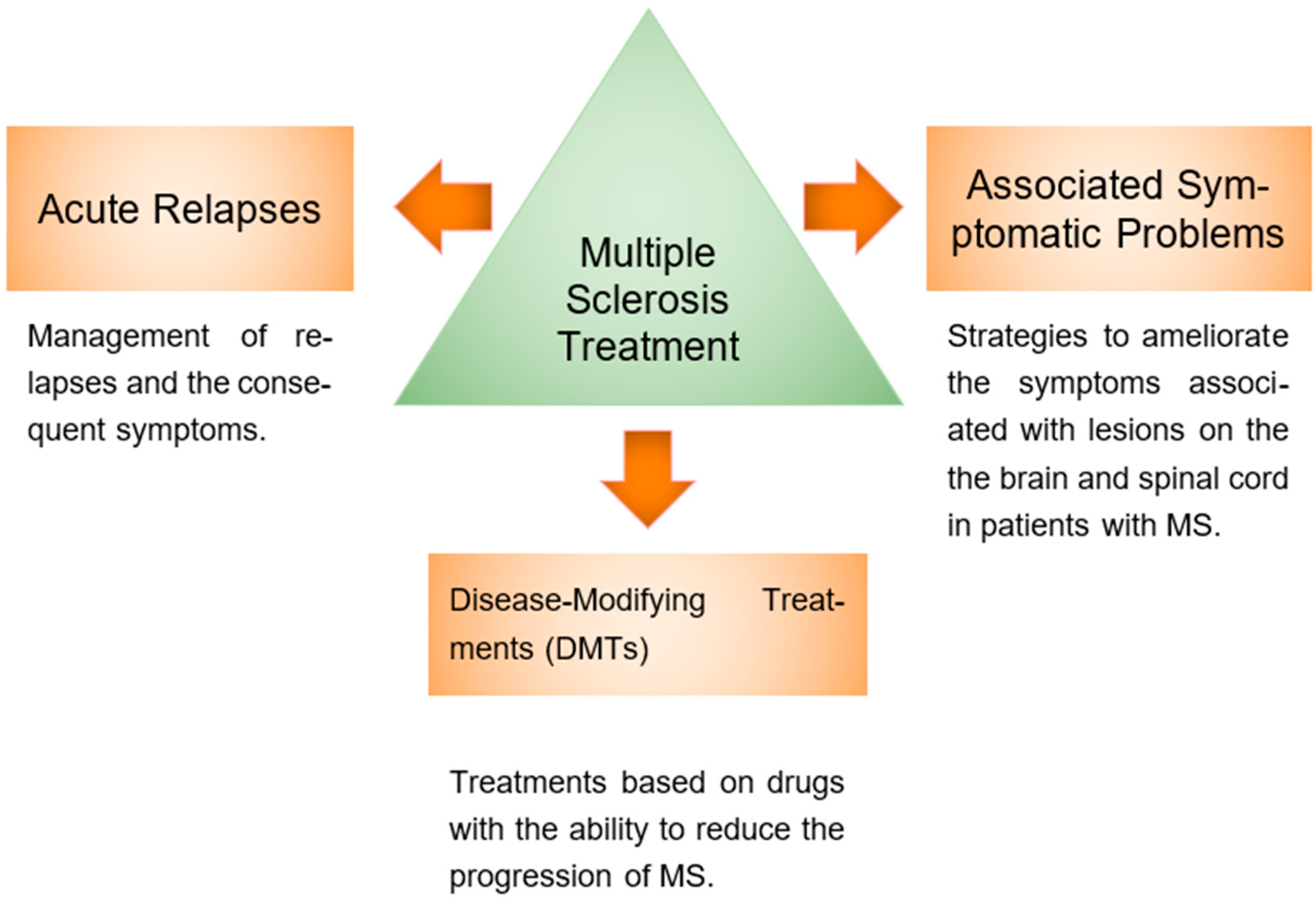
3. Multiple Sclerosis Treatment

The treatment of MS is usually approached along the following three general pathways: acute relapses (1), associated symptomatic problems (2), and Disease-Modifying Treatments (DMTs) (3) (Figure 1) [27]. A summary of multiple sclerosis treatments is presented in Table 2.

3.1. MS Acute Relapses Treatment

Patients with MS commonly experience a relapse with the onset of new symptoms. Managing a relapse requires the differentiation of the nature of the episode; it could be a relapse with signs and symptoms derived from a new focal demyelinating lesion; an exacerbation of existing signs and symptoms; or a fluctuating, short-term modification of symptoms due to external factors that do not represent a relapse [28]. Treatments aimed at treating acute relapses are based on the following two strategies: the first is the use of corticosteroids, and the second is Plasma Exchange (PE) and intravenous[NO_PRINTED_FORM] immunoglobulin G treatment.
Table 2. A summary of multiple sclerosis treatment.
CategoryTreatmentMechanism of ActionCommon Side EffectsReferences
Acute Relapse ManagementCorticosteroidsRestores the blood–brain barrier and reduces metalloproteinase activity and mononuclear trafficking.Gastrointestinal intolerance, insomnia, weight gain, osteoporosis.[29,30,31,32]
Plasma Exchange (PE) and Intravenous Immunoglobulin G (IVIG)Removes circulating antibodies; counteracts antibodies against myelin proteins.Limited clinical efficacy.[27,33,34,35,36]
Symptomatic TreatmentsFatigueAmantadine, modafinil, L-carnitine.Insomnia, dizziness, nausea.[37,38,39,40,41]
DepressionSelective serotonin reuptake inhibitors (SSRIs) and tricyclic antidepressants (TCAs).Nausea, dry mouth, insomnia.[42,43,44]
Bladder dysfunctionOxybutynin and tolterodine.Dry mouth, blurred vision.[44,45]
SpasticityIntrathecal baclofen, tizanidine, botulinum toxin type A, dantrolene, benzodiazepines.Drowsiness, muscle weakness, hepatotoxicity (dantrolene).[46,47,48,49,50,51,52]
Disease-Modifying Therapies (DMTs)Interferon betaModulates T- and B-cell functions; reduces inflammatory cytokines.Flu-like symptoms and injection site reactions.[53,54,55,56]
Glatiramer acetateStimulates specific suppressor T cells.Local reactions and injection site pain.[57,58,59]
FingolimodSphingosine-1-phosphate receptor agonist; inhibits lymphocyte exit from lymph nodes.Infections and bradycardia.[60,61]
TeriflunomideDihydroorotate dehydrogenase inhibitor; blocks lymphocyte proliferation.Alopecia, diarrhea, hypertension.[61,62]
NatalizumabInhibits leukocyte adhesion to endothelium; prevents immune cell migration into the CNS.Risk of progressive multifocal leukoencephalopathy (PML).[55,63,64,65,66]
Dimethyl fumarateModulates proinflammatory T cells; activates antioxidant pathways.Mild infections and gastrointestinal discomfort.[61,67]
Emerging TherapiesAlemtuzumabMonoclonal antibody against CD52; induces cell lysis and prolonged lymphopenia.Autoimmune reactions and infections.[57,68,69]
OcrelizumabMonoclonal antibody against CD20; depletes mature B cells.Respiratory infections and potential increased risk of neoplasms.[70,71,72,73]

3.1.1. Corticosteroids

Traditionally, exacerbations have been controlled using corticosteroids and adrenocorticotropic hormone, since these compounds can reinstate the integrity of the blood–brain barrier, inducing mononuclear trafficking mechanisms. Similarly, they reduce the activity of matrix metalloproteinases, hence preventing neuronal membrane dysfunction [29]. Corticosteroids reduce the recovery time after a relapse episode, but there is not enough evidence to support its long-term effect [31].
Corticosteroids have the following three main effects on the immune system: rapid, nonspecific, and nongenomic effects due to direct interaction with the cell membrane; rapid, specific, nongenomic effects mediated by steroid-selective receptors on the cell; and delayed genomic effects mediated by cytosolic corticosteroid receptors [29].
Side effects are usually associated with the chronic use of corticosteroids. Common problems include gastrointestinal intolerance and dyspepsia, which could require therapy with H2 antagonists. Other less frequent adverse effects may include weight gain, paresthesias, insomnia, and taste disturbance [31]. In order to reduce or prevent adverse effects, supplementation with vitamin D and calcium has been recommended, as well as antiresorptive agents for patients with osteopenia or osteoporosis [32].

3.1.2. Plasma Exchange (PE) and Intravenous Immunoglobulin G (IVIG)

Plasma exchange is a process that separates plasma from red blood cells, white blood cells, and platelets. The blood cells are later returned to the patient, and the plasma is discarded [33]. This procedure is sporadically used as an additional therapy if the relapse is rapidly progressive or severe [27]. It is thought that the positive response to PE could be related to humoral immune mechanisms in MS lesions. However, evidence of its efficacy is restricted [34].
A similar strategy is Intravenous Immunoglobulin G (IVIG) therapy. This therapy is based on plasma pools gathered from various donors. It contains anti-idiotypic antibodies with the ability to counteract circulating antibodies against myelin proteins and restore the antibody concentration in plasma [35]. IVIG is not a first-line therapy for MS because of the lack of evidence of its clinical efficacy. Nonetheless, it could be a second- or third-line therapy if immunomodulatory therapies are not tolerated or not recommended during pregnancy [31,36].

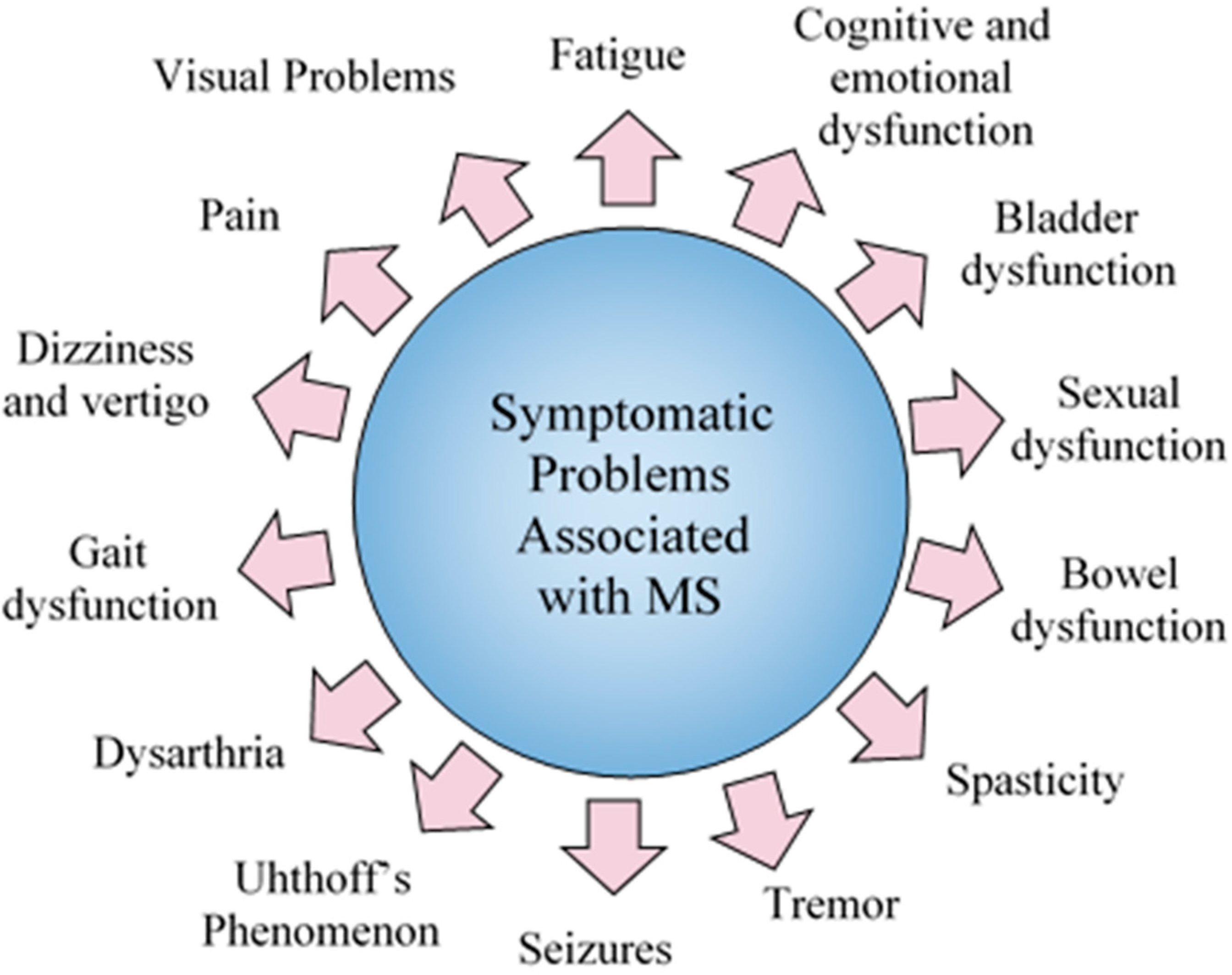
3.2. Symptomatic Problems

MS has a prevalence of disorders, like anxiety, depression, fatigue, and dysfunction of the bladder, bowel, and sexual function. These symptoms are consequence of lesions within the brain and spinal cord (Figure 2) [42]. Here we review the symptoms most associated with MS and the proposed treatments.

3.2.1. Fatigue

Fatigue is the most common symptom of MS. It involves a lack of excessive energy to perform activities during the day, which becomes a disability, as 80% of people with MS lose their jobs due to the fatigue they experience during the first phase of the disease [37]. The association of MS with fatigue is not certain. A drug-based treatment and lifestyle changes, such as a good diet, constant exercise, quitting smoking, and the use of certain drugs (amantadine and modafinil), are recommended. The first drug used for fatigue in patients with MS was amantadine, but the mechanism of action of this drug is unknown [38,39].
Usual medications include amantadine (the most used), aminopyridines, modafinil, and pemoline. However, patients treated with L-carni-tine (ALCAR) have benefited from the reduction of fatigue in chronic neurological diseases. The use of ALCAR as part of treatment fatigue in MS has not been comprehensively reported [40,41].
However, ALCAR (g-trimethyl-h-acetylbutyrrobetaine) plays a key role in the transport of fatty acids from cytosol into the mitochondrial matrix of h-oxidation and has the ability to influence energy metabolism patterns in central and peripheral systems. Its effects in patients with MS are increased level of neurotransmitters. Also, ALCAR synthesis and freed acetylcholine induce choline acetyltransferase activity due to its high affinity and facilitate the release of dopamine. ALCAR is used in patients with MS who cannot tolerate the side effects caused by other drugs. ALCAR is considered more effective than amantadine, because it provides moderate improvements in fatigue [41].

3.2.2. Cognitive and Emotional Dysfunction (Social Functioning)

Cognitive and emotional dysfunctions are important difficulties for patients with MS and their ability to live an economical, productive life, especially because of the lack of both clinical research on the matter and the development of efficient drug therapies. Cognitive decline leads to psychiatric manifestations (e.g., affective disturbances, psychosis, and personality changes), depression, euphoria, emotional lability (crying or laughing suddenly), and suicidal ideation; some occur in more than half of patients who have MS [42,43].
Depression is most commonly observed early in the course of the disease. Pharmacological treatment includes the use of serotonin reuptake inhibitors (SSRIs) such as paroxetine and tricyclic antidepressants (TCAs) like desipramine. As second-line treatments, serotonin norepinephrine reuptake inhibitors (SNRIs), such as venlafaxine, duloxetine, and mirtazapine, are used [44].
Anxiety does not have a specific pharmacological treatment, but it is usually based on benzodiazepines. The disadvantages of this therapy include long-term effects, like dependence [44].

3.2.3. Bladder Dysfunction

Bladder dysfunction is a common problem in 75% of patients with MS. In particular, incontinence is a problem associated with having an overactive bladder, causing concern in those who develop it, and their activities are conditioned. Among anticholinergics, the most widely used are oxybutynin (2.5 to 20 mg twice daily) and tolterodine. Both drugs have similar effects, but tolterodine requires smaller doses. Therefore, tolterodine is considered a first-line therapy for bladder dysfunction [45].
Additional therapies include the use of drug-based treatments to reduce detrusor overactivity, thus ensuring that the bladder does not retain a high volume of urine. It has been found that a mix of anticholinergic medication and intermittent catheterization (suprapubically or urethrally) is an effective but not efficient treatment for patients with increasing disabilities. Another treatment is neural stimulation; in some cases, the progress is good, but it is not likely to be a definitive solution [44,45].

3.2.4. Male Sexual Dysfunction

Sildenafil is the first PDE5 (phosphodiesterase 5) inhibitor to be used as a treatment for erectile dysfunction and high NO guanine monophosphate (cGMP) levels, producing corporal smooth muscle relaxation and, therefore, an erection (at doses of 25.50 to 100 mg). However, side effects include headache, flushing, nasal congestion, and dyspepsia. Other inhibitors of PDE5 are tadalafil and vardenafil (IC-351), but they have a lower effectiveness than sildenafil [74].
An alternative agent is sublingual apomorphine. It acts within the CNS by interacting with D1/D2 receptors within 20 min of ingestion (dose of 2 to 3 mg), which can cause spontaneous erections. However, patients with MS are recommended to follow the advice of their respective physician, since the side effects are unknown [45,75].
Future treatments of sexual dysfunction in patients with MS could involve the enzyme Rho-kinase, which produces relaxation of the smooth muscle. On the other hand, the adrenocorticotrophic hormone (ACTH) and growth-hormone-releasing hormones (GHRHs) are sites of action for neurotransmitters in the erectile response [44,74].

3.2.5. Female Sexual Dysfunction

Some women with MS that experience sexual dysfunction use estrogen creams to sensitize the clitoris and avoid pain during sex. Also, tibolone helps women increase sexual desire, especially after menopause. Methyl testosterone addresses the same needs as those mentioned, but it has adverse effects such as an enlarged clitoris, increased facial hair, and weight fluctuations [44,75].
Pharmacological therapies for women focus on blocking PDE5 by means of alpha-blockers such as phentolamine, causing relaxation in vascular smooth muscle. Sildenafil shows effectiveness in post-menopausal women with sexual dysfunction, but the efficacy for women with MS is still unknown [74]. There are still no effective therapies for women with MS that experience sexual dysfunction [75,76].

3.2.6. Bowel Dysfunction

A diet based on osmotic laxatives, which retain fluid in the intestine, is recommended to manage bowel dysfunction. To increase bowel motility, non-osmotic but stimulating laxatives are used, such as sodium dioctyl sulfosuccinate, senna, and bisacodyl [44,45].
Cisapride helps reduce the transit time in patients with spinal cord lesions. It works as a stimulant laxative, but it was withdrawn from the market because of its association with adverse effects on the cardiac cycle. Some patients with MS have been treated with “biofeedback”, which is a new alternative for treating constipation during the early stages of the disease [45]. On the other hand, surgical interventions are recommended only when a colostomy or an appendicostomy is required [45].

3.2.7. Spasticity

Spasticity is a clinical syndrome that involves muscle hypertonia and exaggerated tendon reflexes because of the inhibition of supraspinal control of reflex activity and spinal programming by MS lesions. This results in functional voluntary movements as a compromise [46].
Physical exercise is advised; however, there are a number of drugs that can be prescribed to assess this problem. As spasticity tends to change over time, a constant reevaluation of the treatment should be considered [46]. In the following, we review some of the most common treatments.
Intrathecal Baclofen (ITB) is a g-Aminobutyric Acid B (GABA-B) receptor agonist used to reduce muscular tone. These receptors are located in laminae I-IV of the spinal cord, at the terminals of primary sensory fibers. Its binding results in the hyperpolarization of neurons [47].
To avoid the low penetrance rate of the blood–brain barrier that the oral administration of baclofen has, ITB delivers the drug into the spinal fluid through a surgically implanted pump. This method requires smaller doses, thus reducing the possible side effects and enhancing the therapeutic response. ITB is usually prescribed for non-ambulatory patients with severe spasticity, as it may cause weakness and loss of mobility in ambulatory patients [48].
ITB is a useful option to relieve pain and improve mobility in patients with MS with generalized spasticity, particularly in the lower extremities [49].
Tizanidine is an imidazoline derivative that acts as an agonist to noradrenergic alpha-2 receptors. It reduces the release of amino acids such as glutamate and aspartate from spinal interneurons and increases presynaptic inhibition of motor neurons [50,51]. Tizanidine is an effective treatment for spasticity. It may reduce muscle pain and does not induce muscle weakness [52].
Botulinum toxin-A (BT) is a neurotoxin from Clostridium botulinum. It inhibits muscle contractions by restricting the liberation of acetylcholine at neuromuscular junctions. It has shown to be useful to reduce the degree of hip adductor spasticity associated with MS [77].
BT injections have also been applied in the early stages of MS, with positive results regarding improved mobility, better functioning in daily activities, pain relief, and posturing hygiene [78].
Dantrolene is a ryanodine receptor-1 antagonist that results in muscle relaxation by inhibiting calcium release from the sarcoplasmic reticulum of the skeletal muscle [79]. This treatment is recommended for spasticity associated with MS, stroke, spinal cord injury, and cerebral palsy [80].
Common side effects may include weakness, nausea, speech difficulty, or dizziness. Hepatotoxicity is a possible risk; hence, continuous monitoring of liver functions is recommended [81].
Benzodiazepines are selective ligands that bind to benzodiazepine receptors and interact with an allosteric modulatory site on the GABA A receptor. These drugs exhibit sedative, anxiolytic, and anticonvulsant activities [82].
The use of benzodiazepines is limited due to adverse effects, such as sleepiness and cognitive impairment [83].
9-δ-Tetrahydrocannabinol and cannabidiol (THC:CBD) is a cannabis-based drug. It interacts with cannabinoid human receptors involved in modulation of muscle tone [84,85].
THC:CBD has been shown to alleviate spasticity caused by MS and its related symptoms, such as cramps, spasms, and pain, as well as bladder function and sleep quality [86].
Tremor is a common disabling symptom associated with MS that may compromise the head, neck, vocal cords, trunk, and limb [87]. The usual medication includes benzodiazepines, cannabinoids, and antispasmodics, such as baclofen and tizanidine (described above), as well as anticonvulsants such as gabapentin and pregabalin [88].
Gabapentin and pregabalin are related compounds, which are derivatives of the inhibitory neurotransmitter γ-Aminobutyric Acid (GABA). The effects are thought to be mediated by the inhibition of calcium currents via high-voltage-activated channels containing the a2d-1 subunit, thus reducing neurotransmitter release and diminishing postsynaptic excitability [89].
Benzodiazepines and anticonvulsants are two of the most used drugs to treat tremor, with clonazepam and gabapentin, respectively, being reported as the most beneficial [88]. However, medical treatment is often unrewarding, which has led to further research on surgical interventions. Surgical options include gamma knife thalamotomy, deep brain stimulation, and radiofrequency thalamotomy [90].

3.2.8. Seizures

Seizures are more likely to occur in MS than in the general population. Epileptic and non-epileptic seizures can be distinguished. Epileptic seizures are rarely and typically occur in the presence of plaques in or near the cerebral cortex, often accompanied by cytotoxic edema. Non-epileptic seizures include tonic spasms and possibly paroxysmal akinesia and dysesthesia. MRI and imaging studies are helpful in determining the nature of the seizure and potential risks [91,92].
Common medications used to treat seizures include oxcarbazepine, carbamazepine, and phenytoin, according to the type of seizure disorder and tolerability of the drug [93].
Oxcarbazepine is an antiepileptic drug approved as an adjunctive therapy or monotherapy for partial seizures in adults and children. It is rapidly reduced to a Monohydroxy Derivative (MHD) by cytosolic arylketone reductases [93].
Carbamazepine and phenytoin are also antiepileptic drugs often prescribed to control seizures in MS. Both block sodium channels by binding to channels at membrane potentials typical of resting neurons, preferentially to an inactivated state, hence allowing for normal neuronal firing while blocking ictal activity. However, side effects such as sleepiness, dizziness, stomach upset, blurred or double vision, and, especially, exacerbation of seizures, raise concern among physicians, and often these drugs have to be discontinued. The efficacy of these drugs may be the result of conductance in the network [94,95,96].

3.2.9. Uhthoff’s Phenomenon

Uhthoff’s phenomenon is defined as a set of short and reversible disruptions in neurological functioning that are related to fluctuations in axonal conduction properties. This characteristic problem with MS may be due to triggering factors that increase the body’s temperature, as there is a slowing in the conduction or blocking of the pores in sodium channels [97].
The usual recommendation is to avoid high-temperature situations. However, drugs such as 4-Aminopyridine (4-AP) can be used to ameliorate symptoms, especially due to its effect on walking ability and speed [98].

3.2.10. Dysarthria

Dysarthria is a speech disorder that affects both the timing and accuracy of the speech process. It results from disturbances in muscular control over speech mechanisms due to damage to the central or peripheral nervous system [99]. The severity of speech deviation could be positively correlated to the severity of neurological involvement, type of disease course, and number of years in progression [100].
Rusz et al. have shown an association between articulation rate and bilateral white and grey matter volume in speakers with spastic dysarthria, which suggests slow speech velocity may be a consequence of pyramidal involvement. Also, their studies revealed that slow oral diadochokinesis in patients with MS may be related to the extent of cerebellar atrophy; the slower oral diadochokinesis may be due to the reduced capacity of the cerebellum to adapt to specific demands of the diadochokinetic task. Speech markers such as those mentioned previously may improve monitoring of disease activity in MS [101].
Paroxysmal ataxia and dysarthria are characterized by brief attacks of slurred speech. These symptoms are usually addressed with membrane-stabilizing medications such as the anticonvulsants listed above. However, levetiracetam has been shown to improve the frequency of ataxia and dysarthria attacks. It is thought that levetiracetam modulates neurotransmitter release via interaction with synaptic vesicle protein 2A and reduces intracellular calcium levels via calcium channel blockade. Common side effects include sedation, headache, dizziness, and neuropsychiatric symptoms [102].

3.2.11. Gait Dysfunction

Gait abnormalities such as decreased length step and cadence, reduced joint movement, and increased variability of most gait parameters are common symptoms of MS. According to the nature of the lesions within the CNS, there may be different patterns of gait deficits, for instance spastic paresis, cerebellar ataxia, sensory ataxia, or even a combination of patterns [103,104]. A significant portion of patients with MS have abnormalities of postural control and gait even in the early stages of the disease, suffering from falls and related injuries, as well as a restriction in their daily activities due to imbalance [104].
Dalfampridine is an approved agent for symptomatic therapy that has been proven to significantly improve the walking speed of patients with MS. It is thought to be an inhibitor of potassium channels in the axonal membrane that prolongs action potentials in demyelinated neurons, therefore improving conduction. It is not considered a disease-modifying therapy but can act as an adjunct. Close monitoring after its prescription is advised, as dalfampridine increases the risk of seizures and is contraindicated in patients with moderate to severe renal dysfunction [105].
Hippotherapy has been studied as a complementary therapy for patients with gait dysfunction. This therapy uses the movement of a horse to provide patients with neuromuscular and sensory stimuli influencing somatosensory, visual, and vestibular systems, as well as anticipatory and reactive postural adjustments. This intervention has shown to be useful in improving walking performance and spatiotemporal gait parameters in people with relapsing-remitting MS [106].

3.2.12. Dizziness and Vertigo

Dizziness and vertigo are frequent impairments associated with MS. Dizziness encompasses faintness or lightheadedness, disequilibrium, and vertigo, which is characterized by the illusion of movement [107].
Vestibular rehabilitation has been used for patients with MS who suffer from dizziness, vertigo, and poor balance. This therapy consists of exercises that direct sensory systems to interact and integrate within the central nervous system to allow for correct spatial cues to position, as well as for head and body motion [108]. Vestibular rehabilitation has been a useful tool for postural control and improving dizziness, as well as fatigue reduction and walking speed enhancement [109].

3.2.13. Pain

In more than 85% of cases, pain is considered a chronic or acute symptom in patients with MS, significantly impacting their daily activities. Pain manifests in various ways. Bagnato et al. identified some of the most common types of pain, including dysesthesia-related extremity pain, trigeminal neuralgia, and Lhermitte’s sign, which are associated with the formation of demyelinating lesions in specific brain areas. Additionally, pain can be related to immunomodulatory treatment, optic neuritis, painful tonic spasms, back pain, and headaches [110].
Furthermore, MS is associated with chronic pain as a dysfunction of the dorsal column–medial lemniscal pathway expressed in hypoesthesia. One mechanism present is nociceptive activation of the unmyelinated C-fibers, which inhibits the processing of non-nociceptive information with beta fibers [111].
Anxiety and depression are associated with pain, where noradrenaline and serotonin are the involved neurotransmitters, which overlap in central nociceptive and affective pathways [112].
Currently, drugs like paracetamol, baclofen, cannabis, diclofenac, naproxen, and ibuprofen are used by patients with MS who have clinical symptoms of pain.
Pain is the result of abnormal impulse transmission, and it is treated with antiepileptic drugs. However, spasticity and pain can be treated with cannabinoids [44,112]. If patients do not receive good treatment for chronic pain, it is possible that they will develop sleep disturbances [113].

3.2.14. Visual Problems

Disturbances in the visual system present in MS allow for the localization of clinical and subclinical manifestations of disease activity. At the same time, optic neuritis is an inflammatory injury that represents a clinically isolated syndrome associated with MS and it is often associated with the earliest clinical stage [114].
Retrograde trans-synaptic retinal ganglion cell degeneration, a response of MS lesions, can cause retinal nerve fiber layer (RNFL) loss. Additionally, RNFL thinning is another response of progressive axonal loss [115].
Sanchez-Dalmau et al. described a protocol to identify visual problems in MS patients using the Visual Functioning Questionnaire (VFQ 25), supplemented with neuro-ophthalmological items to better assess the disease’s impact [116].
Atrophy of the inner retinal layers is the most common visual alteration. It is caused by retrograde trans-synaptic in the optic nerve. Decreased high-contrast visual acuity and low-contrast visual acuity are part of disease [117,118].

3.2.15. Neurorehabilitation

World Health Organization (WHO) defines neurorehabilitation as “an active process by which those disabled by injury or decrease achieves a full recovery”. It is a way for patients with MS to reintegrate into their environment [119]. Treatments such as Acetylcholinesterase Inhibitors (AChEIs), memantine and new approaches like physiotherapy and yoga contribute to activating parts of the body that patients may believe are no longer functional in MS [119].
Acetylcholinesterase Inhibitors (AChEI) and Memantine: These drugs have limited impact and are therefore not widely recommended as part of cognitive therapy for MS [120]. However, Miller et al. reported that this approach can improve memory and verbal learning. Despite this, its potential negative effects are not yet well understood. Similarly, memantine faces the same problem, as its possible effects remain unknown [121].
Currently, neurorehabilitation lacks specific guidelines regarding duration or intensity, as each patient experience different challenges throughout the progression of the disease [44].
Physiotherapy: involves kinesitherapy, physical therapy, massage and hydrotherapy to help patients with MS improve mobility by activating effector capabilities. Gradually, patients regain some functionality, reducing the negative effects of akinesia, though complete restoration of movement is typically not achieved [122]. Some methods used for functional rehabilitation include Proprioceptive Neuromuscular Facilitation (PNF) and Bobath Neurodevelopmental Treatment (NDT). These methods incorporate techniques of coordination, stabilization and relaxation.
Physical activity and Aerobic training: physical activity and aerobic training in patients with MS improve attitude, mood and physical capacity, with benefits in cerebrovascular function. It is recommended to engage in physical activities 2 to 3 times per week, with the duration depending on the degree of disability. Aerobic training also increases muscle strength and helps reduce spasticity or fatigue [122].
Yoga, according to Thakur et al., has a positive impact on patients with MS, reducing pain, fatigue, cortisol levels, and neurogenic bladder dysfunction while improving quality of life. Certain yoga practices like Sukshma Vyayama, as well as deep relaxation, are associated with the alleviation of symptoms associated with neurogenic bladder dysfunction in patients with MS [123].

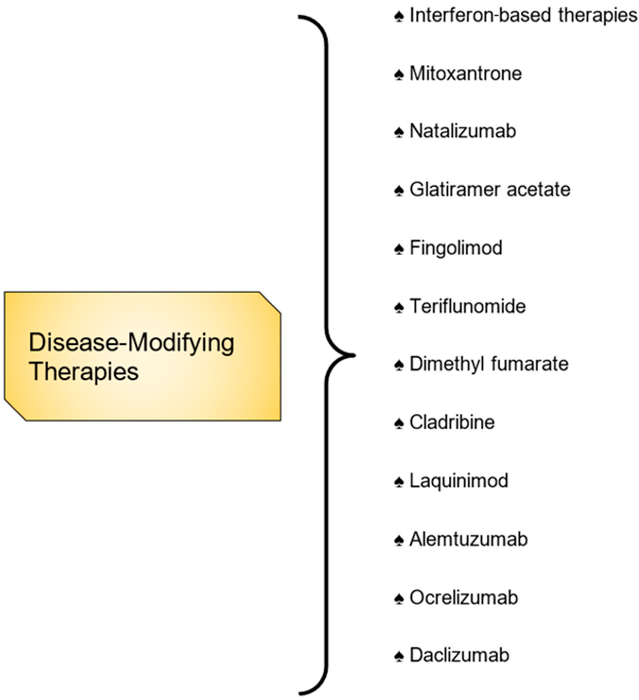
3.3. Disease-Modifying Therapies

Disease-modifying treatments (DMTs) (Figure 3) include interferon-β agents, glatiramer acetate, natalizumab, teriflunomide, etc. Below, we describe the mechanisms of action and efficacy of DMTs based on the severity of the disease [53].
The advantages of DMTs include their ability to reduce clinical and radiological relapses through different mechanisms within the CNS. Another advantage is the extensive clinical research and experimentation conducted in recent years. The disadvantages involve a high risk of infections, which, in extreme cases, require immediate hospitalization because of the immunosuppressive and immunomodulatory effects associated with these treatments [54].

3.3.1. Self-Injectables Therapies

First-generation MS treatments include self-injectable therapies, such as interferon beta and glatiramer acetate (GA). Interferon beta reduces BBB disruption and modulates T-cell, B-cell, and cytokine functions, while GA primarily stimulates T cells [55]. Neutralizing antibodies against interferon beta (IFN-β) are associated with reduced drug efficacy, and IFN-β is understood to downregulate co-stimulatory molecules, adhesion molecules, and MHC class II antigens while also modulating the balance between pro-inflammatory and anti-inflammatory cytokines. Additionally, it inhibits lymphocyte migration across the blood–brain barrier [56].
Despite remaining a first-line treatment, self-injectable therapies face significant competition from oral therapies. Interferon beta is typically administered at a dose of 30 μg once weekly via intramuscular injection or at a dose of 22 to 24 μg every other day via subcutaneous injection [55,56].

3.3.2. Interferon-Based Therapies

Interferons are a family of cytokines that have a variety of functions, such as antiviral, antiproliferative, and antitumor activities, in addition to immunomodulatory effects on the innate and adaptive immune responses. Interferons are classified as type I and type II, according to their interactions with the IFN receptor subunits, peptide mapping, and sequencing homology [124]. Interferons are used as disease-modifying therapies for RRMS. We discuss the IFN-β and peginterferon beta-1a therapies, both widely used to treat MS.
IFN-β is a therapy that reduces the expression of Major Histocompatibility Complex (MHC) molecules on antigen-presenting cells, inhibits T-cell activation, and reduces the release of inflammatory cytokines to inhibit the inflammatory response in MS. IFN-β also increases T-cell suppressor function and enhances the production of growth factors from immune cells [125].
Peginterferon beta-1a is a pegylated form of interferon beta; PEG is a biologically inert polymer chain that is composed of identical repeating ethylene glycol units, commonly used to increase blood residency time, drug exposure, and bioavailability of therapeutic molecules, which traduces into less frequent dosing [126]. Newsome et al. report that peginterferon beta-1a significantly reduces the risk of confirmed disability progression, meaning that this therapy has the potential to delay worsening of disability in patients with RRMS and enhance quality of life [127].

3.3.3. General Immunosuppression

Immunosuppressive drugs are DMTs with high efficacy in modulating the immune system. However, these drugs can cause lymphopenia, opportunistic infections, and a reduced antibody response in some cases. Since not all patients will respond the same way, it is important to carefully consider potential future risks [128]. Drugs like mitoxantrone and natalizumab, which suppress T-cell and B-cell activity, are described below and are among the most commonly used treatments for RRMS.
Mitoxantrone is an immunosuppressive and cytotoxic drug. Its mechanism involves intercalation with DNA and inhibition of both DNA and RNA synthesis, resulting in a reduced number of lymphocytes. As a type II topoisomerase inhibitor, it suppresses the activity of macrophages, T cells, and B cells [55,63].
The usual dose ranges from 5 to 12 mg. Studies have shown that more than 60% of patients with SPMS who took this drug experienced serious infections, leading to hospitalization. Reported infections included viral infections, tonsillitis, endometritis, and urinary tract infections, particularly in patients receiving doses within the 5 to 12 mg range [56].
Mitoxantrone is the only agent approved to treat secondary multiple sclerosis patients, but it is used for RRMS in lower doses [63].
Natalizumab is a monoclonal antibody, widely used as an effective treatment for RRMS. It inhibits leukocyte transmigration across the BBB. This drug prevents the adhesion of antigen 4 integrin and its ligand, Vascular Cell-Adhesion Molecule-1 (VCAM-1), on brain vascular endothelium. This inhibition blocks immune cell transmigration into the CNS, thereby preventing lymphocytes from triggering acute MS lesions [55,63].
Studies in patients with early-stage MS have shown that natalizumab suppresses Cerebrospinal Fluid (CSF) markers of inflammation, neurodegeneration, and B-Cell Chemokine (CXCL13). However, natalizumab carries a high risk of causing Progressive Multifocal Leukoencephalopathy (PML). In 2005, it was withdrawn from the market after cases of PML were reported, including one fatality. The drug was reintroduced a year later, but only as a monotherapy for RRMS. Currently, the risk of PML is low, particularly in patients who receive no more than seven monthly doses [64,65,66].

3.3.4. Oral DMTs

Oral drugs have proven to be highly effective in the treatment of MS, representing a significant advancement in new therapies. Currently, three oral drugs—fingolimod, teriflunomide, and dimethyl fumarate—are established as parts of MS treatment. Additionally, other oral drugs, such as cladribine and laquinimod, remain under close investigation [129].
Glatiramer acetate (Copolymer-1) is a therapeutic tool used for RRMS due to its potential to reduce the frequency of relapses [57]. It is a heterogeneous mixture of not fully characterized synthetic polypeptides, containing L-alanine, L-lysine, L-glutamic acid, and L-tyrosine. The mechanism of action is not completely understood; it is thought to involve modifications of the immune process responsible for the pathogenesis of MS, inducing GA-specific suppressor T cells [58].
The FDA approved Copaxone, a synthetic analogue of GA, in 1996 as a treatment for RRMS, exhibiting, to this day, efficacy and safety [57]. In 2015, Glatopa, the first generic formulation, was produced to reduce the cost of treatment [59].
Greenberg et al. report that both Copaxone and Glatopa provide comparable health outcomes, with borderline statistically significant or borderline insignificant—depending on the exact metric—differences in relapse frequency in favor of Glatopa. The therapeutic costs are similar with both treatments [59].
Fingolimod is the first oral drug used for MS treatment. It is a sphingosine-1-phosphate (S1P) agonist and, in the US, was used as a new generation of disease-modifying therapies. In addition, it is used as a first-line treatment for RRMS because of a reduction in progression of neurological disability with doses of 0.5 mg once a day [60].
Fingolimod acts on four receptor subtypes of sphingosine-1-phosphate (S1P), which are expressed in endothelial cells, lymphocytes, smooth muscle and cardiac myocytes, and neural cells. S1P is an extracellular signaling molecule that regulates B cells and T cells from lymph nodes to the bloodstream [55,61]. Fingolimod functions by targeting S1P receptors, which play a key role in the mechanism that allows lymphocytes to exit lymph nodes and enter the bloodstream. When S1P activity is inhibited, T-cell levels in peripheral blood decrease because T cells are sequestered in lymph nodes, preventing their migration into the bloodstream [56,61].
There have been reports of patients treated with fingolimod who develop infections and diseases, such as PML and, in a few patients between 49 and 63 years old, cancer [65].
Teriflunomide is an oral drug with a once-daily dose approved as treatment for RRMS. It activates T and B lymphocytes and is the only oral drug that has a specific cytostatic effect on proliferating lymphocytes [130].
Teriflunomide non-competitively inhibits Dihydroorotate Dehydrogenase (DHODH), a mitochondrial enzyme involved in de novo pyrimidine synthesis, leading to lymphocyte cell cycle impairment without causing cell death. Teriflunomide reduces neutrophils, lymphocytes, and protein tyrosine kinases, thereby decreasing T-cell proliferation and reducing the production of Interferon-Gamma (IFN-γ), Interleukin-2 (IL-2), and B-Cell Immunoglobulins (Igs) [61,62].
In addition, teriflunomide stimulates T cells and inhibits interleukin-1 beta, matrix metalloproteinases, and cyclo-oxygenase-2 activity. Reported side effects include lymphopenia, nausea, diarrhea, hypertension and, sometimes, alopecia and acute renal failure (less than 1% percent of patients) [55,62].
Dimethyl Fumarate (BG-12) has been used since 2013 as an oral treatment for MS administered as a 240 mg dose taken twice daily. Its activity involves modulating proinflammatory TH1 and TH17 cells. This drug also activates nuclear factor pathways which participate in the physiologic response to oxidative stress [55,61,67].
When administered orally, dimethyl fumarate is metabolized to its active form, Monomethyl Fumarate (MMF). MMF exerts neuroprotective and immunomodulatory effects by inducing the transcription factor nuclear factor (erythroid-derived 2) (Nrf2), with the help of Nuclear Factor Kappa B (NF-kB). The full mechanism of action is not yet fully understood [56,67].
Studies have shown that BG-12 decreases total leukocyte and lymphocyte counts without affecting monocytes, anti-inflammatory subtypes (CD56+NK cells), and neutrophils. BG-12 also has additional immunomodulatory effects, including the suppression of immune responses mediated by Th1 cells [61,67]. Common infections observed in patients with MS treated with BG-12 include nasopharyngeal or influenza-like infections (approximately 2% of patients), urinary tract infections, bronchitis, and gastroenteritis, though these are generally not severe [56].
Cladribine is a deoxyadenosine analogue prodrug, Cladribine (2-chlorodeoxyadenosine), which primarily targets lymphocytes. Its administration results in reductions in CD4+ and CD8+ T cells, with even more pronounced effects on CD19+ B cells. It also causes a significative reduction in the levels of proinflammatory cytokines, cerebrospinal fluid chemokines, and serum [131,132]. Cladribine enters cells via specific membrane nucleoside transporters and is subsequently extruded by ATP-Binding Cassette Subfamily C Member 4 (ABCC4). Once inside the cell, cladribine undergoes phosphorylation to cladribine monophosphate, a reaction catalyzed by Deoxycytidine Kinase (DCK). DCK activity contrasts with that of cytosolic 5’-Nucleotidases (5-NT), which regulate the equilibrium of cellular triphosphorylated nucleotides. Low 5-NT levels confer resistance to cladribine toxicity, creating a balance between DCK and 5-NT. Additionally, c-NT1A and c-NT1B are enzymes involved in cladribine metabolism [131,133].
Cladribine reduces lymphocyte levels in adults with RRMS and is recommended at a dose of 3.5 mg/kg over two years as part of the treatment regimen. Some patients with MS treated with cladribine have reported infections like Herpes zoster [134]. Recent studies suggest that cladribine has the potential to cross the blood–brain barrier in patients with cancer [131].
Laquinimod is a quinoline-3-carboxamide derivative that reduces CD4, CD5, and CD8 lymphocytes, as well as macrophages. It modulates the Th1/Th2 response and decreases the activity of NK cells and the proportion of proinflammatory cytokines (IL4, IL10, and TGFβ). When laquinimod induces axonal damage, demyelination, and oligodendrocyte apoptosis, this suggests that its effects on CNS-resident inflammatory responses in astrocytes contribute to this damage through inhibition of NF-κB signaling [135,136,137].
Currently, laquinimod is used to treat Relapsing-Remitting Multiple Sclerosis (RRMS) and Chronic Progressive Multiple Sclerosis (CPMS). In RRMS, it modulates B cells and exhibits regulatory effects on T cells [56]. One of its advantages lies in the understanding of its mechanisms of action, including the modulation of astrocytic activation, which reduces astrocytic proinflammatory responses while preserving oligodendrocytes, myelin, and axons [138]. Another mechanism involves inhibiting leukocyte entry into the CNS via Very Late Antigen-4 (VLA-4). Additionally, laquinimod has neuroprotective properties by promoting the production of Brain-Derived Neurotrophic Factor (BDNF). It is metabolized primarily by the Cytochrome P450 3A4 (CYP450 3A4) enzyme [135,136].

3.4. Emerging Therapies

Immunopharmacology has proven to be an effective approach for the development of drugs that modulate the immune system and control the progression of diseases. The following drugs represent some of the options that have been investigated as high-efficiency treatment for emergency therapies at various stages of diseases affecting patients with MS [133].

3.4.1. Alemtuzumab

Alemtuzumab is a monoclonal antibody (IgG1) that targets the cell surface glycoprotein CD52, which is expressed on T cells and B cells. It induces cell lysis and leads to prolonged lymphopenia in both B and T cells [57,130]. In rare cases, when patients receive a dose of approximately 12 mg (typically during the first year of the disease), alemtuzumab may cause adverse effects mediated by CD52-dependent Antibody-Dependent Cell-Mediated Cytolysis (ADCC) and Complement-Dependent Cytolysis (CDC). Because of the high levels of CD52, cells become more susceptible to lysis by alemtuzumab. However, further studies are needed to fully understand the effects of CD52 and the mechanisms of action of this drug [68].
Alemtuzumab is not exclusively used for MS; it is also employed in the treatment of other conditions, such as aplastic anemia, immune thrombocytopenic purpura, vasculitis, autoimmune hemolytic anemia, and as an agent in hematopoietic stem cell transplants. However, there are risks associated with alemtuzumab use, including the potential to induce autoimmune diseases such as Goodpasture’s disease and immune thrombocytopenic purpura [69]. D’Amico et al. reported that patients receiving alemtuzumab for other purposes could develop PML [65]. It is also worth noting that the drug’s half-life in MS patients is approximately five days.

3.4.2. Ocrelizumab

Ocrelizumab is a humanized monoclonal antibody that targets cell-surface antigens expressed on memory and mature B cells. Specifically, it binds to CD20, an activated glycosylated phosphoprotein with high affinity for B cells, leading to the depletion of CD20-expressing B cells [70,71].
Ocrelizumab operates through the following three primary mechanisms: the first is complement-dependent cytotoxicity, which is a process that results in membrane breakdown and subsequent cell lysis. The second is antibody-dependent cellular cytotoxicity, which facilitates the immune-mediated destruction of B cells. The third is the induction of apoptosis; CD20 engagement amplifies calcium signaling, promoting programmed cell death [132]. Furthermore, ocrelizumab demonstrates rapid activity during the first four weeks of treatment with doses of 600 mg. When administered as an early treatment, it may decrease neurological damage by B-cell depletion in the peripheral blood [72].
Less than 40 percent of patients with MS treated with ocrelizumab experience respiratory tract infections. Ongoing investigations are focused on the drug’s safety profile, particularly regarding its association with a higher incidence of neoplasms [70,73].

3.4.3. Daclizumab

Daclizumab is a humanized monoclonal antibody of the IgG1 subtype with specificity for the Interleukin (IL)-2 Receptor Alpha (IL-2Rα) Chain (CD25), and it has high efficacy in RRMS, because it blocks the binding of IL-2 to IL-2Rα [139,140].
IL-2 is a proinflammatory cytokine, often referred to as a “T-cell growth factor”, which is produced by activated CD8+ T cells and CD4+ T cells. It plays a crucial role in immune responses and is considered a pleiotropic cytokine [110]. IL-2Rα, the alpha subunit of the IL-2 receptor, is essential for signal transduction, cell growth, survival, death, and differentiation. It functions as an activator of transcription. Although CD25 (IL-2Rα) does not have a specific intracellular signaling function, it increases the receptor’s affinity for IL-2 [140].
Daclizumab blocks IL-2 and IL-2Rα, inhibiting several IL-2-dependent T-cell functions. As a result, it reduces CD25 expression on CD4+ T cells via an FC-receptor mechanism [141].
In patients with MS, daclizumab reduces the risk of progression to permanent disability. However, it is associated with risks, including primary infections, skin rashes, liver toxicity, and lymphadenopathy [140]. Despite these risks, daclizumab has shown clinical benefits in preventing the rejection of allogeneic renal transplants [142].

4. Conclusions

Multiple Sclerosis (MS) is significant due to its complex nature and the wide range of symptoms that patients may experience, including depression, fecal incontinence, cognitive dysfunction, fatigue, and spasticity. Disease progression is crucial in determining the most appropriate treatment strategy, as it helps tailor the approach based on the development of the disease. Commonly used drugs for MS, such as peginterferon beta-1a, glatiramer acetate (Copolymer-1), mitoxantrone, alemtuzumab, and teriflunomide, aim to address the resolution of damage caused by the disease. However, these medications can carry risks, including side effects like nausea, urinary tract infections, diarrhea, and vomiting, among others. Emerging therapies are paving the way for new research paths in MS treatment, as these drugs show high efficacy as immunopharmacological treatments.

Author Contributions

Conceptualization, R.D.Á.-A.; methodology, R.D.Á.-A.; investigation, A.V.A.-R. and K.P.R.-L.; resources, R.D.Á.-A.; data curation, R.D.Á.-A.; writing—original draft preparation, A.V.A.-R. and K.P.R.-L.; writing—review and editing, R.D.Á.-A. and A.V.A.-R.; supervision, R.D.Á.-A.; project administration, R.D.Á.-A.; funding acquisition, R.D.Á.-A. All authors have read and agreed to the published version of the manuscript.

Funding

Rodolfo Daniel Ávila-Avilés was financially supported through the “Investigadoras e Investigadores COMECYT 2024” program, CAT2024-0077.

Informed Consent Statement

This article does not contain data from any studies with human participants or animals performed by any of the authors.

Conflicts of Interest

The authors declare that they have no conflicts of interest.

References

  1. Trapp, B.D.; Peterson, J.; Ransohoff, R.M.; Rudick, R.; Mörk, S.; Bö, L. Axonal transection in the lesions of multiple sclerosis. N. Engl. J. Med. 1998, 338, 278–285. [Google Scholar] [CrossRef] [PubMed]
  2. Trapp, B.D.; Ransohoff, R.M.; Fisher, E.; Rudick, R.A. Neurodegeneration in multiple sclerosis: Relationship to neurological disability. Neuroscientist 1999, 5, 48–57. [Google Scholar] [CrossRef]
  3. Polman, C.H.; O’Connor, P.W.; Havrdova, E.; Hutchinson, M.; Kappos, L.; Miller, D.H.; Phillips, J.T.; Lublin, F.D.; Giovannoni, G.; Wajgt, A.; et al. A Randomized, Placebo-Controlled Trial of Natalizumab for Relapsing Multiple Sclerosis. N. Engl. J. Med. 2006, 354, 899–900. [Google Scholar] [CrossRef] [PubMed]
  4. Brownlee, W.J.; Hardy, T.A.; Fazekas, F.; Miller, D.H. Diagnosis of multiple sclerosis: Progress and challenges. Lancet 2017, 389, 1336–1346. [Google Scholar] [CrossRef] [PubMed]
  5. Thompson, A.J.; Banwell, B.L.; Barkhof, F.; Carroll, W.M.; Coetzee, T.; Comi, G.; Correale, J.; Fazekas, F.; Filippi, M.; Freedman, M.S.; et al. Diagnosis of multiple sclerosis: 2017 revisions of the McDonald criteria. Lancet Neurol. 2018, 17, 162–173. [Google Scholar] [CrossRef]
  6. Prescott, J.; Sr Director, C.P. Communications Naomi Musaji, AJMC ® Perspectives MARCH 2018 3 PERSPECTIVES IN MS PERSPECTIVES IN MS Senior Vice President, Managed Markets. 2018. Available online: www.ajmc.com (accessed on 10 September 2021).
  7. Whitacre, C.C. Sex differences in autoimmune disease. Nat. Immunol. 2001, 2, 777–780. [Google Scholar] [CrossRef] [PubMed]
  8. Bove, R.; Chitnis, T. The role of gender and sex hormones in determining the onset and outcome of multiple sclerosis. Mult. Scler. J. 2014, 20, 520–526. [Google Scholar] [CrossRef] [PubMed]
  9. Zeydan, B.; Kantarci, O.H. Impact of age on multiple sclerosis disease activity and progression. Curr. Neurol. Neurosci. Rep. 2020, 20, 24. [Google Scholar] [CrossRef]
  10. Habbestad, A.; Willumsen, J.S.; Aarseth, J.H.; Grytten, N.; Midgard, R.; Wergeland, S.; Myhr, K.M.; Torkildsen, Ø. Increasing age of multiple sclerosis onset from 1920 to 2022: A population-based study. J. Neurol. 2024, 271, 1610–1617. [Google Scholar] [CrossRef]
  11. Prosperini, L.; Lucchini, M.; Ruggieri, S.; Tortorella, C.; Haggiag, S.; Mirabella, M.; Pozzilli, C.; Gasperini, C. Shift of multiple sclerosis onset towards older age. J. Neurol. Neurosurg. Psychiatry 2022, 93, 1137–1139. [Google Scholar] [CrossRef]
  12. Koch-Henriksen, N.; Thygesen, L.C.; Stenager, E.; Laursen, B.; Magyari, M. Incidence of MS has increased markedly over six decades in Denmark particularly with late onset and in women. Neurology 2018, 90, e1954–e1963. [Google Scholar] [CrossRef]
  13. Brownlee, W.J.; Miller, D.H. Clinically isolated syndromes and the relationship to multiple sclerosis. J. Clin. Neurosci. 2014, 21, 2065–2071. [Google Scholar] [CrossRef] [PubMed]
  14. Lebrun-Frenay, C.; Kantarci, O.; Siva, A.; Azevedo, C.J.; Makhani, N.; Pelletier, D.; Okuda, D.T. Radiologically isolated syndrome. Lancet Neurol. 2023, 22, 1075–1086. [Google Scholar] [CrossRef]
  15. Hardy, T.A.; Miller, D.H. Baló’s concentric sclerosis. Lancet Neurol. 2014, 13, 740–746. [Google Scholar] [CrossRef] [PubMed]
  16. Kraus, D.; Konen, O.; Straussberg, R. Schilder’s disease: Non-invasive diagnosis and successful treatment with human immunoglobulins. Eur. J. Paediatr. Neurol. 2012, 16, 206–208. [Google Scholar] [CrossRef] [PubMed]
  17. McKay, K.A.; Kwan, V.; Duggan, T.; Tremlett, H. Risk factors associated with the onset of relapsing-remitting and primary progressive multiple sclerosis: A systematic review. BioMed Res. Int. 2015, 2015, 817238. [Google Scholar] [CrossRef] [PubMed]
  18. Rodriguez, M. Have we finally identified an autoimmune demyelinating disease? Ann. Neurol. 2009, 66, 572–573. [Google Scholar] [CrossRef] [PubMed]
  19. Wootla, B.; Eriguchi, M.; Rodriguez, M. Is multiple sclerosis an autoimmune disease? Autoimmune Dis. 2012, 2012, 1–12. [Google Scholar] [CrossRef]
  20. Miller, D.; Barkhof, F.; Montalban, X.; Thompson, A.; Filippi, M. Clinically isolated syndromes suggestive of multiple sclerosis, part I: Natural history, pathogenesis, diagnosis, and prognosis. Lancet Neurol. 2005, 4, 281–288. [Google Scholar] [CrossRef] [PubMed]
  21. Chaudhuri, A.; Behan, P.O. Multiple Sclerosis Is Not an Autoimmune Disease. Arch Neurol. 2004, 61, 1610. [Google Scholar] [CrossRef]
  22. Kasper, L.H.; Shoemaker, J. Multiple sclerosis immunology: The healthy immune system vs the MS immune system. Neurology 2010, 74 (Suppl. S1), S2–S8. [Google Scholar] [CrossRef] [PubMed]
  23. Van Oosten, B.W.; Lai, M.; Hodgkinson, S.; Barkhof, F.; Miller, D.H.; Moseley, I.F.; Ader, H.J. Treatment of multiple sclerosis with the monoclonal anti-CD4 antibody cM-T412: Results of a randomized, double-blind, placebo-controlled MR-monitored phase II trial. Neurology 1997, 49, 351–357. [Google Scholar] [CrossRef] [PubMed]
  24. Kebir, H.; Kreymborg, K.; Ifergan, I.; Dodelet-Devillers, A.; Cayrol, R.; Bernard, M.; Giuliani, F.; Arbour, N.; Becher, B.; Prat, A. Human TH 17 lymphocytes promote blood-brain barrier disruption and central nervous system inflammation. Nat. Med. 2007, 13, 1173–1175. [Google Scholar] [CrossRef]
  25. Johnson, A.J.; Suidan, G.L.; McDole, J.; Pirko, I. The CD8 T cell in multiple sclerosis: Suppressor cell or mediator of neuropathology? Int. Rev. Neurobiol. 2007, 79, 73–97. [Google Scholar] [CrossRef] [PubMed]
  26. Khoruts, A.; Miller, S.D.; Jenkins, M.K. Neuroantigen-specific Th2 cells are inefficient suppressors of experimental autoimmune encephalomyelitis induced by effector Th1 cells. J. Immunol. 1995, 155, 5011–5017. [Google Scholar] [CrossRef] [PubMed]
  27. Doshi, A.; Chataway, J. Multiple sclerosis, a treatable disease. Clin. Med. 2016, 16, s53–s59. [Google Scholar] [CrossRef]
  28. Perry, M.; Swain, S.; Kemmis-Betty, S.; Cooper, P. Multiple sclerosis: Summary of NICE guidance. BMJ 2014, 349, g5701. [Google Scholar] [CrossRef]
  29. Frohman, E.M.; Shah, A.; Eggenberger, E.; Metz, L.; Zivadinov, R.; Stüve, O. Corticosteroids for multiple sclerosis: I. application for treating exacerbations. Neurotherapeutics 2007, 4, 618–626. [Google Scholar] [CrossRef]
  30. Ciccone, A.; Beretta, S.; Brusaferri, F.; Galea, I.; Protti, A.; Spreafico, C. Corticosteroids for the long-term treatment in multiple sclerosis. Cochrane Database Syst. Rev. 2008, CD006264. [Google Scholar] [CrossRef] [PubMed]
  31. Ontaneda, D.; Rae-Grant, A.D. Management of acute exacerbations in multiple sclerosis. Ann. Indian Acad. Neurol. 2009, 12, 264. [Google Scholar] [CrossRef]
  32. Tumani, H. Corticosteroids and plasma exchange in multiple sclerosis. J. Neurol. 2008, 255, 36–42. [Google Scholar] [CrossRef] [PubMed]
  33. Khatri, B.O.; McQuillen, M.P.; Hoffmann, R.G.; Harrington, G.J.; Schmoll, D. Plasma exchange in chronic progressive multiple sclerosis. Neurology 1991, 41, 409. [Google Scholar] [CrossRef]
  34. Linker, R.A.; Chan, A.; Sommer, M.; Koziolek, M.; Müller, G.-A.; Paulus, W.; Gold, R. Plasma exchange therapy for steroid-refractory superimposed relapses in secondary progressive multiple sclerosis. J. Neurol. 2007, 254, 1288–1289. [Google Scholar] [CrossRef] [PubMed]
  35. Sorensen, P.S. The role of intravenous immunoglobulin in the treatment of multiple sclerosis. J. Neurol. Sci. 2003, 206, 123–130. [Google Scholar] [CrossRef]
  36. Elovaara, I.; Apostolski, S.; Van Doorn, P.; Gilhus, N.E.; Hietaharju, A.; Honkaniemi, J.; Udd, B. EFNS guidelines for the use of intravenous immunoglobulin in treatment of neurological diseases: EFNS task force on the use of intravenous immunoglobulin in treatment of neurological diseases. Eur. J. Neurol. 2008, 15, 893–908. [Google Scholar] [CrossRef] [PubMed]
  37. Glanz, B.I.; Dégano, I.; Rintell, D.; Chitnis, T.; Weiner, H.L.; Healy, B. Work Productivity in Relapsing Multiple Sclerosis: Associations with Disability, Depression, Fatigue, Anxiety, Cognition, and Health-Related Quality of Life. Value Health 2012, 15, 1029–1035. [Google Scholar] [CrossRef]
  38. Pucci, E.; Brañas Tato, P.; D’Amico, R.; Giuliani, G.; Solari, A.; Taus, C. Amantadine for fatigue in multiple sclerosis. Cochrane Database Syst. Rev. 2007, CD002818. [Google Scholar] [CrossRef] [PubMed]
  39. Rossini, P.M.; Pasqualetti, P.; Pozzilli, C.; Grasso, M.G.; Millefiorini, E.; Graceffa, A.; Caltagirone, C. Fatigue in progressive multiple sclerosis: Results of a randomized, double-blind, placebo-controlled, crossover trial of oral 4-aminopyridine. Mult. Scler. J. 2001, 7, 354–358. [Google Scholar] [CrossRef]
  40. Brenner, P.; Piehl, F. Fatigue and depression in multiple sclerosis: Pharmacological and non-pharmacological interventions. Acta Neurol. Scand. 2016, 134, 47–54. [Google Scholar] [CrossRef] [PubMed]
  41. Tomassini, V.; Pozzilli, C.; Onesti, E.; Pasqualetti, P.; Marinelli, F.; Pisani, A.; Fieshchi, C. Comparison of the effects of acetyl L-carnitine and amantadine for the treatment of fatigue in multiple sclerosis: Results of a pilot, randomized, double-blind, crossover trial. J. Neurol. Sci. 2004, 218, 103–108. [Google Scholar] [CrossRef]
  42. Marrie, R.A.; Zhang, L.; Lix, L.M.; Graff, L.A.; Walker, J.R.; Fisk, J.D.; Patten, S.B.; Hitchon, C.A.; Bolton, J.M.; Sareen, J.; et al. The validity and reliability of screening measures for depression and anxiety disorders in multiple sclerosis. Mult. Scler. Relat. Disord. 2018, 20, 9–15. [Google Scholar] [CrossRef] [PubMed]
  43. Brassington, J.C.; Marsh, N.V. Neuropsychological Aspects of Multiple Sclerosis. Neuropsychol. Rev. 1998, 8, 43–77. [Google Scholar] [CrossRef] [PubMed]
  44. Kesselring, J.; Beer, S. Symptomatic therapy and neurorehabilitation in multiple sclerosis. Lancet Neurol. 2005, 4, 643–652. [Google Scholar] [CrossRef] [PubMed]
  45. DasGupta, R.; Fowler, C.J. Bladder, Bowel and Sexual Dysfunction in Multiple Sclerosis. Ther. Pract. 2003, 63, 153–166. [Google Scholar] [CrossRef]
  46. Kesselring, J. Chapter 14 Complications of Multiple Sclerosis: Fatigue; Spasticity; Ataxia; Pain; and Bowel, Bladder, and Sexual Dysfunction. In Blue Books of Practical Neurology; Elsevier: Amsterdam, The Netherlands, 2003; Volume 27, pp. 217–227. [Google Scholar] [CrossRef]
  47. Krach, L.E. Pharmacotherapy of spasticity: Oral medications and intrathecal baclofen. J. Child Neurol. 2001, 16, 31–36. [Google Scholar] [CrossRef] [PubMed]
  48. Sadiq, S.A.; Wang, G.C. Long–term intrathecal baclofen therapy in ambulatory patients with spasticity. J. Neurol. 2006, 253, 563–569. [Google Scholar] [CrossRef] [PubMed]
  49. Rekand, T.; Grønning, M. Treatment of spasticity related to multiple sclerosis with intrathecal baclofen: A long-term follow-up. J. Rehabil. Med. 2011, 43, 511–514. [Google Scholar] [CrossRef]
  50. Coward, D.M. Tizanidine: Neuropharmacology and mechanism of action. Neurology 1994, 44 (Suppl. S9), S6–S10, discussion S10–S11. [Google Scholar] [PubMed]
  51. Ghanavatian, S.; Derian, A. Tizanidine. In StatPearls [Internet]; StatPearls Publishing: Treasure Island, FL, USA, 2025. [Google Scholar]
  52. Kamen, L.; Henney, H.R., III; Runyan, J.D. A practical overview of tizanidine use for spasticity secondary to multiple sclerosis, stroke, and spinal cord injury. Curr. Med. Res. Opin. 2008, 24, 425–439. [Google Scholar] [CrossRef] [PubMed]
  53. Oh, J.; O’Connor, P.W. Established disease-modifying treatments in relapsing-remitting multiple sclerosis. Curr. Opin. Neurol. 2015, 28, 220–229. [Google Scholar] [CrossRef]
  54. Yong, K.P.; Kim, H.J. Disease modifying therapies and infection risks in multiple sclerosis-a decision-making conundrum. Ann. Transl. Med. 2020, 8, 722. [Google Scholar] [CrossRef] [PubMed]
  55. Wingerchuk, D.M.; Carter, J.L. Multiple sclerosis: Current and emerging disease-modifying therapies and treatment strategies. Mayo Clin. Proc. 2014, 89, 225–240. [Google Scholar] [CrossRef] [PubMed]
  56. Winkelmann, A.; Loebermann, M.; Reisinger, E.C.; Hartung, H.P.; Zettl, U.K. Disease-modifying therapies and infectious risks in multiple sclerosis. Nat. Rev. Neurol. 2016, 12, 217–233. [Google Scholar] [CrossRef] [PubMed]
  57. Weinstock-Guttman, B.; Nair, K.V.; Glajch, J.L.; Ganguly, T.C.; Kantor, D. Two decades of glatiramer acetate: From initial discovery to the current development of generics. J. Neurol. Sci. 2017, 376, 255–259. [Google Scholar] [CrossRef]
  58. Rocco, P.; Eberini, I.; Musazzi, U.M.; Franzè, S.; Minghetti, P. Glatiramer acetate: A complex drug beyond biologics. Eur. J. Pharm. Sci. 2019, 133, 8–14. [Google Scholar] [CrossRef] [PubMed]
  59. Greenberg, B.; Hall, S.; Grabner, M.; Balu, S.; Zhang, X.; Kantor, D. Multiple sclerosis relapse rates and healthcare costs of two versions of glatiramer acetate. Curr. Med. Res. Opin. 2020, 36, 1167–1175. [Google Scholar] [CrossRef]
  60. Scott, L.J. Fingolimod: A review of its use in the management of relapsing-remitting multiple sclerosis. CNS Drugs 2011, 25, 673–698. [Google Scholar] [CrossRef]
  61. Pardo, G.; Jones, D.E. The sequence of disease-modifying therapies in relapsing multiple sclerosis: Safety and immunologic considerations. J. Neurol. 2017, 264, 2351–2374. [Google Scholar] [CrossRef]
  62. He, D.; Zhang, C.; Zhao, X.; Zhang, Y.; Dai, Q.; Li, Y.; Chu, L. Teriflunomide for multiple sclerosis. Cochrane Database Syst. Rev. 2016, 3, CD009882. [Google Scholar] [CrossRef]
  63. Filippini, G.; Del Giovane, C.; Clerico, M.; Beiki, O.; Mattoscio, M.; Piazza, F.; Fredrikson, S.; Tramacere, I.; Scalfari, A.; Salanti, G. Treatment with disease-modifying drugs for people with a first clinical attack suggestive of multiple sclerosis. Cochrane Database Syst. Rev. 2017, 4, CD012200. [Google Scholar] [CrossRef] [PubMed]
  64. Ryerson, L.Z.; Foley, J.; Chang, I.; Kister, I.; Cutter, G.; Metzger, R.R.; Goldberg, J.D.; Li, X.; Riddle, E.; Smirnakis, K.; et al. Risk of natalizumab-associated PML in patients with MS is reduced with extended interval dosing. Neurology 2019, 93, 1452–1462. [Google Scholar] [CrossRef]
  65. D’Amico, E.; Zanghì, A.; Leone, C.; Tumani, H.; Patti, F. Treatment-Related Progressive Multifocal Leukoencephalopathy in Multiple Sclerosis: A Comprehensive Review of Current Evidence and Future Needs. Drug Saf. 2016, 39, 1163–1174. [Google Scholar] [CrossRef] [PubMed]
  66. Kapoor, R.; Ho, P.R.; Campbell, N.; Chang, I.; Deykin, A.; Forrestal, F.; Lucas, N.; Yu, B.; Arnold, D.L.; Freedman, M.S.; et al. Effect of natalizumab on disease progression in secondary progressive multiple sclerosis (ASCEND): A phase 3, randomised, double-blind, placebo-controlled trial with an open-label extension. Lancet Neurol. 2018, 17, 405–415. [Google Scholar] [CrossRef] [PubMed]
  67. Montes Diaz, G.; Fraussen, J.; Van Wijmeersch, B.; Hupperts, R.; Somers, V. Dimethyl fumarate induces a persistent change in the composition of the innate and adaptive immune system in multiple sclerosis patients. Sci. Rep. 2018, 8, 8194. [Google Scholar] [CrossRef]
  68. Ruck, T.; Bittner, S.; Wiendl, H.; Meuth, S.G. Alemtuzumab in Multiple Sclerosis: Mechanism of Action and Beyond. Int. J. Mol. Sci. 2015, 16, 16414–16439. [Google Scholar] [CrossRef] [PubMed]
  69. Riera, R.; Porfírio, G.J.; Torloni, M.R. Alemtuzumab for multiple sclerosis. Cochrane Database Syst. Rev. 2016, 4, CD011203. [Google Scholar] [CrossRef] [PubMed]
  70. Hauser, S.L.; Bar-Or, A.; Comi, G.; Giovannoni, G.; Hartung, H.P.; Hemmer, B.; Lublin, F.; Montalban, X.; Rammohan, K.W.; Selmaj, K.; et al. Ocrelizumab versus Interferon Beta-1a in Relapsing Multiple Sclerosis. N. Engl. J. Med. 2017, 376, 221–234. [Google Scholar] [CrossRef]
  71. Sorensen, P.S.; Blinkenberg, M. The potential role for ocrelizumab in the treatment of multiple sclerosis: Current evidence and future prospects. Ther. Adv. Neurol. Disord. 2016, 9, 44–52. [Google Scholar] [CrossRef]
  72. Barkhof, F.; Kappos, L.; Wolinsky, J.S.; Li, D.; Bar-Or, A.; Hartung, H.P.; Belachew, S.; Han, J.; Julian, L.; Sauter, A.; et al. Onset of clinical and MRI efficacy of ocrelizumab in relapsing multiple sclerosis. Neurology 2019, 93, 1778–1786. [Google Scholar] [CrossRef]
  73. Mulero, P.; Midaglia, L.; Montalban, X. Ocrelizumab: A new milestone in multiple sclerosis therapy. Ther. Adv. Neurol. Disord. 2018, 11, 1756286418773025. [Google Scholar] [CrossRef] [PubMed]
  74. Xiao, Y.; Wang, J.; Luo, H. Sildenafil citrate for erectile dysfunction in patients with multiple sclerosis. Cochrane Database Syst. Rev. 2012, CD009427. [Google Scholar] [CrossRef] [PubMed]
  75. Nortvedt, M.W.; Riise, T.; Myhr, K.-M.; Landtblom, A.-M.; Bakke, A.; Nyland, H.I. Reduced quality of life among multiple sclerosis patients with sexual disturbance and bladder dysfunction. Mult. Scler. J. 2001, 7, 231–235. [Google Scholar] [CrossRef] [PubMed]
  76. Zorzon, M.; Zivadinov, R.; Bosco, A.; Bragadin, L.M.; Moretti, R.; Bonfigli, L.; Morassi, P.; Iona, L.G.; Cazzato, G. Sexual dysfunction in multiple sderosis: A case-control study. 1. Frequency and comparison of groups. Mult. Scler. J. 1999, 5, 418–427. [Google Scholar] [CrossRef] [PubMed]
  77. Hyman, N.; Barnes, M.; Bhakta, B.; Cozens, A.; Bakheit, M.; Kreczy-Kleedorfer, B.; Poewe, W.; Wissel, J.; Bain, P.; Glickman, S.; et al. Botulinum toxin (Dysport®) treatment of hip adductor spasticity in multiple sclerosis: A prospective, randomised, double blind, placebo controlled, dose ranging study. J. Neurol. Neurosurg. Psychiatry 2000, 68, 707–712. [Google Scholar] [CrossRef] [PubMed]
  78. Moccia, M.; Frau, J.; Carotenuto, A.; Butera, C.; Coghe, G.; Barbero, P.; Frontoni, M.; Groppo, E.; Giovannelli, M.; Del Carro, U.; et al. Botulinum toxin for the management of spasticity in multiple sclerosis: The Italian botulinum toxin network study. Neurol. Sci. 2020, 41, 2781–2792. [Google Scholar] [CrossRef] [PubMed]
  79. Muehlschlegel, S.; Sims, J.R. Dantrolene: Mechanisms of neuroprotection and possible clinical applications in the neurointensive care unit. Neurocritical Care 2009, 10, 103–115. [Google Scholar] [CrossRef] [PubMed]
  80. Ratto, D.; Joyner, R.W. Dantrolene. In StatPearls [Internet]; StatPearls Publishing: Treasure Island, FL, USA, 2025. [Google Scholar]
  81. Haselkorn, J.K.; Balsdon Richer, C.; Fry Welch, D.; Herndon, R.M.; Johnson, B.; Little, J.W.; Miller, J.R.; Rosenberg, J.H.; Seidle, M.E. Multiple Sclerosis Council for Clinical Practice Guidelines, Overview of spasticity management in multiple sclerosis. Evidence-based management strategies for spasticity treatment in multiple sclerosis. J. Spinal Cord Med. 2005, 28, 167–199. [Google Scholar] [CrossRef] [PubMed]
  82. Bateson, A.N. The benzodiazepine site of the GABAA receptor: An old target with new potential? Sleep Med. 2004, 5, S9–S15. [Google Scholar] [CrossRef] [PubMed]
  83. Markota, M.; Rummans, T.A.; Bostwick, J.M.; Lapid, M.I. Benzodiazepine use in older adults: Dangers, management, and alternative therapies. In Mayo Clinic Proceedings; Elsevier: Amsterdam, The Netherlands, 2016; Volume 91, pp. 1632–1639. [Google Scholar] [CrossRef]
  84. Barnes, M.P. Sativex®: Clinical efficacy and tolerability in the treatment of symptoms of multiple sclerosis and neuropathic pain. Expert Opin. Pharmacother. 2006, 7, 607–615. [Google Scholar] [CrossRef] [PubMed]
  85. Patti, F.; Messina, S.; Solaro, C.; Amato, M.P.; Bergamaschi, R.; Bonavita, S.; Bossio, R.B.; Morra, V.B.; Costantino, G.F.; Cavalla, P.; et al. Efficacy and safety of cannabinoid oromucosal spray for multiple sclerosis spasticity. J. Neurol. Neurosurg. Psychiatry 2016, 87, 944–951. [Google Scholar] [CrossRef] [PubMed]
  86. Patti, F.; Chisari, C.G.; Solaro, C.; Benedetti, M.D.; Berra, E.; Bianco, A.; Bossio, R.B.; Buttari, F.; Castelli, L.; Cavalla, P.; et al. Effects of THC/CBD oromucosal spray on spasticity-related symptoms in people with multiple sclerosis: Results from a retrospective multicenter study. Neurol. Sci. Off. J. Ital. Neurol. Soc. Ital. Soc. Clin. Neurophysiol. 2020, 41, 2905–2913. [Google Scholar] [CrossRef]
  87. Alusi, S.H. Evaluation of three different ways of assessing tremor in multiple sclerosis. J. Neurol. Neurosurg. Psychiatry 2000, 68, 756–760. [Google Scholar] [CrossRef] [PubMed]
  88. Meador, W.; Salter, A.R.; Rinker, J.R. Symptomatic management of multiple sclerosis–associated tremor among participants in the NARCOMS registry. Int. J. MS Care 2016, 18, 147–153. [Google Scholar] [CrossRef]
  89. Tzellos, T.G.; Papazisis, G.; Toulis, K.A.; Sardeli, C.; Kouvelas, D. A2δ ligands gabapentin and pregabalin: Future implications in daily clinical practice. Hippokratia 2010, 14, 71. [Google Scholar] [PubMed]
  90. Raju, S.S.; Niranjan, A.; Monaco, E.A.; Flickinger, J.C.; Lunsford, L.D. Stereotactic radiosurgery for medically refractory multiple sclerosis–related tremor. J. Neurosurg. 2018, 128, 1214–1221. [Google Scholar] [CrossRef] [PubMed]
  91. Spatt, J.; Chaix, R.; Mamoli, B. Epileptic and non-epileptic seizures in multiple sclerosis. J. Neurol. 2001, 248, 2–9. [Google Scholar] [CrossRef] [PubMed]
  92. Kelley, B.J.; Rodriguez, M. Seizures in patients with multiple sclerosis. CNS Drugs 2009, 23, 805–815. [Google Scholar] [CrossRef] [PubMed]
  93. Flesch, G.; Czendlik, C.; Renard, D.; Lloyd, P. Pharmacokinetics of the monohydroxy derivative of oxcarbazepine and its enantiomers after a single intravenous dose given as racemate compared with a single oral dose of oxcarbazepine. Drug Metab. Dispos. 2011, 39, 1103–1110. [Google Scholar] [CrossRef] [PubMed]
  94. Farooque, P.; Detyniecki, K.; Mattson, R.H. Epilepsy; Antiepileptic drug profiles. In Encyclopedia of the Neurological Sciences; Elsevier: Amsterdam, The Netherlands, 2014; pp. 81–92. [Google Scholar] [CrossRef]
  95. Dagiasi, I.; Vall, V.; Kumlien, E.; Burman, J.; Zelano, J. Treatment of epilepsy in multiple sclerosis. Seizure 2018, 58, 47–51. [Google Scholar] [CrossRef] [PubMed]
  96. Thomas, E.A.; Petrou, S. Network-specific mechanisms may explain the paradoxical effects of carbamazepine and phenytoin. Epilepsia 2013, 54, 1195–1202. [Google Scholar] [CrossRef]
  97. Frohman, T.C.; Davis, S.L.; Beh, S.; Greenberg, B.M.; Remington, G.; Frohman, E.M. Uhthoff’s phenomena in MS—Clinical features and pathophysiology. Nat. Rev. Neurol. 2013, 9, 535–540. [Google Scholar] [CrossRef] [PubMed]
  98. Mathiesen, H.K.; Sorensen, P.S. Prolonged-release fampridine improves walking in a proportion of patients with multiple sclerosis. Expert Rev. Neurother. 2013, 13, 1309–1317. [Google Scholar] [CrossRef]
  99. Piacentini, V.; Mauri, I.; Cattaneo, D.; Gilardone, M.; Montesano, A.; Schindler, A. Relationship between quality of life and dysarthria in patients with multiple sclerosis. Arch. Phys. Med. Rehabil. 2014, 95, 2047–2054. [Google Scholar] [CrossRef]
  100. Hartelius, L.; Runmarker, B.; Andersen, O. Prevalence and characteristics of dysarthria in a multiple-sclerosis incidence cohort: Relation to neurological data. Folia Phoniatr. Logop. 2000, 52, 160–177. [Google Scholar] [CrossRef]
  101. Rusz, J.; Vaneckova, M.; Benova, B.; Tykalova, T.; Novotny, M.; Ruzickova, H.; Uher, T.; Andelova, M.; Novotna, K.; Friedova, L.; et al. Brain volumetric correlates of dysarthria in multiple sclerosis. Brain Lang. 2019, 194, 58–64. [Google Scholar] [CrossRef]
  102. Goodwin, S.J.; Carpenter, A.F. Successful treatment of paroxysmal ataxia and dysarthria in multiple sclerosis with levetiracetam. Mult. Scler. Relat. Disord. 2016, 10, 79–81. [Google Scholar] [CrossRef]
  103. Stevens, V.; Goodman, K.; Rough, K.; Kraft, G.H. Gait impairment and optimizing mobility in multiple sclerosis. Phys. Med. Rehabil. Clin. 2013, 24, 573–592. [Google Scholar] [CrossRef] [PubMed]
  104. Cameron, M.H.; Nilsagard, Y. Balance, gait, and falls in multiple sclerosis. In Handbook of Clinical Neurology; Elsevier: Amsterdam, The Netherlands, 2018; Volume 159, pp. 237–250. [Google Scholar] [CrossRef]
  105. Egeberg, M.D.; Oh, C.Y.; Bainbridge, J.L. Clinical overview of dalfampridine: An agent with a novel mechanism of action to help with gait disturbances. Clin. Ther. 2012, 34, 2185–2194. [Google Scholar] [CrossRef] [PubMed]
  106. Moraes, A.G.; Neri SG, R.; Motl, R.W.; Tauil, C.B.; von Glehn Silva, F.; Corrêa, É.C.; de David, A.C. Effect of hippotherapy on walking performance and gait parameters in people with multiple sclerosis. Mult. Scler. Relat. Disord. 2020, 43, 102203. [Google Scholar] [CrossRef] [PubMed]
  107. Marrie, R.A.; Cutter, G.R.; Tyry, T. Substantial burden of dizziness in multiple sclerosis. Mult. Scler. Relat. Disord. 2013, 2, 21–28. [Google Scholar] [CrossRef]
  108. Tjernström, F.; Zur, O.; Jahn, K. Current concepts and future approaches to vestibular rehabilitation. J. Neurol. 2016, 263, 65–70. [Google Scholar] [CrossRef] [PubMed]
  109. García-Muñoz, C.; Cortés-Vega, M.D.; Heredia-Rizo, A.M.; Martín-Valero, R.; García-Bernal, M.I.; Casuso-Holgado, M.J. Effectiveness of Vestibular Training for Balance and Dizziness Rehabilitation in People with Multiple Sclerosis: A Systematic Review and Meta-Analysis. J. Clin. Med. 2020, 9, 590. [Google Scholar] [CrossRef] [PubMed]
  110. Bagnato, F.; Centonze, D.; Galgani, S.; Grasso, M.G.; Haggiag, S.; Strano, S. Painful and involuntary multiple sclerosis. Expert Opin. Pharmacother. 2011, 12, 763–777. [Google Scholar] [CrossRef]
  111. Scherder, R.J.; Kant, N.; Wolf, E.T.; Pijnenburg, B.; Scherder, E. Sensory Function and Chronic Pain in Multiple Sclerosis. Pain Res. Manag. 2018, 2018, 1924174. [Google Scholar] [CrossRef] [PubMed]
  112. Scherder, R.; Kant, N.; Wolf, E.; Pijnenburg, A.; Scherder, E. Pain and Cognition in Multiple Sclerosis. Pain Med. 2017, 18, 1987–1998. [Google Scholar] [CrossRef] [PubMed]
  113. Kesselring, J. Neurorehabilitation in multiple sclerosis—What is the evidence-base? J. Neurol. 2004, 251 (Suppl. S4), IV25–IV29. [Google Scholar] [CrossRef]
  114. Costello, F. Vision Disturbances in Multiple Sclerosis. Semin. Neurol. 2016, 36, 185–195. [Google Scholar] [CrossRef] [PubMed]
  115. Noval, S.; Contreras, I.; Muñoz, S.; Oreja-Guevara, C.; Manzano, B.; Rebolleda, G. Optical coherence tomography in multiple sclerosis and neuromyelitis optica: An update. Mult. Scler. Int. 2011, 2011, 1–11. [Google Scholar] [CrossRef] [PubMed]
  116. Sanchez-Dalmau, B.; Martinez-Lapiscina, E.H.; Pulido-Valdeolivas, I.; Zubizarreta, I.; Llufriu, S.; Blanco, Y.; Sola-Valls, N.; Sepulveda, M.; Guerrero, A.; Alba, S.; et al. Predictors of vision impairment in Multiple Sclerosis. PLoS ONE 2018, 13, e0195856. [Google Scholar] [CrossRef] [PubMed]
  117. Nij Bijvank, J.A.; Petzold, A.; Coric, D.; Tan, H.S.; Uitdehaag, B.; Balk, L.J.; van Rijn, L.J. Quantification of Visual Fixation in Multiple Sclerosis. Investig. Ophthalmol. Vis. Sci. 2019, 60, 1372–1383. [Google Scholar] [CrossRef] [PubMed]
  118. Balk, L.J.; Coric, D.; Nij Bijvank, J.A.; Killestein, J.; Uitdehaag, B.M.; Petzold, A. Retinal atrophy in relation to visual functioning and vision-related quality of life in patients with multiple sclerosis. Mult. Scler. 2018, 24, 767–776. [Google Scholar] [CrossRef] [PubMed]
  119. World Health Organization. Neurological Disorders [OP]: Public Health Challenges (1.a ed.). 2006. Available online: https://www.who.int/publications/i/item/9789241563369 (accessed on 10 September 2021).
  120. Calabresi, P.A. Diagnosis and management of multiple sclerosis. Am. Fam. Physician 2004, 70, 1935–1944. [Google Scholar] [PubMed]
  121. Miller, E.; Morel, A.; Redlicka, J.; Miller, I.; Saluk, J. Pharmacological and Non-pharmacological Therapies of Cognitive Impairment in Multiple Sclerosis. Curr. Neuropharmacol. 2018, 16, 475–483. [Google Scholar] [CrossRef] [PubMed]
  122. Kubsik-Gidlewska, A.M.; Klimkiewicz, P.; Klimkiewicz, R.; Janczewska, K.; Woldańska-Okońska, M. Rehabilitation in multiple sclerosis. Adv. Clin. Exp. Med. Off. Organ Wroc. Med. Univ. 2017, 26, 709–715. [Google Scholar] [CrossRef] [PubMed]
  123. Thakur, P.; Mohammad, A.; Rastogi, Y.R.; Saini, R.V.; Saini, A.K. Yoga as an intervention to manage multiple sclerosis symptoms. J. Ayurveda Integr. Med. 2020, 11, 114–117. [Google Scholar] [CrossRef] [PubMed]
  124. de Padilla, C.M.L.; Niewold, T.B. The type I interferons: Basic concepts and clinical relevance in immune-mediated inflammatory diseases. Gene 2016, 576, 14–21. [Google Scholar] [CrossRef] [PubMed]
  125. Javed, A.; Reder, A.T. Therapeutic role of beta-interferons in multiple sclerosis. Pharmacol. Ther. 2006, 110, 35–56. [Google Scholar] [CrossRef] [PubMed]
  126. Kieseier, B.C.; Calabresi, P.A. PEGylation of Interferon-β-1a. CNS Drugs 2012, 26, 205–214. [Google Scholar] [CrossRef] [PubMed]
  127. Newsome, S.D.; Kieseier, B.C.; Liu, S.; You, X.; Kinter, E.; Hung, S.; Sperling, B. Peginterferon beta-1a reduces disability worsening in relapsing–remitting multiple sclerosis: 2-year results from ADVANCE. Ther. Adv. Neurol. Disord. 2017, 10, 41–50. [Google Scholar] [CrossRef] [PubMed]
  128. Giovannoni, G. Disease-modifying treatments for early and advanced multiple sclerosis: A new treatment paradigm. Curr. Opin. Neurol. 2018, 31, 233–243. [Google Scholar] [CrossRef] [PubMed]
  129. Kim, W.; Zandoná, M.E.; Kim, S.H.; Kim, H.J. Oral disease-modifying therapies for multiple sclerosis. J. Clin. Neurol. 2015, 11, 9–19. [Google Scholar] [CrossRef] [PubMed]
  130. Bar-Or, A.; Pachner, A.; Menguy-Vacheron, F.; Kaplan, J.; Wiendl, H. Teriflunomide and its mechanism of action in multiple sclerosis. Drugs 2014, 74, 659–674. [Google Scholar] [CrossRef] [PubMed]
  131. Cook, S.; Leist, T.; Comi, G.; Montalban, X.; Giovannoni, G.; Nolting, A.; Hicking, C.; Galazka, A.; Sylvester, E. Safety of cladribine tablets in the treatment of patients with multiple sclerosis: An integrated analysis. Mult. Scler. Relat. Disord. 2019, 29, 157–167. [Google Scholar] [CrossRef]
  132. Giovannoni, G.; Comi, G.; Cook, S.; Rammohan, K.; Rieckmann, P.; Soelberg Sørensen, P.; Vermersch, P.; Chang, P.; Hamlett, A.; Musch, B.; et al. A placebo-controlled trial of oral cladribine for relapsing multiple sclerosis. N. Engl. J. Med. 2010, 362, 416–426. [Google Scholar] [CrossRef] [PubMed]
  133. Jacobs, B.M.; Ammoscato, F.; Giovannoni, G.; Baker, D.; Schmierer, K. Cladribine: Mechanisms and mysteries in multiple sclerosis. J. Neurol. Neurosurg. Psychiatry 2018, 89, 1266–1271. [Google Scholar] [CrossRef] [PubMed]
  134. Deeks, E.D. Cladribine Tablets: A Review in Relapsing MS. CNS Drugs 2018, 32, 785–796. [Google Scholar] [CrossRef] [PubMed]
  135. Thöne, J.; Linker, R.A. Laquinimod in the treatment of multiple sclerosis: A review of the data so far. Drug Des. Dev. Ther. 2016, 10, 1111–1118. [Google Scholar] [CrossRef] [PubMed]
  136. Preiningerova, J. Oral laquinimod therapy in relapsing multiple sclerosis. Expert Opin. Investig. Drugs 2009, 18, 985–989. [Google Scholar] [CrossRef]
  137. Kieseier, B.C. Defining a role for laquinimod in multiple sclerosis. Ther. Adv. Neurol. Disord. 2014, 7, 195–205. [Google Scholar] [CrossRef] [PubMed]
  138. Varrin-Doyer, M.; Zamvil, S.S.; Schulze-Topphoff, U. Laquinimod, an up-and-coming immunomodulatory agent fortreatment of multiple sclerosis. Exp. Neurol. 2014, 262 Pt A, 66–71. [Google Scholar] [CrossRef]
  139. Baldassari, L.E.; Rose, J.W. Daclizumab: Development, Clinical Trials, and Practical Aspects of Use in Multiple Sclerosis. Neurother. J. Am. Soc. Exp. NeuroTherapeutics 2017, 14, 842–858. [Google Scholar] [CrossRef] [PubMed]
  140. Bielekova, B. Daclizumab Therapy for Multiple Sclerosis. Cold Spring Harb. Perspect. Med. 2019, 9, a034470. [Google Scholar] [CrossRef] [PubMed]
  141. Milo, R. The efficacy and safety of daclizumab and its potential role in the treatment of multiple sclerosis. Ther. Adv. Neurol. Disord. 2014, 7, 7–21. [Google Scholar] [CrossRef] [PubMed]
  142. Lugaresi, A.; di Ioia, M.; Travaglini, D.; Pietrolongo, E.; Pucci, E.; Onofrj, M. Risk-benefit considerations in the treatment of relapsing-remitting multiple sclerosis. Neuropsychiatr. Dis. Treat. 2013, 9, 893–914. [Google Scholar] [CrossRef]
Figure 1. Multiple sclerosis treatment: Approaches to multiple sclerosis include acute relapses, associated symptomatic problems, and disease-modifying therapies (DMTs).
Neuroglia 06 00006 g001
Figure 2. Symptomatic problems associated with MS: common dysfunctions associated with MS brain and spinal cord lesions.
Neuroglia 06 00006 g002
Figure 3. Disease-modifying therapies: commonly used drugs associated with the treatment of MS that reduce the progression of patients’ disease or disability.
Neuroglia 06 00006 g003
Table 1. 2017 McDonald Criteria for Diagnosis of Multiple Sclerosis, from [5].
Clinical PresentationAdditional Criteria
In patients with an attack * at onset
≥2 attacks and clinical evidence of ≥2 lesions or ≥2 attacks and clinical evidence of 1 lesion + evidence of previous attacks with lesions in different anatomical areas implicated.No additional tests to demonstrate dissemination across space and time.
≥2 attacks and clinical evidence of 1 lesion.Dissemination across space demonstrated by an additional clinical attack implicating a different CNS site or by ≥1 MS-typical T2 lesions in ≥2 areas of CNS: periventricular, cortical, juxtacortical, infratentorial, or spinal cord.
1 attack and clinical evidence of ≥2 lesions.Dissemination across time demonstrated by an additional clinical attack, or concurrent enhancing and non-enhancing MS-typical MRI lesions, or hyperintense lesions on T2-weighted MRI, or enhancing MRI lesion compared to baseline scan, or cerebrospinal fluid oligoclonal bands.
1 attack and clinical evidence of 1 lesion.Dissemination across space demonstrated by an additional clinical attack involving a different CNS site or by ≥1 MS-typical T2 lesions in ≥2 CNS locations: periventricular, cortical, juxtacortical, infratentorial or spinal cord; and dissemination across time determined by an additional clinical attack or concurrent enhancing and non-enhancing MS-typical MRI lesions, or hyperintense lesions on T2-weighted MRI, or enhancing MRI lesion compared to baseline scan or cerebrospinal fluid oligoclonal bands.
Patients with steady progression of disease since onset
1 year of disease progression (retrospective or prospective)Dissemination across space demonstrated by two of the following: 1 or more MS-typical hyperintense lesions on T2-weighted MRI (periventricular, cortical, juxtacortical or infratentorial), 2 or more T2 spinal cord lesions, or cerebrospinal fluid oligoclonal bands. No distinction between symptomatic and asymptomatic MRI lesions is required.
* An attack, relapse, exacerbation and clinically isolated syndrome (in the first episode) all describe a clinical onset with an acute or subacute episode of neurological disruption due to a single white-matter lesion.
Disclaimer/Publisher’s Note: The statements, opinions and data contained in all publications are solely those of the individual author(s) and contributor(s) and not of MDPI and/or the editor(s). MDPI and/or the editor(s) disclaim responsibility for any injury to people or property resulting from any ideas, methods, instructions or products referred to in the content.

