Review

BMAL1 in Astrocytes: A Protective Role in Alzheimer’s and Parkinson’s Disease

by
David Brash-Arias
1,
Luis I. García
2,*,
Gonzalo Aranda-Abreu
2,
Rebeca Toledo-Cárdenas
2,
César Pérez-Estudillo
2 and
Donaji Chi-Castañeda
2,*
1
Doctorado en Investigaciones Cerebrales, Instituto de Investigaciones Cerebrales, Universidad Veracruzana, Xalapa 91190, Mexico
2
Instituto de Investigaciones Cerebrales, Universidad Veracruzana, Xalapa 91190, Mexico
*
Authors to whom correspondence should be addressed.
Neuroglia 2025, 6(1), 1; https://doi.org/10.3390/neuroglia6010001
Submission received: 27 October 2024 / Revised: 29 November 2024 / Accepted: 30 December 2024 / Published: 2 January 2025

Abstract

:
Astrocyte activation is a critical aspect of brain health and disease, and the central circadian clock protein BMAL1 has emerged as a regulator of astrogliosis and inflammatory gene expression. Bmal1 deletion in astrocytes reprograms endolysosomal transcriptional pathways, inducing endocytosis, lysosomal degradation, and autophagic activity. This regulation of proteostasis by BMAL1 implicates circadian clock proteins in neurodegenerative diseases. Studies suggest that astrocyte activation is a complex process with diverse phenotypes beyond classic markers such as GFAP, exhibiting neurotoxic and neuroprotective effects. Deletion of Bmal1 in astrocytes has shown protective effects in models of Alzheimer’s disease (AD) and Parkinson’s disease (PD), influencing Aβ accumulation and α-syn pathology, respectively, through a state of protective astrocyte activation that mitigates tauopathy and α-syn pathology, possibly through the induction of the chaperone protein BAG3. These findings suggest that BMAL1 is crucial in regulating astrocytic function and neuroprotection in neurodegenerative diseases. This review explores the relationship between circadian dysfunction and the development/progression of AD and PD. Furthermore, it recapitulates the most recent findings on manipulating the clock protein BMAL1 and its potential protective effects in astrocytes.

1. Introduction

Circadian rhythms are oscillating biological changes occurring approximately every 24 h, allowing an organism to anticipate and adapt to daily external environmental changes induced by the Earth’s rotation on its axis [1]. These rhythms are generated by an endogenous circadian clock that can be regulated by external signals, such as the light–dark cycle, temperature, and food cues [2,3]. Circadian rhythms manifest at all levels of organization, from gene expression to multisystem functional coordination, which is essential for physiological homeostasis [1,4].
At the molecular level, the central circadian clock consists of a transcriptional-translational feedback loop (TTFL) that tunes gene expression and cellular function. This clock includes the basic helix–loop–helix transcription factor BMAL1, which heterodimerizes with CLOCK or NPAS2 to drive tissue-specific transcriptional processes. Bmal1 drives the expression of various negative feedback repressors, including Period (Per1, Per2), Cryptochrome (Cry1, Cry2), and nuclear receptor subfamily 1 Nr1d1 and Nr1d2 (REV-ERB) proteins, which inhibit Bmal1-mediated transcription and undergo post-transcriptional regulation within a daily cycle [5]. The cycle ends when the de novo transcription expression levels of CRY and PER decrease below a threshold, and the CRY/PER complex is targeted for proteasomal degradation by E3-ubiquitin ligases, releasing the BMAL1/CLOCK heterodimer complexes to restart the cycle [6,7,8,9].
The circadian clock regulates several biological functions, such as the cell cycle, cellular metabolism, inflammation, and redox balance. However, disruptions in circadian rhythms have been associated with several neurodegenerative disorders [2,10,11]. Alongside disturbances in sleep patterns and circadian rhythms, irregular expression of circadian genes has been identified in both patients and animal models of neurodegenerative diseases [12,13,14]. Recent research has demonstrated alterations in circadian rhythm dysregulation and the onset and progression of tauopathies and synucleinopathies operating at cellular and molecular levels [3,15,16,17].
Alzheimer’s disease (AD) and other tauopathies are characterized by extracellular deposition of β-amyloid (Aβ) plaques in the brain parenchyma and the cerebral vasculature, as well as the intraneuronal aggregation of neurofibrillary tangles consisting of tau, a microtubule-associated protein that regulates the cytoskeleton of all neurons [18,19,20]. These conditions are associated with the gradual loss of synapses, as well as cognitive decline, and predict AD pathology progression [18,19,20]. AD may impact circadian rhythms by modulating the methylation of clock genes in humans [14] and promoting clock protein degradation, leading to alterations in body temperature and activity patterns in AD mouse models [21]. The relationship between sleep/wake cycles and AD pathology is bidirectional, as sleep deprivation can exacerbate both Aβ and tau accumulation in transgenic Aβ mouse models, such as 5xFAD, ISF, APP/PS1, and APPswe/PS1dE9 [13,22,23,24].
Parkinson’s disease (PD) is characterized by the accumulation of alpha-synuclein (α-syn), a protein believed to regulate the presynaptic function of the SNARE complex and vesicular dynamics at physiological concentrations. However, its excessive accumulation has been associated with cytotoxicity in multiple forms, leading to the development of synucleinopathies [25,26,27]. The most prevalent circadian alteration behaviorally observed in PD patients is the progressive impairment of the qualitative and quantitative aspects of sleep/wake cycles [28]. Like other non-motor symptoms of PD, such as constipation, olfactory deficit, and depression, there is a growing body of evidence suggesting that some sleep disturbances in PD patients precede the onset of motor symptoms by 10 to 15 years [29,30].
Astrocytes are essential cells for the homeostatic balance of the central nervous system (CNS). They maintain ionic, metabolic, and neurotransmitter balance; glutamate uptake; synaptic pruning; and trophic and metabolic support to neurons, among other crucial functions [31,32]. These cells play a vital role in neurodegenerative diseases, both in the early stages and during their progression, as dysfunctional astrocytes can contribute to neurodegeneration [33,34,35].
Neurons and glial cells in the brain, inside and outside the suprachiasmatic nucleus (SCN), contain functional circadian clocks, though the role of glial clocks remains incompletely understood [36]. Notably, astrocytes express clock genes and demonstrate vigorous circadian activity in both cell cultures and within the SCN [36,37,38]. Interestingly, the central circadian clock protein BMAL1 has also been implicated in astrocytic function [11,39,40].
Astrocyte activation is a ubiquitous response to brain injury, characterized by reactive astrogliosis, due to increased expression of glial fibrillary acidic protein (GFAP) and hypertrophy of astrocytic processes [41,42]. In recent years, it has been found that BMAL1 regulates astrogliosis synergistically through an autonomous cellular mechanism and a non-autonomous cellular signal from neurons [43]. More recently, it has been discovered that BMAL1 acts as a potent regulator of astrogliosis and neuroprotective activity, mediated by BMAL1 and the expression of BCL2-associated athanogene 3 (BAG3), a macroautophagy chaperone. Despite these findings, it remains unknown whether manipulation of BMAL1 can positively influence neurodegenerative diseases, such as tauopathies and synucleinopathies [44].
With this in mind, this review presents the most recent findings related to studying BMAL1 and its signaling pathways on CNS astrocytes, as well as their potential protective role in AD and PD.

2. Circadian Rhythms and Astrocytes

The initial clue that SCN astrocytes possess clock-like characteristics arose from the observation that GFAP-immunoreactive processes in rodent SCN are more widespread during the circadian day [45]. Given that neuronal metabolism in the SCN reaches its highest activity during this period, these changes may be a response to neuronal signaling or could be driven by an independent clock within astrocytes. The detection of autonomous TTFL activity in cortical astrocytes supports the possibility that an internal clock regulates this activity in astrocytes [36].
Astrocytes in the SCN can also utilize adenosine triphosphate (ATP) as a gliotransmitter to help regulate circadian rhythms [39]. In this sense, it has been demonstrated that both SCN astrocytes in vitro and intact rat SCN show circadian rhythms in ATP accumulation [46]. Mutations in clock genes within cultured astrocytes lead to weakened ATP rhythms, which appear to rely on functional IP3-dependent intracellular calcium signaling [39]. In SCN astrocytes, extracellular ATP levels exhibit an inverse phase relationship with cytosolic calcium, while being in sync with mitochondrial calcium [47]. Although the processes by which astrocytes rhythmically release ATP have not yet been elucidated, the evidence suggests that astrocytic ATP rhythms have functional implications. For instance, the responsiveness of astrocytes to daily glucocorticoid fluctuations enables them to influence pain signaling by rhythmically releasing ATP, which targets purinergic receptors on microglia [48].
Glutamate uptake does not exhibit significant variation throughout the day in astrocytes. However, glutamine synthetase, a non-neuronal enzyme essential for providing neurons with the glutamate precursor, exhibits significantly lower activity in the mouse SCN during the circadian night. This indicates that astrocytes might regulate glutamate metabolism at different times of the day, thereby controlling the availability of glutamate to neurons [49]. Glutamate toxicity plays a central role in ischemic brain injury but has also been implicated in the development and exacerbation of neurodegenerative diseases, such as AD and PD [50]. Astrocytic clock proteins play a role in regulating glutamate metabolism, and disruptions in these clocks may contribute to excitotoxicity. In fact, neuronal vulnerability to excitotoxic cell death and the severity of ischemic stroke seem to follow circadian patterns [51,52,53].
Recent approaches have started to uncover the variety of astrocyte activation phenotypes beyond GFAP, as astrocytes activated by different stimuli exhibit distinct transcriptional profiles. These profiles are linked to a range of phenotypes, ranging from neurotoxic to neurotrophic responses [32,54].
Circadian rhythms of gene expression in in vitro astrocytes can be disrupted by the deletion of Per, Cry, Clock, and Bmal1, highlighting the dependence on the core astrocytic clock [39]. Following this, researchers have clarified the role of the circadian cycle in modulating glial responses to daily fluctuations in neuronal activity and environmental signals. Furthermore, the astrocyte-specific deletion of Bmal1, which impairs circadian gene expression rhythms in SCN astrocytes, significantly affects behavioral rhythms in mice [37,38,55].
A recent study aimed to elucidate the circadian role of astrocytes in the SCN revealed that the onset of de novo oscillations was discovered in the arrhythmic SCN of mice lacking Cry1 and Cry2. The expression of Cry1 in neurons activates TTFL cycles in brain slices and affects circadian rest–activity patterns in mice [56]. However, restoring Cry function, specifically in SCN astrocytes, effectively initiates TTFL cycles and circadian behavior [38,57]. Thus, signals under the control of astrocytic autonomous cellular clocks may deliver specific time-of-day information to neurons. This ensures proper circadian regulation of neuronal function in the SCN, including signaling with behaviorally relevant targets [58].
Global deletion of Bmal1, and even deletion of Bmal1 in astrocytes, leads to the development of astrogliosis. Nonetheless, glial activation appears to be regulated by the positive limb of the circadian clock, as it can be mimicked by the dual deletion of Bmal1 binding partners NPAS2 and CLOCK, whereas Per1/2 mutant mice do not show signs of gliosis. The control of astrocytic activation by the clock seems to be autonomous and influences the capacity of astrocytes to promote neuronal survival in vitro [43]. BMAL1 in astrocytes seems to mediate astrocytic activation by altering protein glutathionylation [50], one of the various reversible redox regulatory mechanisms that protect proteins against irreversible oxidation and regulate their function under oxidative stress conditions [59].
Although astrocyte activation and circadian clock dysfunction are two hallmarks and often coexisting features of neurological diseases, the specific pathways controlled by this mechanism in astrocytes are barely known [43]. Moreover, the study of the circadian genes in astrocytes has recently gained relevance for the potential development of therapeutic targets against circadian dysregulation and the development of AD and PD.

3. The Role of Bmal1 in Alzheimer’s and Parkinson’s Disease

Bmal1 acts as a key regulator of the mammalian circadian clock. It is regarded as the sole indispensable clock gene responsible for controlling rhythmic behaviors and is crucial for sustaining physiological functions at the cellular and organ levels (Figure 1) [60,61]. Evidence suggests that the elimination of Bmal1 can accelerate aging. In a Bmal1 knockout (KO) mice model, symptoms of premature aging were observed, manifested by organ atrophy, sarcopenia, cataracts, and a shorter lifespan [62].
In addition, BMAL1 participates in preserving redox homeostasis [64,65] and plays a crucial role in regulating inflammatory responses [3], insulin sensitivity [66], and mitochondrial functions [67]. The elimination of Bmal1 suppresses 24-hour activity patterns, leading to circadian rhythm disorders and age-related diseases [68].
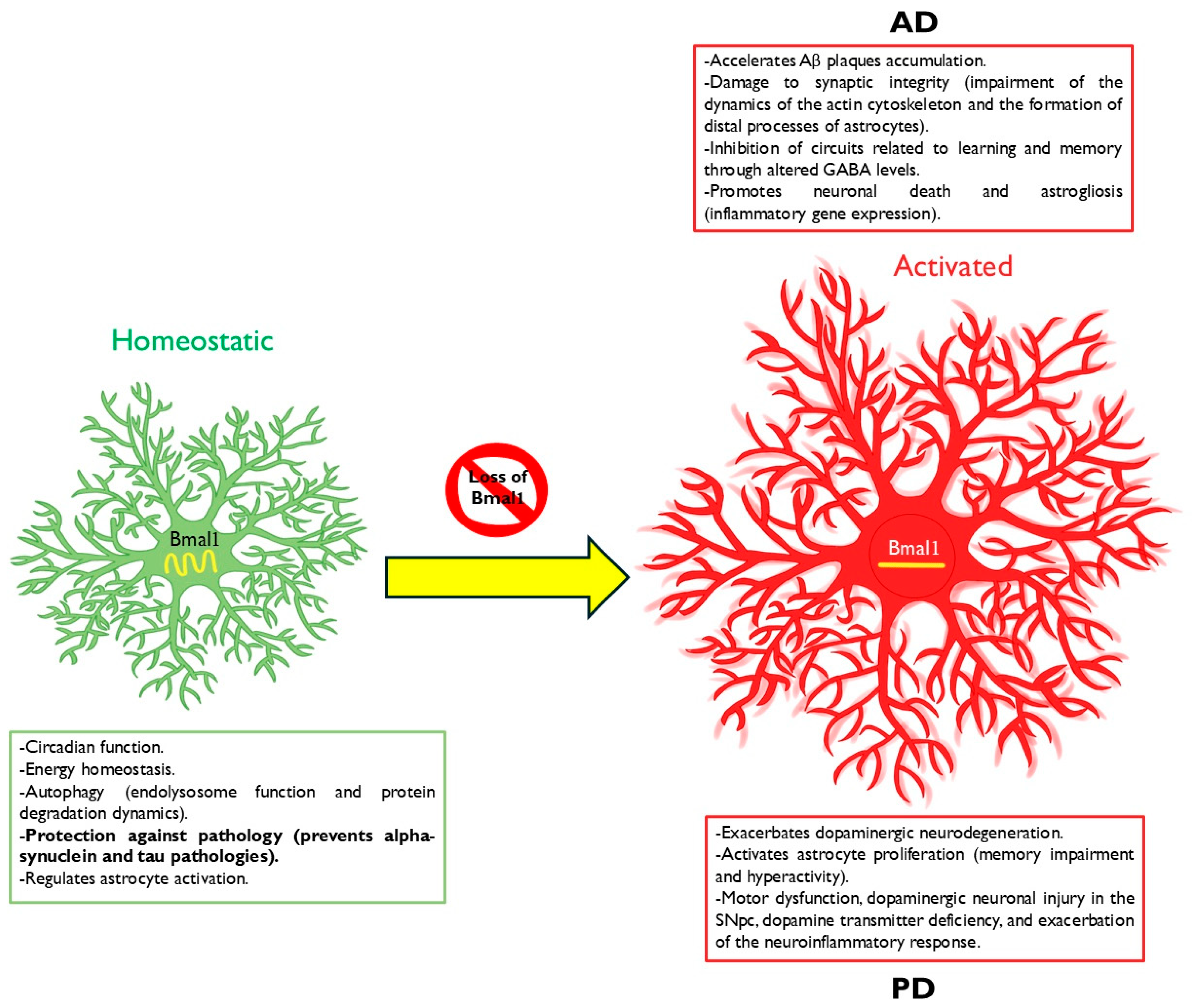
Since the link between circadian dysfunction, particularly involving Bmal1, and the initiation and advancement of neurodegenerative disorders, such as AD and PD, was established, numerous studies have been undertaken. These studies initially aimed to manipulate Bmal1 in different models to explore both the underlying pathophysiological mechanisms and the outcomes of such manipulation, with the subsequent goal of identifying new pharmacological targets. Although numerous studies have focused on manipulating Bmal1 in different cell types, this review focuses on the latest discoveries regarding the manipulation of Bmal1 in astrocytes (Figure 2).

3.1. BMAL1 in Alzheimer’s Disease

The disruption of normal circadian rhythms is a common hallmark of Alzheimer’s disease, often manifesting as increased daytime sleep and nighttime awakenings [69]. Bmal1, Cry1, and Per1 have been reported to have rhythmic expression in the human pineal gland, and these rhythms are lost in patients with preclinical and clinical AD [70]. Numerous studies suggest a bidirectional relationship between Bmal1 dysfunction and Aβ accumulation. Aβ has been shown to induce BMAL1 degradation in mouse models of AD [13]. Conversely, abnormal expression of Bmal1 mRNA and protein in the hippocampus can be caused by Aβ [71], and the loss of Bmal1 has been associated with an increase in amyloid plaque formation [72]. These observations indicate that changes in BMAL1 levels may be either a contributing factor or a result of AD pathology [63].
The impact of Bmal1 deletion on Aβ dynamics and plaque deposition is influenced by the degree of the deletion. A global deletion of Bmal1, including the SCN, disrupts the regular daily variations of Aβ in the hippocampal interstitial fluid, accelerating plaque aggregation [72].
Recent studies have found that the loss of Bmal1, specifically in astrocytes, does not entirely explain the heightened plaque burden seen in whole-brain Bmal1-KO mice. This suggests that the involvement of astrocyte activation in AD is multifaceted and may encompass mechanisms unrelated to Aβ [73]. Additionally, Bmal1 deficiency has been associated with sleep disorders [74,75], which impair Aβ clearance and elevate inflammatory cytokines, further contributing to plaque formation [76].
Furthermore, Bmal1 deletion has been shown to induce pericyte dysfunction, leading to a decline in the integrity of the blood–brain barrier (BBB) in an age-dependent manner. This results in decreased cerebral blood flow and the accumulation of blood-derived neurotoxins [77]. Disruptions in typical circadian rhythms also amplify the daily fluctuations of Bmal1 and other clock genes while promoting the hyperphosphorylation of soluble tau protein, associating Bmal1 rhythms with AD pathology [78].

3.2. Astrocyte BMAL1 in Alzheimer’s Disease

Periplaque astrocyte activation occurs in AD patients and animal models alongside the development of Aβ pathology [79,80,81,82], likely influencing astrocyte interactions with Aβ aggregates, which are critical to plaque removal [83,84,85,86,87,88]. While the impact of disrupted astrocytic clocks on Aβ clearance remains unclear, the circadian regulation of astrocytic Aβ metabolic proteins and reactive gliosis may affect the clearance functions of astrocytes and alter the accumulation of Aβ and tau in the brains of individuals with AD [50].
Another intriguing possibility is that astrocytic clocks influence AD pathogenesis through sleep modulation. Sleep deprivation has been shown to hasten the accumulation of amyloid plaques in mice and elevate Aβ levels in both the cerebrospinal fluid of humans [89] and the interstitial fluid of mice [22]. Additionally, lack of sleep can raise tau levels in the brain and promote tau pathology in mice [24]. Astrocytes play a significant role in the regulation of sleep [90], although their circadian role in sleep regulation is poorly understood.
Many studies on the deletion of Bmal1 in astrocytes specifically address Bmal1 deficiency in astrocytes from certain brain regions, such as the SCN, or in astrocytes from mice with a global deletion of Bmal1 [91]. Increasing evidence indicates that Bmal1 deficiency in astrocytes is closely linked to pathological factors that contribute to AD. In an in vitro study, Bmal1−/− astrocytes displayed shorter actin filaments, reduced cortactin expression, and lower Rho-GTP levels, which impaired actin cytoskeleton dynamics and hindered the formation of distal processes in astrocytes, ultimately negatively affecting synaptic integrity [92].
Emerging insights indicate that the deletion of Bmal1 in astrocytes impacts neurons, as astrocytic Bmal1 is crucial for preventing the accumulation of extracellular gamma-aminobutyric acid (GABA), which facilitates communication between astrocytes and neurons [93]. One study found that administering GABA receptor antagonists restored cognitive functions in Bmal1 KO mice, likely because the deficiency of Bmal1 in astrocytes may significantly inhibit circuits related to learning and memory by altering GABA levels [91]. Furthermore, astrocyte-specific deletion of Bmal1 leads to increased neuronal death, astrogliosis, and the upregulation of inflammatory gene expression in vitro [43].
Recent findings have revealed that the loss of Bmal1 in astrocytes does not lead to an increased plaque burden in mice with global Bmal1 KO, indicating that the activation state of astrocytes in AD brains has intricate effects, highlighting the need for further research to investigate their contributions to AD that are not related to Aβ [73].

3.3. BMAL1 in Parkinson’s Disease

Numerous clinical symptoms of PD indicate dysfunction of circadian rhythms, including disruptions in rest and activity cycles, irregular patterns of blood pressure fluctuations, and atypical rhythms of melatonin secretion [71]. Bmal1 is one of the critical molecules of the circadian cycle and is related to the pathology and symptoms of PD. Research in human studies and animal models has demonstrated that reduced levels of Bmal1 mRNA are associated with PD. Notably, lower Bmal1 mRNA levels have been observed in the leukocytes of patients with PD, indicating that the disease can be related to dysfunction of both the central and peripheral biological clock, which may reflect the severity of PD [93].
A substantial amount of literature discusses circadian disruption in PD, which includes fragmentation of daily behavioral rhythms and dampening of the expression rhythms of Bmal1 and other core circadian clock genes in peripheral blood mononuclear cells (PBMCs) from PD patients [12,93,94,95,96]. In murine models, circadian disruption caused by light exposure [97] and the germline deletion of Bmal1 through the use of the mitochondrial toxin 1-methyl-4-phenyl-1,2,3,6-tetrahydropyridine (MPTP) have both been found to aggravate the degeneration of dopaminergic neurons [3].
One possible explanation for the changes in BMAL1 expression is that dopamine (DA) can influence the activity of the BMAL1/CLOCK complex. Therefore, reduced DA levels in PD impact this key element of the molecular clock [12]. After melatonin treatment, the Bmal1 mRNA levels were increased in PD patients, reinforcing a close link between Bmal1 and PD [98].
Additionally, Bmal1 plays a role in regulating neuroinflammation in the brain to preserve the functionality of the DA signaling pathway. Concurrently, disruption of this balance has been suggested as a contributing factor to the onset of PD [3]. In addition to DA signaling, Bmal1 deletion in mice has been shown to activate astrocyte proliferation [73], leading to abnormal pathological phenotypes, such as memory impairment and hyperactivity [99]. Conversely, elevated BMAL1 expression alters astrocyte function by inhibiting aerobic glycolysis [100].
BMAL1 has been shown to participate in the regulation of Parkinson’s disease pathogenesis. In a mouse model of PD induced by MPTP, inactivation of BMAL1 resulted in injury to dopaminergic neurons in the substantia nigra pars compacta (SNpc), a deficiency in DA transmission, motor dysfunction, and an intensification of the neuroinflammatory response. These findings suggest that BMAL1 may play a protective role in the survival of DA neurons by regulating neuroinflammation mediated by microglia [3]. Treatment with 6-hydroxydopamine in an animal model of PD resulted in a reduction in SIRT1 levels and an increase in acetylated BMAL1 levels, leading to abnormal antioxidant activity. These findings suggest that the SIRT1-BMAL1 pathway regulates abnormal antioxidant responses in PD [71].
In a study based on circadian dysfunction in a mouse model of Parkinson’s disease with α-syn overexpression, Liu and colleagues discovered that the degradation of the BMAL1 protein remained unchanged after α-syn overexpression while its mRNA level decreased. The miR-155, one of the microRNAs that regulate α-syn expression and is known for its role in neuroinflammation and the development of CNS diseases, was upregulated by overexpression of α-syn. At the same time, the negative regulation of Bmal1 was partially reversed by transfection with an miR-155 inhibitor. Additionally, α-syn overexpression induced circadian rhythm disruption and negatively regulated BMAL1 expression by decreasing Bmal1 mRNA stability through miR-155. These findings established that α-syn overexpression induces biorhythm alteration and downregulated BMAL1 expression by decreasing the stability of Bmal1 mRNA via miR-155 [101].
A recent study investigated the impact of postnatal and cell-type-specific deletion of Bmal1 on the viability of DA neurons in the substantia nigra pars compacta (SNpc) using an α-syn mouse model. The results revealed that BMAL1 governs cell-specific transcriptional programs essential for the survival of DA neurons, indicating that maintaining BMAL1 expression could have neuroprotective effects in PD [102].
Although the present study aims to analyze the existing information on BMAL1 in astrocytic cells, it should be noted that the literature does not provide evidence of the role of this protein in astrocytes, particularly in PD models. For this reason, research related to this field of study is of utmost importance for a better understanding of the subject.

4. Astrocyte Bmal1 Deletion: A Potential Protective Role Against AD and PD

A range of harmful stimuli trigger astrocyte activation and are vital for both brain health and the progression of disease. The central circadian clock protein BMAL1 has been found to regulate astrogliosis and inflammatory gene expression both in vitro and in vivo synergistically through an autonomous cellular mechanism and a minor non-autonomous neuronal signal by disrupting protein glutathionylation mediated by glutathione-S-transferase enzymatic activity. Additionally, Lananna et al. discovered that BMAL1 regulates astrocyte activation and function in vivo, elucidating the prominent role of the circadian clock and its influence on brain function and neurodegenerative diseases [43].
In cell cultures, Bmal1-deficient astrocytes show increased endocytosis and lysosome-dependent protein cleavage. In vivo, brains with specific Bmal1 deletion in astrocytes have shown accumulation of autophagosome-like structures within astrocytes, according to electron microscopy. Considering the established connection between neurodegeneration and endolysosomal dysfunction with aging, this research identified BMAL1 as a critical regulator of these essential astrocyte functions in health and disease [103].
The elimination/reduction of Bmal1 in astrocytes has been shown to reprogram endolysosomal transcriptional pathways and induce endocytosis, lysosomal degradation, and autophagic activity within these cells. Although the precise mechanism through which Bmal1 affects these astrocyte functions remains unclear, the deletion of Bmal1 may impact numerous genes related to the endolysosomal system. These findings bolster the evidence that circadian clock proteins are involved in neurodegenerative diseases by implicating BMAL1 in the regulation of proteostasis within astrocytes. The potential for downstream genes of Bmal1 to temporally regulate proteostasis presents new therapeutic opportunities for targeting these processes in the context of the disease [104].
For several years, classic markers such as GFAP have helped define astrocyte reactivity. Still, they must fully describe the diversity of responses depending on the type of injury, disease progression, and interaction with other cells. The term “astrogliosis” does not reflect the variability of “reactive astrocytes”, whose properties vary depending on the stimulus and evolve during the disease, reflecting the diversity of signals received. Some signals may be harmful, while others may be protective. Recent studies have started to reveal the variety of astrocyte activation phenotypes that extend apart from GFAP, with unique gene expressions associated with effects ranging from neurotoxic to neurotrophic [32,43,54,104].
In an experiment using mice with global and brain-specific Bmal1 KO, many genes associated with reactive astrogliosis, such as Nr1d1, Fabp7, and S100a6, were significantly regulated, demonstrating that Bmal1 in astrocytes regulates these genes autonomously from cells. For this purpose, and given its reproducibility and frequency change in experiments, the astrocytic markers to elucidate this activation state were Fabp7, Gfap, Mmp14, and Cxcl5 [73].
It was also discovered that astrocytic Bmal1 modulates genes with contradictory effects on Aβ accumulation. For instance, deactivation of the clock-controlled gene Chi3l1 improved Aβ pathology, likely by promoting Aβ phagocytosis by glial cells, and BMAL1 KO significantly reduced Chi3l1 expression, suggesting that this astrocyte activation state might exacerbate Aβ pathology. Likewise, Mmp14, an enzyme responsible for degrading Aβ40, was notably overexpressed in this context [73].
Recently, Kanan and colleagues investigated how postnatal and cell-specific deletion of Bmal1 affects DA neuron viability in the SNpc using a mouse model of α-synuclein neurodegeneration. They observed that Bmal1 modulates distinct cell-autonomous transcriptional programs in DA neurons, which are essential for their survival. This implies that Bmal1 independently regulates DA neuron viability in the SNpc and that maintaining BMAL1 expression may offer neuroprotection in Parkinson’s disease. These findings were not replicated by disrupting light-driven circadian behavioral rhythms or through glia-specific Bmal1 deletion [102].
In an investigation using transgenic mice models of tauopathy and Parkinson’s disease with astrocytic deletion of Bmal1, researchers discovered a protective effect driven by BMAL1-mediated astrogliosis and expression of BAG3, a macroautophagy chaperone. The overexpression of astrocytic BAG3 itself was shown to protect against various pathologies, with BAG3 being highly expressed in a specific subset of disease-associated activated astrocytes in human brain samples from AD patients. These findings suggest that early astrocyte activation can mitigate both α-synuclein and tauopathy pathology in vivo. Furthermore, the loss of BMAL1 in astrocytes triggered a protective state of astrocyte activation, highlighting the potential role of this molecular pathway in shielding against the progression of neurodegenerative diseases [44].
Multiple studies have demonstrated that BMAL1 is capable of influencing NRF2 activity and the expression of downstream genes [99,105,106], indicating that these two transcription factors may cooperate to regulate protective astrocyte responses in the context of protein aggregation.
The findings by Sheehan and colleagues indicate that Bmal1 deletion triggers a distinct type of astrocyte reactivity, characterized by strong GFAP expression and traditional morphological alterations, yet accompanied by the activation of a unique transcriptional profile. In the APP/PS1 mice model of AD, which is mainly characterized by a notable elevation of β-amyloid production, such astrocytic activation considerably reduced plaque formation. It remains protective against tau and α-synuclein pathology, implying that astrocyte activation caused by Bmal1 deletion triggers specific cellular adaptations. These adaptations effectively suppress the aggregation of intraneuronal tau and α-synuclein while leaving extracellular Aβ deposition unaffected.
Additionally, BAG3 has been recognized as a crucial downstream mediator of the neuroprotective phenotype associated with Bmal1 deletion. The loss of Bmal1 in astrocytes modifies the expression of various genes, including Apoe and Chi3l1, which may play significant roles in enhancing neuroprotection. The exact functions of these genes within the framework of neurodegenerative diseases present promising avenues for further research. Investigating how these gene expressions interact with cellular pathways could yield valuable insights into potential therapeutic strategies for conditions such as AD and other tauopathies [44].

5. Discussion and Future Perspectives

It is clear that central clock genes play a vital role in regulating essential cellular functions across various organs, and disruption of normal circadian rhythm can contribute to the onset or exacerbation of AD and PD. The significance of astrocyte function in AD, PD, and other neurodegenerative disorders is receiving growing attention. The diverse roles of astrocytes in the brain suggest various mechanisms through which they may contribute to disease pathology. Astrocytes are integral to critical circadian clock functions, and disruptions to these circadian rhythms can manifest distinct phenotypes that have detrimental and beneficial effects on neurodegeneration.
Profiling astrocyte responses to tau and Aβ pathology has uncovered that their “reactive” state exhibits signatures indicative of cellular dysfunction and inflammation alongside adaptive protective responses. If these adaptive responses can be effectively harnessed, they may slow the aggregation of tau and α-synuclein and mitigate the progression of both diseases [104]. Transcriptomic profiling studies of astrocytes in neurodegenerative conditions indicate multiple modes of astrocyte activation, which vary depending on the disease state and may involve differential expression of GFAP [104,107,108,109,110]. This complexity suggests that astrocyte reactivity cannot be accurately captured by a simple binary classification system [44].
The influence of astrocyte activation in AD is not a topic that seems close to being fully elucidated. It is still uncertain whether astrocyte activation primarily worsens neuroinflammation and accelerates the disease’s progression or if it shifts the balance toward beneficial functions, such as promoting Aβ clearance and supporting neuronal viability. This duality of astrocyte function complicates the interpretation of their role in AD pathology, as evidence suggests both detrimental and protective effects depending on the context of their activation ambiguity surrounding astrocyte activation while emphasizing the complexity of their roles in AD [111]. More knowledge is needed to study astrogliosis over different temporal courses and investigate astrocytic inflammatory profiles and their toxic and protective processes.
Numerous research efforts have associated astrocyte activation with an increased plaque burden [112,113,114], while others have linked astrocyte activation to a decrease in plaque burden [115,116]. These findings highlight that the multiple settings and transcriptional profiles of activated astrocytes are implicated in AD through markedly different processes and functions. There remains a critical need to identify astrocyte genes that either mitigate or exacerbate Aβ pathology and to investigate how these genes are expressed across various reactive states [73]. In summary, some of the findings presented in this review suggest that the astrocytic circadian clock may regulate the activation and downstream genes that can influence the generation, deposition, and clearance of Aβ [73].
Recent studies examining the role of Bmal1 in transgenic Parkinsonian mice have found that neuronal Bmal1 modulates transcriptional pathways associated with PD and oxidative phosphorylation. Neuronal deletion of Bmal1 led to the spontaneous death of dopaminergic neurons, indicating that BMAL1 contributes to the survival of dopaminergic neurons. These findings indicate that environmental or pathological factors leading to decreased expression of BMAL1 may initiate or worsen neurodegeneration and Parkinsonian phenotypes [102].
There is a need to expand the knowledge linking circadian dysfunction in synucleinopathies and Parkinsonian models by manipulating astrocyte-specific Bmal1. While some studies have found adverse effects from neuronal Bmal1 deletion [102] or have studied the indirect impact of global and microglial Bmal1 in deletion on DA signaling [3,73,99,100,117], astrocytic Bmal1 deletion in specific brain regions, such as the SCN or SNpc, in models with α-syn aggregates or Parkinsonian genotypes and phenotypes, either in vivo or in vitro, has yet to be fully explored.
The effects of manipulating Bmal1 in models of neurological diseases largely depend on the cell type and context. While this review highlighted findings on Bmal1 manipulation in astrocytes, it is essential to note that neuronal Bmal1 deletion has shown primarily adverse effects in neurodegeneration models, both in vivo and in vitro. Different studies have demonstrated that the deletion of neuronal Bmal1 increases neuronal sensitivity to oxidative stress and contributes to neuronal death [99]. In contrast, Bmal1 deletion in glial cells appears to produce more variable effects, often providing protective benefits [44]. Consequently, Bmal1 seems to play a crucial role in ensuring overall neuronal survival [102].
Alternatively, mice with neuron-specific Bmal1 KO have demonstrated arrhythmic behaviors [118], and this loss of rhythm could induce astrocyte activation. Hence, it is necessary to examine the influence of non-autonomous cells on glial activation in the brain [43].
Global deletion of Bmal1 has been shown to induce oxidative stress in the brain and lead to synaptic loss [99]. Additionally, it exacerbates MPTP-induced loss of dopaminergic neurons [3], accelerates the formation of amyloid plaques [72], and lowers the seizure threshold [119,120], demonstrating neurodegenerative and adverse effects.
Conversely, earlier studies demonstrated that whole-brain Bmal1 deletion mitigated stroke damage [121] and spinal cord injury [122]. Along with these promising results, it has been recently found that astrocyte-specific Bmal1 deletion induces astrogliosis [43] and can prevent tau and α-syn aggregation in an in vivo model, partly through the regulation of the chaperone protein BAG3 or changes in lysosomal function [44,103]. Additionally, other groups have demonstrated that microglia-specific Bmal1 deletion reduces inflammatory activation and decreases damage caused by ischemic stroke [77].
The specific role of astrocytic Bmal1 deletion in disrupting circadian rhythms remains unclear. While BMAL1 is known for its circadian regulation, it also carries out several non-circadian cellular functions [10]. For instance, astrocytic BMAL1 regulates the expression of non-rhythmic transcripts [43], underscoring its diverse functional role. It is hypothesized that the loss of intrinsic transcriptional rhythms in astrocytes may lead to chronic cellular adaptations, resulting in astrocyte activation and the engagement of specific signaling pathways. Further research is needed to explore how mutations in other clock genes, or non-genetic disruptions of circadian rhythms, influence tauopathies and synucleinopathies [44].
It is possible that Bmal1 KO astrocytes indirectly facilitate the degradation of protein aggregates in neurons, potentially by delivering chaperone proteins to neurons via exosomes [110]. Based on these findings, further investigation is necessary to elucidate the specific mechanisms by which astrocytes influence the aggregation and propagation of intraneuronal proteins involved in various neurodegenerative disorders.
Despite significant advances in understanding the relationship between circadian dysfunction and global and astrocytic manipulation of Bmal1 in neurodegenerative models, additional research is needed to determine how other clock gene mutations or non-genetic circadian alterations impact tauopathies, such as AD and synucleinopathies such as PD. Thus far, Bmal1 has recently been identified as a key regulator of neuroprotective activity by mediating astrocyte reactivity, and manipulation of astrocytic Bmal1 and its downstream pathways has revealed protective effects against tau and α-syn aggregation, positioning the central circadian clock gene Bmal1 as a potential neuroprotective target.

Author Contributions

D.B.-A., L.I.G. and D.C.-C.: conceptualization, writing—original draft. G.A.-A., R.T.-C. and C.P.-E.: writing—review and editing. All authors have read and agreed to the published version of the manuscript.

Funding

D.B.-A. is supported by CONAHCYT-Mexico fellowship 510113.

Conflicts of Interest

The authors have no conflicts of interest to declare.

References

  1. Bass, J.; Lazar, M.A. Circadian time signatures of fitness and disease. Science 2016, 354, 994–999. [Google Scholar] [CrossRef] [PubMed]
  2. Bass, J.; Takahashi, J.S. Circadian integration of metabolism and energetics. Science 2010, 330, 1349–1354. [Google Scholar] [CrossRef] [PubMed]
  3. Liu, W.W.; Wei, S.Z.; Huang, G.D.; Liu, L.B.; Gu, C.; Shen, Y.; Wang, X.H.; Xia, S.T.; Xie, A.M.; Hu, L.F.; et al. BMAL1 regulation of microglia-mediated neuroinflammation in MPTP-induced Parkinson’s disease mouse model. FASEB J. 2020, 34, 6570–6581. [Google Scholar] [CrossRef] [PubMed]
  4. Fifel, K.; De Boer, T. The circadian system in Parkinson’s disease, multiple system atrophy, and progressive supranuclear palsy. Handb. Clin. Neurol. 2021, 179, 301–313. [Google Scholar] [CrossRef]
  5. Mohawk, J.A.; Green, C.B.; Takahashi, J.S. Central and peripheral circadian clocks in mammals. Annu. Rev. Neurosci. 2012, 35, 445–462. [Google Scholar] [CrossRef]
  6. Yoo, S.H.; Mohawk, J.A.; Siepka, S.M.; Shan, Y.; Huh, S.K.; Hong, H.K.; Kornblum, I.; Kumar, V.; Koike, N.; Xu, M.; et al. Competing E3 ubiquitin ligases govern circadian periodicity by degradation of CRY in nucleus and cytoplasm. Cell 2013, 152, 1091–1105. [Google Scholar] [CrossRef]
  7. Hirano, A.; Fu, Y.H.; Ptáček, L.J. The intricate dance of post-translational modifications in the rhythm of life. Nat. Struct. Mol. Biol. 2016, 23, 1053–1060. [Google Scholar] [CrossRef]
  8. Hastings, M.H.; Smyllie, N.J.; Patton, A.P. Molecular-genetic Manipulation of the Suprachiasmatic Nucleus Circadian Clock. J. Mol. Biol. 2020, 432, 3639–3660. [Google Scholar] [CrossRef]
  9. Schurhoff, N.; Toborek, M. Circadian rhythms in the blood-brain barrier: Impact on neurological disorders and stress responses. Mol. Brain 2023, 16, 5. [Google Scholar] [CrossRef]
  10. Yu, E.A.; Weaver, D.R. Disrupting the circadian clock: Gene-specific effects on aging, cancer, and other phenotypes. Aging 2011, 3, 479–493. [Google Scholar] [CrossRef]
  11. Musiek, E.S.; Holtzman, D.M. Mechanisms linking circadian clocks, sleep, and neurodegeneration. Science 2016, 354, 1004–1008. [Google Scholar] [CrossRef] [PubMed]
  12. Breen, D.P.; Vuono, R.; Nawarathna, U.; Fisher, K.; Shneerson, J.M.; Reddy, A.B.; Barker, R.A. Sleep and circadian rhythm regulation in early Parkinson disease. JAMA Neurol. 2014, 71, 589–595. [Google Scholar] [CrossRef] [PubMed]
  13. Song, H.; Moon, M.; Choe, H.K.; Han, D.H.; Jang, C.; Kim, A.; Cho, S.; Kim, K.; Mook-Jung, I. Aβ-induced degradation of BMAL1 and CBP leads to circadian rhythm disruption in Alzheimer’s disease. Mol. Neurodegener. 2015, 10, 13. [Google Scholar] [CrossRef] [PubMed]
  14. Cronin, P.; McCarthy, M.J.; Lim, A.S.P.; Salmon, D.P.; Galasko, D.; Masliah, E.; Jager, P.L.D.; Bennett, D.A.; Desplats, P. Circadian alterations during early stages of Alzheimer’s disease are associated with aberrant cycles of DNA methylation in BMAL1. Alzheimers Dement. 2017, 13, 689–700. [Google Scholar] [CrossRef] [PubMed]
  15. Pilleri, M.; Levedianos, G.; Weis, L.; Gasparoli, E.; Facchini, S.; Biundo, R.; Formento-Dojot, P.; Antonini, A. Heart rate circadian profile in the differential diagnosis between Parkinson disease and multiple system atrophy. Park. Relat. Disord. 2014, 20, 217–221. [Google Scholar] [CrossRef]
  16. Raggi, A.; Neri, W.; Ferri, R. Sleep-related behaviors in Alzheimer’s disease and dementia with Lewy bodies. Rev. Neurosci. 2015, 26, 31–38. [Google Scholar] [CrossRef]
  17. Vallelonga, F.; Di Stefano, C.; Merola, A.; Romagnolo, A.; Sobrero, G.; Milazzo, V.; Burrello, A.; Burrello, J.; Zibetti, M.; Veglio, F.; et al. Blood pressure circadian rhythm alterations in alpha-synucleinopathies. J. Neurol. 2019, 266, 1141–1152. [Google Scholar] [CrossRef]
  18. Wang, Y.; Mandelkow, E. Tau in physiology and pathology. Nat. Rev. Neurosci. 2016, 17, 5–21. [Google Scholar] [CrossRef]
  19. Busche, M.A.; Hyman, B.T. Synergy between amyloid-β and tau in Alzheimer’s disease. Nat. Neurosci. 2020, 23, 1183–1193. [Google Scholar] [CrossRef]
  20. Chang, C.W.; Shao, E.; Mucke, L. Tau: Enabler of diverse brain disorders and target of rapidly evolving therapeutic strategies. Science 2021, 371, eabb8255. [Google Scholar] [CrossRef]
  21. Ju, Y.S.; Ooms, S.J.; Sutphen, C.; Macauley, S.L.; Zangrilli, M.A.; Jerome, G.; Fagan, A.M.; Mignot, E.; Zempel, J.M.; Claassen, J.A.H.R.; et al. Slow wave sleep disruption increases cerebrospinal fluid amyloid-β levels. Brain 2017, 140, 2104–2111. [Google Scholar] [CrossRef] [PubMed]
  22. Kang, J.E.; Lim, M.M.; Bateman, R.J.; Lee, J.J.; Smyth, L.P.; Cirrito, J.R.; Fujiki, N.; Nishino, S.; Holtzman, D.M. Amyloid-beta dynamics are regulated by orexin and the sleep-wake cycle. Science 2009, 326, 1005–1007. [Google Scholar] [CrossRef] [PubMed]
  23. Sprecher, K.E.; Koscik, R.L.; Carlsson, C.M.; Zetterberg, H.; Blennow, K.; Okonkwo, O.C.; Sager, M.A.; Asthana, S.; Johnson, S.C.; Benca, R.M.; et al. Poor sleep is associated with CSF biomarkers of amyloid pathology in cognitively normal adults. Neurology 2017, 89, 445–453. [Google Scholar] [CrossRef] [PubMed]
  24. Holth, J.K.; Fritschi, S.K.; Wang, C.; Pedersen, N.P.; Cirrito, J.R.; Mahan, T.E.; Finn, M.B.; Manis, M.; Geerling, J.C.; Fuller, P.M.; et al. The sleep-wake cycle regulates brain interstitial fluid tau in mice and CSF tau in humans. Science 2019, 363, 880–884. [Google Scholar] [CrossRef]
  25. Bendor, J.T.; Logan, T.P.; Edwards, R.H. The function of α-synuclein. Neuron 2013, 79, 1044–1066. [Google Scholar] [CrossRef]
  26. Du, X.Y.; Xie, X.X.; Liu, R.T. The Role of α-Synuclein Oligomers in Parkinson’s Disease. Int. J. Mol. Sci. 2020, 21, 8645. [Google Scholar] [CrossRef]
  27. Brash-Arias, D.; García, L.I.; Pérez-Estudillo, C.A.; Rojas-Durán, F.; Aranda-Abreu, G.E.; Herrera-Covarrubias, D.; Chi-Castañeda, D. The Role of Astrocytes and Alpha-Synuclein in Parkinson’s Disease: A Review. NeuroSci 2024, 5, 71–86. [Google Scholar] [CrossRef]
  28. Gros, P.; Videnovic, A. Overview of Sleep and Circadian Rhythm Disorders in Parkinson Disease. Clin. Geriatr. Med. 2020, 36, 119–130. [Google Scholar] [CrossRef]
  29. Maetzler, W.; Liepelt, I.; Berg, D. Progression of Parkinson’s disease in the clinical phase: Potential markers. Lancet Neurol. 2009, 8, 1158–1171. [Google Scholar] [CrossRef]
  30. Torres-Pasillas, G.; Chi-Castañeda, D.; Carrillo-Castilla, P.; Marín, G.; Hernández-Aguilar, M.E.; Aranda-Abreu, G.E.; Manzo, J.; García, L.I. Olfactory Dysfunction in Parkinson’s Disease, Its Functional and Neuroanatomical Correlates. NeuroSci 2023, 4, 134–151. [Google Scholar] [CrossRef]
  31. Sofroniew, M.V.; Vinters, H.V. Astrocytes: Biology and pathology. Acta Neuropathol. 2010, 119, 7–35. [Google Scholar] [CrossRef] [PubMed]
  32. Zamanian, J.L.; Xu, L.; Foo, L.C.; Nouri, N.; Zhou, L.; Giffard, R.G.; Barres, B.A. Genomic analysis of reactive astrogliosis. J. Neurosci. 2012, 32, 6391–6410. [Google Scholar] [CrossRef] [PubMed]
  33. Wakabayashi, K.; Hayashi, S.; Yoshimoto, M.; Kudo, H.; Takahashi, H. NACP/alpha-synuclein-positive filamentous inclusions in astrocytes and oligodendrocytes of Parkinson’s disease brains. Acta Neuropathol. 2000, 99, 14–20. [Google Scholar] [CrossRef] [PubMed]
  34. Braak, H.; Sastre, M.; Del Tredici, K. Development of alpha-synuclein immunoreactive astrocytes in the forebrain parallels stages of intraneuronal pathology in sporadic Parkinson’s disease. Acta Neuropathol. 2007, 114, 231–241. [Google Scholar] [CrossRef]
  35. Acioglu, C.; Li, L.; Elkabes, S. Contribution of astrocytes to neuropathology of neurodegenerative diseases. Brain Res. 2021, 1758, 147291. [Google Scholar] [CrossRef]
  36. Prolo, L.M.; Takahashi, J.S.; Herzog, E.D. Circadian rhythm generation and entrainment in astrocytes. J. Neurosci. 2005, 25, 404–408. [Google Scholar] [CrossRef]
  37. Tso, C.F.; Simon, T.; Greenlaw, A.C.; Puri, T.; Mieda, M.; Herzog, E.D. Astrocytes Regulate Daily Rhythms in the Suprachiasmatic Nucleus and Behavior. Curr. Biol. 2017, 27, 1055–1061. [Google Scholar] [CrossRef]
  38. Brancaccio, M.; Edwards, M.D.; Patton, A.P.; Smyllie, N.J.; Chesham, J.E.; Maywood, E.S.; Hastings, M.H. Cell-autonomous clock of astrocytes drives circadian behavior in mammals. Science 2019, 363, 187–192. [Google Scholar] [CrossRef]
  39. Marpegan, L.; Swanstrom, A.E.; Chung, K.; Simon, T.; Haydon, P.G.; Khan, S.K.; Liu, A.C.; Herzog, E.D.; Beaulé, C. Circadian regulation of ATP release in astrocytes. J. Neurosci. 2011, 31, 8342–8350. [Google Scholar] [CrossRef]
  40. Rath, M.F.; Rohde, K.; Fahrenkrug, J.; Møller, M. Circadian clock components in the rat neocortex: Daily dynamics, localization and regulation. Brain Struct. Funct. 2013, 218, 551–562. [Google Scholar] [CrossRef]
  41. Sofroniew, M.V. Astrogliosis. Cold Spring Harb. Perspect. Biol. 2014, 7, a020420. [Google Scholar] [CrossRef] [PubMed]
  42. Yang, Z.; Wang, K.K. Glial fibrillary acidic protein: From intermediate filament assembly and gliosis to neurobiomarker. Trends Neurosci. 2015, 38, 364–374. [Google Scholar] [CrossRef] [PubMed]
  43. Lananna, B.V.; Nadarajah, C.J.; Izumo, M.; Cedeño, M.R.; Xiong, D.D.; Dimitry, J.; Tso, C.F.; McKee, C.A.; Griffin, P.; Sheehan, P.W.; et al. Cell-Autonomous Regulation of Astrocyte Activation by the Circadian Clock Protein BMAL1. Cell Rep. 2018, 25, 1–9.e5. [Google Scholar] [CrossRef] [PubMed]
  44. Sheehan, P.W.; Nadarajah, C.J.; Kanan, M.F.; Patterson, J.N.; Novotny, B.; Lawrence, J.H.; King, M.W.; Brase, L.; Inman, C.E.; Yuede, C.M.; et al. An astrocyte BMAL1-BAG3 axis protects against alpha-synuclein and tau pathology. Neuron 2023, 111, 2383–2398.e7. [Google Scholar] [CrossRef] [PubMed]
  45. Servière, J.; Lavialle, M. Astrocytes in the mammalian circadian clock: Putative roles. Prog. Brain Res. 1996, 111, 57–73. [Google Scholar] [CrossRef]
  46. Womac, A.D.; Burkeen, J.F.; Neuendorff, N.; Earnest, D.J.; Zoran, M.J. Circadian rhythms of extracellular ATP accumulation in suprachiasmatic nucleus cells and cultured astrocytes. Eur. J. Neurosci. 2009, 30, 869–876. [Google Scholar] [CrossRef]
  47. Burkeen, J.F.; Womac, A.D.; Earnest, D.J.; Zoran, M.J. Mitochondrial calcium signaling mediates rhythmic extracellular ATP accumulation in suprachiasmatic nucleus astrocytes. J. Neurosci. 2011, 31, 8432–8440. [Google Scholar] [CrossRef]
  48. Koyanagi, S.; Kusunose, N.; Taniguchi, M.; Akamine, T.; Kanado, Y.; Ozono, Y.; Masuda, T.; Kohro, Y.; Matsunaga, N.; Tsuda, M.; et al. Glucocorticoid regulation of ATP release from spinal astrocytes underlies diurnal exacerbation of neuropathic mechanical allodynia. Nat. Commun. 2016, 7, 13102. [Google Scholar] [CrossRef]
  49. Leone, M.J.; Beaule, C.; Marpegan, L.; Simon, T.; Herzog, E.D.; Golombek, D.A. Glial and light-dependent glutamate metabolism in the suprachiasmatic nuclei. Chronobiol. Int. 2015, 32, 573–578. [Google Scholar] [CrossRef]
  50. McKee, C.A.; Lananna, B.V.; Musiek, E.S. Circadian regulation of astrocyte function: Implications for Alzheimer’s disease. Cell Mol. Life Sci. 2020, 77, 1049–1058. [Google Scholar] [CrossRef]
  51. Hassler, C.; Burnier, M. Circadian variations in blood pressure: Implications for chronotherapeutics. Am. J. Cardiovasc. Drugs 2005, 5, 7–15. [Google Scholar] [CrossRef] [PubMed]
  52. Manfredini, R.; Boari, B.; Smolensky, M.H.; Salmi, R.; Cecilia, O.I.; Malagoni, A.M.; Haus, E.; Manfredini, F. Circadian variation in stroke onset: Identical temporal pattern in ischemic and hemorrhagic events. Chronobiol. Int. 2005, 22, 417–453. [Google Scholar] [CrossRef] [PubMed]
  53. Karmarkar, S.W.; Tischkau, S.A. Influences of the circadian clock on neuronal susceptibility to excitotoxicity. Front. Physiol. 2013, 4, 313. [Google Scholar] [CrossRef]
  54. Liddelow, S.A.; Guttenplan, K.A.; Clarke, L.E.; Bennett, F.C.; Bohlen, C.J.; Schirmer, L.; Bennett, M.L.; Münch, A.E.; Chung, W.S.; Peterson, T.C.; et al. Neurotoxic reactive astrocytes are induced by activated microglia. Nature 2017, 541, 481–487. [Google Scholar] [CrossRef]
  55. You, S.; Fulga, T.A.; Van Vactor, D.; Jackson, F.R. Regulation of Circadian Behavior by Astroglial MicroRNAs in Drosophila. Genetics 2018, 208, 1195–1207. [Google Scholar] [CrossRef]
  56. Maywood, E.S.; Elliott, T.S.; Patton, A.P.; Krogager, T.P.; Chesham, J.E.; Ernst, R.J.; Beránek, V.; Brancaccio, M.; Chin, J.W.; Hastings, M.H. Translational switching of Cry1 protein expression confers reversible control of circadian behavior in arrhythmic Cry-deficient mice. Proc. Natl. Acad. Sci. USA 2018, 115, E12388–E12397. [Google Scholar] [CrossRef]
  57. Patton, A.P.; Smyllie, N.J.; Chesham, J.E.; Hastings, M.H. Astrocytes Sustain Circadian Oscillation and Bidirectionally Determine Circadian Period, but Do Not Regulate Circadian Phase in the Suprachiasmatic Nucleus. J. Neurosci. 2022, 42, 5522–5537. [Google Scholar] [CrossRef]
  58. Hastings, M.H.; Brancaccio, M.; Gonzalez-Aponte, M.F.; Herzog, E.D. Circadian Rhythms and Astrocytes: The Good, the Bad, and the Ugly. Annu. Rev. Neurosci. 2023, 46, 123–143. [Google Scholar] [CrossRef]
  59. Anashkina, A.A.; Poluektov, Y.M.; Dmitriev, V.A.; Kuznetsov, E.N.; Mitkevich, V.A.; Makarov, A.A.; Petrushanko, I.Y. A novel approach for predicting protein S-glutathionylation. BMC Bioinform. 2020, 21 (Suppl. S11), 282. [Google Scholar] [CrossRef]
  60. Bunger, M.K.; Wilsbacher, L.D.; Moran, S.M.; Clendenin, C.; Radcliffe, L.A.; Hogenesch, J.B.; Simon, M.C.; Takahashi, J.S.; Bradfield, C.A. Mop3 is an essential component of the master circadian pacemaker in mammals. Cell 2000, 103, 1009–1017. [Google Scholar] [CrossRef]
  61. Welz, P.S.; Zinna, V.M.; Symeonidi, A.; Koronowski, K.B.; Kinouchi, K.; Smith, J.G.; Guillén, I.M.; Castellanos, A.; Furrow, S.; Aragón, F.; et al. BMAL1-Driven Tissue Clocks Respond Independently to Light to Maintain Homeostasis. Cell 2019, 177, 1436–1447.e12. [Google Scholar] [CrossRef] [PubMed]
  62. Kondratov, R.V.; Kondratova, A.A.; Gorbacheva, V.Y.; Vykhovanets, O.V.; Antoch, M.P. Early aging and age-related pathologies in mice deficient in BMAL1, the core componentof the circadian clock. Genes Dev. 2006, 20, 1868–1873. [Google Scholar] [CrossRef] [PubMed]
  63. Fan, R.; Peng, X.; Xie, L.; Dong, K.; Ma, D.; Xu, W.; Shi, X.; Zhang, S.; Chen, J.; Yu, X.; et al. Importance of Bmal1 in Alzheimer’s disease and associated aging-related diseases: Mechanisms and interventions. Aging Cell 2022, 21, e13704. [Google Scholar] [CrossRef] [PubMed]
  64. Chhunchha, B.; Kubo, E.; Singh, D.P. Clock Protein Bmal1 and Nrf2 Cooperatively Control Aging or Oxidative Response and Redox Homeostasis by Regulating Rhythmic Expression of Prdx6. Cells 2020, 9, 1861. [Google Scholar] [CrossRef] [PubMed]
  65. Xie, M.; Tang, Q.; Nie, J.; Zhang, C.; Zhou, X.; Yu, S.; Sun, J.; Cheng, X.; Dong, N.; Hu, Y.; et al. BMAL1-Downregulation Aggravates Porphyromonas gingivalis-Induced Atherosclerosis by Encouraging Oxidative Stress. Circ. Res. 2020, 126, e15–e29. [Google Scholar] [CrossRef]
  66. Shi, S.Q.; Ansari, T.S.; McGuinness, O.P.; Wasserman, D.H.; Johnson, C.H. Circadian disruption leads to insulin resistance and obesity. Curr. Biol. 2013, 23, 372–381. [Google Scholar] [CrossRef]
  67. Li, E.; Li, X.; Huang, J.; Xu, C.; Liang, Q.; Ren, K.; Bai, A.; Lu, C.; Qian, R.; Sun, N. BMAL1 regulates mitochondrial fission and mitophagy through mitochondrial protein BNIP3 and is critical in the development of dilated cardiomyopathy. Protein Cell 2020, 11, 661–679. [Google Scholar] [CrossRef]
  68. Ray, S.; Valekunja, U.K.; Stangherlin, A.; Howell, S.A.; Snijders, A.P.; Damodaran, G.; Reddy, A.B. Circadian rhythms in the absence of the clock gene Bmal1. Science 2020, 367, 800–806. [Google Scholar] [CrossRef]
  69. Mattis, J.; Sehgal, A. Circadian Rhythms, Sleep, and Disorders of Aging. Trends Endocrinol. Metab. 2016, 27, 192–203. [Google Scholar] [CrossRef]
  70. Niu, L.; Zhang, F.; Xu, X.; Yang, Y.; Li, S.; Liu, H.; Le, W. Chronic sleep deprivation altered the expression of circadian clock genes and aggravated Alzheimer’s disease neuropathology. Brain Pathol. 2022, 32, e13028. [Google Scholar] [CrossRef]
  71. Wang, Y.; Lv, D.; Liu, W.; Li, S.; Chen, J.; Shen, Y.; Wang, F.; Hu, L.F.; Liu, C.F. Disruption of the Circadian Clock Alters Antioxidative Defense via the SIRT1-BMAL1 Pathway in 6-OHDA-Induced Models of Parkinson’s Disease. Oxid. Med. Cell Longev. 2018, 2018, 4854732. [Google Scholar] [CrossRef] [PubMed]
  72. Kress, G.J.; Liao, F.; Dimitry, J.; Cedeno, M.R.; FitzGerald, G.A.; Holtzman, D.M.; Musiek, E.S. Regulation of amyloid-β dynamics and pathology by the circadian clock. J. Exp. Med. 2018, 215, 1059–1068. [Google Scholar] [CrossRef] [PubMed]
  73. McKee, C.A.; Lee, J.; Cai, Y.; Saito, T.; Saido, T.; Musiek, E.S. Astrocytes deficient in circadian clock gene Bmal1 show enhanced activation responses to amyloid-beta pathology without changing plaque burden. Sci. Rep. 2022, 12, 1796. [Google Scholar] [CrossRef] [PubMed]
  74. Akladious, A.; Azzam, S.; Hu, Y.; Feng, P. Bmal1 knockdown suppresses wake and increases immobility without altering orexin A, corticotrophin-releasing hormone, or glutamate decarboxylase. CNS Neurosci. Ther. 2018, 24, 549–563. [Google Scholar] [CrossRef]
  75. Qiu, P.; Jiang, J.; Liu, Z.; Cai, Y.; Huang, T.; Wang, Y.; Liu, Q.; Nie, Y.; Liu, F.; Cheng, J.; et al. BMAL1 knockout macaque monkeys display reduced sleep and psychiatric disorders. Natl. Sci. Rev. 2019, 6, 87–100. [Google Scholar] [CrossRef]
  76. Ettcheto, M.; Olloquequi, J.; Sánchez-López, E.; Busquets, O.; Cano, A.; Manzine, P.R.; Beas-Zarate, C.; Castro-Torres, R.D.; García, M.L.; Bulló, M.; et al. Benzodiazepines and Related Drugs as a Risk Factor in Alzheimer’s Disease Dementia. Front. Aging Neurosci. 2020, 11, 344. [Google Scholar] [CrossRef]
  77. Nakazato, R.; Hotta, S.; Yamada, D.; Kou, M.; Nakamura, S.; Takahata, Y.; Tei, H.; Numano, R.; Hida, A.; Shimba, S.; et al. The intrinsic microglial clock system regulates interleukin-6 expression. Glia 2017, 65, 198–208. [Google Scholar] [CrossRef]
  78. Huang, J.; Peng, X.; Fan, R.; Dong, K.; Shi, X.; Zhang, S.; Yu, X.; Yang, Y. Disruption of Circadian Clocks Promotes Progression of Alzheimer’s Disease in Diabetic Mice. Mol. Neurobiol. 2021, 58, 4404–4412. [Google Scholar] [CrossRef]
  79. Tagarelli, A.; Piro, A.; Tagarelli, G.; Lagonia, P.; Quattrone, A. Alois Alzheimer: A hundred years after the discovery of the eponymous disorder. Int. J. Biomed. Sci. 2006, 2, 196–204. [Google Scholar] [CrossRef]
  80. Duyckaerts, C.; Potier, M.C.; Delatour, B. Alzheimer disease models and human neuropathology: Similarities and differences. Acta Neuropathol. 2008, 115, 5–38. [Google Scholar] [CrossRef]
  81. Olabarria, M.; Noristani, H.N.; Verkhratsky, A.; Rodríguez, J.J. Concomitant astroglial atrophy and astrogliosis in a triple transgenic animal model of Alzheimer’s disease. Glia 2010, 58, 831–838. [Google Scholar] [CrossRef] [PubMed]
  82. Bouvier, D.S.; Jones, E.V.; Quesseveur, G.; Davoli, M.A.; Ferreira, T.A.; Quirion, R.; Mechawar, N.; Murai, K.K. High-Resolution Dissection of Reactive Glial Nets in Alzheimer’s Disease. Sci. Rep. 2016, 6, 24544. [Google Scholar] [CrossRef] [PubMed]
  83. Koistinaho, M.; Lin, S.; Wu, X.; Esterman, M.; Koger, D.; Hanson, J.; Higgs, R.; Liu, F.; Malkani, S.; Bales, K.R.; et al. Apolipoprotein E promotes astrocyte colocalization and degradation of deposited amyloid-beta peptides. Nat. Med. 2004, 10, 719–726. [Google Scholar] [CrossRef] [PubMed]
  84. Basak, J.M.; Verghese, P.B.; Yoon, H.; Kim, J.; Holtzman, D.M. Low-density lipoprotein receptor represents an apolipoprotein E-independent pathway of Aβ uptake and degradation by astrocytes. J. Biol. Chem. 2012, 287, 13959–13971. [Google Scholar] [CrossRef]
  85. Thal, D.R. The role of astrocytes in amyloid β-protein toxicity and clearance. Exp. Neurol. 2012, 236, 1–5. [Google Scholar] [CrossRef]
  86. Xiao, Q.; Yan, P.; Ma, X.; Liu, H.; Perez, R.; Zhu, A.; Gonzalez, E.; Burchett, J.M.; Schuler, D.R.; Cirrito, J.R.; et al. Enhancing astrocytic lysosome biogenesis facilitates Aβ clearance and attenuates amyloid plaque pathogenesis. J. Neurosci. 2014, 34, 9607–9620. [Google Scholar] [CrossRef]
  87. Söllvander, S.; Nikitidou, E.; Brolin, R.; Söderberg, L.; Sehlin, D.; Lannfelt, L.; Erlandsson, A. Accumulation of amyloid-β by astrocytes result in enlarged endosomes and microvesicle-induced apoptosis of neurons. Mol. Neurodegener. 2016, 11, 38. [Google Scholar] [CrossRef]
  88. Liu, C.C.; Hu, J.; Zhao, N.; Wang, J.; Wang, N.; Cirrito, J.R.; Kanekiyo, T.; Holtzman, D.M.; Bu, G. Astrocytic LRP1 Mediates Brain Aβ Clearance and Impacts Amyloid Deposition. J. Neurosci. 2017, 37, 4023–4031. [Google Scholar] [CrossRef]
  89. Lucey, B.P.; Hicks, T.J.; McLeland, J.S.; Toedebusch, C.D.; Boyd, J.; Elbert, D.L.; Patterson, B.W.; Baty, J.; Morris, J.C.; Ovod, V.; et al. Effect of sleep on overnight cerebrospinal fluid amyloid β kinetics. Ann. Neurol. 2018, 83, 197–204. [Google Scholar] [CrossRef]
  90. Haydon, P.G. Astrocytes and the modulation of sleep. Curr. Opin. Neurobiol. 2017, 44, 28–33. [Google Scholar] [CrossRef]
  91. Barca-Mayo, O.; Pons-Espinal, M.; Follert, P.; Armirotti, A.; Berdondini, L.; De Pietri Tonelli, D. Astrocyte deletion of Bmal1 alters daily locomotor activity and cognitive functions via GABA signalling. Nat. Commun. 2017, 8, 14336. [Google Scholar] [CrossRef] [PubMed]
  92. Ali, A.A.H.; Schwarz-Herzke, B.; Rollenhagen, A.; Anstötz, M.; Holub, M.; Lübke, J.; Rose, C.R.; Schnittler, H.J.; von Gall, C. Bmal1-deficiency affects glial synaptic coverage of the hippocampal mossy fiber synapse and the actin cytoskeleton in astrocytes. Glia 2020, 68, 947–962. [Google Scholar] [CrossRef] [PubMed]
  93. Cai, Y.; Liu, S.; Sothern, R.B.; Xu, S.; Chan, P. Expression of clock genes Per1 and Bmal1 in total leukocytes in health and Parkinson’s disease. Eur. J. Neurol. 2010, 17, 550–554. [Google Scholar] [CrossRef] [PubMed]
  94. Li, T.; Cheng, C.; Jia, C.; Leng, Y.; Qian, J.; Yu, H.; Liu, Y.; Wang, N.; Yang, Y.; Al-Nusaif, M.; et al. Peripheral Clock System Abnormalities in Patients with Parkinson’s Disease. Front. Aging Neurosci. 2021, 13, 736026. [Google Scholar] [CrossRef] [PubMed]
  95. Videnovic, A.; Noble, C.; Reid, K.J.; Peng, J.; Turek, F.W.; Marconi, A.; Rademaker, A.W.; Simuni, T.; Zadikoff, C.; Zee, P.C. Circadian melatonin rhythm and excessive daytime sleepiness in Parkinson disease. JAMA Neurol. 2014, 71, 463–469. [Google Scholar] [CrossRef]
  96. Videnovic, A.; Golombek, D. Circadian and sleep disorders in Parkinson’s disease. Exp. Neurol. 2013, 243, 45–56. [Google Scholar] [CrossRef]
  97. Lauretti, E.; Di Meco, A.; Merali, S.; Praticò, D. Circadian rhythm dysfunction: A novel environmental risk factor for Parkinson’s disease. Mol. Psychiatry 2017, 22, 280–286. [Google Scholar] [CrossRef]
  98. Delgado-Lara, D.L.; González-Enríquez, G.V.; Torres-Mendoza, B.M.; González-Usigli, H.; Cárdenas-Bedoya, J.; Macías-Islas, M.A.; de la Rosa, A.C.; Jiménez-Delgado, A.; Pacheco-Moisés, F.; Cruz-Serrano, J.A.; et al. Effect of melatonin administration on the PER1 and BMAL1 clock genes in patients with Parkinson’s disease. Biomed. Pharmacother. 2020, 129, 110485. [Google Scholar] [CrossRef]
  99. Musiek, E.S.; Lim, M.M.; Yang, G.; Bauer, A.Q.; Qi, L.; Lee, Y.; Roh, J.H.; Ortiz-González, X.; Dearborn, J.T.; Culver, J.P.; et al. Circadian clock proteins regulate neuronal redox homeostasis and neurodegeneration. J. Clin. Investig. 2013, 123, 5389–5400. [Google Scholar] [CrossRef]
  100. Yoo, I.D.; Park, M.W.; Cha, H.W.; Yoon, S.; Boonpraman, N.; Yi, S.S.; Moon, J.S. Elevated CLOCK and BMAL1 Contribute to the Impairment of Aerobic Glycolysis from Astrocytes in Alzheimer’s Disease. Int. J. Mol. Sci. 2020, 21, 7862. [Google Scholar] [CrossRef]
  101. Liu, J.Y.; Xue, J.; Wang, F.; Wang, Y.L.; Dong, W.L. α-Synuclein-Induced Destabilized BMAL1 mRNA Leads to Circadian Rhythm Disruption in Parkinson’s Disease. Neurotox. Res. 2023, 41, 177–186. [Google Scholar] [CrossRef]
  102. Kanan, M.F.; Sheehan, P.W.; Haines, J.N.; Gomez, P.G.; Dhuler, A.; Nadarajah, C.J.; Wargel, Z.M.; Freeberg, B.M.; Nelvagal, H.R.; Izumo, M.; et al. Neuronal deletion of the circadian clock gene Bmal1 induces cell-autonomous dopaminergic neurodegeneration. JCI Insight 2024, 9, e162771. [Google Scholar] [CrossRef]
  103. McKee, C.A.; Polino, A.J.; King, M.W.; Musiek, E.S. Circadian clock protein BMAL1 broadly influences autophagy and endolysosomal function in astrocytes. Proc. Natl. Acad. Sci. USA 2023, 120, e2220551120. [Google Scholar] [CrossRef]
  104. Jiwaji, Z.; Tiwari, S.S.; Avilés-Reyes, R.X.; Hooley, M.; Hampton, D.; Torvell, M.; Johnson, D.A.; McQueen, J.; Baxter, P.; Sabari-Sankar, K.; et al. Reactive astrocytes acquire neuroprotective as well as deleterious signatures in response to Tau and Aß pathology. Nat. Commun. 2022, 13, 135. [Google Scholar] [CrossRef]
  105. Lee, J.; Moulik, M.; Fang, Z.; Saha, P.; Zou, F.; Xu, Y.; Nelson, D.L.; Ma, K.; Moore, D.D.; Yechoor, V.K. Bmal1 and β-cell clock are required for adaptation to circadian disruption, and their loss of function leads to oxidative stress-induced β-cell failure in mice. Mol. Cell Biol. 2013, 33, 2327–2338. [Google Scholar] [CrossRef]
  106. Early, J.O.; Menon, D.; Wyse, C.A.; Cervantes-Silva, M.P.; Zaslona, Z.; Carroll, R.G.; Palsson-McDermott, E.M.; Angiari, S.; Ryan, D.G.; Corcoran, S.E.; et al. Circadian clock protein BMAL1 regulates IL-1β in macrophages via NRF2. Proc. Natl. Acad. Sci. USA 2018, 115, E8460–E8468. [Google Scholar] [CrossRef]
  107. Diaz-Castro, B.; Gangwani, M.R.; Yu, X.; Coppola, G.; Khakh, B.S. Astrocyte molecular signatures in Huntington’s disease. Sci. Transl. Med. 2019, 11, eaaw8546. [Google Scholar] [CrossRef]
  108. Habib, N.; McCabe, C.; Medina, S.; Varshavsky, M.; Kitsberg, D.; Dvir-Szternfeld, R.; Green, G.; Dionne, D.; Nguyen, L.; Marshall, J.L.; et al. Disease-associated astrocytes in Alzheimer’s disease and aging. Nat. Neurosci. 2020, 23, 701–706. [Google Scholar] [CrossRef]
  109. Escartin, C.; Galea, E.; Lakatos, A.; O’Callaghan, J.P.O.; Petzold, G.C.; Serrano-Pozo, A.; Steinhäuser, C.; Volterra, A.; Carmignoto, G.; Agarwal, A.; et al. Reactive astrocyte nomenclature, definitions, and future directions. Nat. Neurosci. 2021, 24, 312–325. [Google Scholar] [CrossRef]
  110. Abjean, L.; Ben Haim, L.; Riquelme-Perez, M.; Gipchtein, P.; Derbois, C.; Palomares, M.A.; Petit, F.; Hérard, A.S.; Gaillard, M.C.; Guillermier, M.; et al. Reactive astrocytes promote proteostasis in Huntington’s disease through the JAK2-STAT3 pathway. Brain 2023, 146, 149–166. [Google Scholar] [CrossRef]
  111. Arranz, A.M.; De Strooper, B. The role of astroglia in Alzheimer’s disease: Pathophysiology and clinical implications. Lancet Neurol. 2019, 18, 406–414. [Google Scholar] [CrossRef]
  112. Ceyzériat, K.; Ben Haim, L.; Denizot, A.; Pommier, D.; Matos, M.; Guillemaud, O.; Palomares, M.A.; Abjean, L.; Petit, F.; Gipchtein, P.; et al. Modulation of astrocyte reactivity improves functional deficits in mouse models of Alzheimer’s disease. Acta Neuropathol. Commun. 2018, 6, 104. [Google Scholar] [CrossRef]
  113. Reichenbach, N.; Delekate, A.; Plescher, M.; Schmitt, F.; Krauss, S.; Blank, N.; Halle, A.; Petzold, G.C. Inhibition of Stat3-mediated astrogliosis ameliorates pathology in an Alzheimer’s disease model. EMBO Mol. Med. 2019, 11, e9665. [Google Scholar] [CrossRef]
  114. Wojtas, A.M.; Sens, J.P.; Kang, S.S.; Baker, K.E.; Berry, T.J.; Kurti, A.; Daughrity, L.; Jansen-West, K.R.; Dickson, D.W.; Petrucelli, L.; et al. Astrocyte-derived clusterin suppresses amyloid formation in vivo. Mol. Neurodegener. 2020, 15, 71. [Google Scholar] [CrossRef]
  115. Kraft, A.W.; Hu, X.; Yoon, H.; Yan, P.; Xiao, Q.; Wang, Y.; Gil, S.C.; Brown, J.; Wilhelmsson, U.; Restivo, J.L.; et al. Attenuating astrocyte activation accelerates plaque pathogenesis in APP/PS1 mice. FASEB J. 2013, 27, 187–198. [Google Scholar] [CrossRef]
  116. Xu, Z.; Xiao, N.; Chen, Y.; Huang, H.; Marshall, C.; Gao, J.; Cai, Z.; Wu, T.; Hu, G.; Xiao, M. Deletion of aquaporin-4 in APP/PS1 mice exacerbates brain Aβ accumulation and memory deficits. Mol. Neurodegener. 2015, 10, 58. [Google Scholar] [CrossRef]
  117. Zheng, Y.; Pan, L.; Wang, F.; Yan, J.; Wang, T.; Xia, Y.; Yao, L.; Deng, K.; Zheng, Y.; Xia, X.; et al. Neural function of Bmal1: An overview. Cell Biosci. 2023, 13, 1. [Google Scholar] [CrossRef]
  118. Izumo, M.; Pejchal, M.; Schook, A.C.; Lange, R.P.; Walisser, J.A.; Sato, T.R.; Wang, X.; Bradfield, C.A.; Takahashi, J.S. Differential effects of light and feeding on circadian organization of peripheral clocks in a forebrain Bmal1 mutant. Elife 2014, 3, e04617. [Google Scholar] [CrossRef]
  119. Gerstner, J.R.; Smith, G.G.; Lenz, O.; Perron, I.J.; Buono, R.J.; Ferraro, T.N. BMAL1 controls the diurnal rhythm and set point for electrical seizure threshold in mice. Front. Syst. Neurosci. 2014, 8, 121. [Google Scholar] [CrossRef]
  120. Wu, H.; Liu, Y.; Liu, L.; Meng, Q.; Du, C.; Li, K.; Dong, S.; Zhang, Y.; Li, H.; Zhang, H. Decreased expression of the clock gene Bmal1 is involved in the pathogenesis of temporal lobe epilepsy. Mol. Brain 2021, 14, 113. [Google Scholar] [CrossRef]
  121. Lembach, A.; Stahr, A.; Ali, A.A.H.; Ingenwerth, M.; von Gall, C. Sex-Dependent Effects of Bmal1-Deficiency on Mouse Cerebral Cortex Infarction in Response to Photothrombotic Stroke. Int. J. Mol. Sci. 2018, 19, 3124. [Google Scholar] [CrossRef] [PubMed]
  122. Slomnicki, L.P.; Myers, S.A.; Ohri, S.S.; Parsh, M.V.; Andres, K.R.; Chariker, J.H.; Rouchka, E.C.; Whittemore, S.R.; Hetman, M. Improved locomotor recovery after contusive spinal cord injury in Bmal1−/− mice is associated with protection of the blood spinal cord barrier. Sci. Rep. 2020, 10, 14212. [Google Scholar] [CrossRef] [PubMed]
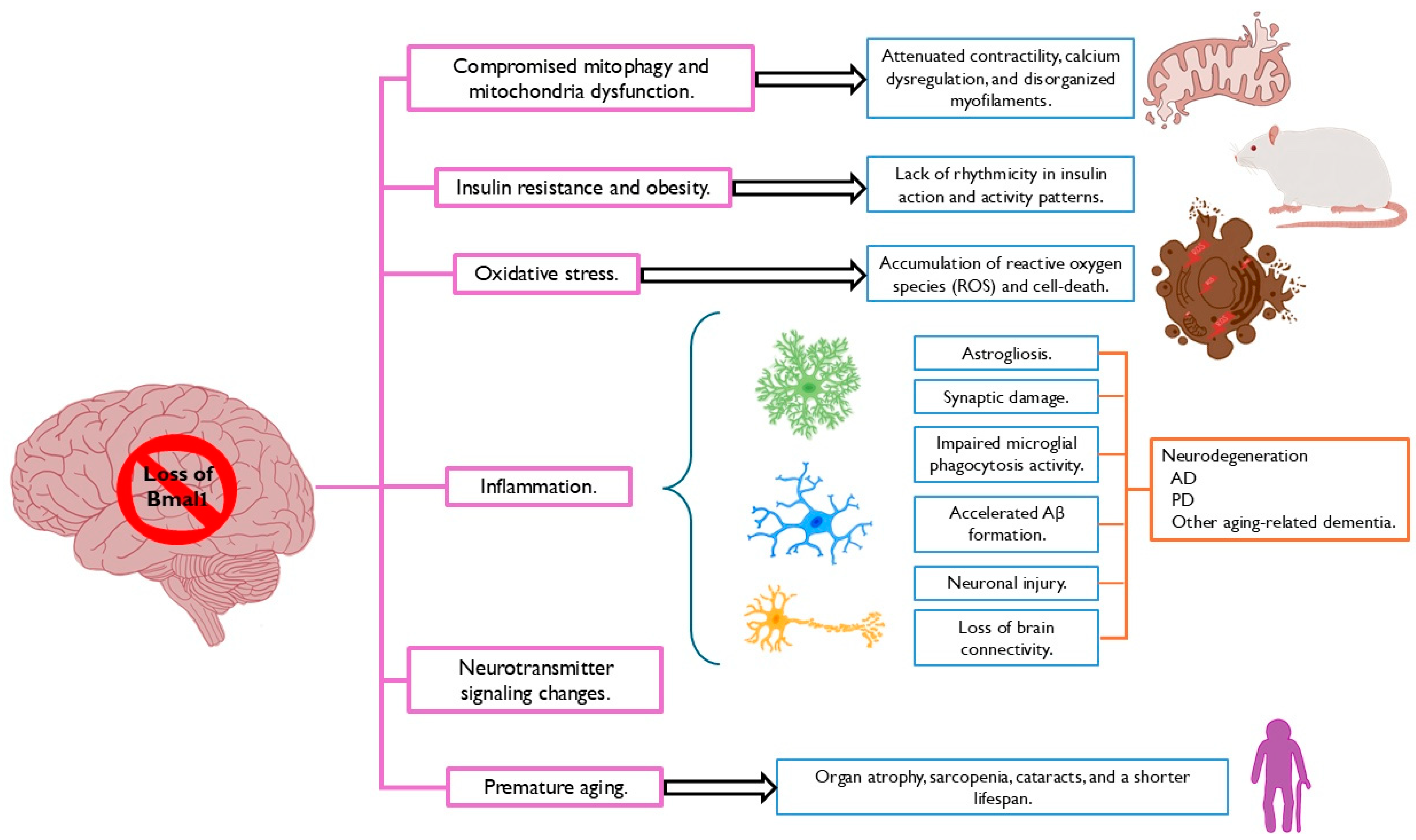
Figure 1. Loss of Bmal1 contributes to the neurodegeneration associated with Alzheimer’s and Parkinson’s disease. The Bmal1 deficiency in the brain has adverse effects on diverse types of cells, including the astrocytes. These implications lead to alterations in multiple critical physiological processes that constitute the pathological basis of Alzheimer’s and Parkinson’s disease. Modified from Fan et al., 2022 [63].
Neuroglia 06 00001 g001
Figure 2. Implications of astrocytic Bmal1 in both homeostatic conditions and Alzheimer’s and Parkinson’s disease. Aβ, β-amyloid plaques; AD, Alzheimer’s disease; GABA, gamma-aminobutyric acid; PD, Parkinson’s disease; SNpc, substantia nigra pars compacta.
Neuroglia 06 00001 g002
Disclaimer/Publisher’s Note: The statements, opinions and data contained in all publications are solely those of the individual author(s) and contributor(s) and not of MDPI and/or the editor(s). MDPI and/or the editor(s) disclaim responsibility for any injury to people or property resulting from any ideas, methods, instructions or products referred to in the content.