Review

Microglial Dyshomeostasis: A Common Substrate in Neurodevelopmental and Neurodegenerative Diseases

1
UCLA Neuroscience Undergraduate Interdepartmental Program, Los Angeles, CA 90095, USA
2
Department of Neurology, David Geffen School of Medicine, University of California Los Angeles, Los Angeles, CA 90095, USA
*
Author to whom correspondence should be addressed.
These authors contributed equally to this work.
Neuroglia 2024, 5(2), 119-128; https://doi.org/10.3390/neuroglia5020009
Submission received: 29 March 2024 / Revised: 1 May 2024 / Accepted: 6 May 2024 / Published: 12 May 2024

Abstract

:
Neurodevelopmental disorders such as autism spectrum disorder (ASD) and attention-deficit hyperactivity disorder (ADHD) are clinically distinct, yet share synaptic dysfunction as a common brain pathophysiology. Neurodegenerative diseases such as Huntington’s disease (HD) entail a neuroinflammatory cascade of molecular and cellular events which can contribute to the death of neurons. Emerging roles for supportive glial cells such as microglia and astrocytes in the ongoing regulation of neural synapses and brain excitability raise the possibility that some of the synaptic pathology and/or inflammatory events could be a direct consequence of malfunctioning glial cells. Focusing on microglia, we cross-examined 12 recently published studies in which microglial dysfunction was induced/identified in a cell-autonomous manner and its functional consequence on neural development, brain volume, functional connectivity, inflammatory response and synaptic regulation were evaluated; in many cases, the onset of symptoms relevant to all three neurodevelopmental disorders were assessed behaviorally. Challenging the classic notion of microglial activation as an inflammatory response to neuropathology, our compilation clarifies that microglial dyshomeostasis itself can consequently disrupt neural homeostasis, leading to neuropathology and symptom onset. This further warranted defining the molecular signatures of context-specific microglial pathology relevant to human diseases.

1. Introduction

Neurodevelopmental disorders (e.g., ASD and ADHD) and neurodegenerative diseases (e.g., HD) encompass human conditions which chronically alter/affect the central nervous system (CNS). Although they do not overlap in their clinical presentation, genetics and age of onset, common cellular and molecular events in these divergent diseases might offer new insights into human neurological conditions. Neuropathologies (e.g., decreased brain volume in HD, decreased brain connectivity in ASD, etc.) and their mechanisms (e.g., increased immature synaptic densities in ASD and striatal neural death in HD) have long been examined in these conditions using human and animal models. The majority of such studies focus on understanding neuronal dysfunctions to explain disease onset and progression [1,2,3]. However, a paradigm shift in delineating the causes for disease pathophysiology is being substantiated by growing evidence that non-neuronal cells are integral to homeostatic functions in the CNS and the ensuing behavioral outcomes [4,5]. In particular, the knowledge that microglia, classically described as the resident immune cells of the CNS that acutely activate to produce neuroinflammatory responses, are now known to serve ongoing neural excitability and synapse regulatory functions during development and aging [6]. Such evidence offers the premise that microglial dysfunctions can be causative to human neurological conditions such as ASD, ADHD and HD.
Microglia, widely regarded as the resident macrophages of the nervous system, sparked considerable debate with their discovery and classification in the realm of neuroscience. The timing of the discovery of microglia according to neuroscientists is contested by historians. Metchnikoff created the classification system of phagocytes in 1892 and called it the macrophage system (MS) [7]. It encompassed all the cells in the lymph nodes and nonlymphoid tissues, including the brain. The macrophages of the brain were assumed to have the same progenitors as all lymphatic phagocytes and acquired their unique dendritic features upon final differentiation [8]. Despite being a distinctive cell population, Metchnikoff similarly assumed that the brain’s resident immune cells had the same function as all other mononuclear phagocytes: maintaining homeostasis, clearing apoptotic cells and recognizing pathogens. This belief system persisted for decades until Pio del Rio-Hortega began studying the macrophage population within the CNS. In 1932, Hortega proposed that the CNS macrophages were of mesodermal origin based on their similarities to other blood and immune cells. However, he subsequently recognized that their embryonic progenitors had over eleven cell markers (now more) that were not present in mononuclear phagocytes [9]. This prompted further investigation, and in 1939, he named the cells “microglia” and refined his definition of the cells to be non-neuronal and non-astrocytic, however, still integral to the cell composition of the CNS [9]. Ever since this combination of hallmark discoveries by Metchnikoff and Hortega, phagocyte biologists and neuroscientists alike have continued to challenge the MS framework, especially in the context of discussing microglia. A better understanding of microglial function and dysregulation mean that their role in the CNS is beginning to be clarified.
Considered to be the resident immune cells of the CNS, microglia are known to play important roles in maintaining brain homeostasis by responding to injury or infection through a distinct state change [6]. This understanding stemmed from microglial homeostatic states characterized by their ramified morphology and ongoing surveillance of their environment for potential threats [6]. Then again in response to injury/infection, they transform into an amoeboid shape, phagocytose cellular debris, secrete proinflammatory cytokines and promote tissue repair; these events are attributed to microglial activation [6]. However, it is important to note that the distinction between a homeostatic versus activated state of microglia is complex since microglia can undergo activation as part of normal physiological processes as well [10]. This understanding further complicates the boundary between normal versus pathological activation in chronic diseases [6]. Here, by cross-examining the evidence for causative roles of microglia in chronic diseases including ASD, ADHD and HD, we attempt to clarify the notion of microglial dysfunction and its contribution to neurological pathologies.

2. Microglial Causative Role in Neurodevelopmental Disorders

We considered the neurodevelopmental disorders namely, autism spectrum disorder or ASD and attention-deficit hyperactivity disorder or ADHD, which are both characterized by an impairment in brain development, with challenges in social interaction, communication and cognitive abilities [11]. Additionally, ASD presents with compulsivity and repetitive behaviors; ADHD manifests as persistent patterns of inattention, hyperactivity and impulsivity, affecting academic performance and interpersonal relationships [11]. Both ASD and ADHD are believed to be caused by a combination of genetics (e.g., ASD risk genes including FMR1, SHANK3, MECP2, CUL3 and CDKL5) and environmental factors [11]. The principal pathophysiology of ASD involves abnormalities in brain structure and connectivity [12] and in ADHD, there is a significant dysregulation of the monoaminergic neurotransmitter systems [13].
We closely examined seven recent studies [11,12,13,14,15,16,17], which tested a causal link between microglial dysfunction and pathogenesis in neurodevelopmental disorders; five of these studies employed the targeted genetic manipulation of microglial proteins in mice to test its consequence on synaptic integrity (structure and function), brain connectivity, neuroinflammation and social behavior with relevance to ASD or ADHD; one study used a murine model of ADHD to examine the therapeutic action of a clinically approved methylphenidate (MPH) via microglial pathways; one study conducted a functional analysis of microglial activation in an ADHD patient cohort (see summary in Figure 1 and Table 1).

2.1. Autism Spectrum Disorder

Microglia are characterized by a high expression of the chemokine receptor CX3CR1, also called the fractalkine receptor, implicated in microglia-neuron communication [18]. Hence, CX3CR1 promoter activity has been leveraged for the genetic manipulation of microglia to probe their functional contributions in the CNS. For example, Zhan et al. (2014) investigated the effects of CX3CR1 deletion on excitatory synaptic maturity, functional brain connectivity and social behavior in the medial prefrontal cortex in juvenile and adult mice [12]. They found that CX3CR1-deficient mice exhibited decreased excitatory synapses and functional brain connectivity; concomitantly, social interaction decreased and repetitive behaviors increased. The observed decrease in excitatory synapses and weakened functional connectivity in CX3CR1-deficient mice may be a consequence of dysregulated microglial phagocytosis suggesting a causal link between microglial dysfunction, synaptic abnormalities and the behavioral deficits observed in these animals.
Microglia play a crucial role in synaptic maturation [19]. Transmembrane protein 59 (TMEM59), also known as dendritic cell factor 1 (DCF1), has been recently shown to regulate microglial function and neuroinflammation [20,21]. Although the selective loss of neuronal TMEM59 has been shown to induce ASD-like behaviors such as impaired social interaction and elevated self-grooming in mice [21], Meng et al. (2022) investigated the TMEM59 role in ASD by selective ablation in microglia during a critical period of synapse development in mice at postnatal days 12–14 [11]. The ablation of microglial TMEM59 resulted in a reduction in cortical network activity. Specifically, the frequency of spontaneous inhibitory synaptic currents were decreased, which can ultimately disrupt the balance of excitation-inhibition [22,23] and functional connectivity in cortical regions implicated in ASD (insular, limbic, parietal and anterior cingulate cortices) [24]. This suggests that microglial TMEM59 is essential for the normal development and functioning of inhibitory synapses and supports the possibility that microglial TMEM59 reduction may partly drive the excitation-inhibition imbalance observed in ASD.
The structural integrity and functional properties of synapses further depend on continuous protein renewal, which in turn depends on the rate-limiting steps in protein synthesis [25]. Xu et al. (2020) examined the eukaryotic translation initiation factor 4E (eIF4E) involved in the rate-limiting step in protein synthesis using a transgenic mouse model of ASD [14]. They showed that eIF4E overexpression in microglia led to increased excitatory synaptic density and impaired dendritic spine maturation in medial prefrontal cortex pyramidal and CA1 hippocampal neurons [14]. Additionally, in organotypic hippocampal cultures, ATP-induced microglial process motility was defective and preferentially decreased in cultures from male mice overexpressing eIF4E (MG4E); of note, males were four times more likely than females to be diagnosed with ASD [14]. Consistently, synaptic engulfment measured as Homer1 protein immunoreactivity in microglia was significantly lower in male MG4E mice relative to the control, indicating that impaired microglial motility decreases synaptic pruning contributing to increased synaptic densities. Together, these findings clarify that the dysregulation of microglial eIF4E impairs the structural integrity and functional maturation of synapses in ASD [26,27].
In addition to protein synthesis, the structural and functional integrity of synapses relies on protein clearance mechanisms such as autophagy. Kim et al. (2017) used a conditional knockout of autophagy-related 7 (Atg7) protein expression in microglia to test its effects on synaptic pruning. Such microglial dysfunction led to ASD-like phenotypes in mice, characterized by decreased social interaction and increased repetitive behaviors [15]. This was further accompanied by decreased synaptic pruning by microglia in the somatosensory 2 (S2) brain region. Importantly, microglial dysfunction via autophagy pathways is implicated in ASD pathogenesis [15].
These findings support a causative role for dysregulated microglial proteins such as CX3CR1, TMEM59, eIF4E and ATG-7 in the onset and aggravation of synaptic pathology, disrupted brain connectivity and ASD symptomatology. The potential for microglia-targeted therapies in alleviating neurocognitive deficits in ASD is also highlighted [28].

2.2. Attention-Deficit Hyperactivity Disorder

Studies linking microglial activation and cognitive impairments highlight microglial roles in ADHD pathophysiology. Yokokura et al. (2023) explored the role of microglia in individuals with ADHD by employing positron emission tomography (PET) with the 11CPK11195 tracer [16]. This tracer is highly selective to translocator protein (TSPO), which is upregulated in activated microglia [29]. The study involved psychotropic-naïve individuals with ADHD and age- and sex-matched typically developing (TD) controls. The study results showed an increased binding potential of the 11CPK11195 tracer in the dorsolateral prefrontal cortex (DLPFC) and orbitofrontal cortex (OFC) of individuals with ADHD compared to TD subjects. Importantly, this elevated microglial activation in the DLPFC was associated with diminished processing speed and sustained attention performance, establishing a connection between increased microglial activation and cognitive impairments in ADHD. Moreover, the research uncovered specificity in positive correlations between microglial activation and decreased D1 dopamine receptors (D1R) in the DLPFC and OFC of individuals with ADHD. These results present direct evidence that microglia activation is a dysfunctional phenomenon seen in ADHD patients; however, this contrasts with the reduced microglial activation noted in ASD.
Sanches et al. (2023), explored microglial involvement in ADHD-related retinal dysfunction using a rat model for ADHD [13]. Retinal changes and vision problems (color vision deficiency, monocular vision loss and strabismus) have been linked with ADHD [13]. They noted an increased percentage of activated microglia (CD68+ Iba-1+ cells) and a concurrent decrease in CX3CR1 levels, likely present in a variety of retinal cells (photoreceptors, bipolar cells, ganglion cells, horizontal cells and amacrine cells). The retina of the ADHD model also showed an upregulation of proinflammatory cytokines, IL-6 and TNF-a [13]. The use of a clinically approved methylphenidate (MPH) treatment in ADHD on the mutant mice significantly rescued microglial activation, as well as cytokine and chemokine changes, while not altering synaptic or astrocyte and blood vessel changes. Hence, the therapeutic actions of MPH were largely limited to reversing increased microglial activation and the consequential cytokine release. By demonstrating microglial activation as a pathophysiology even in the peripheral retina, these results substantiate a more ubiquitous role for microglia in ADHD pathology.
Given their function in timely release of inflammatory cytokines, microglia are critical cellular participants in inflammasome formation. Inflammasomes are multimeric cytosolic protein complexes of the innate immune system that assemble to sense and mediate the activation of inflammatory responses [30]. Chuang et al. (2021) focused on microglia inflammasome components in the context of ADHD during neural development [17]. They employed genetically modified mice with a brain-wide deletion of inflammasome-related genes, including caspase-1, interleukin-1 receptor (IL1R), gasdermin D (GSDMD) and NOD-like receptor family pyrin domain containing 3 (NLRP3) and evaluated ADHD-like behaviors. Specifically, they measured attention performance using the open field assay and 5-choice serial reaction time task (5-CSRTT), from which they observed inappropriate climbing behaviors, such as climbing up stranger mouse cylinders or escaping from test chambers, which suggest hyperactivity and inattention. Their study revealed that mice lacking Caspase-1 and NLRP3 exhibited increased climbing behaviors, indicative of ADHD-like behaviors, while the deletion of other inflammasome components (IL1R and GSDMD) was ineffective. The pharmacological administration of selective inhibitors of NLRP3 and Caspase-1 were similarly found to induce inappropriate climbing behaviors, suggesting the therapeutic potential of targeting microglial inflammasomes in ADHD treatment. Furthermore, they determined that the disruption of inflammasome formation through the Caspase-1 knockout reduced the microglial expression of mannose receptor C type 2 (Mrc2), a gene involved in phagocytosis. This observation coupled with the increase in neuron number in the thalamic reticular nucleus (TRN) implicates altered microglial phagocytosis as contributing to ADHD pathology [17]. These findings provide evidence that heightened microglial inflammatory responses (increased activation and release of proinflammatory cytokines) coupled with reduced microglial homeostatic functions (phagocytosis) contribute to the onset and symptomatology of ADHD. However, the limited literature and variety of models employed to investigate the role of microglia in ADHD makes it difficult to draw overarching conclusions regarding the role of microglial activation in ADHD pathology. In summary, the evidence implicates altered homeostatic functions of microglia during development and substantiates microglia activation as the cause of proinflammatory events in ADHD pathology and symptomology and further highlights the therapeutic merit of reversing such activation.

3. Microglial Causative Role in Neurodegenerative Diseases

Neurodegenerative diseases, HD considered here, involve late symptom onset due to progressive damage to neuronal structure and function, which eventually results in the death of affected neurons [5]. HD is a hereditary neurodegenerative disorder caused by an abnormal expansion of CAG trinucleotide repeats specifically within the huntingtin (HTT) gene [5]. A mutant HTT (mHTT) gene is characterized by the presence of 37 or more CAG repeats; the mutant gene can produce misfolded proteins which accumulate primarily throughout the striatum and cerebral cortex, causing neuronal dysfunction and degeneration [5]. Besides motor impairments, symptoms of HD can include cognitive decline, depression and anxiety.
In the HD context, we closely examined five recent studies [31,32,33,34,35], which tested a causal link between microglial dysfunction and pathogenesis in HD; one of these studies employed PET imaging to reveal the association between microglial activation and HD neuronal volume loss; two studies used cell lines to examine the consequences of proinflammatory responses of microglia in HD; two studies altered microglial proteins to reveal their role in worsening or rescuing HD pathology (see summary in Figure 1 and Table 1).
Table 1. Summary table overviewing experimental references used throughout this review.
StudyAnimal Model (s)Microglial DysfunctionFunctional Changes
ASD [11]Tmem59 KO mice↓ synaptic pruning↓ synaptic inhibition,
↑ excitatory synaptic density
ASD [12]CX3CR1 KO mice↓ synaptic pruning↓ functional connectivity,
↓ social interaction
ADHD [13]AHR & WKY rats↑ proinflammatory↓ retinal layer thinning
ASD [14]Syn-R26 mice↓ synaptic pruning↓ synaptic inhibition,
↑ excitatory synaptic density
ASD [15]Atg7 KO mice↓ synaptic pruning↑ immature synaptic density, ↓ functional connectivity
ADHD [16]Human↑ activation↓ dopamine receptor 1
ADHD [17]Casp1 KO mice↓ microglial phagocytosis↑ increased neuronal numbers in the thalamus
HD [31]R6/2 male mice↑ neurotoxicity ↓ striatal volume loss
HD [32]R6/2 male mice w/CB2 KO↑ activation↑ neurodegeneration
HD [33]BV2 mouse↑ proinflammatory↑ neurodegeneration
HD [34]Human↑ proinflammatory ↑ neurodegeneration
HD [35]Human↑ activation↓ dopamine receptor 2 binding
↑ increase, ↓ decrease.

Huntington’s Disease

In HD patients and in the widely studied R6/2 transgenic mouse model for HD, medium spiny neurons (MSNs) in the striatum degenerate, accompanied by significant striatal tissue atrophy. Such atrophy is further associated with microglial activation in the caudate-putamen nuclei and in the globus pallidus as confirmed by multiple immunohistochemistry (IHC) studies on postmortem HD brain samples (see review [36]). Functional studies using PET imaging of HD patients by Pavese et al. (2006) confirmed an association between striatal neuron loss and microglial activation [35]. Supporting this, a causal link between microglial activation and brain volumetric deficits was clarified in the transgenic R6/2 HD mice wherein depletion of a colony-stimulating factor 1 receptor inhibitor (CSF1Ri) in the microglia prevented depletion in the extracellular matrix and striatal volume [31]. Indeed, mHTT gene expression in microglia is known to drive its activation and neurotoxic effects. Functional positron emission tomography (PET) studies have shown that microglial activation can be detected via 11C-(R)-PK11195 (PK) well before symptom manifestation in HD patients and is considered to drive neurodegeneration [31]. For example, the mHTT-expressing microglia upregulated the expression of the transcription factor PU.1, which is a putative mechanism for an overall increase in proinflammatory gene expression [33].
Microglial activation can cause a cascade of downstream neurotoxic effects involving the release of proinflammatory factors further corresponding with HD severity. An examination of isogenic lines of human pluripotent stem cell-derived microglia by O’Regan et al. (2021) revealed that upon application of lipopolysaccharide (LPS), used to stimulate the innate neuroimmune response, HD microglia released greater levels of proinflammatory cytokines [34]. The secretion of inflammation-associated cytokines assessed by multiplex ELISA of the supernatants showed a percent increase in a polyglutamine repeat in a length-dependent manner [34]. The production of reactive oxygen species (ROS) associated with cellular damage and death was also found to be increased [34]. The downstream effects of increases in proinflammatory factors are characterized by excitotoxic conditions, including microglial phagocytosis. This can be visualized by confocal microscopy analysis exhibiting neuronal debris present in Iba-1 (microglia-specific marker) positive cells [32].
Despite these neurotoxic effects of microglial activation, the possibility that pre-symptomatically, microglia are neuroprotective and aid in ameliorating striatal neural dysfunction is also supported. For example, the analysis of pre-symptomatic R6/2 mouse striatum samples identified increased levels of neuroprotective cannabinoid receptor-type 2 (CB2) expression. A similar examination of HD patients’ brains revealed increased levels of CB2 receptors in the caudate putamen compared to controls, with selective expression in microglia, and not in astrocytes [32]. Remarkably, administering the CB2 agonist HU-308 to mice injected with quinolinic acid, an excitotoxin, resulted in a notable reduction in striatal degeneration, attributed to a decrease in microglial activation [32]. These reports lend credence to the functional dichotomy of microglia in both neuroprotective and neurotoxic roles in HD, although specific molecular and cellular contexts for this switch are yet to be clarified. Additionally, microglial neuroprotective roles via the endocannabinoid pathways offer promising therapeutic strategies for degenerative diseases such as HD that are aggravated by runaway excitotoxicity.

4. Conclusions and Future Perspectives

The interplay between neuronal and non-neuronal components in the CNS has long been a focal point of research to unravel the complexities of neurological pathologies. The scope of this review focused on identifying recent studies which investigated the causal impact of microglial mechanisms in neurodevelopmental (ASD and ADHD) and neurodegenerative diseases, specifically, HD; however, we acknowledge that not all such studies have been included in this review. Contrary to the established role of microglia as the resident immune cells in the CNS, microglial dysfunctions were demonstrated to play a causative role in driving the pathology and symptoms in neurodevelopmental disorders and neurodegenerative diseases.
In ASD, microglial deficits stemmed from reduced motility and phagocytic ability, causing an overabundance of immature neural synapses influencing behavioral outputs. In ADHD, abnormal microglial activation contributed to the release of proinflammatory factors and the onset of ADHD-like behaviors. Interestingly, in the chronic neurodegenerative HD, microglial activity is modified at the pre-symptomatic and symptomatic stages. While pre-symptomatically, microglial changes might aid in neuroprotection, an as yet unclear switch to a neurotoxic state appears to promote excessive phagocytosis and reduced cerebral volume. In both of these roles, microglia seem to become hyper-functional, suggesting a rather over-compensatory role. A common theme of microglial dyshomeostasis emerged from these studies, prompting strategies targeting microglia to reverse or delay disease progression. In support of this possibility, treatment using minocycline, a tetracycline derivative, in a BTBR ASD mouse model restored microglia to their homeostatic state and rescued ASD-related sociability changes and repetitive behaviors [37]. As also noted earlier, a further study treated HD patients with cannabinoids which act on cannabinoid receptors, and found that this improved HD related motor symptoms via microglial mechanisms [38]. Therapeutic outcomes are not limited to the neuropathology of diseases discussed herein. Additionally, the administration of a dopamine D1R agonist to schizophrenia patients, which ameliorated attention deficits, also reduced microglial activation [39]. In Alzheimer’s disease (AD), the in vivo targeted inhibition of BACE-1, an amyloid-precursor-protein-cleaving enzyme in microglia, reduced amyloid plaque development, further demonstrating prolonged benefits in delaying disease progression [40]. In summary, microglia are not mere immune-activated cell types, but a pervasive pathological substrate in neurological diseases. Their dyshomeostasis involving altered efficacy as both immune modulators and regulators of neural development and functions consequently results in symptom development/aggravation. Therefore, a need for the careful characterization of the molecular mechanisms underlying microglial dysfunctions and strategies to leverage them for therapeutic interventions is explicitly substantiated.

Author Contributions

This review was developed as part of an undergraduate honors class in neuroscience at UCLA. V.A.F. and D.M. coordinated the review conceptualization and interpretation and prepared the review draft. All authors contributed to the literature review and manuscript preparation. S.V. supervised the literature review and edited the manuscript. All authors have read and agreed to the published version of the manuscript.

Funding

This research received no external funding.

Institutional Review Board Statement

Not applicable.

Informed Consent Statement

Not applicable.

Data Availability Statement

No new data was created.

Acknowledgments

We thank the UCLA Neuroscience Undergraduate Interdepartmental Program, Stephanie White and Martina Wiedau for supporting SV.

Conflicts of Interest

The authors declare no conflicts of interest.

References

  1. Kasparek, T.; Theiner, P.; Filova, A. Neurobiology of ADHD From Childhood to Adulthood: Findings of Imaging Methods. J. Atten. Disord. 2015, 19, 931–943. [Google Scholar] [CrossRef] [PubMed]
  2. Varghese, M.; Keshav, N.; Jacot-Descombes, S.; Warda, T.; Wicinski, B.; Dickstein, D.L.; Harony-Nicolas, H.; De Rubeis, S.; Drapeau, E.; Buxbaum, J.D.; et al. Autism Spectrum Disorder: Neuropathology and Animal Models. Acta Neuropathol. (Berl.) 2017, 134, 537–566. [Google Scholar] [CrossRef] [PubMed]
  3. Waldvogel, H.J.; Kim, E.H.; Tippett, L.J.; Vonsattel, J.-P.G.; Faull, R.L.M. The Neuropathology of Huntington’s Disease. Curr. Top. Behav. Neurosci. 2015, 22, 33–80. [Google Scholar] [CrossRef] [PubMed]
  4. Lukens, J.R.; Eyo, U.B. Microglia and Neurodevelopmental Disorders. Annu. Rev. Neurosci. 2022, 45, 425–445. [Google Scholar] [CrossRef] [PubMed]
  5. Crotti, A.; Glass, C.K. The Choreography of Neuroinflammation in Huntington’s Disease. Trends Immunol. 2015, 36, 364–373. [Google Scholar] [CrossRef] [PubMed]
  6. Napoli, I.; Neumann, H. Microglial Clearance Function in Health and Disease. Neuroscience 2009, 158, 1030–1038. [Google Scholar] [CrossRef] [PubMed]
  7. van Furth, R.; Cohn, Z.A.; Hirsch, J.G.; Humphrey, J.H.; Spector, W.G.; Langevoort, H.L. The Mononuclear Phagocyte System: A New Classification of Macrophages, Monocytes, and Their Precursor Cells. Bull. World Health Organ. 1972, 46, 845–852. [Google Scholar] [PubMed]
  8. Ginhoux, F.; Merad, M. Ontogeny and Homeostasis of Langerhans Cells. Immunol. Cell Biol. 2010, 88, 387–392. [Google Scholar] [CrossRef] [PubMed]
  9. del Río-Hortega, P. Microglia. Cytol. Cell. Pathol. Nerv. Syst. 1932, 2, 483–534. [Google Scholar]
  10. Lull, M.E.; Block, M.L. Microglial Activation and Chronic Neurodegeneration. Neurotherapeutics 2010, 7, 354–365. [Google Scholar] [CrossRef]
  11. Meng, J.; Han, L.; Zheng, N.; Wang, T.; Xu, H.; Jiang, Y.; Wang, Z.; Liu, Z.; Zheng, Q.; Zhang, X.; et al. Microglial Tmem59 Deficiency Impairs Phagocytosis of Synapse and Leads to Autism-Like Behaviors in Mice. J. Neurosci. 2022, 42, 4958–4979. [Google Scholar] [CrossRef] [PubMed]
  12. Zhan, Y.; Paolicelli, R.C.; Sforazzini, F.; Weinhard, L.; Bolasco, G.; Pagani, F.; Vyssotski, A.L.; Bifone, A.; Gozzi, A.; Ragozzino, D.; et al. Deficient Neuron-Microglia Signaling Results in Impaired Functional Brain Connectivity and Social Behavior. Nat. Neurosci. 2014, 17, 400–406. [Google Scholar] [CrossRef] [PubMed]
  13. Sanches, E.S.; Boia, R.; Leitão, R.A.; Madeira, M.H.; Fontes-Ribeiro, C.A.; Ambrósio, A.F.; Fernandes, R.; Silva, A.P. Attention-Deficit/Hyperactivity Disorder Animal Model Presents Retinal Alterations and Methylphenidate Has a Differential Effect in ADHD versus Control Conditions. Antioxidants 2023, 12, 937. [Google Scholar] [CrossRef] [PubMed]
  14. Xu, Z.-X.; Kim, G.H.; Tan, J.-W.; Riso, A.E.; Sun, Y.; Xu, E.Y.; Liao, G.-Y.; Xu, H.; Lee, S.-H.; Do, N.-Y.; et al. Elevated Protein Synthesis in Microglia Causes Autism-like Synaptic and Behavioral Aberrations. Nat. Commun. 2020, 11, 1797. [Google Scholar] [CrossRef] [PubMed]
  15. Kim, H.-J.; Cho, M.-H.; Shim, W.H.; Kim, J.K.; Jeon, E.-Y.; Kim, D.-H.; Yoon, S.-Y. Deficient Autophagy in Microglia Impairs Synaptic Pruning and Causes Social Behavioral Defects. Mol. Psychiatry 2017, 22, 1576–1584. [Google Scholar] [CrossRef] [PubMed]
  16. Yokokura, M.; Takebasashi, K.; Takao, A.; Nakaizumi, K.; Yoshikawa, E.; Futatsubashi, M.; Suzuki, K.; Nakamura, K.; Yamasue, H.; Ouchi, Y. In Vivo Imaging of Dopamine D1 Receptor and Activated Microglia in Attention-Deficit/Hyperactivity Disorder: A Positron Emission Tomography Study. Mol. Psychiatry 2021, 26, 4958–4967. [Google Scholar] [CrossRef] [PubMed]
  17. Chuang, H.-C.; Nichols, E.K.; Rauch, I.; Chang, W.-C.; Lin, P.M.; Misra, R.; Kitaoka, M.; Vance, R.E.; Saijo, K. Lytic Cell Death in Specific Microglial Subsets Is Required for Preventing Atypical Behavior in Mice. eNeuro 2021, 8, ENEURO.0342-20.2020. [Google Scholar] [CrossRef] [PubMed]
  18. Reshef, R.; Kudryavitskaya, E.; Shani-Narkiss, H.; Isaacson, B.; Rimmerman, N.; Mizrahi, A.; Yirmiya, R. The Role of Microglia and Their CX3CR1 Signaling in Adult Neurogenesis in the Olfactory Bulb. eLife 2017, 6, e30809. [Google Scholar] [CrossRef] [PubMed]
  19. Dixon, M.A.; Greferath, U.; Fletcher, E.L.; Jobling, A.I. The Contribution of Microglia to the Development and Maturation of the Visual System. Front. Cell. Neurosci. 2021, 15, 659843. [Google Scholar] [CrossRef]
  20. Wang, Q.; Liu, Y.; Zhou, J. Neuroinflammation in Parkinson’s Disease and Its Potential as Therapeutic Target. Transl. Neurodegener. 2015, 4, 19. [Google Scholar] [CrossRef]
  21. Liu, Z.; Ning, J.; Zheng, X.; Meng, J.; Han, L.; Zheng, H.; Zhong, L.; Chen, X.-F.; Zhang, X.; Luo, H.; et al. TMEM59 Interacts with TREM2 and Modulates TREM2-Dependent Microglial Activities. Cell Death Dis. 2020, 11, 678. [Google Scholar] [CrossRef] [PubMed]
  22. Bruining, H.; Hardstone, R.; Juarez-Martinez, E.L.; Sprengers, J.; Avramiea, A.-E.; Simpraga, S.; Houtman, S.J.; Poil, S.-S.; Dallares, E.; Palva, S.; et al. Measurement of Excitation-Inhibition Ratio in Autism Spectrum Disorder Using Critical Brain Dynamics. Sci. Rep. 2020, 10, 9195. [Google Scholar] [CrossRef]
  23. Canitano, R.; Palumbi, R. Excitation/Inhibition Modulators in Autism Spectrum Disorder: Current Clinical Research. Front. Neurosci. 2021, 15, 753274. [Google Scholar] [CrossRef]
  24. Siegel-Ramsay, J.E.; Romaniuk, L.; Whalley, H.C.; Roberts, N.; Branigan, H.; Stanfield, A.C.; Lawrie, S.M.; Dauvermann, M.R. Glutamate and Functional Connectivity—Support for the Excitatory-Inhibitory Imbalance Hypothesis in Autism Spectrum Disorders. Psychiatry Res. Neuroimaging 2021, 313, 111302. [Google Scholar] [CrossRef] [PubMed]
  25. Cohen, L.D.; Ziv, T.; Ziv, N.E. Synapse Integrity and Function: Dependence on Protein Synthesis and Identification of Potential Failure Points. Front. Mol. Neurosci. 2022, 15, 1038614. [Google Scholar] [CrossRef]
  26. Guang, S.; Pang, N.; Deng, X.; Yang, L.; He, F.; Wu, L.; Chen, C.; Yin, F.; Peng, J. Synaptopathology Involved in Autism Spectrum Disorder. Front. Cell. Neurosci. 2018, 12, 470. [Google Scholar] [CrossRef]
  27. Amorim, I.S.; Lach, G.; Gkogkas, C.G. The Role of the Eukaryotic Translation Initiation Factor 4E (eIF4E) in Neuropsychiatric Disorders. Front. Genet. 2018, 9, 424981. [Google Scholar] [CrossRef] [PubMed]
  28. Zhang, W.; Chen, H.; Ding, L.; Huang, J.; Zhang, M.; Liu, Y.; Ma, R.; Zheng, S.; Gong, J.; Piña-Crespo, J.C.; et al. Microglial Targeted Therapy Relieves Cognitive Impairment Caused by Cntnap4 Deficiency. Explor. Beijing China 2023, 3, 20220160. [Google Scholar] [CrossRef]
  29. Su, Z.; Herholz, K.; Gerhard, A.; Roncaroli, F.; Du Plessis, D.; Jackson, A.; Turkheimer, F.; Hinz, R. [11C]-(R)PK11195 Tracer Kinetics in the Brain of Glioma Patients and a Comparison of Two Referencing Approaches. Eur. J. Nucl. Med. Mol. Imaging 2013, 40, 1406–1419. [Google Scholar] [CrossRef]
  30. Guo, H.; Callaway, J.B.; Ting, J.P.-Y. Inflammasomes: Mechanism of Action, Role in Disease, and Therapeutics. Nat. Med. 2015, 21, 677–687. [Google Scholar] [CrossRef]
  31. Crapser, J.D.; Ochaba, J.; Soni, N.; Reidling, J.C.; Thompson, L.M.; Green, K.N. Microglial Depletion Prevents Extracellular Matrix Changes and Striatal Volume Reduction in a Model of Huntington’s Disease. Brain 2020, 143, 266–288. [Google Scholar] [CrossRef] [PubMed]
  32. Palazuelos, J.; Aguado, T.; Pazos, M.R.; Julien, B.; Carrasco, C.; Resel, E.; Sagredo, O.; Benito, C.; Romero, J.; Azcoitia, I.; et al. Microglial CB2 Cannabinoid Receptors Are Neuroprotective in Huntington’s Disease Excitotoxicity. Brain J. Neurol. 2009, 132, 3152–3164. [Google Scholar] [CrossRef] [PubMed]
  33. Crotti, A.; Benner, C.; Kerman, B.; Gosselin, D.; Lagier-Tourenne, C.; Zuccato, C.; Cattaneo, E.; Gage, F.H.; Cleveland, D.W.; Glass, C.K. Mutant Huntingtin Promotes Autonomous Microglia Activation via Myeloid Lineage-Determining Factors. Nat. Neurosci. 2014, 17, 513–521. [Google Scholar] [CrossRef] [PubMed]
  34. O’Regan, G.C.; Farag, S.H.; Casey, C.S.; Wood-Kaczmar, A.; Pocock, J.M.; Tabrizi, S.J.; Andre, R. Human Huntington’s Disease Pluripotent Stem Cell-Derived Microglia Develop Normally but Are Abnormally Hyper-Reactive and Release Elevated Levels of Reactive Oxygen Species. J. Neuroinflamm. 2021, 18, 94. [Google Scholar] [CrossRef] [PubMed]
  35. Pavese, N.; Gerhard, A.; Tai, Y.F.; Ho, A.K.; Turkheimer, F.; Barker, R.A.; Brooks, D.J.; Piccini, P. Microglial Activation Correlates with Severity in Huntington Disease: A Clinical and PET Study. Neurology 2006, 66, 1638–1643. [Google Scholar] [CrossRef] [PubMed]
  36. Palpagama, T.H.; Waldvogel, H.J.; Faull, R.L.M.; Kwakowsky, A. The Role of Microglia and Astrocytes in Huntington’s Disease. Front. Mol. Neurosci. 2019, 12, 258. [Google Scholar] [CrossRef]
  37. Luo, Y.; Lv, K.; Du, Z.; Zhang, D.; Chen, M.; Luo, J.; Wang, L.; Liu, T.; Gong, H.; Fan, X. Minocycline Improves Autism-Related Behaviors by Modulating Microglia Polarization in a Mouse Model of Autism. Int. Immunopharmacol. 2023, 122, 110594. [Google Scholar] [CrossRef]
  38. Saft, C.; von Hein, S.M.; Lücke, T.; Thiels, C.; Peball, M.; Djamshidian, A.; Heim, B.; Seppi, K. Cannabinoids for Treatment of Dystonia in Huntington’s Disease. J. Huntingt. Dis. 2018, 7, 167–173. [Google Scholar] [CrossRef] [PubMed]
  39. Girgis, R.R.; Van Snellenberg, J.X.; Glass, A.; Kegeles, L.S.; Thompson, J.L.; Wall, M.; Cho, R.Y.; Carter, C.S.; Slifstein, M.; Abi-Dargham, A.; et al. A Proof-of-Concept, Randomized Controlled Trial of DAR-0100A, a Dopamine-1 Receptor Agonist, for Cognitive Enhancement in Schizophrenia. J. Psychopharmacol. Oxf. Engl. 2016, 30, 428–435. [Google Scholar] [CrossRef]
  40. Singh, N.; Das, B.; Zhou, J.; Hu, X.; Yan, R. Targeted BACE-1 Inhibition in Microglia Enhances Amyloid Clearance and Improved Cognitive Performance. Sci. Adv. 2022, 8, eabo3610. [Google Scholar] [CrossRef]
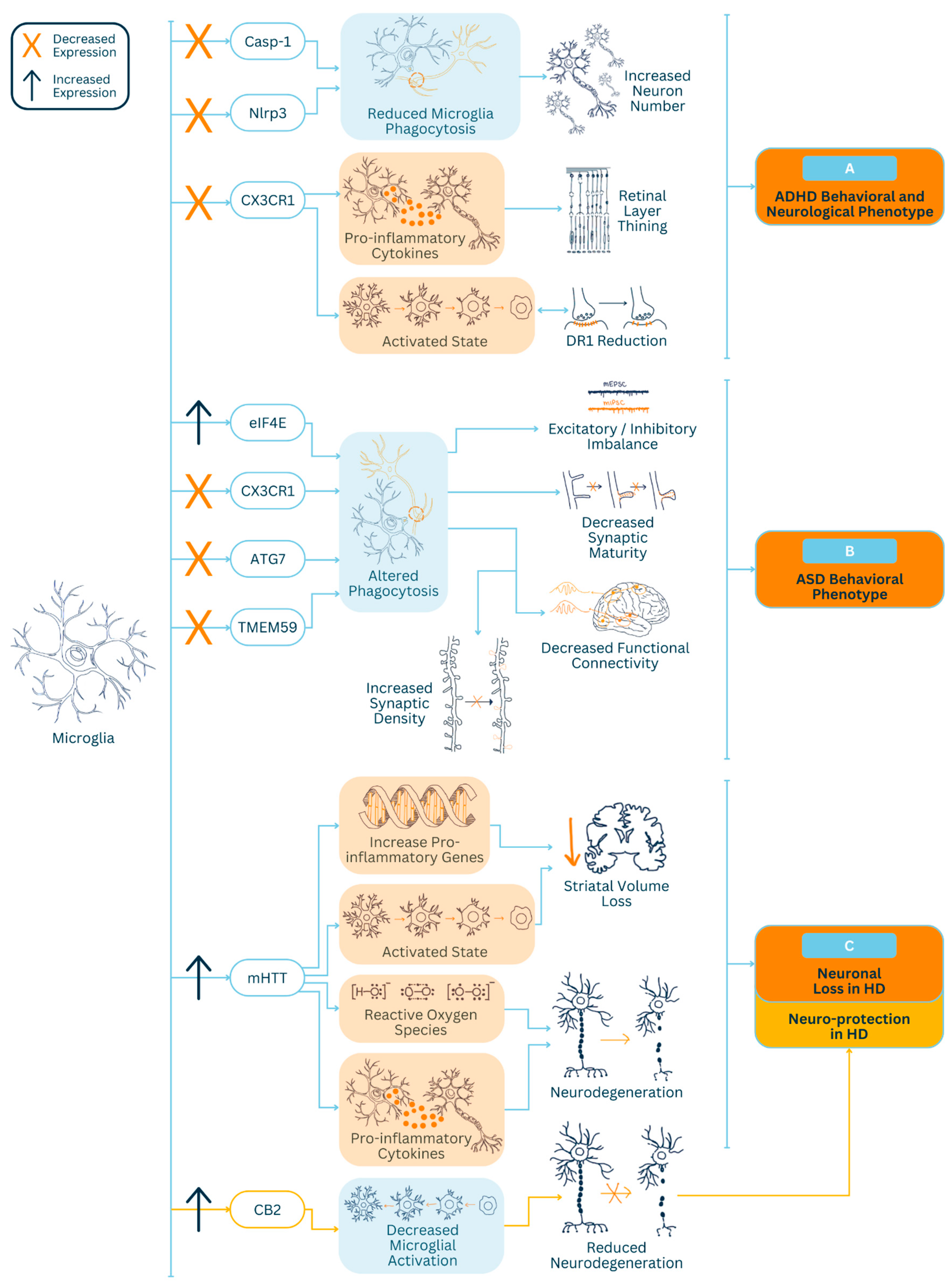
Figure 1. Microglia-mediated mechanisms contributing to (A) attention-deficit hyperactivity disorder (ADHD), (B) autism spectrum disorder (ASD) and (C) Huntington’s disease (HD). Light blue arrows indicate the contribution to pathophysiology of disease/disorder. Light yellow arrows indicate protection against the pathophysiology of disease/disorder. Light blue boxes indicate homeostatic microglial changes. Light orange boxes indicate activated microglial changes. Orange X symbols indicate decreased expression of a gene. Dark blue up arrows indicate increased expression of a gene. C-X3-C motif chemokine receptor 1 (CX3CR1). Dopamine receptor 1 level (DR1). Caspase-1 (Casp-1). NLR family pyrin domain-containing 3 (Nlrp3).
Neuroglia 05 00009 g001
Disclaimer/Publisher’s Note: The statements, opinions and data contained in all publications are solely those of the individual author(s) and contributor(s) and not of MDPI and/or the editor(s). MDPI and/or the editor(s) disclaim responsibility for any injury to people or property resulting from any ideas, methods, instructions or products referred to in the content.