Review

Understanding the Relevance of Aging-Related Tau Astrogliopathy (ARTAG)

Institute of Neurology, Medical University Vienna, AKH 4J, A-1090 Vienna, Austria
Neuroglia 2018, 1(2), 339-350; https://doi.org/10.3390/neuroglia1020023
Submission received: 9 October 2018 / Revised: 19 October 2018 / Accepted: 22 October 2018 / Published: 29 October 2018

Abstract

:
Aging-related tau astrogliopathy (ARTAG) is an umbrella term that encompasses a spectrum of morphological abnormalities seen in astrocytes of the aging brain using immunostaining for pathological forms of the microtubule-associated protein tau. Morphologies of ARTAG include thorn-shaped astrocytes (TSA), and additionally granular/fuzzy astrocytes (GFA) characterized by fine granular tau immunoreactivity extending into the astrocytic processes. Thorn-shaped astrocytes can be present in the same brain in subpial, subependymal, perivascular, and white and gray matter locations together with GFAs, which are seen in the gray matter. Primary tauopathies show ARTAG-related morphologies as well, moreover, GFA has been proposed to present a conceptual link between brain ageing and primary tauopathies. Sequential distribution patterns have been recognized for subpial, white and gray matter ARTAG. This either suggests the involvement of astrocytes in the propagation of tau pathology or reflects the consequence of a long-term pathogenic process such as barrier dysfunction, local mechanical impact, or early response to neuronal degeneration. The concept of ARTAG facilitated communication among neuropathologists and researchers, informed biomarker researchers with focus on tau-related indicators and motivated further exploration of the significance of astrocytic lesions in various neurodegenerative conditions.

1. Introduction: What Is ARTAG?

Aging-related tau astrogliopathy (ARTAG) is an umbrella term that encompasses a spectrum of morphological abnormalities seen in astrocytes using immunostaining for pathological forms of the microtubule-associated protein tau, mainly in the aging brain [1]. This term was introduced to harmonize the nomenclature and evaluation strategies for the different morphological forms of tau immunoreactive astrocytes previously described by several authors. ARTAG includes morphologies described originally as thorn-shaped astrocytes (TSA) as well as fine granular tau immunoreactivity extending into the astrocytic processes in the gray matter, now called granular/fuzzy astrocytes (GFA) [1].
Ikeda and colleagues were the first to describe TSAs in the subpial or subependymal regions of the gray and white matter, and frequently in the depths of the gyri, as well as in the basal forebrain and brainstem, in aged individuals [2,3,4]. This was followed by a study from Schultz et al. reporting a high prevalence of TSAs in the aged human medial temporal lobe, particularly at the level of the amygdala [5]. Interestingly, TSA-like morphologies have been described in aged gorillas, and particularly in baboons, but not in other primates [6,7,8,9]. This is possibly due to differences in the tau sequence or the lack of sufficient neuropathological studies focusing on tau pathologies in animals [10]. Diffuse granular tau immunoreactivity in astrocytic processes has been described in the context of a study on a peculiar constellation of tau pathology in aged demented individuals [11]. However, the term GFA was introduced only in the consensus paper on ARTAG [1].
In TSAs, tau immunoreactivity is localized in the astrocytic perikarya with extension into the proximal parts of the astrocytic processes, with inclusions also in the astrocytic end feet at the glia limitans around blood vessels and at the pial surface [1]. The processes are thick and short and thus reminiscent of thorns. In contrast, GFAs exhibit fine granular immunoreactivity of branching processes with a few dilations, and the perinuclear soma is densely immunoreactive in most of these astrocytes [1]. These two types of tau immunoreactive astrocytes can both be present in the same brain. Thorn-shaped astrocytes are seen mostly in subpial, subependymal, or perivascular areas, as well as in the white, and less so, in the gray matter. Granular/fuzzy astrocytes are observed in the gray matter. In both the white and gray matter, TSAs and GFAs may build clusters.
For the evaluation of ARTAG a simple strategy has been proposed [1]:
  • Identify the morphologic and distribution types of ARTAG based on parenchymal localization of TSA and GFA: i.e., subpial, subependymal, perivascular, white matter, and gray matter.
  • Identify involvement of gross anatomical regions such as the medial temporal lobe, further lobes of the brain, subcortical structures, and the brainstem.
  • Document the severity of ARTAG pathology; in particular, whether this is seen in occasional or in numerous astrocytes and whether clusters or widespread distribution is noted.
  • Finally, particularly for scientific discovery studies, detailed anatomical mapping is recommended.
In a consecutive study, digital images were evaluated by a group of researchers to evaluate whether these strategies are reproducible [12]. This study revealed the challenging issues of always being able to readily differentiate and clearly classify tau-positive astrocytic lesions. Still, this motivates further exploration of the significance of astrocytic lesions in neurodegenerative disorders and further consensus meetings to reach high agreement. Otherwise, the comparability of research studies will be questionable.

2. ARTAG and Primary Tauopathies

Morphologies of ARTAG may be seen in so-called primary tauopathies as well. Indeed TSA as described by Ikeda et al. [2,3,4] were similar in morphology to the tau-positive astrocytes described by Nishimura et al. in progressive supranuclear palsy (PSP) [13]. The concept of GFAs has only recently been added to the spectrum of primary tauopathy-related astroglial tau pathologies [14]. Primary tauopathies are biochemically, genetically, clinically and neuropathologically heterogeneous neurodegenerative disorders characterized by the abnormal deposition of tau protein in different cell types of the central nervous system, including neurons and neuroglia. Tauopathies are classified based on the distribution and spectrum of cell types involved and also on a biochemical level. In spite of showing a wide spectrum of biochemical modifications, currently the most widely accepted classification focuses on the predominance of four-repeat (4R) or three-repeat (3R) isoforms of the tau protein, or the presence of both [15].
Importantly, TSAs and GFA-like astrocyte morphologies are common, but not the distinguishing astrocytic morphologies seen in primary tauopathies. The most characteristic astrocytic tau pathologies in primary tauopathies comprise tufted astrocytes in PSP, astrocytic plaques in corticobasal degeneration (CBD), globular glial inclusions in globular glial tauopathies (GGT), and ramified astrocytes in Pick’s disease (PiD) [15]. 4R-tauopathies comprise PSP, CBD and GGT, while PiD is a 3R-predominant tauopathy [15]. A further disease affecting the limbic system showing characteristic grains in the neuronal dendrites, hence called argyrophilic grain disease (AGD) also exhibits tau immunoreactive astrocytes in the medial temporal lobe often termed bushy astrocytes in the literature [16,17]. However, these are now also called GFAs due to their similarity to gray matter ARTAG [1]. Finally, the mixed 3R + 4R disease primary tauopathy (PART), which shows neurofibrillary tangles as in Alzheimer`s disease (AD), but without significant amyloid-β plaques, does not show the specific tau pathology of astrocytes, but can be associated with ARTAG [14,18]. FTDP-17, or hereditary frontotemporal dementia associated with mutations in the MAPT gene, shows a wide variety of tau pathologies and various constellations of tau isoforms [19]. Although astrocytic tau pathology has been readily recognized in several hereditary conditions [9,20], the descriptions vary considerably making comparisons difficult. Tau pathologies resembling ARTAG are also recognized [20]. Interestingly, subpial ARTAG was prominently seen in a recently reported 49-year old demented individual with MAPT gene duplication [21,22], suggesting that an imbalance of tau homeostasis may contribute to the early development of an otherwise age-related pathology.
Since GFA-like morphologies are seen in primary tauopathies, we introduced the concept that, analogously to the pretangles, which might be a preceding form of neurofibrillary tangles, the first step of astrocytic pathology might be the fine granular accumulation in astrocytic processes [14,20]. These tau deposits are then potentially redistributed to distal or proximal segments of the astrocytic cytoskeleton and eventually aggregate and become detectable using silver-stainings or anti-ubiquitin antibodies [14,23]. This concept allows the speculation that pure detection of single astrocytes with fine granular phospho-tau immunoreactivity in the human brain might represent an early preclinical form of primary tauopathy or ARTAG, or eventually the first moment of a response to a neurodegenerative event [14,20].
Ferrer et al. [24,25] showed that the biochemical signature of astroglial tau pathology in the elderly in both white and gray matter (i.e., representing ARTAG) differs in some aspects from that of other astrocytic tau pathologies in primary tauopathies. For example, astroglial tau pathologies in the white matter and gray matter in aging brains were not consistently detectable using tau truncated at aspartic acid 421 (tau-C3), or conformational tau modifications at amino acids 312 to 322 (antibody MC1), or phospho-specific anti-tau antibody Ser262 [24,25].

3. ARTAG and Various Disorders Including Chronic Traumatic Encephalopathy

A peculiar aspect of ARTAG is its relation to chronic traumatic encephalopathy (CTE). Astrocytic tau pathology that resembles TSAs (although usually termed astrocytic tangles), is an important component of the morphological alterations reported in CTE, a disorder associated with mild repetitive brain trauma and progressive neurological deterioration [26,27]. Examples of overlapping aspects of CTE and ARTAG include accumulation of subpial, perivascular and gray matter astrocytes in basal brain regions, but also in dorsolateral lobar areas, overrepresentation of males, or association with ventricular enlargement [14,26,28,29,30]. Importantly, the definition of CTE-associated lesions emphasizes the presence of neuronal tau pathology [26]. Hence, the presence of pure subpial or cortical clusters of astrocytic tau immunoreactivities such as seen in ARTAG should not be at once interpreted as CTE. A study on potential sequential distribution of ARTAG (see below) [31], however, raises an interesting point. Can it be that, at least in some cases, these represent the earliest stage, preceding neuronal tau accumulation, of CTE type pathology? Further studies on CTE cases with early stage tau pathology [30] might be able to address this point.
A wide range of disorders can associate with ARTAG, which could suggest that it is a non-specific condition. Indeed, gray matter ARTAG has been reported in prion diseases, Lewy body disorders, psychiatric conditions (i.e., here mostly restricted to the amygdala), multiple system atrophy, and amyotrophic lateral sclerosis (i.e., here also in the spinal cord) [14,28,32]. However, it might also reflect a response to early neuronal degeneration irrespective of the predominating proteinopathy. Since it is not seen in all cases with some form of neuronal degeneration, a yet unidentified driving force or additional factor need to be considered.

4. Sequential Distribution of ARTAG

In the human brain, hierarchical or stereotypical involvement of anatomical regions (i.e., stages or phases) have been described for several neurodegeneration-related protein pathologies [33]. These focus only on neuronal (tau, α-synuclein, TDP-43) or extracellular (amyloid-β) protein depositions. A recent study evaluated frequencies and hierarchical clustering of anatomical involvement and used conditional probability and logistic regression to model the sequential distribution of ARTAG and astroglial tau pathologies across different brain regions [31]. It has been emphasized that ARTAG does not show such clear stages as neuronal protein pathologies, or in other words not all cases can be put in one box. Therefore, first patterns have to be recognized and then the sequential distribution becomes more visible. Except for subependymal ARTAG, the following sequential patterns have been described for different ARTAG types [31].

4.1. Subpial ARTAG (Thorn-Shaped Astrocytes Morphology)

Pattern 1 (Figure 1): Basal brain regions show subpial ARTAG first (stage 1) followed by a bidirectional sequence rostral (lobar, stage 2a) or caudal (brainstem, stage 2b), which, however, are usually affected together (stage 3).
Pattern 2 (Figure 1): Subpial ARTAG is initiated in lobar regions (stage 1a) or in the brainstem (stage 1b) followed by the involvement of both (stage 2) preceding basal brain regions (stage 3).
Pattern 3 (Figure 1): This is seen only in CBD, since the morphology of subpial tau accumulation is different. One form, characterized by the immunoreactivity of astrocytic end-feets but not the cell body, is seen regularly in CBD. However, typical TSA morphology can be recognized as well in some CBD cases. Thus, in CBD subpial tau immunoreactivity of astrocytic feet in lobar areas is the predominant pathology independently of subpial ARTAG in basal brain regions (together representing stage 1) and both are followed by the involvement of the brainstem, representing stage 2. We termed this a “masked” bidirectional sequence [31]. This means that pattern 1 as described above, seen in non-CBD cases with the typical subpial TSA morphologies is masked by the predominant end-feet tau immunoreactivity appearing in the lobar subpial location in CBD.

4.2. White Matter ARTAG (Thorn-Shaped Astrocytes Morphology)

Pattern 1 (Figure 1): This is similar to pattern 1 of subpial ARTAG; thus, basal brain regions (stage 1) are followed by the involvement of lobar regions (stage 2a), or brainstem (stage 2b), and then all regions are involved (stage 3).
Pattern 2 (Figure 1): Lobar white matter ARTAG seems to be independent from involvement of the basal brain region. In this case lobar involvement (stage 1) is followed by the involvement of the basal brain regions (stage 2a) or occasionally the brainstem (stage 2b) and then all regions are involved (stage 3).

4.3. Gray Matter ARTAG (Granular/Fuzzy Astrocytes Morphology)

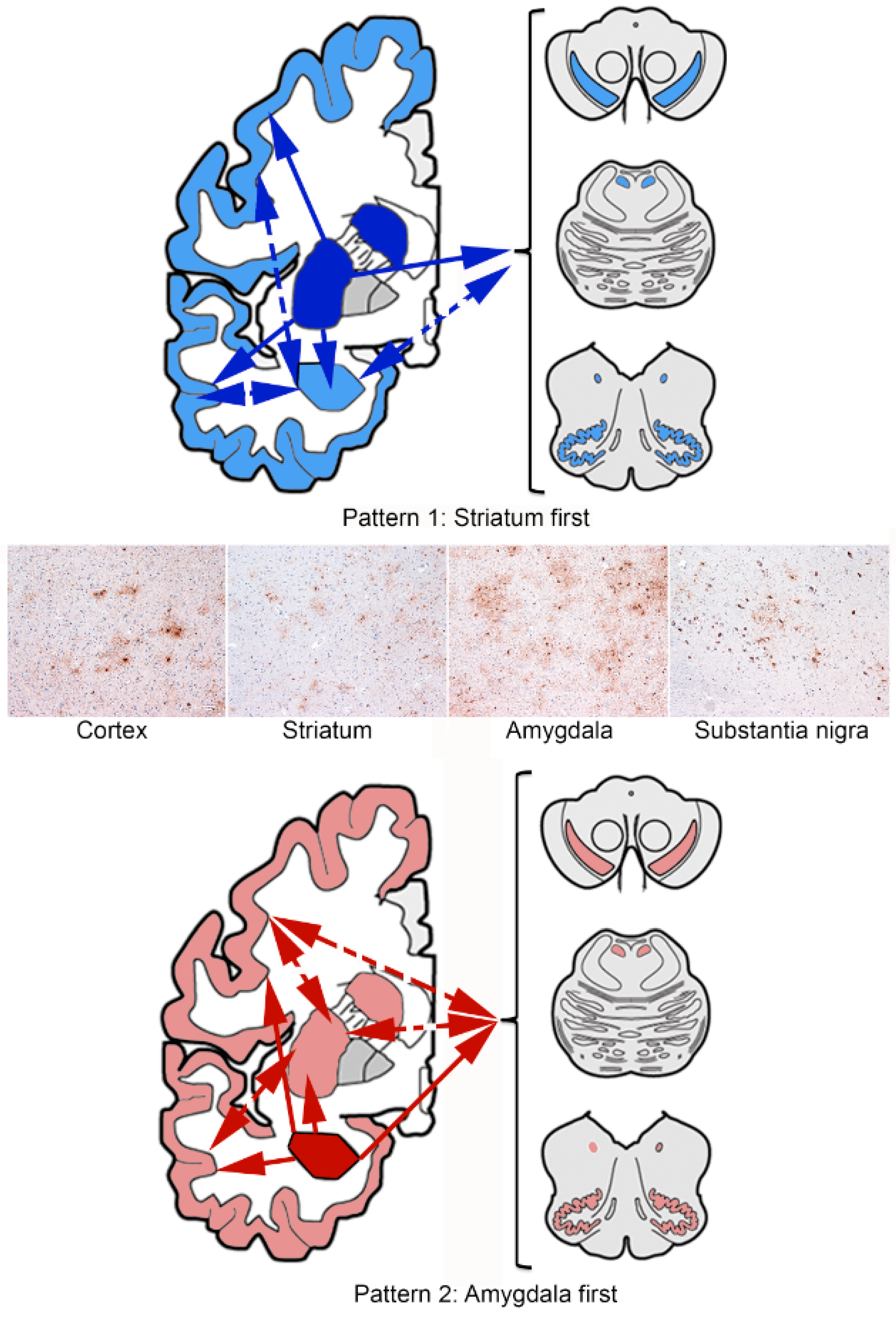
Pattern 1, Figure 2 (striatum first): The striatal pathway (stage 1) proceeds either towards the amygdala (stage 2a), cortex (stage 2b), or rarely to the brainstem (stage 2c), followed by stage 3a (striatum + amygdala + cortex), or stage 3b (striatum + amygdala + brainstem), and eventually involves all regions (stage 4). The constellation of striatum + cortex + brainstem has not been observed, hence there is no stage 3c.
Pattern 2, Figure 2 (amygdala first): The amygdala (stage 1) precedes the involvement of the striatum (stage 2a), the cortex (stage 2b) or the brainstem (stage 2c). This is followed by three combinations of stage 3 (a: amygdala + striatum + cortex; b: amygdala + striatum + brainstem; c: amygdala +cortex + brainstem) and is eventually followed by the involvement of all regions (stage 4).
A sequential pattern of astrocytic tau pathology can be better recognized for CBD and PSP [31]. This included the combined evaluation of both GFAs and astrocytic plaques CBD, and GFAs and tufted astrocytes (PSP). In CBD a four-stage sequence was proposed: frontal (including premotor) and parietal cortex (stage 1) is followed by temporal and occipital cortex (stage 2), with parallel movement into subcortical areas, including either, or both, the striatum and the amygdala (stage 3), followed by the brainstem (stage 4) including the substantia nigra followed by the pons and medulla oblongata. In PSP striatum (stage 1) to cortical (frontal-parietal to temporal to occipital) areas (stage 2a and b, respectively) to the amygdala (stage 3) and to the brainstem (stage 4), including the substantia nigra followed by the pons and medulla oblongata, sequence was recognized. Interestingly, the striatal pattern as summarized above for gray matter ARTAG is reminiscent of the combined pattern of tufted astrocytes and GFAs seen in PSP. Therefore, theoretically some cases with gray matter ARTAG in these regions could represent a preclinical form of PSP. Some of these cases might even not proceed to the full-blown neuropathological phenotype either due to a yet unidentified host response that does not allow this, or due to the presence of another predominating neurodegenerative condition.
That study addressed also whether in the same region any type of ARTAG precedes another type or neuronal tau pathology [31]. It has been suggested that in the amygdala, subpial, white matter, and perivascular areas, ARTAG appear together and precede the appearance of subependymal ARTAG. On the other hand, gray matter ARTAG is independent from these. Interestingly, based on the conditional probability values, gray matter ARTAG might precede the presence of dendritic tau-positive grains. This observation would be in line with those showing that in certain regions astrocytic tau pathology may come before neuronal tau pathology (see below) [14,31,34].

5. Considerations on Pathogenesis

Historical studies have identified neuroglia as highly important for barrier function [35]. Importantly, specific types of ARTAG tend to develop at interface regions. Interestingly, tufted astrocytes in PSP and astrocytic plaques in CBD are also often located near the blood vessels [36]; moreover, in a familial disorder with astrocyte-predominant tauopathy, perivascular accumulations are noted as well [37]. Furthermore, tau-containing astrocytes do not always match the distribution of tau-containing neurons in tauopathies [9].
A recent study evaluated the astrocytic markers connexin-43 (Cx43) and aquaporin-4 (AQP4) in relation to ARTAG [38]. A dramatic increase of Cx43 density of immunoreactivity was seen in ARTAG cases and types correlating strongly with tau positive astrocytes, irrespective of the presence of neuronal tau pathology or reactive gliosis measured by glial fibrillar acidic protein (GFAP) density. This could suggest a response to blood-brain barrier dysfunction. However, since this was seen also in the gray matter, it might be that Cx43 expression may promote neuronal survival, for example, by sensing and reducing elevated levels of extracellular glutamate. Therefore, it can be theorized that gray matter ARTAG reflects the efforts of astroglia perceiving early neurodegeneration and leading to tau accumulation in astrocytes as a response of an overwhelming pathogenic process. On the other hand, ARTAG can reflect effective take-up of locally produced and released neuronal tau, thus preventing its accumulation in neurons. Indeed, astrocytes have been found to highly express an array of phagocytic receptors and can phagocytize synapses [39] or axonal mitochondria [40] in the brain.
Aquaporin-4 density of immunoreactivity was increased only in the white and gray matter, and was associated with increased ARTAG density only in white matter and perivascular areas [38]. Aquaporin-4 is a member of the water-channel proteins expressed in the foot processes of glial cells surrounding capillaries, and it is associated with water transfer into and out of the brain parenchyma [41]. Thus, the presence of ARTAG associated with increased AQP4 density in the white matter further supports the notion that pathogenic events are associated with the blood-brain barrier.
And what can we learn about pathogenesis from the sequential distribution patterns? In two studies we reported that GFA-like morphologies appear in cortical areas without local neuronal tau pathology and without obvious clinical symptoms related to this region in primary tauopathies [14,31]. Indeed, this may reach up to 30% of PSP and PiD cases in the occipital lobe [31], which is usually less affected by neuronal tau pathology. The concept that astroglial pathology precedes neuronal tau pathology has also been discussed in presymptomatic cases showing CBD–type pathology [34]. Thus, these tau positive astrocytes might phagocytize pathological tau derived from the endings of projecting neurons, or this may simply represent local astroglial upregulation of tau as a response to a yet unidentified event. We can speculate that GFA astrocytes have the role of scouts in regions not yet affected by neuronal tau pathology. These concepts seem to be supported by animal inoculation studies as well. By injecting pathological tau extracted from post-mortem brains of AD, PSP, and CBD patients into different brain regions of non-transgenic mice, differences in tau strain potency between disorders have been identified [42]. This study found a significant inverse correlation between neuronal and astrocytic tau pathology, supporting the notion of transmission of pathological tau seeding from neurons to neighboring astrocytes. As an alternative mechanism, they proposed that astrocytic tau pathology might spread from one astrocyte to another, possibly through astrocytic gap junction networks [42]. A recent study using tau-enriched fractions of brain homogenates from pure ARTAG (with no associated tauopathy) inoculated into wild-type mice generated intracytoplasmic hyper-phosphorylated tau inclusions in astrocytes, oligodendrocytes and neurons [43]. It has been proposed that ARTAG-related tau might have a cardinal role in seeding tau to neurons and glial cells [43]. Further aspects are highlighted by observations in a tau transgenic mouse model of astrocytic tau pathologies, suggesting its contribution to glial degeneration [44]. As a functional consequence of astrocytic tau pathology, neuronal degeneration can occur in the absence of neuronal tau inclusions [45].
Does the sequential pattern always mean cell-cell-spreading of tau pathology? It might be feasible for gray matter ARTAG, although this needs to be clarified. For subpial, subependymal, white matter, and perivascular ARTAG, however, sequential involvement of regions might reflect consequences of a permanent (or repeated) pathogenic process. For example, subpial ARTAG initiated in basal regions proceeding towards the convexity of the brain (lobar areas), or dorsolateral parts of the brainstem, might indicate a pathogenesis related to the circulation of the cerebrospinal fluid [31]. In contrast, the existence of a second pattern of subpial ARTAG initiated in the dorsolateral lobar areas and dorsolateral parts of the brainstem, suggests a local mechanical inducing factor such as the role of mild traumatic brain injury in some cases [31].

6. What Is the Clinical Relevance of ARTAG?

To understand this, it is crucial to recognize the different ARTAG types. The possibility that TSA may have clinical significance was first discussed by Munoz and colleagues [46]. In a cohort of patients with a non-fluent variant of primary progressive aphasia associated with AD pathology, they detected “argyrophilic thorny astrocyte clusters (ATACs)” and observed them in the frontal, temporal, and parietal cortices and in subcortical white matter in [46]. Further reports also linked TSAs to symptomatology; however, not all found an association between ATACs and focal syndromes [47,48]. Recent studies however, have shown that white matter ARTAG in lobar regions is frequently associated with AD-related pathology [14]. This suggests that a subset of AD cases have additional pathogenic components; for example, hypoperfusion in the white matter, which can eventually be associated with focal symptoms. These concepts merit further confirmation.
A peculiar constellation of tau pathology was reported in elderly patients with dementia with or without parkinsonism [11]. Diffuse granular tau immunoreactivity in astrocytic processes (retrospectively these could be called GFAs) was described as the most characteristic feature [11]. The study emphasized additional neuronal pathologies, including threads and diffuse neuronal cytoplasmic tau immunoreactivity (pretangle-like). A subsequent study found these pathologies and suggested four different patterns based on the anatomical distribution of the tau astrogliopathy and its combination with neuronal tau pathology [49]: (1) medial temporal lobe type; (2) amygdala type; (3) limbic-basal ganglia-nigral type with neuronal tauopathy; and (4) hippocampus-dentate gyrus-amygdala type with neuronal tauopathy. It has been suspected that these might represent stages of the same process whereas other might be distinct conditions. Accumulation of TSAs in the dentate gyrus of the hippocampus were recognized by others as well [49,50]. Mathematical modeling of hippocampal tau immunolabeling patterns suggested that some forms of tau astrogliopathy in the elderly involve hippocampal subregions in a different pattern from that of primary tauopathies [51].
A recent study highlighted an interesting aspect of ARTAG. A study on individuals 90 years or older found an association with cortical but not limbic or brainstem ARTAG, independent of AD pathology, with cognitive decline [52]. Thus, the non-AD dementia group showed more hippocampal sclerosis, cortical ARTAG, TDP-43 and Lewy body pathology, while the cognitive resilient group had less of these [52]. Moreover, the authors found that cortical ARTAG independent of both limbic and brainstem ARTAGs is very rare (4%, 7/185). They speculated on an outward spread of ARTAG from limbic to the brainstem areas and then to the neocortical areas, and that neuronal tau pathology and astrocytic tau pathology are related in the oldest-old [52].
In summary, ARTAG most likely reflects the various impacts that individuals suffer during life, be it barrier dysfunction, mechanical impact, perfusion disturbance or a yet unidentified neurodegenerative event including propagation of pathological tau. Depending on the type and location of ARTAG it might be a sign of a reduced threshold that might lead to, or be associated with, decompensation of cognitive functions. And, especially when combined with other pathologies, perhaps with different pathogenesis, an additive effect might be seen, and individuals reach this threshold for cognitive decompensation more easily.

7. Perspectives

The accumulation of neurodegeneration-related proteins in astrocytes is not unique for tauopathies. Therefore, the term protein astrogliopathy (PAG) has been introduced to encompass different protein accumulations in astroglia in distinct neurodegenerative conditions. This emphasizes the yet unidentified role of astrocytes in the protein pathology of neurodegenerative diseases. Indeed, the variability of astrocytes associated with specific roles is being recognized [53]. Markers are developed [9,54,55,56] that still need to be linked to the involvement of specific glial cell populations affected by protein pathology. Eventually, these markers can be translated into bodily fluid biomarkers or probes for neuroimaging. These will help to understand the dynamics of astrocytic responses in various neurodegenerative conditions. The role of astrocytes in the processing and propagation, and the exact cytopathological mechanism of neurodegeneration-related proteins, are still not understood across the full spectrum. All these aspects position astrocytes in the center of current research on neurodegenerative conditions. Harmonizing the nomenclature of astrocytic tau pathologies leading to the definition of ARTAG enhanced these studies and further motivated researchers.

Funding

This research received no external funding.

Conflicts of Interest

The author declares no conflict of interest.

References

  1. Kovacs, G.G.; Ferrer, I.; Grinberg, L.T.; Alafuzoff, I.; Attems, J.; Budka, H.; Cairns, N.J.; Crary, J.F.; Duyckaerts, C.; Ghetti, B.; et al. Aging-related tau astrogliopathy (ARTAG): Harmonized evaluation strategy. Acta Neuropathol. 2016, 131, 87–102. [Google Scholar] [CrossRef] [PubMed]
  2. Ikeda, K. Glial fibrillary tangles and argyrophilic threads: Classification and disease specificity. Neuropathology 1996, 16, 71–77. [Google Scholar] [CrossRef]
  3. Ikeda, K.; Akiyama, H.; Arai, T.; Nishimura, T. Glial tau pathology in neurodegenerative diseases: Their nature and comparison with neuronal tangles. Neurobiol. Aging 1998, 19, S85–S91. [Google Scholar] [CrossRef]
  4. Ikeda, K.; Akiyama, H.; Kondo, H.; Haga, C.; Tanno, E.; Tokuda, T.; Ikeda, S. Thorn-shaped astrocytes: Possibly secondarily induced tau-positive glial fibrillary tangles. Acta Neuropathol. 1995, 90, 620–625. [Google Scholar] [CrossRef] [PubMed]
  5. Schultz, C.; Ghebremedhin, E.; Del Tredici, K.; Rub, U.; Braak, H. High prevalence of thorn-shaped astrocytes in the aged human medial temporal lobe. Neurobiol. Aging 2004, 25, 397–405. [Google Scholar] [CrossRef]
  6. Schultz, C.; Dehghani, F.; Hubbard, G.B.; Thal, D.R.; Struckhoff, G.; Braak, E.; Braak, H. Filamentous tau pathology in nerve cells, astrocytes, and oligodendrocytes of aged baboons. J. Neuropathol. Exp. Neurol. 2000, 59, 39–52. [Google Scholar] [CrossRef] [PubMed]
  7. Schultz, C.; Hubbard, G.B.; Rub, U.; Braak, E.; Braak, H. Age-related progression of tau pathology in brains of baboons. Neurobiol. Aging 2000, 21, 905–912. [Google Scholar] [CrossRef]
  8. Perez, S.E.; Raghanti, M.A.; Hof, P.R.; Kramer, L.; Ikonomovic, M.D.; Lacor, P.N.; Erwin, J.M.; Sherwood, C.C.; Mufson, E.J. Alzheimer’s disease pathology in the neocortex and hippocampus of the western lowland gorilla (Gorilla gorilla gorilla). J. Comp. Neurol. 2013, 521, 4318–4338. [Google Scholar] [CrossRef] [PubMed]
  9. Ferrer, I. Astrogliopathy in Tauopathies. Neuroglia 2018, 1, 126–150. [Google Scholar] [CrossRef]
  10. Holzer, M.; Craxton, M.; Jakes, R.; Arendt, T.; Goedert, M. Tau gene (MAPT) sequence variation among primates. Gene 2004, 341, 313–322. [Google Scholar] [CrossRef] [PubMed]
  11. Kovacs, G.G.; Molnar, K.; Laszlo, L.; Strobel, T.; Botond, G.; Honigschnabl, S.; Reiner-Concin, A.; Palkovits, M.; Fischer, P.; Budka, H. A peculiar constellation of tau pathology defines a subset of dementia in the elderly. Acta Neuropathol. 2011, 122, 205–222. [Google Scholar] [CrossRef] [PubMed]
  12. Kovacs, G.G.; Xie, S.X.; Lee, E.B.; Robinson, J.L.; Caswell, C.; Irwin, D.J.; Toledo, J.B.; Johnson, V.E.; Smith, D.H.; Alafuzoff, I.; et al. Multisite Assessment of Aging-Related Tau Astrogliopathy (ARTAG). J. Neuropathol. Exp. Neurol. 2017, 76, 605–619. [Google Scholar] [CrossRef] [PubMed] [Green Version]
  13. Nishimura, M.; Namba, Y.; Ikeda, K.; Oda, M. Glial fibrillary tangles with straight tubules in the brains of patients with progressive supranuclear palsy. Neurosci. Lett. 1992, 143, 35–38. [Google Scholar] [CrossRef]
  14. Kovacs, G.G.; Robinson, J.L.; Xie, S.X.; Lee, E.B.; Grossman, M.; Wolk, D.A.; Irwin, D.J.; Weintraub, D.; Kim, C.F.; Schuck, T.; et al. Evaluating the Patterns of Aging-Related Tau Astrogliopathy Unravels Novel Insights into Brain Aging and Neurodegenerative Diseases. J. Neuropathol. Exp. Neurol. 2017, 76, 270–288. [Google Scholar] [CrossRef] [PubMed]
  15. Kovacs, G.G. Invited review: Neuropathology of tauopathies: Principles and practice. Neuropathol. Appl. Neurobiol. 2015, 41, 3–23. [Google Scholar] [CrossRef] [PubMed]
  16. Botez, G.; Probst, A.; Ipsen, S.; Tolnay, M. Astrocytes expressing hyperphosphorylated tau protein without glial fibrillary tangles in argyrophilic grain disease. Acta Neuropathol. 1999, 98, 251–256. [Google Scholar] [CrossRef] [PubMed]
  17. Tolnay, M.; Clavaguera, F. Argyrophilic grain disease: A late-onset dementia with distinctive features among tauopathies. Neuropathology 2004, 24, 269–283. [Google Scholar] [CrossRef] [PubMed] [Green Version]
  18. Crary, J.F.; Trojanowski, J.Q.; Schneider, J.A.; Abisambra, J.F.; Abner, E.L.; Alafuzoff, I.; Arnold, S.E.; Attems, J.; Beach, T.G.; Bigio, E.H.; et al. Primary age-related tauopathy (PART): A common pathology associated with human aging. Acta Neuropathol. 2014, 128, 755–766. [Google Scholar] [CrossRef] [PubMed]
  19. Ghetti, B.; Oblak, A.L.; Boeve, B.F.; Johnson, K.A.; Dickerson, B.C.; Goedert, M. Invited review: Frontotemporal dementia caused by microtubule-associated protein tau gene (MAPT) mutations: A chameleon for neuropathology and neuroimaging. Neuropathol. Appl. Neurobiol. 2015, 41, 24–46. [Google Scholar] [CrossRef] [PubMed] [Green Version]
  20. Kovacs, G.G.; Lee, V.M.; Trojanowski, J. Protein astrogliopathies in human neurodegenerative diseases and aging. Brain Pathol. 2017, 27, 675–690. [Google Scholar] [CrossRef] [PubMed]
  21. Alexander, J.; Kalev, O.; Mehrabian, S.; Traykov, L.; Raycheva, M.; Kanakis, D.; Drineas, P.; Lutz, M.I.; Strobel, T.; Penz, T.; et al. Familial early-onset dementia with complex neuropathologic phenotype and genomic background. Neurobiol. Aging 2016, 42, 199–204. [Google Scholar] [CrossRef] [PubMed]
  22. Le Guennec, K.; Quenez, O.; Nicolas, G.; Wallon, D.; Rousseau, S.; Richard, A.C.; Alexander, J.; Paschou, P.; Charbonnier, C.; Bellenguez, C.; et al. 17q21.31 duplication causes prominent tau-related dementia with increased MAPT expression. Mol. Psychiatry 2017, 22, 1119–1125. [Google Scholar] [CrossRef] [PubMed]
  23. Ikeda, C.; Yokota, O.; Nagao, S.; Ishizu, H.; Oshima, E.; Hasegawa, M.; Okahisa, Y.; Terada, S.; Yamada, N. The Relationship Between Development of Neuronal and Astrocytic Tau Pathologies in Subcortical Nuclei and Progression of Argyrophilic Grain Disease. Brain Pathol. 2016, 26, 488–505. [Google Scholar] [CrossRef] [PubMed]
  24. Ferrer, I.; Lopez-Gonzalez, I.; Carmona, M.; Arregui, L.; Dalfo, E.; Torrejon-Escribano, B.; Diehl, R.; Kovacs, G.G. Glial and neuronal tau pathology in tauopathies: Characterization of disease-specific phenotypes and tau pathology progression. J. Neuropathol. Exp. Neurol. 2014, 73, 81–97. [Google Scholar] [CrossRef] [PubMed]
  25. Lopez-Gonzalez, I.; Carmona, M.; Blanco, R.; Luna-Munoz, J.; Martinez-Mandonado, A.; Mena, R.; Ferrer, I. Characterization of thorn-shaped astrocytes in white matter of temporal lobe in Alzheimer’s disease brains. Brain Pathol. 2013, 23, 144–153. [Google Scholar] [CrossRef] [PubMed]
  26. McKee, A.C.; Cairns, N.J.; Dickson, D.W.; Folkerth, R.D.; Keene, C.D.; Litvan, I.; Perl, D.P.; Stein, T.D.; Vonsattel, J.P.; Stewart, W.; et al. The first NINDS/NIBIB consensus meeting to define neuropathological criteria for the diagnosis of chronic traumatic encephalopathy. Acta Neuropathol. 2016, 131, 75–86. [Google Scholar] [CrossRef] [PubMed]
  27. McKee, A.C.; Daneshvar, D.H. The neuropathology of traumatic brain injury. Handb. Clin. Neurol. 2015, 127, 45–66. [Google Scholar] [PubMed] [Green Version]
  28. Liu, A.K.; Goldfinger, M.H.; Questari, H.E.; Pearce, R.K.; Gentleman, S.M. ARTAG in the basal forebrain: Widening the constellation of astrocytic tau pathology. Acta Neuropathol. Commun. 2016, 4, 59. [Google Scholar] [CrossRef] [PubMed]
  29. McKee, A.C.; Stein, T.D.; Kiernan, P.T.; Alvarez, V.E. The neuropathology of chronic traumatic encephalopathy. Brain Pathol. 2015, 25, 350–364. [Google Scholar] [CrossRef] [PubMed]
  30. McKee, A.C.; Stern, R.A.; Nowinski, C.J.; Stein, T.D.; Alvarez, V.E.; Daneshvar, D.H.; Lee, H.S.; Wojtowicz, S.M.; Hall, G.; Baugh, C.M.; et al. The spectrum of disease in chronic traumatic encephalopathy. Brain 2013, 136, 43–64. [Google Scholar] [CrossRef] [PubMed]
  31. Kovacs, G.G.; Xie, S.X.; Robinson, J.L.; Lee, E.B.; Smith, D.H.; Schuck, T.; Lee, V.M.; Trojanowski, J.Q. Sequential stages and distribution patterns of aging-related tau astrogliopathy (ARTAG) in the human brain. Acta Neuropathol. Commun. 2018, 6, 50. [Google Scholar] [CrossRef] [PubMed] [Green Version]
  32. Kovacs, G.G.; Rahimi, J.; Strobel, T.; Lutz, M.I.; Regelsberger, G.; Streichenberger, N.; Perret-Liaudet, A.; Hoftberger, R.; Liberski, P.P.; Budka, H.; et al. Tau pathology in Creutzfeldt-Jakob disease revisited. Brain Pathol. 2017, 27, 332–344. [Google Scholar] [CrossRef] [PubMed]
  33. Brettschneider, J.; Del Tredici, K.; Lee, V.M.; Trojanowski, J.Q. Spreading of pathology in neurodegenerative diseases: A focus on human studies. Nat. Rev. Neurosci. 2015, 16, 109–120. [Google Scholar] [CrossRef] [PubMed]
  34. Ling, H.; Kovacs, G.G.; Vonsattel, J.P.; Davey, K.; Mok, K.Y.; Hardy, J.; Morris, H.R.; Warner, T.T.; Holton, J.L.; Revesz, T. Astrogliopathy predominates the earliest stage of corticobasal degeneration pathology. Brain 2016, 139, 3237–3252. [Google Scholar] [CrossRef] [PubMed] [Green Version]
  35. Chvátal, A.; Verkhratsky, A. An Early History of Neuroglial Research: Personalities. Neuroglia 2018, 1, 245–281. [Google Scholar] [CrossRef]
  36. Shibuya, K.; Yagishita, S.; Nakamura, A.; Uchihara, T. Perivascular orientation of astrocytic plaques and tuft-shaped astrocytes. Brain Res. 2011, 1404, 50–54. [Google Scholar] [CrossRef] [PubMed]
  37. Ferrer, I.; Legati, A.; Garcia-Monco, J.C.; Gomez-Beldarrain, M.; Carmona, M.; Blanco, R.; Seeley, W.W.; Coppola, G. Familial behavioral variant frontotemporal dementia associated with astrocyte-predominant tauopathy. J. Neuropathol. Exp. Neurol. 2015, 74, 370–379. [Google Scholar] [CrossRef] [PubMed]
  38. Kovacs, G.G.; Yousef, A.; Kaindl, S.; Lee, V.M.; Trojanowski, J.Q. Connexin-43 and Aquaporin-4 are markers of ARTAG-related astroglial response. Neuropathol. Appl. Neurobiol. 2017. [Google Scholar] [CrossRef]
  39. Chung, W.S.; Clarke, L.E.; Wang, G.X.; Stafford, B.K.; Sher, A.; Chakraborty, C.; Joung, J.; Foo, L.C.; Thompson, A.; Chen, C.; et al. Astrocytes mediate synapse elimination through MEGF10 and MERTK pathways. Nature 2013, 504, 394–400. [Google Scholar] [CrossRef] [PubMed] [Green Version]
  40. Davis, C.H.; Kim, K.Y.; Bushong, E.A.; Mills, E.A.; Boassa, D.; Shih, T.; Kinebuchi, M.; Phan, S.; Zhou, Y.; Bihlmeyer, N.A.; et al. Transcellular degradation of axonal mitochondria. Proc. Natl. Acad. Sci. USA 2014, 111, 9633–9638. [Google Scholar] [CrossRef] [PubMed] [Green Version]
  41. Tang, G.; Yang, G.Y. Aquaporin-4: A Potential Therapeutic Target for Cerebral Edema. Int. J. Mol. Sci. 2016, 17, 1413. [Google Scholar] [CrossRef] [PubMed]
  42. Narasimhan, S.; Guo, J.L.; Changolkar, L.; Stieber, A.; McBride, J.D.; Silva, L.V.; He, Z.; Zhang, B.; Gathagan, R.J.; Trojanowski, J.Q.; et al. Pathological Tau Strains from Human Brains Recapitulate the Diversity of Tauopathies in Nontransgenic Mouse Brain. J. Neurosci. 2017, 37, 11406–11423. [Google Scholar] [CrossRef] [PubMed] [Green Version]
  43. Ferrer, I.; Garcia, M.A.; Gonzalez, I.L.; Lucena, D.D.; Villalonga, A.R.; Tech, M.C.; Llorens, F.; Garcia-Esparcia, P.; Martinez-Maldonado, A.; Mendez, M.F.; et al. Aging-related tau astrogliopathy (ARTAG): Not only tau phosphorylation in astrocytes. Brain Pathol. 2018. [Google Scholar] [CrossRef] [PubMed]
  44. Higuchi, M.; Ishihara, T.; Zhang, B.; Hong, M.; Andreadis, A.; Trojanowski, J.; Lee, V.M. Transgenic mouse model of tauopathies with glial pathology and nervous system degeneration. Neuron 2002, 35, 433–446. [Google Scholar] [CrossRef]
  45. Forman, M.S.; Lal, D.; Zhang, B.; Dabir, D.V.; Swanson, E.; Lee, V.M.; Trojanowski, J.Q. Transgenic mouse model of tau pathology in astrocytes leading to nervous system degeneration. J. Neurosci. 2005, 25, 3539–3550. [Google Scholar] [CrossRef] [PubMed]
  46. Munoz, D.G.; Woulfe, J.; Kertesz, A. Argyrophilic thorny astrocyte clusters in association with Alzheimer’s disease pathology in possible primary progressive aphasia. Acta Neuropathol. 2007, 114, 347–357. [Google Scholar] [CrossRef] [PubMed] [Green Version]
  47. Bigio, E.H.; Mishra, M.; Hatanpaa, K.J.; White, C.L., 3rd; Johnson, N.; Rademaker, A.; Weitner, B.B.; Deng, H.X.; Dubner, S.D.; Weintraub, S.; et al. TDP-43 pathology in primary progressive aphasia and frontotemporal dementia with pathologic Alzheimer disease. Acta Neuropathol. 2010, 120, 43–54. [Google Scholar] [CrossRef] [PubMed] [Green Version]
  48. Mesulam, M.; Wicklund, A.; Johnson, N.; Rogalski, E.; Leger, G.C.; Rademaker, A.; Weintraub, S.; Bigio, E.H. Alzheimer and frontotemporal pathology in subsets of primary progressive aphasia. Ann. Neurol. 2008, 63, 709–719. [Google Scholar] [CrossRef] [PubMed] [Green Version]
  49. Kovacs, G.G.; Milenkovic, I.; Wohrer, A.; Hoftberger, R.; Gelpi, E.; Haberler, C.; Honigschnabl, S.; Reiner-Concin, A.; Heinzl, H.; Jungwirth, S.; et al. Non-Alzheimer neurodegenerative pathologies and their combinations are more frequent than commonly believed in the elderly brain: A community-based autopsy series. Acta Neuropathol. 2013, 126, 365–384. [Google Scholar] [CrossRef] [PubMed]
  50. Lace, G.; Ince, P.G.; Brayne, C.; Savva, G.M.; Matthews, F.E.; de Silva, R.; Simpson, J.E.; Wharton, S.B. Mesial temporal astrocyte tau pathology in the MRC-CFAS ageing brain cohort. Dement. Geriatr. Cogn. Disord. 2012, 34, 15–24. [Google Scholar] [CrossRef] [PubMed]
  51. Milenkovic, I.; Petrov, T.; Kovacs, G.G. Patterns of hippocampal tau pathology differentiate neurodegenerative dementias. Dement. Geriatr. Cogn. Disord. 2014, 38, 375–388. [Google Scholar] [CrossRef] [PubMed]
  52. Robinson, J.L.; Corrada, M.M.; Kovacs, G.G.; Dominique, M.; Caswell, C.; Xie, S.X.; Lee, V.M.; Kawas, C.H.; Trojanowski, J.Q. Non-Alzheimer’s contributions to dementia and cognitive resilience in the 90+ Study. Acta Neuropathol. 2018. [Google Scholar] [CrossRef] [PubMed]
  53. Verkhratsky, A.; Oberheim Bush, N.A.; Nedergaard, M.; Butt, A. The Special Case of Human Astrocytes. Neuroglia 2018, 1, 21–29. [Google Scholar] [CrossRef] [Green Version]
  54. Herculano-Houzel, S.; Dos Santos, S.E. You Do Not Mess with the Glia. Neuroglia 2018, 1, 193–219. [Google Scholar] [CrossRef]
  55. Ferrer, I. Diversity of astroglial responses across human neurodegenerative disorders and brain aging. Brain Pathol. 2017, 27, 645–674. [Google Scholar] [CrossRef] [PubMed]
  56. Verkhratsky, A.; Zorec, R.; Parpura, V. Stratification of astrocytes in healthy and diseased brain. Brain Pathol. 2017, 27, 629–644. [Google Scholar] [CrossRef] [PubMed]
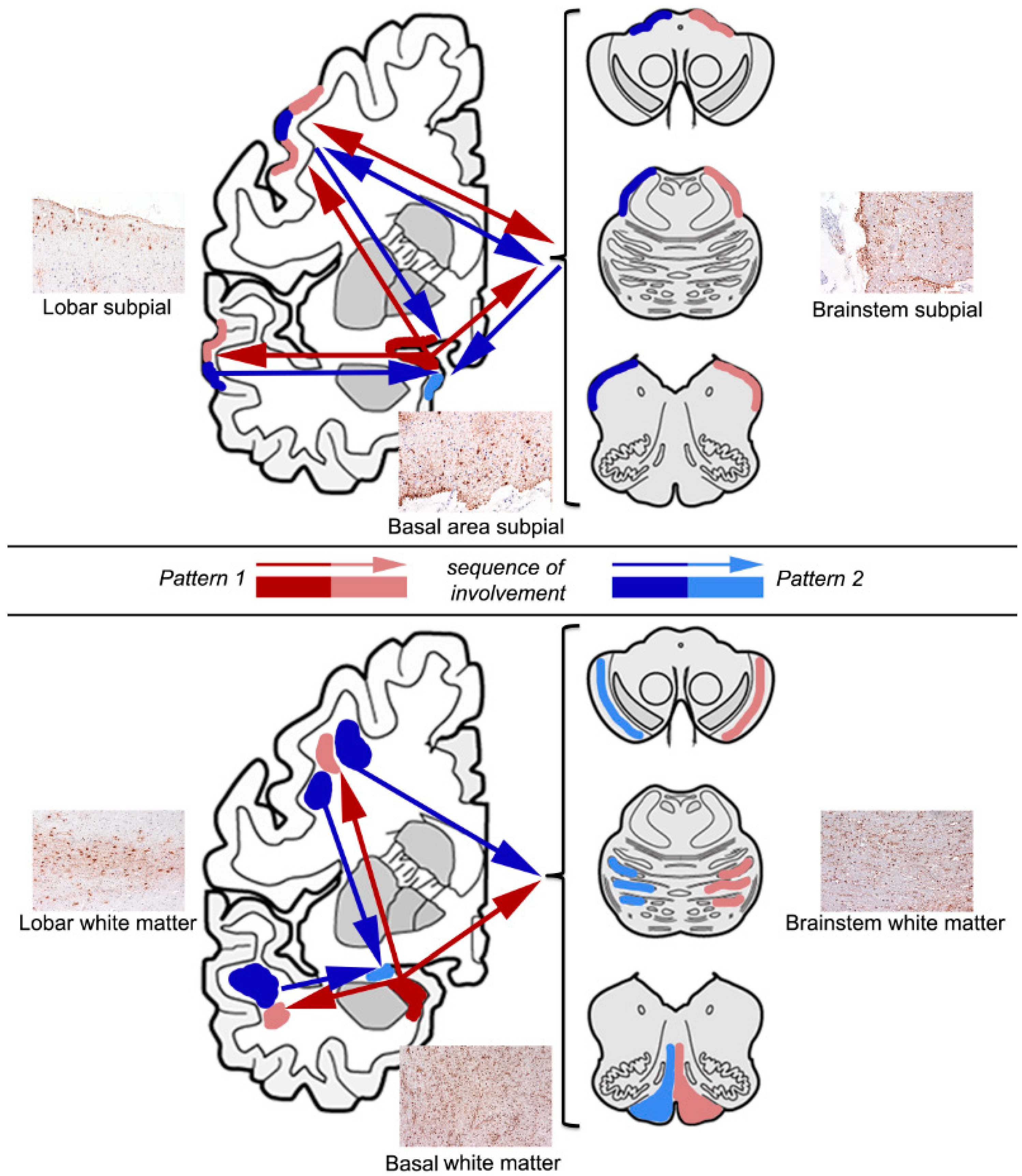
Figure 1. Representative images and sequential distribution patterns of subpial (upper panel) and white matter (lower panel) types of aging-related tau astrogliopathy (ARTAG) in the human brain. For both, two major patterns are seen; one beginning in the basal brain areas, in particular the amygdala (indicated by red arrows), and a second initiated in lobar areas and/or the brainstem (indicated by blue arrows). The arrowheads point towards the direction of sequential involvement and a deeper color represents an earlier stage of involvement. A double-headed arrow means both regions can be involved together as a specific stage of sequential involvement. The deeper color represents the first stage.
Neuroglia 01 00023 g001
Figure 2. Representative images and sequential distribution patterns of gray matter ARTAG in the human brain. One of these begins in the striatum (upper panel), and another in the amygdala (lower panel), followed by the cortical and brainstem regions. A double-headed arrow with a dashed line means both regions can be involved together as a specific stage of sequential involvement. The deeper color represents the first stage.
Neuroglia 01 00023 g002