Perspective

Interlaminar Glia and Other Glial Themes Revisited: Pending Answers Following Three Decades of Glial Research

Unit of Applied Neurobiology (UNA, CEMIC-CONICET), Buenos Aires 1053, Argentina
Neuroglia 2018, 1(1), 7-20; https://doi.org/10.3390/neuroglia1010003
Submission received: 23 January 2018 / Revised: 20 February 2018 / Accepted: 22 February 2018 / Published: 1 March 2018

Abstract

:
This review aims to highlight the various significant matters in glial research stemming from personal work by the author and associates at the Unit of Applied Neurobiology (UNA, CEMIC-CONICET), and some of the pending questions. A reassessment and further comments on interlaminar astrocytes—an astroglial cell type that is specific to humans and other non-human primates, and is not found in rodents, is presented. Tentative hypothesis regarding their function and future possible research lines that could contribute to further the analysis of their development and possible role(s), are suggested. The possibility that they function as a separate entity from the “territorial” astrocytes, is also considered. In addition, the potential significance of our observations on interspecies differences in in vitro glial cell dye coupling, on glial diffusible factors affecting the induction of this glial phenotype, and on their interference with the cellular toxic effects of cerebrospinal fluid obtained from l-DOPA treated patients with Parkinson´s disease, is also considered. The major differences oberved in the cerebral cortex glial layout between human and rodents—the main model for studying glial function and pathology—calls for a careful assessment of known and potential species differences in all aspects of glial cell biology. This is essential to provide a better understanding of the organization and function of human and non-human primate brain, and of the neurobiological basis of their behavior.

1. Introduction

Following retirement from active laboratory work, a change in the experimental line of research at the Unit of Applied Neurobiology (UNA, CEMIC-CONICET) laboratories—at present aimed at studying more decisively neurocognitive issues—has provided the opportunity to propose this sort of brief account and reassessment of unresolved and pending questions on “glial issues” that were dealt with in our neurobiological laboratory in recent decades. This personal viewpoint is intended to stress and revisit several aspects of our own observations on glial physiology and comparative studies, which are aimed at encouraging further research in the field.
Thorough updates, comprehensive reviews, and inspirational thoughts on other related aspects of glial physiology can be found in Kettenmann and Ransom (2005), Verkhratsky and Butt (2013), and Verkhratsky and Nedergaard (2018) [1,2,3], as well as in numerous individual articles, for it, seems evident that experimental research on neuroglia has entered an era of further fertile analysis. Yet, as it will be considered later, caution and weighed decisions should be exerted in attempting interspecies extrapolations—in the present context referred to brain glia—and building our understanding of human and non-human primate brain organization and phyisology based on what can be considered “general mammalian” characteristics, stemming from non-comparative Rodentia approaches. This may limit our views on the neurobiological basis of primate brain evolution and behavior.

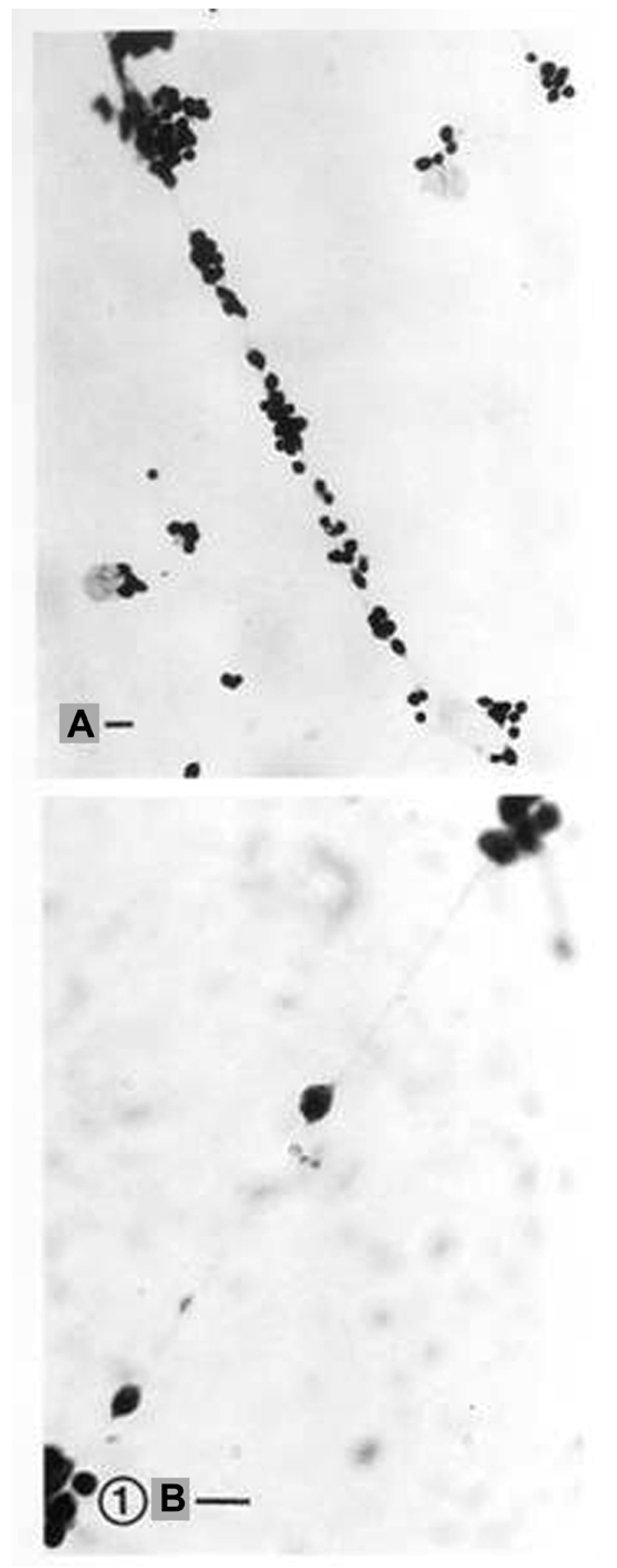
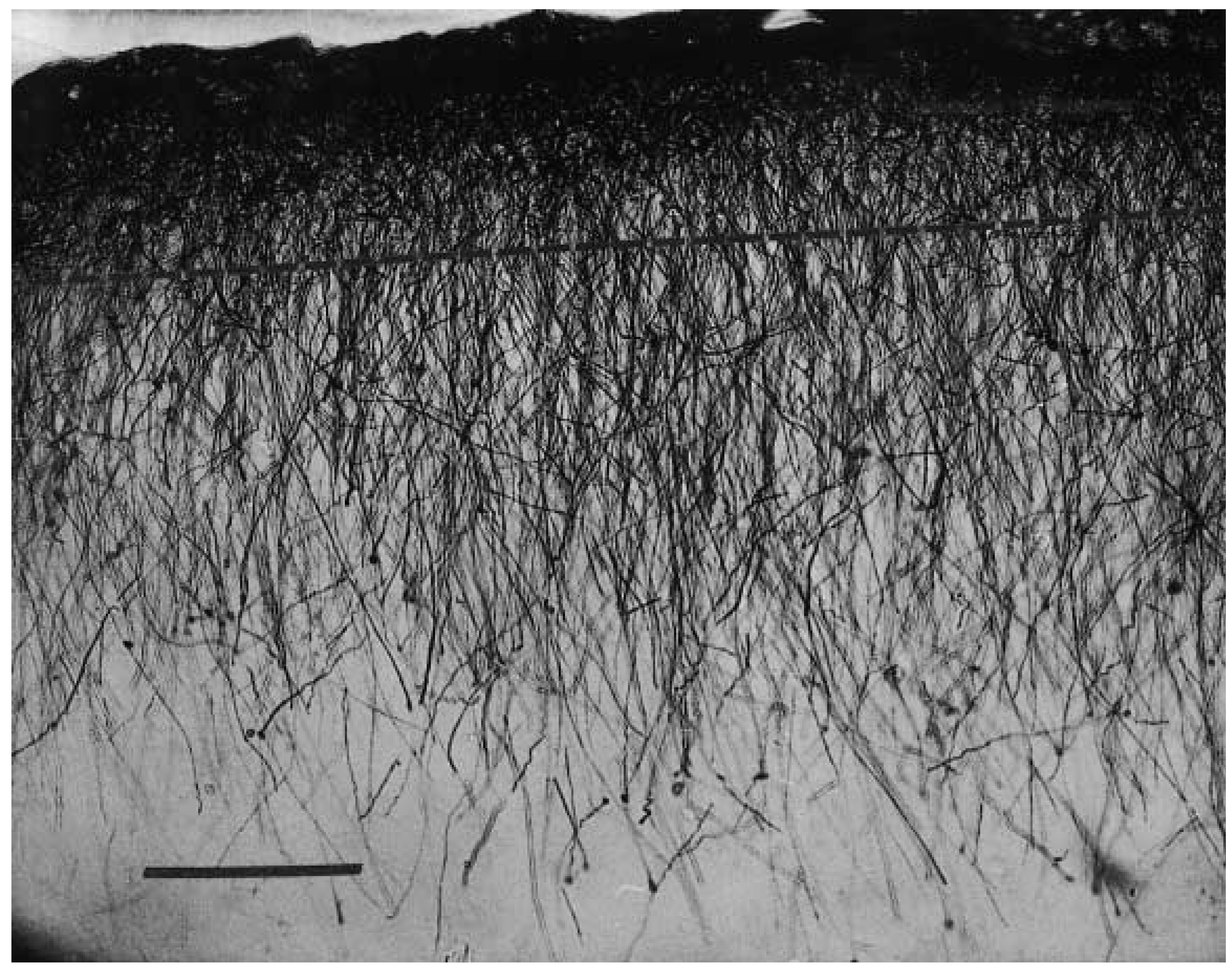
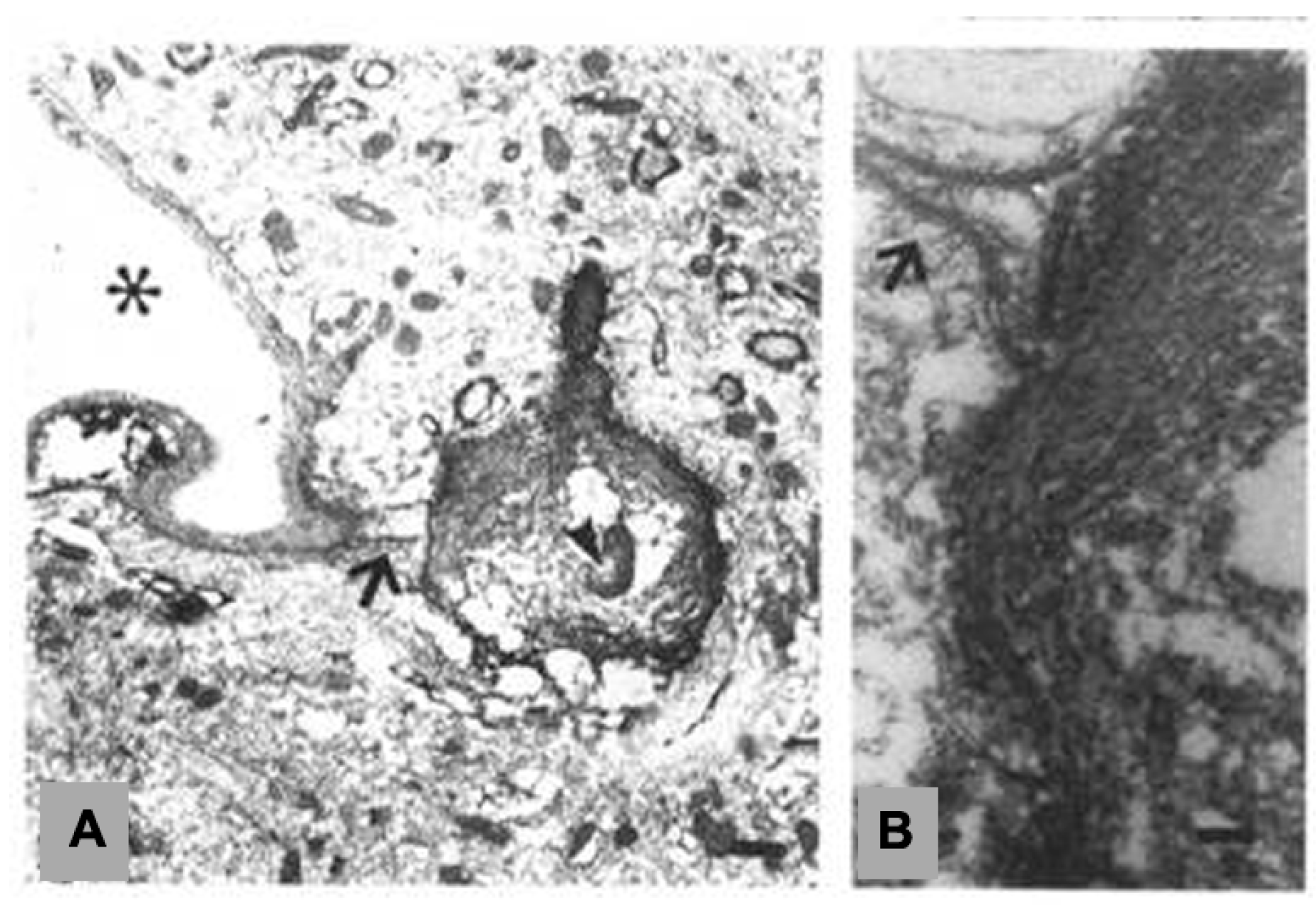
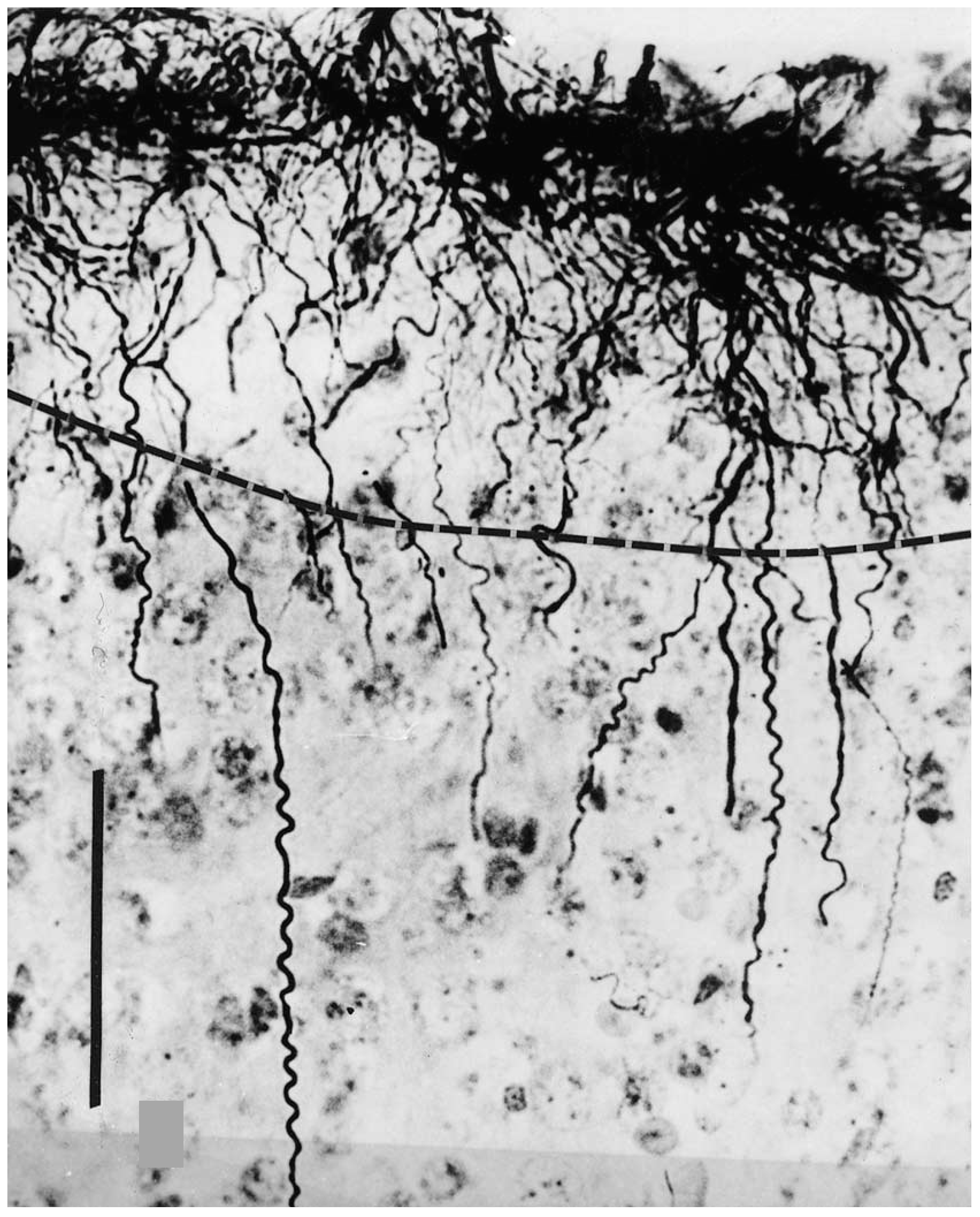
2. Interlaminar Astrocytes and Primate Brain Evolution

Interlaminar glia or interlaminar astrocytes (Figure 1) essentially represent a primate evolutionary development [4,5,6,7,8] linked to the split between prosimians and Anthropoidea. They are characterized by a cell soma placed in lamina I—in general next to the glia limitans—of the cerebral cortex, and long, descending, cell processes that could extend for about 1 mm, thus traversing more than one cortical laminae. Remaining characteristics are mentioned below.
The expression of interlaminar glia among orders and species, according to our screening of available samples and histological identification procedures, is incipient—they take form of isolated events—in the prosimian lemur (no brain samples were available at that time from galago and tarsier), it is absent in Callithricidae (marmosets and the tamarins), constant and yet variable in its palisade expression in Ceboidea (New World monkeys), and fully expressed in Cercopithecidae (Old World monkeys) and Hominoidea (great apes and humans) [4,5,6,7,8]. Their original development in primates possibly relates to point mutation or epigenesis—predating some of the factors that are hypothetically linked to the later increase in brain size (e.g., expensive tissue hypothesis of Aiello and Wheeler [10]; socio-ecological hypothesis of Dunbar [11] and other convergent hypotheses on brain size evolution in humans). The author considers that its emergence is probably associated with the less promoted—in terms of evolutionary impact on brain function—development of columnar organization of the cerebral cortex, which remains to be further comparatively explored in terms of distributed neural circuits (or modules) and of its impact on cerebral cortex information efficiency (response speed, unit assembly synchrony, cognitive fluidity). In this respect, the possibility that they contribute to a “non-territorial” management of the cerebral cortex intercellular space and intercellular interactions should be analyzed. Consequently, whether they are coupled or not to “local–territorial–astrocytes” could add to the characterization of their physiological role(s) and integration into the cerebral cortex processing.
It seems apparent that brain evolution among anthropoid primates has proceeded in a series of continuous structural and functional—neurotransmitter and receptor dynamics for once—deletions and aggregates, which were not exclusively confined to neurons (see e.g., [12]). Among them, appearance of interlaminar astrocytes would represent an evolutionary—“primate-specific”—brain cell trait added to the “general mammalian” brain glial cell family, as suggested by Colombo and colleagues [4,7,8]. Although functional insertion of interlaminar astrocytes into cerebral cortex organization remains highly speculative (see below), their ontogenetic development and some interactions and responses following experimental procedures and pathologies, provide grounds for further research inquiries and analysis.
One important obstacle to experiments aimed at advancing into the general understanding of human brain evolution and organization, resides in the limited access to extant anthropoid species for comparative research purposes—for interlaminar astrocytes are not a “general mammalian” event. In order to minimize the need for more general invasive protocols, perhaps the use of biopsic material would help to bypass such limitation, besides the possibility of novel methodological imaging approaches. Overcoming these problems is imperative, because the positive selection of interlaminar glia in the primate order calls for a full characterization and understanding of its role in cerebral cortex function.
It may be opportune to state upfront that although other laboratory species provide valuable insights into “general mammalian” brain organization and evolution, the above-mentioned limitation generates an unavoidable and objective conceptual “gap” when extrapolating results from non-primate mammals to primates. Such consideration arises since the subtle, bioelectric, ionic/molecular dynamic interactions with cellular receptors operating in a particular brain organization is what provides the finely tuned scaffolding—besides structural or hardware connectivity—for the generation and expression of the characteristic complex and fluid human cognitive and emotional (manifest or introspective) behaviors. In this regard, further advances in the comparative—neuronal and glial—analysis within the primate order—specifically with genetically close extant species—seems critically needed. Perhaps, following the historically theoretical preeminence of the “neuronal doctrine”, most studies have been performed on these cells in anthropoid species, generating a clear gap with respect to advances made on glial cells.
In such respect, although not intended to be reviewed here, numerous studies regarding associated neuronal and behavioral issues have been performed in primates by several authors. In particular, for example, some recent studies on the comparative distribution of neurotransmitter receptors in the brains of humans and extant primate species were reported by Zilles and colleagues [13,14], as well as comparative genetic studies by Pu et al. [15], Muntané et al. [16] and Mitchell and Silver [17]. Yet, the ethical limitations on the experimental use of primate species, and the needed minimization of invasive actions to be taken on them, as well as added limitations imposed on the inclusion in experimental protocols of extant species of great apes—mostly those genetically closer as Pan troglodytes and Pan paniscus (chimpanzee and bonobo, respectively)—calls for developing imaginative and minimally invasive research tools and procedures. The “handicap” of glial cells in terms of lacking readily in situ detectable bioelectric signals adds to the limitations on this field of the neurosciences.
At any rate, as pointed out by several authors (e.g., [4,12,18]), it has become unavoidable to include the spectrum of glial cells into theoretical constructions of brain evolution and organization. Certainly, this stage has been built following studies based on the access to laboratory rodent species. But time has come to work on new approaches and theoretical models to avoid known limitations to expand glial research into primates.
Developmental studies tracing the ontogenetic cellular origin of interlaminar glia remain missing. In the human brain, the developmental expression of interlaminar glia takes place during early postnatal life, following a period of “physiological astrogliosis” by 20–40 days of postnatal life, and attaining the adult-like configuration of interlaminar astrocytes by the second month of life [19]. Genetic analysis following the isolation of these cells could instigate detailed studies of their evolutionary origin and cell lineage.
The soma of interlaminar glial cells is closely apposed to the glia limitans and its short superficial processes are probably functionally associated with the subarachnoid space [9]. This layout suggests that their most superficial aspect could be linked to exchange with the pial vasculature/subarachnoid space, while their distal ending—usually a slender bulbous formation—has been shown [20] to be connected to a blood vessel (Figure 2) or “floating” in the intercellular space. Hence, a role in “fast sink” operations appears as a possibility, besides ion exchange through the membrane of its long interlaminar processes. This glial morphotype rather than engulfing synaptic terminals and intimately interacting with them as the type of layout reported by Grosche et al. [21] for parenchymal astroglia, interlaminar processes would appear to “navigate” in the extracellular space, perhaps monitoring and regulating ionic/molecular imbalances. In this regard, their membrane dynamic characteristics remain to be determined. It would be of significant interest to establish whether this type of glia is independent from the astroglial syncytia, and whether they form a parallel network that is perhaps interconnected at the subpial level. However, our attempts to analyze possible dye coupling of interlaminar glia—in samples from a non-human primate—failed due to technical reasons (previous exposure to an antifreeze medium for transport of fresh sections affected membrane permeable characteristics), and the limited number of sections to work with.
The possibility of vesicular transport as reported by Potokar et al. [22] for rodent glia should also be considered.
Attainment of final length, density and palisade display of interlaminar processes (Figure 1) depends on species and subspecies characteristics: for example, some New World monkeys could present a non-systematic, patchy and more scattered or unpredictable palisade, such as is the case of Cebus paella (tufted capuchin), while a more typical palisade occurs in Saimiri boliviensis (black-capped squirrel monkey). According to experimental data, the presence and length of interlaminar processes are significantly affected by interaction with thalamic cortical afferents, at least in areas that are related to the visual system [23] and spinal cord somatosensory input [24].
What are the signals involved in determining the characteristics of the palisade? Physiological signals driving morphological changes of interlaminar processes under the reported conditions remain undetermined. Whether effects on interlaminar glia (Figure 3 and Figure 4) represent a trophic or regulatory role of thalamic afferents, or an indirect one through their cerebral cortex projections on neuronal activity [25,26], remains an open question. The disruption of the interlaminar cortical palisade after 11–13 months following spinal cord transection—with a lack of evidence of additional astrogliosis—and a somewhat “wavy” individual process display [24], suggests a rather long-term impact on the rearrangement of the local neuropil, or that it acquired a new steady state condition following lesioning, with loss or perturbation of the original columnar arrangement, and perhaps sharing an expanded spatial monitoring due to local disruption of the columnar modules.
Quite interestingly, following cortical lesioning or, most clearly, under cerebral cortex pathological conditions (such as Alzheimer’s disease or advanced Down’s syndrome), or aging, interlaminar glia do not show reactive forms (in contrast to parenchymal astrocytes), but rather disappear [19,27], tending to lose their characteristic, ordered, lay out of long interlaminar processes, or to acquire increased bulbous endings (Figure 3 and Figure 4, and [28]).
The description of the “wavy” terminal (15–30 µm in length) segment [5,19] in interlaminar glia (Figure 5, Figure 6 and Figure 7) usually shows a tortuous “corkscrew” shape that was tentatively interpreted as a local increase in membrane surface, as it also was considered to be its normally slender bulbous ending (10–15 µm in diameter), in some cases being associated to blood vessels [20]. The presence of a mitochondrion embedded in its terminal implies a specific local energy requirement associated with ionic/molecular exchange mechanisms or possibly general terminal structural maintenance. These bulbous terminals acquire larger and sometimes even “massive” proportions (30 µm in diameter) with ageing and in Alzheimer´s disease, thus possibly representing a maladaption to such conditions, also observed, for example, in Albert Einstein´s brain samples [6,29].
Attempts to immunohistochemically label interlaminar processes with other cytoskeletal or neurotrasmitter markers, besides glial fibrillary acidic protein (GFAP), failed systematically in our hands, except at early postnatal ages, as illustrated (Figure 6), in which they could be labelled with a-vimentin.
The role of interlaminar glia in the regulation of the extracellular space and intercellular interactions is also unknown, although some speculative hypotheses were proposed by Reisin and Colombo [30], which were linked to the columnar organization of the neocortex “as proposed by Jakob and Onelli (1913), and later formalized by Lorente de No (1938) and electrophysiologically characterized by Powell and Mountcastle (1959) and Hubel and Wiesel (1965)” cf. [30]. To take this work forward requires studies on fresh tissue sections and/or using procedures, such as “tissue printing”. We have successfully applied tissue printing procedures aimed at developing a new approach to analyze the ionic/molecular dynamics of single interlaminar processes [31], based on previous procedures developed in rats [32]. Unfortunately, these studies could not be continued, but this less invasive procedure—if based on surgical biopsies—could partially overcome the limited access to human and non-human primate brain samples.

3. Analysis of Astroglia in the In Vitro Conditions

Applying lucifer yellow dye coupling procedures comparative studies were performed in human, non-human primate, and rat cerebral cortex astroglial cell cultures. Their hypothetical role in cerebral cortex modularity was analyzed following initial observations on cell coupling in the cerebral cortical and striatal cells from Cebus apella (tufted capuchin) monkeys, demonstrating its functional preservation following freezing and thawing procedures [33]. Later, a comparison between rat, monkey, and human cell cultures from regional brain samples that were obtained at early ages and stored deep frozen, was undertaken by Lanosa et al. [34]. Rat striatum and cerebral cortex showed different coupling characteristics. Significant differences were observed in dye coupling between rodent and human brain cortical cells in in vitro conditions (Figure 8), which were suggestive of a comparatively reduced diffusion of dye coupling in human samples. It remains to be proven that these observations hold true in situ using brain slices. Should it be confirmed, it could be speculated that in Homo there is less tendency to spread cell coupling, which would aid in limiting spatial compromise of ionic/molecular perturbations and hence contribute more precise spatial (modular) characteristics, through reduction of glial territories.
For the aforementioned experiments, the ages that were examined were developmentally early, as determined by the availability of samples from human and non-human primates, using cerebral cortex astroglial culture stocks of rat (five-day-old male), monkey (three-month-old male) and human (six-month-old female; intrasurgical sample) from postnatal origin, kept frozen under N2-atmosphere until they were used [34]. Hence, an effect of age cannot be overlooked. Yet, the results open up an intriguing additional potential difference in glial functional (coupling) characteristics among mammalian species.

4. Glial Cells in Subcortical White Matter: Elements of a Subcortical, Distributed Information Neural Control Circuit?

An additional issue regarding the roles of glial cells in the regulation of information transfer is whether the so called subcortical “interstitial” (neuronal and glial) cells are solely residual (neurons) and maintenance (glia) of projection and association fibers, or they are part of regionally distributed, subcortical, neuron–glial networks, with a role in the control of information transfer probability through such axonal fibers. This matter has been further discussed in [35,36], and the regulation of timing and signal transfer probability in the subcortical white matter alternatively considered. This possibility calls for adequate experimental testing.

5. Diffusible Factors Released by Astroglia

According to observations by Colombo and Napp [37,38], and Hunter and Hatten [39], conditioned medium by confluent embryonic cell cultures of astroglia showed evidence of releasing diffusible inducers of radial glia and neuritogenesis in the in vitro settings. Our contribution further stressed the compatibility and potential usefulness of this in vitro model for the ex vivo analysis of factors affecting the molecular dynamics that are involved in neuronal migration. Figure 9 illustrates characteristic neuronal adhesion to radial glia and their leading and trailing processes. These radial glial cells expressed laminin and 401-R antigens.
In pathological conditions, such as Parkinson’s disease, when considering that cerebrospinal fluid (CSF) from l-DOPA treated Parkinson’s disease patients is dystrophic to neuronal cultures, possible diffusible messengers released into the CSF affecting astroglial cells [40] may intervene in the progression of associated brain pathology. Figure 10 illustrates the effect of CSF preincubation with cultured control glia on the otherwise deleterious effects of cerebrospinal fluid from patients with Parkinson’s disease treated with l-DOPA.
The reported trophic influences of glia [37,38,39,40,41] prompted its implementation in a cell transplantation chymera in a non-human primate in the in vivo system, based on the intracarotid administration of the neurotoxin1-methyl-1-4-phenyl-1,2,3,6-tetrahydropyridine (MPTP)-induced parkinsonism in Cebus apella monkeys [42]. Following bilateral astroglial transplantation, significant performance improvement in a spatial delayed response task was observed, although it failed to modify perseveration in an object retrieval detour task, or to improve motor clinical rating. These observations suggest the possibility of dissociating brain circuits that are subserving various motor and cognitive performances. It should be added that the experimental design should take into consideration cognitive premorbid training effects, to avoid any interference with the interpretation of changes due to transplantation proper [43].

6. Environmental Effects on Glial Response to Injury and Dye Coupling

The reported effects of environmental enrichment (EE) on neuronal events [44,45,46] suggested the need to analyze glial cell responses to such contingencies. We have studied this in two different protocols. One, in terms of recovery after cortical mechanical lesioning [47], with results suggesting that the exposure to EE conditions prior to injury attenuates the post-lesional astroglial GFAP-response in the perilesional cortex of rats, and, hence, could modulate post-lesional reactive components. This requires further studies to characterize the mechanisms that are involved.
In the second protocol, we analyzed glial dye coupling in fresh tissue sections from rat motor cortex, after varying periods of EE. These studies reported several observations, but in terms of EE on cell coupling in cortical laminae II–III, 30 days of EE resulted in a significant increase in the area of cell coupling, but not in the number of coupled cells, with a concomitant decrease in cell density, suggesting a volume increase in intercellular—gliopil—space [48]. These results can be interpreted as a spatial expansion of the glial net, perhaps involving an increase in glial connectivity. Further studies should clarify the mechanisms underlying such gliopil expansion (e.g., astrocyte hypertrophy), and whether changes in neuronal and vascular elements also take part in the process following EE.

7. Summary and Conclusions

Significant recent advances have been made in our understanding of general mammalian characteristics of glial cells and their roles in brain functional organization. However, care must be taken when extrapolating data to primate species. The absence of interlaminar glia in rodent species is an excellent case in point of such limitations. Critical functional knowledge must be based on primate species if we are to understand the role of glia in the complex organization of human and primate brains. In particular, “general mammalian” glial functional characteristics need to be critically assessed in light of interspecies differences in order to appreciate their significance in the cognitive and emotional processes that underlie primate and human behavior. For this purpose, continuously improved experimental procedures in primate species, such as in vitro cell culture, ex vivo organotypic brain slice preparations, and in vivo functional brain imaging and limited brain sampling procedures, would provide efficient means to accomplish such critical aims in highly protected species.

Acknowledgments

Contribution by Fundación Conectar to the development of the present review is gratefully acknowledged. Dedicated research involvement of colleagues and technical support personnel at the Unit of Applied Neurobiology (UNA, CEMIC-CONICET) during several demanding years, and sustained financial suport to the UNA research projects by various local and foreign granting agencies, is also gratefully acknowledged.

Conflicts of Interest

The author declares no conflict of interest.

References

  1. Kettenmann, H.; Ransom, B.R. Neuroglia; Oxford University Press: New York, NY, USA, 2005; ISBN 0-19-515222-0. [Google Scholar]
  2. Verkhratsky, A.; Butt, A. Glial Physiology and Pathophysiology; Wiley-Blackwell: Oxford, UK, 2013; ISBN 978-0-470-97852-8. [Google Scholar]
  3. Verkhratsky, A.; Nedergaard, M. Physiology of Astroglia. Physiol. Rev. 2018, 98, 239–389. [Google Scholar] [CrossRef] [PubMed]
  4. Colombo, J.A. Interlaminar astroglial processes in the cerebral cortex of adult monkeys but not of adult rats. Acta Anat. 1996, 155, 57–62. [Google Scholar] [CrossRef] [PubMed]
  5. Colombo, J.A. A columnar-supporting mode of astroglial architecture in the cerebral cortex of adult primates? Neurobiology 2001, 9, 1–16. [Google Scholar] [CrossRef] [PubMed]
  6. Colombo, J.A. The interlaminar glia: From serendipity to hypothesis. Brain Struct. Funct. 2017, 222, 1109–1129. [Google Scholar] [CrossRef] [PubMed]
  7. Colombo, J.A.; Fuchs, E.; Härtig, W.; Marotte, L.R.; Puissant, V. “Rodent-like” and “primate-like” types of astroglial architecture in the adult cerebral cortex of mammals: A comparative study. Anat. Embryol. 2000, 201, 111–120. [Google Scholar] [CrossRef] [PubMed]
  8. Colombo, J.A.; Sherwood, C.; Hof, P. Interlaminar astroglial processes in the cerebral cortex of great apes. Anat. Embryol. 2004, 429, 391–394. [Google Scholar] [CrossRef] [PubMed]
  9. Colombo, J.A.; Lipina, S.; Yáñez, A.; Puissant, V. Postnatal development of interlaminar astroglial processes in the cerebral cortex of primates. Int. J. Dev. Neurosci. 1997, 15, 823–833. [Google Scholar] [CrossRef]
  10. Aiello, L.C.; Wheeler, P.T. The Expensive-Tissue Hypotesis: The brain and the digestive system in human and primate evolutiom. Curr. Anthropol. 1995, 36, 199–221. [Google Scholar] [CrossRef]
  11. Dunbar, R.I.M. The social brain hypothesis and its implications for social evolution. Ann. Hum. Biol. 2009, 36, 562–572. [Google Scholar] [CrossRef] [PubMed]
  12. Robertson, J.M. Astrocytes and the evolution of the human brain. Med. Hypotheses 2014, 82, 236–239. [Google Scholar] [CrossRef] [PubMed]
  13. Zilles, K.; Schlaug, G.; Matelli, M.; Luppino, G.; Schleicher, A.; Qü, M.; Dabringhaus, A.; Seitz, R.; Roland, P.E. Mapping of human and macaque sensorimotor areas by integrating architectonic, transmitter receptor, MRI and PET data. J. Anat. 1995, 187, 515–537. [Google Scholar] [PubMed]
  14. Zilles, K.; Palomero-Gallagher, N. Multiple transmitter receptors in regions and layers of the human cerebral cortex. Front. Neuroanat. 2017, 11, 1–26. [Google Scholar] [CrossRef] [PubMed]
  15. Pu, M.M.; Yao, J.; Cao, X. Genomics: Disclose the influence of human specific genetic variation on the evolution and development of cerebral cortex. Hereditas 2016, 38, 957–970. [Google Scholar] [PubMed]
  16. Muntané, G.; Santpere, G.; Verendeev, A.; Sherwood, C. Interhemispheric gene expression differences in the cerebral cortex of humans and macaque monkeys. Brain Struct. Funct. 2017, 222, 3241–3254. [Google Scholar] [CrossRef] [PubMed]
  17. Mitchell, C.; Silver, D.L. Enhancing our brains: Genomic mechanisms underlying cortical evolution. Semin. Cell Dev. Biol. 2017. [Google Scholar] [CrossRef] [PubMed]
  18. Oberheim, N.A.; Takano, T.; Han, X.; He, W.; Lin, J.H.; Wang, F.; Xu, Q.; Wyatt, J.D.; Pilcher, W.; Ojemann, J.G.; et al. Uniquely hominid features of adult human astrocytes. J. Neurosci. 2009, 29, 3276–3287. [Google Scholar] [CrossRef] [PubMed]
  19. Colombo, J.A.; Reisin, H.D.; Jones, M.; Bentham, C. Development of interlaminar astroglial processes in the cerebral cortex of control and Down’s syndrome human cases. Exp. Neurol. 2005, 193, 207–217. [Google Scholar] [CrossRef] [PubMed]
  20. Colombo, J.A.; Gayol, S.; Yáñez, A.; Marco, P. Immunocytochemical and electron microscope observations on astroglial interlaminar processes in the primate neocortex. J. Neurosci. Res. 1997, 48, 352–357. [Google Scholar] [CrossRef]
  21. Grosche, J.; Matyash, V.; Moller, T.; Verkhratsky, A.; Reichenbach, A.; Kettenmann, H. Microdomains for neuron–glia interaction: Parallel fiber signaling to Bergmann glial cells. Nat. Neurosci. 1999, 2, 139–143. [Google Scholar] [CrossRef] [PubMed]
  22. Potokar, M.; Kreft, M.; Andersson, J.D.; Pangrsic, T.; Chowdhury, H.H.; Pekny, M.; Zorec, R. Cytoskeleton and vesicle mobility in astrocytes. Traffic 2007, 8, 12–20. [Google Scholar] [CrossRef] [PubMed]
  23. Colombo, J.A.; Yáñez, A.; Lipina, S. Disruption of immunoreactive glial fibrillary acidic protein patterns in the Cebus apella striate cortex following loss of visual input. J. Brain Res. 1999, 39, 447–451. [Google Scholar]
  24. Reisin, H.; Colombo, J.A. Glial changes in primate cerebral cortex following long-term sensory deprivation. Brain Res. 2004, 1000, 179–182. [Google Scholar] [CrossRef] [PubMed]
  25. Peters, A.; Feldman, M.L. The projection of the lateral geniculate nucleus to area 17 of the rat cerebral cortex. IV Terminations upon spiny dendrites. J. Neurocytol. 1977, 6, 669–689. [Google Scholar] [CrossRef] [PubMed]
  26. Biane, J.S.; Takashima, Y.; Scanziani, M.; Conner, J.M.; Tuszynski, M.H. Thalamocortical projections onto behaviorally relevant neurons exhibit plasticity during adult motor learning. Neuron 2016, 89, 1173–1179. [Google Scholar] [CrossRef] [PubMed]
  27. Colombo, J.A.; Quinn, B.; Puissant, V. Disruption of astroglial interlaminar processes in Alzheimer’s disease. Brain Res. Bull. 2002, 58, 235–242. [Google Scholar] [CrossRef]
  28. Colombo, J.A.; Yañez, A.; Lipina, S. Interlaminar astroglial processes in the cerebral cortex of non-human primates: Response to injury. J. Brain Res. 1997, 38, 503–512. [Google Scholar]
  29. Colombo, J.A.; Reisin, H.D.; Miguel-Hidalgo, J.J.; Rajkowska, G. Cerebral cortex astroglia and the brain of a genius: A propos of A. Einstein’s. Brain Res. Rev. 2006, 52, 257–263. [Google Scholar] [CrossRef] [PubMed]
  30. Reisin, H.D.; Colombo, J.A. Considerations on the astroglial architecture and the columnar organization of the cerebral cortex. Cell. Mol. Neurobiol. 2002, 22, 633–644. [Google Scholar] [CrossRef] [PubMed]
  31. Colombo, J.A.; Napp, M.I.; Yañez, A.; Reisin, H. Tissue printing of astroglial interlaminar processes from human and non-human primate cerebral cortex. Brain Res. Rev. 2001, 55, 561–565. [Google Scholar] [CrossRef]
  32. Barres, B.A.; Koroshetz, W.J.; Chun, L.L.Y.; Corey, D.P. Ion channel expression by white matter glia: The type 1 astrocyte. Neuron 1990, 5, 527–544. [Google Scholar] [CrossRef]
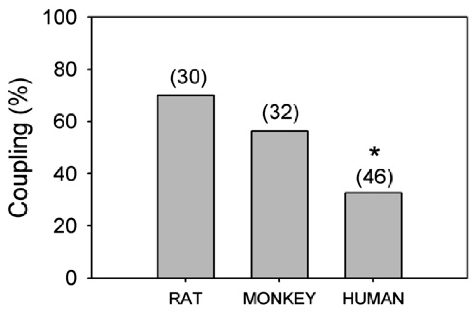
  33. Gayol, S.; Pannicke, T.; Reichenbach, E.; Colombo, J.A. Cell–cell coupling in cultures of striatal and cortical astrocytes of the monkey Cebus apella. J. Brain Res. 1999, 4, 473–478. [Google Scholar]
  34. Lanosa, X.A.; Reisin, H.D.; Santacroce, I.; Colombo, J.A. Astroglial dye-coupling: An in vitro analysis of regional and interspecies differences in rodents and primates. Brain Res. 2008, 1240, 82–86. [Google Scholar] [CrossRef] [PubMed]
  35. Colombo, J.A.; Bentham, C. Immunohistochemical analysis of subcortical white matter astroglia of infant and adult primates, with a note on resident neurons. Brain Res. 2006, 1100, 93–103. [Google Scholar] [CrossRef] [PubMed]
  36. Colombo, J.A. Cellular complexity in subcortical white matter: A distributed control circuit? Brain Struct. Funct. 2018, 223, 981–985. [Google Scholar] [CrossRef] [PubMed]
  37. Colombo, J.A.; Napp, M.I. Ex vivo astroglial-induced radial glia express in vivo markers. J. Neurosci. Res. 1996, 46, 674–677. [Google Scholar] [CrossRef]
  38. Colombo, J.A.; Napp, M.I. Forebrain and midbrain astrocytes promotes neuritogenesis in cultured chromaffin cells. Restor. Neurol. Neurosci. 1994, 7, 111–117. [Google Scholar] [PubMed]
  39. Hunter, K.E.; Hatten, M.E. Radial glial cell transformation to astrocytes in bidirectional regulation by a diffusible factor in embryonic forebrain. Proc. Natl. Acad. Sci. USA 1995, 92, 2061–2065. [Google Scholar] [CrossRef] [PubMed]
  40. Colombo, J.A.; Napp, M.I. Cerebrospinal fluid from l-dopa-treated Parkinson’s disease patients is dystrophic for various neural cell types ex vivo: Effects of astroglia. Exp. Neurol. 1998, 154, 452–463. [Google Scholar] [CrossRef] [PubMed]
  41. Uceda, G.; Colombo, J.A.; Michelena, P.; López, M.G.; García, A.G. Rat striatal astroglia induce morphological and neurochemical changes in adult bovine, adrenergic-enriched adrenal chromaffin cells in vitro. Restor. Neurol. Neurosci. 1995, 8, 129–136. [Google Scholar] [PubMed]
  42. Lipina, S.J.; Colombo, J.A. Dissociated functional recovery in parkinsonian monkeys following transplantation of astroglial cells. Brain Res. 2001, 911, 176–180. [Google Scholar] [CrossRef]
  43. Lipina, S.J.; Colombo, J.A. Premorbid exercising in specific cognitive tasks prevents impairment of performance in parkinsonian monkeys. Brain Res. 2007, 1134, 180–186. [Google Scholar] [CrossRef] [PubMed]
  44. Diamond, M.C.; Krech, D.; Rosenzweig, M.R. The effects of an enriched environment on the histology of the rat cerebral cortex. J. Comp. Neurol. 1964, 123, 111–120. [Google Scholar] [CrossRef] [PubMed]
  45. Globus, A.; Rosenzweig, M.R.; Bennett, E.L.; Diamond, M.C. Effects of differential experience on dendritic spine counts in rat cerebral cortex. J. Comp. Physiol. Psychol. 1973, 82, 175–181. [Google Scholar] [CrossRef] [PubMed]
  46. Sirevaag, A.M.; Greenough, W.T. Differential rearing effects on rat visual cortex synapses. III. Neuronal and glial nuclei, boutons, dendrites, and capillaries. Brain Res. 1987, 424, 320–332. [Google Scholar] [CrossRef]
  47. Lanosa, X.A.; Santacroce, I.; Colombo, J.A. Exposure to environmental enrichment prior to a cerebral cortex stab wound attenuates the postlesional astroglia response in rats. Neuron Glia Biol. 2011, 7, 1–13. [Google Scholar] [CrossRef] [PubMed]
  48. Santacroce, I. Plasticity of Astroglial Networks in the Cerebral Cortex of the Rat: Response to Environmental Enrichment and Physicochemical Variables. Ph.D. Thesis, University of Buenos Aires, Buenos Aires, Argentina, 2017. [Google Scholar]
Figure 1. Coronal section from the striate cerebral cortex obtained from an adult Saimiri boliviensis. Note dense packing of glial fibrillary acidic protein-immunoreactivity (GFAP-IR) astroglial processes, the relative height of the band of processes and the frequent appearance of slender bulbous endings. Broken line indicates the limit of lamina I. Scale bar: 100 µm. Adapted from Figure 1 in [9].
Neuroglia 01 00003 g001
Figure 2. Interlaminar bulbous ending in an aged human cerebral cortex sample. Note in (A) a mitochondrion, and bridge (arrow) connecting with blood vessel (asterisk) (also in (B)). In (B), note the multilamellar structure. Scale bar: 50 µm in (A) and 200 nm in (B). Adapted from Figure 4 in Colombo et al. [20].
Neuroglia 01 00003 g002
Figure 3. Interlaminar GFAP-IR events observed in coronal (A,B) and tangential (flattened) (C,D) sections of the striate cortex from control, intact (C) and three months visually deprived (D) adult Cebus apella monkey. Asterisk and broken line in (A) indicate the limit of lamina I. Vascular elements in (C,D) are marked by an asterisk. Note paucity of events in sections on the right side. Scale bar: 100 µm. Adapted from Colombo et al. [23].
Neuroglia 01 00003 g003
Figure 4. Morphology and spatial arrangement of GFAP-IR interlaminar processes in the somatosensory cortex of control and long-term spinal cord-transected Macaca individuals. (A) Control; (B) Operated. Note extreme departure from the rectilinear trajectory and several ‘‘terminal masses’’ (arrowheads). Large arrow indicates direction of the cortical surface. Scale bar: 40 µm. Adapted from Reisin and Colombo [24].
Neuroglia 01 00003 g004
Figure 5. Striate cerebral cortex sample from 2.5-months-old Saimiri boliviensis. Glial fibrillary acidic protein immunostaining with Nissl stain. Scale bar: 100 µm. Adapted from Figure 2 in [9]. Note marked wavy configuration of the long (interlaminar) cellular processes.
Neuroglia 01 00003 g005
Figure 6. Intrasurgical sample. Human frontal cerebral cortex from a seven-year-old. Vimentin immunohistochemistry. Long processes with “corkscrew” appearance. Pia mater is towards the top of the figure asterisk indicates cortical surface. Scale bar: 20 µm. Adapted from Figure 5 in [9].
Neuroglia 01 00003 g006
Figure 7. Tangential section of striate cortex from an adult Saimiri boliviensis monkey, reacted for GFAP with Nissl counterstain, note wavy terminal segments and slender bulbous terminals decorating them. Scale bar: 20 µm. Adapted from Figure 5 in [5].
Neuroglia 01 00003 g007
Figure 8. Interspecies analysis of astroglial coupling levels following lucifer yellow pressure injections in astroglia-enriched cultures from rat, monkey and human cerebral cortex, expressed as the percentage of cells coupled out of the total cells essayed (percentage of cell coupling). Numbers in parentheses indicate sample size. Significant differences were detected using Pearson’s chi-square test (* p = 0.006). Adapted from Figure 3 in [34].
Neuroglia 01 00003 g008
Figure 9. (A) Adhesion of cerebral cortex primary fetal (embryonic day 17 (E17)) cells onto elongated (“radial-like”) processes of subcultured cerebral cortex glia exposed to cerebral cortex astroglial conditioned medium during 24 h, 3 h after seeding primary cells. Note in (B) evidence of leading and trailing processes of primary cells, adherent to a cell process. Induced processes were laminin-positive and Rat-401 antisera-positive. Adapted from Figure 1 in [37]. Scale bar: 10 µm (A,B).
Neuroglia 01 00003 g009
Figure 10. (AC) Effect of astroglial conditioning of three different sources of cerebrospinal fluid (CSF) from Parkinson’s disease patients on neuronal processes. Percentage of emitting cells in rat primary cultures of striatum (dotted bars) or ventral mesencephalon (horizontally dashed bars) after 24 h in culture with CSF before (pre) glial conditioning or after (post) 24 h conditioning with fetal mesencephalic astroglia. Pearson’s chi-square test; ** p < 0.01; *** p < 0.001. Adapted from Figure 6 in [40].
Neuroglia 01 00003 g010

