Abstract
This in vitro investigation evaluated the marginal fit of three pressable glass-ceramic onlay materials: a conventional monolithic lithium disilicate (IPS e.max Press, EM, ivoclar vivadent AG, Schaan, Liechtenstein) and two zirconia-reinforced glass-ceramics (Vita Ambria, VA, VITA Zahnfabrik, Bad Säckingen, Germany; Celtra Press, CP, Sirona Dentsply, Milford, CT, USA). A typodont maxillary first premolar was prepared for an intensive onlay design by a single operator using a milling surveyor. The master die was duplicated with silicone impressions to create 72 identical epoxy resin dies. Seventy-two onlays (n = 24 per material) were fabricated and adhesively cemented to their respective dies. Vertical marginal gaps were recorded under a stereo-electron microscope before and after thermomechanical loading (TML) in a chewing simulator. Data were analyzed with one-way ANOVA and Tukey’s post hoc tests for intergroup comparisons and paired t-tests for pre- versus post-TML values. All groups showed a significant increase in marginal gap following TML. VA exhibited mean gaps of 46.41 µm before and 57.28 µm after loading (p = 0.001). EM demonstrated 41.16 µm before and 46.63 µm after TML (p = 0.002). CP showed 45.70 µm before and 55.99 µm after TML (p = 0.003). Among the three materials, EM maintained the most accurate marginal adaptation both before and after simulated chewing. Despite the increases, all post-loading values remained within the clinically acceptable threshold for marginal discrepancy. These findings indicated that thermomechanical fatigue adversely affected the marginal integrity of pressable glass-ceramic onlays, including zirconia-reinforced formulations. Nevertheless, zirconia-reinforced ceramics (VA and CP) achieved marginal gaps comparable to conventional lithium disilicate and remained within acceptable clinical limits. IPS e.max Press provided the best overall fit, suggesting it may offer superior long-term marginal stability for onlay restorations.
1. Introduction
Ceramic restorations have gained popularity due to patients’ growing desire for esthetically beautiful restorations and worries about employing direct resin composites for severely deteriorated posterior teeth. Tissue loss is considerably less for partial crowns and occlusal onlays, while full crowns in both anterior and posterior regions may need to remove as much as 70% of the hard tissue in the clinical crown [1,2]. For a long time, the gold standard for posterior tooth rehabilitation was cast gold partial coverage restorations. However, a key concern in restorative dentistry is the potential for greater preservation of residual tooth structure brought about by novel adhesive technology and ceramic materials [3]. However, a variety of ceramic systems and production techniques have been introduced by the manufacturing of dentistry solutions [4]. They include industrially manufactured machinable ceramic blocks for chair-side and laboratory CAD/CAM systems, as well as pressable ingot ceramics made using the lost-wax technique, in place of the conventional powder slurry manufacturing process [3]. Hot pressing is the preferred technique for creating glass-ceramic restorations because it produces restorations with superior mechanical strength, edge quality, low porosity, and fitting accuracy compared to computer-aided design/computer-aided manufacturing (CAD/CAM)-milled materials [5,6,7].
The collection of ceramic materials known as glass-ceramics, which were employed extensively in the past, is a significant subject of research [8,9]. Since the development of the lithium disilicate glass-ceramic group and its fabrication to accomplish full-contour monolithic restorations along with cementation using adhesive resin cement, it has been demonstrated to be a good option in higher stress scenarios [10]. About 70% of the IPS e.max Press’s microstructure is made up of crystals that are 3–6 μm long and encased in a glassy matrix [11,12]. Leucite, zirconia, and lithium disilicate crystals are examples of filler particles that can be added to the foundation glass composition to meet the expanding need for ceramic restorations [4,13].
In the past decade, several companies have developed derivatives of lithium disilicate to enhance esthetics, strength, and clinical performance. One of the most significant advancements is zirconia-reinforced glass-ceramics, which combine the translucency of lithium-based ceramics with the increased strength offered by zirconia reinforcement [8,14].
A key subgroup within this category are lithium silicate (Li2O·SiO2) ceramics, which are strengthened with approximately 10% tetragonal zirconia and nanoscale lithium phosphate. Notable examples include Suprinity and Celtra Duo, both of which demonstrate superior esthetics, improved machinability, and higher flexural strength compared with conventional lithium disilicate materials [15,16]. Building on these developments, a pressable variant—Celtra Press—was later introduced. This material incorporates about 10% zirconia and 0.5 μm nanoscale lithium phosphate, providing enhanced mechanical properties relative to the machinable Celtra Duo blocks. These improvements expand its clinical indications, allowing use in veneers, inlays, onlays, single crowns, and even multi-unit bridges [17,18].
Comparative studies of zirconia-reinforced lithium silicate (ZLS) ceramics versus traditional lithium disilicate have revealed several advantages of ZLS ceramics. These include improved surface polishability, greater edge strength, enhanced machinability, and reduced brittleness [19,20]. Additionally, researchers have found that adjusting the crystal structure or heat-treatment process of ZLS ceramics can significantly affect their mechanical, chemical, and physical properties [21].
In addition to Celtra Press, the current study also evaluated Vita Ambria, a zirconia-reinforced lithium disilicate produced by VITA Zahnfabrik. This material contains 8–12% zirconia oxide and features lithium disilicate crystals measuring between 2.5 and 3.5 μm. This combination results in a durable glass-ceramic characterized by complete zirconia dissolution within the glass matrix. Vita Ambria is designed for use in crowns, onlays, and veneers, making it a promising formulation for pressable glass-ceramics [22].
Despite advances in these materials, clinical evidence on marginal adaptation remains inconsistent. Studies evaluating the fit accuracy of CAD/CAM ceramic crowns have reported highly variable outcomes [23,24,25]. Marginal discrepancies are clinically significant because they can lead to microleakage, cement dissolution, recurrent caries, and periodontal inflammation. These concerns are particularly relevant for onlay and inlay restorations, which are subjected to concentrated functional stresses [26,27]. A systematic review reported an annual failure rate of 0% to 7.5% for ceramic onlays and inlays, highlighting the critical role of material selection and precise marginal fit [28].
Most existing studies have concentrated on CAD/CAM-milled lithium disilicate or have evaluated materials in isolation, limiting our understanding of how pressable zirconia-reinforced ceramics perform under thermomechanical stress. This gap underscores the need for comparative assessments of newer pressable systems, such as Celtra Press and Vita Ambria, particularly in terms of marginal adaptation and potential clinical reliability.
Addressing this gap, the present study aimed to evaluate the marginal gaps of maxillary premolars restored with IPS e.max Press, pressable Vita Ambria, and Celtra Press onlays, before and after thermomechanical loading. The hypotheses were (1) there are no significant differences in marginal gaps among the three examined materials (IPS e.max Press, pressable Vita Ambria, and Celtra Press) and (2) thermomechanical loading does not significantly affect marginal adaptation.
2. Materials and Methods
This study was authorized by the Faculty of Dental Medicine (Cairo) Research Ethics Committee at Al-Azhar University under protocol number EC. Ref No: 679/272. Table 1 includes different ceramic materials that were assessed for this investigation.

Table 1.
Ceramic materials tested in this study.
Before the main experiment, we conducted a pilot study using descriptive statistics (2 samples in each group). The pilot study revealed mean differences in marginal gap values among the materials of roughly 10–15 µm and within-group standard deviations ranging from 6 to 8 µm. When these differences were standardized, they reflected a large effect size (Cohen’s d ≈ 1.1–1.3). Based on previous findings (ref. [29,30]), the G power statistical power analysis tool (ref. [31]) (version 3.1.9.4) was used to determine the sample size. A total sample size of N = 72 was sufficient to detect a significant effect size (d) =1.2 when the null hypothesis test was conducted with an actual power (1-β error) of 0.8 (power = 80%) and a significance level (α error) of 0.05 (5%) set. The value of d was derived from our own pilot study (observed mean differences and variability in marginal gaps among ceramic groups), rather than solely from published studies. Three equal main groups of ceramic onlay restorations (n = 24 in each group) were judged based on the type of ceramic material used: the (EM) group for lithium disilicate (IPS e.max Press), (CP) group for Celtra Press (zirconia-reinforced lithium silicate), and (VA) group for zirconia-reinforced lithium disilicate (Vita Ambria).
2.1. Master Die Fabrication and Onlay Preparations
A definite region of the jaw with definite parameters was selected to decrease variations between teeth size and anatomy. For this reason, the maxillary first premolar typodont tooth was chosen and prepared to be used as the master die. The parallelometer (Paraflex, Bego, Bremer, Germany) was adjusted both horizontally and vertically to mount and install the typodont tooth inside a custom-made cupper holder and counter metal ring. An auto-polymerized mix of polymethyl methacrylate was used to hold and secure this tooth during preparation inside the custom-made cupper holder where the cervical line of this tooth was coronal to the top surface of the holder by about 2 mm.
A silicon putty template guide (Zeta plus; C-Silicone, Zhermack, Italy) was used to determine the total reduction during the typodont tooth preparation [32]. To facilitate consistent preparation, a single prosthodontist with extensive experience created the onlay design using a milling surveyor (Paraskop®M, Bego, Bremer, Germany). The pulpal floor was prepared to a depth of 1.5 mm from the occlusal cavo-surface border of the preparation in order to meet the following criteria for creating the mesial-occlusal-distal (MOD) cavity: A 1.5 mm occlusal reduction was performed on the functional cusp. The gingival floor preparations measured 1.5 mm in width and 1.5 mm in depth. The width of the occlusal isthmus accounted for one-third of the intercuspal distance. The sharp angles of all internal lines were rounded [33]. A flow chart of the experimental steps of the study is illustrated in Figure 1.
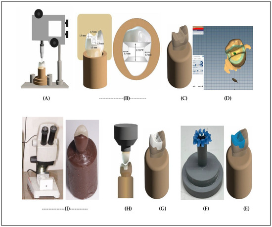
Figure 1.
Flow chart illustrating the experimental steps of the study: (A) parallelometer used to secure the typodont tooth (master die) in the custom holder, and a milling surveyor used to perform onlay tooth preparation; (B) putty template guide used to control the preparation dimensions of onlay “lateral view and top view”; (C) the epoxy resin die (Replica) after duplication by silicon material; (D) CAD design of onlay after scanning of epoxy die; (E) milled wax pattern “CAD wax”; (F) spruing of CAD wax; (G) pressable ceramic onlay; (H) cementation of onlay using static load “5 kg”; (I) stereomicroscopic marginal gap evaluation of onlay after cementation on epoxy die.
2.2. Duplication of the Master Die
A silicon mold was produced using replicating silicon (Replisil 22N; Zubler, Dallas, TX, USA) after the master die was surrounded by a metal ring. Following the manufacturer’s directions, the epoxy resin (Kema poxy 150; CMB International, Giza, Egypt) was mixed, poured into the silicone mold, vibrated, and allowed to fully polymerize [34,35]. A total of seventy-two epoxy dies (Replica) were produced.
2.3. Restoration Construction
Seventy-two biogeneric wax patterns were produced with the aid of computer-aided design and computer-aided manufacturing (CAD/CAM) in the following sequence. In order to eliminate optical highlights from the die’s surface and improve the accuracy of the optical impressions obtained by producing a consistently reflecting surface, the master die was sprayed with powder Shera scan spray (Shera werkstoff-technologie, Lemförde, Germany). The Ceramill Map 400 scanner (Amann Girrbach, Vorarlberg, Austria) was then used to create an optical impression. Following an assessment of the scan’s clarity, the information was saved to create CAD software from the manufacturer (Ceramill Mind Software 3.6, Austria). On the computer screen, a 3D virtual overlay was produced, with the margins automatically generated and manually adjusted as necessary. The wax patterns had a minimum radial thickness of 1.5 mm, a minimum occlusal thickness of 1.5 mm, a cement gap of 50 μm, and a border thickness of 120 μm. Milling of the CAD wax patterns was completed using wax material (Ceramill D-wax, Amman Girrbach, Vorarlberg, Austria) with (CAD/CAM) technology using the 5-axis milling machine (Ceramill motion 2 Amann Girrbach, CAD/CAM System, Vorarlberg, Austria).
In all groups, before the investment step, the properly seated onlay wax pattern was checked visually and finished on the analogous epoxy die. Using sprues measuring 3 mm in diameter and 3 mm in length, all completed wax patterns were affixed to the base of a cylindrical silicon ring. They were then invested using a phosphate-bonded investment material in accordance with the manufacturer’s instructions (e.max ingots were used; PressVEST Speed, Ivoclar Vivadent, Ellwangen, Germany). Thereafter, following the manufacturer’s directions on each material, all wax patterns within the silicon ring were heated and burned out to create a mold ready to press the ceramic ingots. Ceramic ingots of e.max Press ingots “EM” (LT A2), Celtra Press ingots “CP” (LT A2), and Vita Ambria Press ingots “VA” (T-A2) were hot-pressed in a specialized pressing furnace (Programat EP 3010; Ivoclar Vivadent, Schaan/Liechtenstein, Switzerland) according to the press parameters of each material, following the manufacturers’ orientations to inject and fill the empty silicon mold. Once cooled, 110 μm aluminum oxide particles (Cobra; Renfert) were used in a sandblasting machine (Basic eco; Renfert) at a pressure of 0.3 MPa to remove the investment. Glaze firing programs were selected according to the specific manufacturer’s recommendation for each type of onlay material.
2.4. Cementation of Restorations
The intaglio of the onlay restoration was washed well with water and allowed to air dry after being etched for 20 s with 9.5% hydrofluoric acid (porcelain etchant; Bisco, Inc., Schaumburg, IL, USA). After a minute, the air-thinned silane (Porcelain primer; Bisco, Inc., Schaumburg, IL, USA) was brushed onto the etched surface. Onlay restorations were fixed using self-adhesive resin cement (Theracem; Bisco, Inc., Schaumburg, IL, USA). The manufacturer’s instructions were followed for the activation, mixing, and polymerization. Specifically-made cementation apparatus was used to seat each restoration on its corresponding epoxy resin die for five minutes at room temperature while applying a load of (49 N) [36]. After cementation, onlay restorations over dies were kept in distilled water at 37 °C up to marginal evaluation.
2.5. Marginal Gap Evaluation
As previously mentioned [37], the vertical marginal distance between the epoxy die’s finish line and onlay margins was measured (Figure 2) using a handheld digital microscope with an integrated camera mounted on a stereomicroscopic stand (Dino-Lite Pro, Olympus, Tokyo, Japan) and connected to a personal computer with 40× magnification. Prior to starting measurements, the calibration instructions for the microscope were correctly followed. To overcome the difficulties of repeating the measurements of the marginal fit at the same distance and angle from the microscope capturing unit, a Sample Positioning Device (SPD) including a calibrated gauge, fixable base, and reading marks was created. Digital images were taken for each mesial, distal, bucco-occlusal, and lingual surface. Open-source software for processing and interpreting scientific photographs (ImageJ 1.53t, National Institute of Health, USA) was used to finish the digital analysis. Measurements and qualitative assessments of the gaps were performed by microns at the five predetermined points on the epoxy die for each surface, providing total of twenty readings for every restoration. The average marginal gap value for every restoration was determined by taking the mean of all readings.
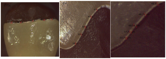
Figure 2.
Vertical marginal gaps between the epoxy die’s finish line and onlay margins.
2.6. Thermomechanical Loading
Following the determination of the marginal gap, each sample was put through a thermomechanical cyclic loading process using a servomotor-driven, multimodal, cyclic load ROBOTA chewing simulator (Model ACH-09075DC-T, Germany) that was integrated with a thermo-cyclic protocol operated on a servomotor. Based on the recommendations made by International Organization for Standardization (ISO TR 11405 [38]), the samples were subjected to 120,000 preloaded cycles accompanied with 10,000 thermal cycles (between 5° and 55° Celsius), 60 s of dwell time, and 98 N of load [39]. Load force of the upper movable compartment of the material testing machine was applied centrally on the occlusal surface of the onlay restoration over the die, including the palatal cusp. The marginal gap was then reevaluated after that.
2.7. Statistical Analysis
The statistical analysis was carried out using SPSS software (SPSS, Chicago, IL, USA, version 20). By examining the data’s distribution and applying the Shapiro–Wilk and Kolmogorov–Smirnov tests, the data’s normalcy was evaluated. The various groups were analyzed using one-way analysis of variances (ANOVA) and subsequently examined with Tukey’s post hoc multiple comparisons test. The paired t-test was utilized to analyze the two conditions before and after thermomechanical loading. A notable threshold of p ≤ 0.05 was utilized.
3. Results
Descriptive statistics presented as mean values and standard deviations of the marginal gaps measured in μm are documented for all groups in Table 2. Regarding the marginal fit of onlay restorations, significant statistical differences in the marginal gaps were observed among the groups following thermomechanical loading “TML” (p < 0.05), whereas before TML, these marginal gaps were deemed not significantly different. When comparing the groups before thermomechanical loading “TML”, a higher mean value was observed in the zirconia-reinforced lithium disilicate Vita Ambria “VA” group (46.42 ± 8.75 μm) compared to the lithium disilicate e.max “EM” group (41.16 ± 5.09 μm) and zirconia-reinforced lithium silicate Celtra Press “CP” group (45.7 ± 6.35 μm). However, this difference did not become statistically significant (p = 0.086) as shown in Table 2. After subjecting these various onlay restorations to TML, we found that the mean vertical marginal gap value in the VA group increased to (57.28 ± 3.27 μm), whereas the mean vertical marginal gap in the EM and CP groups increased to (46.63 ± 1.76 μm and 55.99 ± 1.91 μm, respectively). This difference between the main groups was statistically significant (p = 0.00) after TML.
Table 2.
Descriptive statistics of marginal gap (µm), comparison in between groups One way ANOVA followed by Tukey, s post hoc multiple comparison test. Comparison within each group before and after Thermomechanical Loading (TML) using paired t-test.
In terms of TML’s impact, the EM group’s vertical marginal gap mean value increased statistically significantly from 41.16 ± 5.09 µm (p = 0.002) prior to loading to 46.63 ± 1.76 µm following TML. Similarly, in the VA group, a significant increase was observed after TML (57.28 ± 3.27 µm after TML versus 46.42 ± 8.75 µm before loading) (p = 0.001). Moreover, restorations of the CP group exhibited a significant increase in marginal gap values of 45.7 μm before TML and 55.99 μm after TML with (p = 0.003). This means there was a significant difference within the groups after TML (Table 2 and Figure 3).
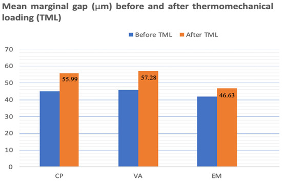
Figure 3.
Column chart illustrating mean marginal gap between groups in µm before TML (blue column) and after TML (orange column).
4. Discussion
This experimental study evaluated how thermomechanical loading (TML) influences the marginal fit of onlay restorations made from three lithium-based glass-ceramics: IPS e.max Press, pressable Vita Ambria, and Celtra Press. Because marginal and internal adaptation are critical for the durability and performance of ceramic restorations [40], the researchers tested two null hypotheses. The first proposed that marginal gap values would not differ significantly among the three materials. This was only partly supported: before TML there was no significant difference, but after TML a statistically significant difference emerged. The second hypothesis stated that TML would not affect marginal gaps. This was rejected, as TML produced a significant change in gap measurements. The results demonstrate that thermomechanical stress can compromise marginal precision and that the ceramic material influences the magnitude of this effect. Additional variables—such as the restorative material, die material, preparation geometry, and cementation protocol—can further impact the marginal gap of fixed dental prostheses [41]. These findings emphasize the need to consider both material selection and functional loading when planning long-lasting, esthetically pleasing ceramic restorations.
This study used shrink-free epoxy resin to create standardized dies that accurately replicate natural teeth. The resin provides a modulus of elasticity close to dentin (≈12.9 GPa) and forms dentin-like bonds with luting agents, ensuring reliable comparison of onlay preparations across groups [34,35]. For precise duplication of the master die, REPLISIL 22 N silicone was selected because of its low viscosity, ability to capture fine details, and excellent flexibility and recovery after deflasking and used in accordance with the manufacturer’s guidelines. To maintain uniformity in fabricating the pressable ceramic onlays, a single “biogeneric copy” design was produced with CAD/CAM software (Ceramill Mind Software 3.6, Amann Girrbach AG, Koblach, Austria). All wax patterns were milled from solid blanks using CAD/CAM technology, minimizing distortion common in conventional wax-up methods and ensuring consistent final restorations [42,43].
The present findings show that all three lithium-based glass-ceramic onlay materials demonstrated marginal gaps well within accepted clinical limits, even after thermomechanical loading (TML). Mean marginal gaps increased slightly after TML—from 46.42 ± 8.75 µm to 57.28 ± 3.27 µm for Vita Ambria (VA), 41.16 ± 5.09 µm to 46.63 ± 1.76 µm for IPS e.max Press (EM), and 45.7 ± 6.35 µm to 55.99 ± 1.91 µm for Celtra Press (CP). Although the post-TML differences among the three groups reached statistical significance, all values remained below the commonly cited threshold of 120 µm for fixed dental prostheses [44] and even below the more stringent <100 µm criterion recommended in the recent literature [45]. These results indicate that the tested ceramics maintain clinically acceptable marginal integrity under simulated functional stresses. Several methodological choices likely contributed to the reliability of these measurements. A stabilizing instrument was employed during cementation to minimize operator-related variability, such as uneven finger pressure and inconsistent cement flow. In addition, direct stereomicroscopic evaluation was selected as a measurement technique because it is less time-consuming and involves fewer procedural steps than alternative methods, thereby reducing the potential for cumulative measurement error [37]. Overall, the data suggest that, despite minor increases in gap width following TML, all three materials provide a marginal fit compatible with long-term clinical performance. This supports their suitability for durable, esthetic onlay restorations when appropriate laboratory and cementation protocols are followed.
According to manufacturer data, Celtra Press contains less than 10% zirconia and 0.5 μm nanoscale lithium phosphate, while Vita Ambria contains more than 10% zirconia oxide with lithium disilicate crystals of 2.5 to 3.5 μm. The non-significant difference of the greater marginal gap of Vita Ambria than Celtra Press before and after TML might be attributed to a higher content of zirconia oxide and large crystal size in Vita Ambria, which in turn might result in difficult finishing of the restoration and margins. Following TML, there was a significant difference in the marginal gap values between the three groups; the Vita Ambria and Celtra Press groups showed a larger marginal gap than the e.max group. This might be attributed to the difference in concentration of nucleating agents (multi-component system), as Vita Zahnfabrik innovated pressable glass-ceramic (Vita Ambria) strengthened with (8–12 wt.%) ZrO2 particles; in addition, Celtra Press had a high content of P2O5 (4.9 wt%) and ZrO2 (9.3 wt%) and lower SiO2 compared to IPS e.max Press [46]. However, the high ZrO2 content in the glass-ceramics results in an increase in the viscosity and is associated with limited flowability of ingots during the pressing process [47,48]; thus, this might result in poor fitting margins. Moreover, in another previous study, IPS e.max showed a higher value of the Weibull coefficient than the other types of ceramics; thus, this material might result in a greater marginal fit [49].
Results of current study were consistent with a former study that attributed higher marginal gap values after TML to temperature fluctuations (5–55 °C), water absorption, and microleakage in the composite resin cement, leading to the degradation and wear of the luting cement, resulting in an increased marginal gap [50]. Additionally, variations in thermal expansion coefficients, particularly at multiple interfaces like the die material, resin cement, and restorative material, exert stress on the cement, further increasing the gap [26]. These factors might collectively contribute to the widening of the marginal gap after thermocycling.
Results of this study contradict the findings of El-Hamid et al. [51] who reported that there is no significantly higher marginal gap and internal fit for IPS e.max Press and Vita Ambria with endocrown restoration. They attributed their results to the bonding of the 10-Methacryloyloxygexyl Di-Hydrogen phosphate (10-MDP) incorporated into the cement with the zirconia oxide, which enhances the restoration’s bonding and reduces the marginal gap and internal fit.
In order to simulate intraoral activities and evaluate innovative dental materials under accelerated settings, thermocycling and dynamic loading are essential. In order to simulate temperature changes that could occur in the oral cavity due to hot and cold extremes, the aged sample onlay restorations were put through 10,000 thermal cycles and 120,000 cycles of cyclic loading in a thermocycling machine. The goal of this simulation was to mimic masticatory conditions in the posterior region of the oral cavity for a whole year [39].
Accurate marginal fit is essential for the longevity of ceramic onlays, as poor adaptation can lead to microleakage, secondary caries, and periodontal complications. IPS e.max Press offers well-documented marginal accuracy and mechanical strength, making it reliable for high-stress posterior restorations. Vita Ambria and Celtra Press, as newer zirconia-reinforced lithium silicate ceramics, provide improved strength and translucency, but their pressability and marginal fit may be influenced by zirconia and nanoscale fillers. Clinicians should balance marginal adaptation, mechanical performance, and esthetics when selecting a material. For cases prioritizing proven fit and durability, IPS e.max Press may be preferred, whereas Vita Ambria and Celtra Press offer enhanced esthetics and sufficient strength for single-unit onlays. Understanding these trade-offs can guide evidence-based material selection tailored to patient needs.
Limitations of the Present Study
Although this study took into account a standardization approach, this study has several limitations. Marginal gap measurements were performed using a stereomicroscope at ×40 magnification, which, while acceptable, is less precise than micro-computed tomography (micro-CT) and may not detect subtle discrepancies. The study was conducted in vitro using epoxy resin dies, which do not fully replicate the complex oral environment, including saliva, occlusal forces, and long-term wear. Only single-unit onlays were evaluated, limiting generalizability to multi-unit restorations or other tooth types. Despite efforts to standardize procedures, inherent limitations of in vitro testing remain. Future studies using higher-resolution imaging and long-term clinical trials are recommended to validate and expand upon these findings.
5. Conclusions
Within the limitations of this study, the following conclusions were drawn:
- Lithium disilicate (IPS e.max Press) onlay restorations showed more precise marginal fit than two groups of zirconia-reinforced lithium-based ceramic onlays.
- Thermomechanical loading (TML) revealed significant marginal gap values of two zirconia-reinforced lithium-based ceramic onlays compared to lithium disilicate.
- For three lithium-based ceramic onlays, thermomechanical loading (TML) significantly raised the marginal gap; however, the values of this marginal gap fell within the clinically acceptable range.
Author Contributions
A.H.A., M.F.M., S.A.A., W.A.A., M.I.A., K.M.H., H.M.E.S., F.B., and A.A.M. contributed to the concept of the research, study design, data collection, statistical analysis, writing of the original draft, and reading and editing of the final paper. All authors have read and agreed to the published version of the manuscript.
Funding
This research received no external funding.
Institutional Review Board Statement
This research was approved by the Research Ethics Committee of the Faculty of Dental Medicine at Al-Azhar University under protocol number EC. Reference Number: 679/272. The Research Ethics Committee at Al-Azhar University’s Faculty of Dental Medicine waived the requirement for informed consent. All procedures were carried out in line with the Helsinki declaration.
Data Availability Statement
The data that support the findings of this study are available on request from the corresponding author. The data are not publicly available due to privacy or ethical restrictions.
Conflicts of Interest
The authors claim they do not have any competing interests.
Abbreviations
VA: Vita Ambria; CP: Celtra Press; IPS e. max: monolithic, lithium disilicate ceramic; EM: e.max press; TML: thermomechanical loading; CAD: computer-aided design; CAM: computer-aided manufacturing; Li2O3Si: Lithium silicate; ZLS: zirconia-reinforced lithium silicate glass-ceramic; MOD: mesial-occlusal-distal; SPD: sample positioning device; FDP: fixed dental prosthesis; 10-MDP: 10-Methacryloyloxygexyl Di-Hydrogenphosphat.
References
- Edelhoff, D.; Sorensen, J.A. Tooth structure removal associated with various preparation designs for posterior teeth. Int. J. Periodontics Restor. Dent. 2002, 22, 241–249. [Google Scholar]
- Monaco, C.; Arena, A.; Štelemėkaitė, J.; Evangelisti, E.; Baldissara, P. In vitro 3D and gravimetric analysis of removed tooth structure for complete and partial preparations. J. Prosthodont. Res. 2019, 63, 173–178. [Google Scholar] [CrossRef]
- Griffis, E.; Alraheam, I.A.; Boushell, L.; Donovan, T.; Fasbinder, D.; Sulaiman, T.A. Tooth-cusp preservation with lithium disilicate onlay restorations: A fatigue resistance study. J. Esthet. Restor. Dent. 2020, 34, 512–518. [Google Scholar] [CrossRef]
- Phark, J.H.; Sillas, D.J. Microstructural considerations for novel lithium disilicate glass ceramics: A review. J. Esthet. Restor. Dent. 2022, 34, 92–103. [Google Scholar] [CrossRef]
- Vasiliu, R.-D.; Porojan, S.D.; Porojan, L. Vitro study of comparative evaluation of marginal and internal fit between heat-pressed and CAD-CAM monolithic glass-ceramic restorations after thermal aging. Materials 2020, 13, 4239. [Google Scholar] [CrossRef]
- Bukhari, A. Mechanical and Optical Properties of Machinable and Pressable Glass Ceramic. Ph.D. Thesis, Boston University, Boston, MA, USA, 2020. [Google Scholar]
- Alharbi, N.A.B. Physico-Mechanical Characterisation of a Novel and Commercial CAD/CAM Composite Blocks. Ph.D. Thesis, The University of Manchester, Manchester, UK, 2020. [Google Scholar]
- Silva, L.H.; Lima, E.; Miranda, R.B.; Favero, S.S.; Lohbauer, U.; Cesar, P.F. Dental ceramics: A review of new materials and processing methods. Braz. Oral Res. 2017, 31, 133–145. [Google Scholar] [CrossRef]
- Taha, D.; Wahsh, M. Assessment of marginal adaptation and fracture resistance of endocrown restorations utilizing different machinable blocks subjected to thermomechanical aging. J. Esthet. Restor. Dent. 2018, 30, 319–328. [Google Scholar] [CrossRef] [PubMed]
- McLaren, E.A.; Figueira, J. Updating classifications of ceramic dental materials: A guide to material selection. Compendium 2015, 36, 739–745. [Google Scholar]
- Alkadi, L.T. IPS e. max CAD and IPS e. max Press: Fracture Mechanics Characterization. Ph.D. Thesis, University of British Columbia, Vancouver, BC, Canada, 2014. [Google Scholar]
- Al-Johani, H. Effects of Etching Duration on the Surface Roughness, Surface Loss, Flexural Strength, and Shear Bond Strength to a Resin Cement of e. max CAD Glass Ceramic. Ph.D. Thesis, Indiana University, Bloomington, IN, USA, 2017. [Google Scholar]
- Zhang, Y.; Vardhaman, S.; Rodrigues, C.; Lawn, B. A Critical Review of Dental Lithia-Based Glass–Ceramics. J. Dent. Res. 2023, 102, 245–253. [Google Scholar] [CrossRef]
- Gracis, S.; Thompson, V.; Ferencz, J.; Silva, N.; Bonfante, E. A new classification system for all-ceramic and ceramic-like restorative materials. Int. J. Prosthodont. 2016, 28, 227–235. [Google Scholar] [CrossRef] [PubMed]
- Rinke, S.; Pfitzenreuter, T.; Leha, A.; Roediger, M.; Ziebolz, D. Clinical evaluation of chairside-fabricated partial crowns composed of zirconia-reinforced lithium silicate ceramics: 3-year results of a prospective practice-based study. J. Esthet. Restor. Dent. 2020, 32, 226–235. [Google Scholar] [CrossRef]
- Sartori, N.; Tostado, G.; Phark, J.; Takanashi, K.; Lin, R.; Duarte, S. CAD/CAM high-strength glass ceramic. Quintessence Dent. Technol. 2015, 38, 39–54. [Google Scholar]
- Matsuzaki, F.; Sekine, H.; Honma, S.; Takanashi, T.; Furuya, K.; Yajima, Y.; Yoshinari, M. Translucency and flexural strength of monolithic translucent zirconia and porcelain layered zirconia. Dent. Mater. 2015, 34, 910–917. [Google Scholar] [CrossRef]
- Elsaka, S.E.; Elnaghy, A.M. Mechanical properties of zirconia reinforced lithium silicate glass ceramic. Dent. Mater. 2016, 32, 908–914. [Google Scholar] [CrossRef]
- Attia, M. Effect of different processing techniques on the marginal and internal fit of monolithic lithium disilicate and zirconia-reinforced lithium silicate restorations. Egypt. Dent. J. 2022, 68, 2497–2507. [Google Scholar] [CrossRef]
- Rezaie, H.R.; Rizi, H.B.; Khamseh, M.M.R.; Öchsner, A. Dental restorative materials. In A Review on Dental Materials; Springer International Publishing: Cham, Switzerland, 2020; pp. 47–171. [Google Scholar]
- Bahgat, S.F.A.; Basheer, R.R.; Sayed, S.M.E. Effect of zirconia addition to lithium disilicate ceramic on translucency and bond strength using different adhesive strategies. Dent. J. 2015, 61, 4519–4533. [Google Scholar]
- Zahnfabrik, V. VITA AMBRIA® PRESS SOLUTIONS, Technical and Scientific Documentation. 2020. Available online: https://www.google.com.hk/url?sa=t&source=web&rct=j&opi=89978449&url=https://mam.vita-zahnfabrik.com/portal/ecms_mdb_download.php%3Fid%3D100097%26sprache%3Den%26fallback%3Den%26cls_session_id%3D%26neuste_version%3D1&ved=2ahUKEwiL27bZ6OSRAxVic_UHHRWaCZQQFnoECBkQAQ&usg=AOvVaw2D3PYeELSn2VG9yA5Ont_H (accessed on 14 December 2025).
- Boitelle, P.; Mawussi, B.; Tapie, L.; Fromentin, O. A systematic review of CAD/CAM fit restoration evaluations. J. Oral Rehabil. 2014, 41, 853–874. [Google Scholar] [CrossRef]
- Neves, F.D.; Prado, C.J.; Prudente, M.S.; Carneiro, T.A.; Zancope, K.; Davi, L.R.; Mendonca, G.; Cooper, L.; Soares, C. Micro-computed tomography evaluation of marginal fit of lithium disilicate crowns fabricated by using chairside CAD/CAM systems or the heat-pressing technique. J. Prosthet. Dent. 2014, 112, 1134–1140. [Google Scholar] [CrossRef]
- Baig, M.R.; Gonzalez, M.A.G.; Abu Kasim, N.H.; Abu Kassim, N.L.; Farook, M.S. Effect of operators’ experience and cement space on the marginal fit of an in-office digitally produced monolithic ceramic crown system. Quintessence Int. 2016, 47, 181–191. [Google Scholar]
- Stappert, C.F.; Chitmongkolsuk, S.; Silva, N.R.; Att, W.; Strub, J.R. Effect of mouth-motion fatigue and thermal cycling on the marginal accuracy of partial coverage restorations made of various dental materials. Dent. Mater. 2008, 24, 1248–1257. [Google Scholar] [CrossRef]
- Benic, G.I.; Sailer, I.; Zeltner, M.; Gütermann, J.N.; Özcan, M.; Mühlemann, S. Randomized controlled within-subject evaluation of digital and conventional workflows for the fabrication of lithium disilicate single crowns. Part. III: Marginal and internal fit. J. Prosthet. Dent. 2019, 121, 426–431. [Google Scholar] [CrossRef]
- Al-Haj, N.Ö.H.; Molinero-Mourelle, M.; Joda, T.P. Clinical Performance of Partial and Full-Coverage Fixed Dental Restorations Fabricated from Hybrid Polymer and Ceramic CAD/CAM Materials: A Systematic Review and Meta-Analysis. J. Clin. Med. 2020, 9, 2107. [Google Scholar] [CrossRef]
- Al-Akhali, M.; Chaar, M.S.; Elsayed, A.; Samran, A.; Kern, M. Fracture resistance of ceramic and polymer-based occlusal veneer restorations. J. Mech. Behav. Biomed. Mater. 2017, 74, 245–250. [Google Scholar] [CrossRef]
- Al-Jlaihawi, Z.G.A.-K. Study of the Physical Properties of a Novel Lithium Aluminosilicate Dental Glass Ceramic; Sheffield Hallam University: Sheffield, UK, 2020. [Google Scholar]
- Uttley, J. Power analysis, sample size, and assessment of statistical assumptions-Improving the evidential value of lighting research. Leukos 2019, 15, 143–162. [Google Scholar] [CrossRef]
- Aminian, A.; Brunton, P.A. A comparison of the depths produced using three different tooth preparation techniques. J. Prosthet. Dent. 2003, 89, 19–22. [Google Scholar] [CrossRef] [PubMed]
- Lin, C.L.; Chang, Y.H.; Liu, P.R. Multi-factorial analysis of a cusp-replacing adhesive premolar restoration: A finite element study. J. Dent. 2008, 36, 194–203. [Google Scholar] [CrossRef]
- Gujjarlapudi, M.C.; Reddy, S.V.; Madineni, P.K.; Ealla, K.; Nunna, V.N.; Manne, S.D. Comparative evaluation of few physical properties of epoxy resin, resin-modified gypsum and conventional type IV gypsum die materials: An in vitro study. J. Contemp. Dent. Pract. 2012, 13, 48–54. [Google Scholar] [CrossRef]
- Yucel, M.T.; Yondem, I.; Aykent, F.; Eraslan, O. Influence of the supporting die structures on the fracture strength of all-ceramic materials. Clin. Oral. Investig. 2012, 16, 1105–1110. [Google Scholar] [CrossRef]
- DeLong, R.; Douglas, W.H. An artificial oral environment for testing dental materials. IEEE Trans. Biomed. Eng. 1991, 38, 339–345. [Google Scholar] [CrossRef]
- Nawafleh, N.A.; Mack, F.; Evans, J.; Mackay, J.; Hatamleh, M.M. Accuracy and reliability of methods to measure marginal adaptation of crowns and FDPs: A literature review. J. Prosthodont. 2013, 22, 419–428. [Google Scholar] [CrossRef]
- ISO/TR 11405:1994; Dental Materials—Guidance on Testing of Adhesion to Tooth Structure. International Organization for Standardization: Geneva, Switzerland, 1994.
- Nawafleh, N.; Hatamleh, M.; Elshiyab, S.; Mack, F. Lithium disilicate restorations fatigue testing parameters: A systematic review. J. Prosthet. Dent. 2016, 25, 116–126. [Google Scholar] [CrossRef]
- Boitelle, P.T.; Mawussi, L.; Olivier, B.F. Evaluation of the marginal fit of CAD-CAM zirconia copings: Comparison of 2D and 3D measurement methods. J. Prosthet. Dent. 2018, 119, 75–81. [Google Scholar] [CrossRef] [PubMed]
- Alassar, R.M.; Samy, A.M.; Abdel-Rahman, F.M. Effect of cavity design and material type on fracture resistance and failure pattern of molars restored by computer-aided design/computer-aided manufacturing inlays/onlays. Dent. Res. J. 2021, 18, 14. [Google Scholar] [CrossRef]
- Homsy, F.R.; Özcan, M.; Khoury, M.; Majzoub, Z.A.K. Comparison of fit accuracy of pressed lithium disilicate inlays fabricated from wax or resin patterns with conventional and CAD-CAM technologies. J. Prosthet. Dent. 2018, 120, 530–536. [Google Scholar] [CrossRef] [PubMed]
- Revilla-León, M. Marginal and internal gap of handmade, milled and 3D printed additive manufactured patterns for pressed lithium disilicate onlay restorations. Eur. J. Prosthodont. Restor. Dent. 2018, 26, 31–38. [Google Scholar] [PubMed]
- Mclean, J.W.; von Fraunhofer, J.A. The estimation of cement film thickness by an in vivo technique. Br. Dent. J. 1971, 131, 107–111. [Google Scholar] [CrossRef]
- Keshvad, A.; Hooshmand, T.; Asefzadeh, F.; Khalilinejad, F.; Alihemmati, M.; van Noort, R. Marginal Gap, Internal Fit, and Fracture Load of Leucite-Reinforced Ceramic Inlays Fabricated by CEREC inLab and Hot-Pressed Techniques. J. Prosthodont. 2011, 20, 535–540. [Google Scholar] [CrossRef]
- Hallmann, L.; Ulmer, P.; Gerngross, M.D.; Jetter, J.; Mintrone, M.; Lehmann, F.; Kern, M. Properties of hot-pressed lithium silicate glass-ceramics. Dent. Mater. 2019, 35, 713–729. [Google Scholar] [CrossRef]
- Tsitrou, E.A.; Northeast, S.E.; van Noort, R. Brittleness index of machinable dental materials and its relation to the marginal chipping factor. J. Dent. 2007, 35, 897–902. [Google Scholar] [CrossRef]
- Apel, E.; van’t Hoen, C.; Rheinberger, V.; Höland, W. Influence of ZrO2 on the crystallization and properties of lithium disilicate glass-ceramics derived from a multi-component system. J. Eur. Ceram. Soc. 2007, 27, 1571–1577. [Google Scholar] [CrossRef]
- Lee, S. Estimation and comparison of Weibull parameters for reliability assessments of Douglas-fir wood. Int. J. Basic Appl. Sci. 2014, 14, 18–21. [Google Scholar]
- Güngör, M.B.; Nemli, S.K. The Effect of Resin Cement Type and Thermomechanical Aging on the Retentive Strength of Custom Zirconia Abutments Bonded to Titanium Inserts. Int. J. Oral Maxillofac. Implants 2018, 33, 523–529. [Google Scholar] [CrossRef] [PubMed]
- ElHamid, A.R.; Masoud, G.I.; Younes, A.A. Assessment of fracture resistance, marginal and internal adaptation of endocrown using two different heat–press ceramic materials: An in-vitro study. Tanta Dent. J. 2023, 20, 196–202. [Google Scholar] [CrossRef]


Disclaimer/Publisher’s Note: The statements, opinions and data contained in all publications are solely those of the individual author(s) and contributor(s) and not of MDPI and/or the editor(s). MDPI and/or the editor(s) disclaim responsibility for any injury to people or property resulting from any ideas, methods, instructions or products referred to in the content. |
© 2025 by the authors. Licensee MDPI, Basel, Switzerland. This article is an open access article distributed under the terms and conditions of the Creative Commons Attribution (CC BY) license.