Article

CaCO3-Infused Carbon Fiber Aerogels: Synthesis and Characterization

1
National Institute for Research and Development in Electrochemistry and Condensed Matter, Dr. A. Păunescu–Podeanu Street, No. 144, 300569 Timișoara, Romania
2
Research Institute for Renewable Energy, Politehnica University Timisoara, 138 Gavril Musicescu, 300501 Timisoara, Romania
3
Department of Microbiology, “Iuliu Hațieganu” University of Medicine and Pharmacy, Louis Pasteur Street, No. 6, 400012 Cluj-Napoca, Romania
*
Authors to whom correspondence should be addressed.
Ceramics 2024, 7(2), 777-795; https://doi.org/10.3390/ceramics7020051
Submission received: 16 April 2024 / Revised: 23 May 2024 / Accepted: 30 May 2024 / Published: 6 June 2024
(This article belongs to the Special Issue Advances in Ceramics, 2nd Edition)

Abstract

:
Carbon aerogels represent a distinctive category of high surface area materials derived from sol-gel chemistry. Functionalizing these aerogels has led to the development of composite aerogels with the potential for a wider range of applications. In this study, the technique of lyophilization was employed to fabricate aerogel composites consisting of inorganic salts and cellulosic fibers. Cellulose carbonization can occur through chemical dehydration by heat treatment in an inert atmosphere. X-ray diffraction analysis spectra and scanning electron microscopy images indicate that the formed polymeric composites contain partially carbonized cellulose fibers, amorphous carbon, and calcium carbonates. CaCO3 primarily forms through the reaction of CaCl2, which moistens cellulose or amorphous carbon fibers with CO2 in ammonia fumes. The water loss in 3D structures was analyzed using thermogravimetric analysis, Fourier Transform Infrared Spectroscopy, and ultraviolet-visible-near-infrared spectroscopy. Depending on the synthesis method, 3D structures can be created from partially or completely dehydrated cellulose fibers. The aerogels were examined for their ability to support the growth of bacterial biofilm and then adorned with metal silver and AgCl to produce bactericidal products. Due to their open pores and CaCO3 content, these aerogels can serve as durable and environmentally friendly thermal insulators with bactericidal properties, as well as a medium for absorbing acidic gases.

Graphical Abstract

1. Introduction

Aerogels are open-cell, 3-dimensional assemblies of organic or inorganic particles of extremely low density and high surface area [1]. Carbon aerogels represent an intriguing three-dimensional (3D) monolithic porous material distinguished by exceptional physicochemical attributes. These properties encompass low density, extensive surface area, abundant pore structure, heightened electrical conductivity, chemical robustness, environmental compatibility, modifiable surface chemistry, and adjustable structural characteristics. Carbon aerogels have multiple applications in environmental remediation through adsorption and catalysis [2,3]. Such versatile attributes equip carbon aerogels with outstanding adsorption and catalytic capabilities, rendering them indispensable in environmental chemistry applications. They find widespread utility in purging pollutants, such as oils, toxic organic solvents, dyes, and heavy metal ions within aquatic settings, as well as volatile organic compounds (VOCs), carbon oxide (CO2, CO), nitrogen oxide (NOx), and hydrogen sulfide (H2S) in the atmosphere. Carbon aerogels can be obtained from the carbonization process of organic aerogels. Previously, several organic aerogels, such as melamine–formaldehyde, resorcinol–formaldehyde, polyisocyanurates, polyimide, polyurethane, and polyamide poly-benzoxazine, have been employed to synthesize nanoporous carbon networks [4,5].
Aerogels based on graphene represent a particular class of aerogels due to their large specific surfaces, low density, and electric conductivity. Graphene oxide is commonly used as a precursor to these. Graphene oxide is produced through the oxidation of graphite, which introduces oxygen-containing groups (such as hydroxyl, epoxide, carbonyl, and carboxylic acid) onto the carbon surface [6]. These functional groups on the surface offer the opportunity for reaction with different cross-linkers (such as isocyanates), leading to the formation of covalently bonded three-dimensional networks of graphene oxide. Alternatively, in another method, graphene oxide undergoes hydrothermal treatment or freeze-drying to achieve physically interconnected three-dimensional networks through π–π interactions [7]. The acidity provided by the deprotonation of oxygenated species in acetonitrile played a crucial role in the formation of the resorcinol–formaldehyde [8] network; in the latter, the hydroxyl and carboxylic acid groups present in GO reacted with aromatic tri-isocyanates to form the poly(urethane-amide) network [9,10]. In another approach, ascorbic acid has been utilized as a reducing agent to create porous networks of rGO.
In a similar study [11], recycled paper-based carbon material effectively removes antibiotics from water, despite lacking a specific structure. The study suggests that this cheap, effective method could be applied to the medical field, potentially enhancing antibiotic adsorption.
The combination of cellulose and graphene [12] can lead to the production of aerogels with better mechanical properties. For instance, Shruthi et al. reported the graphene oxide-induced gelation of cellulose networks with exceptional mechanical properties [13] These covalently or physically linked GO networks are further subjected to thermal treatment under inert conditions to form electrically conductive rGO/graphene networks.
The researchers in [14] successfully developed a method to make aerogels from bacterial cellulose with specific properties, like density, strength, and thermal insulation. These aerogels have potential uses in various biomedical applications, such as wound dressings, acting like antibacterial bio-composites. Gan et al. provided a concise overview of carbon aerogel (CA) synthesis for environmental remediation, focusing on CA-based adsorption and catalysis [15]. They discussed various drying and carbonization techniques for CAs, along with their applications in oil/water separation, organic compound removal, CO2 capture, and catalytic degradation of pollutants.
In a recent publication, Keshavarz et al. presented a comprehensive review of aerogel synthesis, encompassing carbon-based material aerogels, specifically for CO2 adsorption [16]. The most common methods for synthesizing aerogels include the use of supercritical drying. Supercritical drying of wet gel with carbon dioxide prevents the delicate lattice from collapsing while retaining the desirable physical features of high surface area and porosity. In another study the authors [17] introduce a new method to create a special carbon aerogel using bacteria and biomass material. This aerogel is highly compressible and bounces back to its original shape even after repeated squishing. Organic brominated and iodinated by-products formed during chlorination can have an impact on human health, while ozonation by-products, such as bromate, can be carcinogenic [18].
A recent study [19] analyzed the effectiveness of carbon aerogels doped with silver in removing bromide and iodide from natural waters. The study also investigated the influence of operating parameters and the mechanisms involved in the process, demonstrating the use of aerogels in wastewater treatment.
Our team has demonstrated the production of silver nanowire-coated foams based on polyurethane, which exhibit both the capability to absorb H2S from the air and the aptitude to electrochemically activate this composite [20]. Thus, synthetic aerogels provided the foundation for low-cost H2S sensors. We constructed a similar form of foam decorated with silver nanowires, which were used as pressure sensors [21].
In this new study, lyophilization was used to create aerogel composites made of inorganic salts and cellulosic fibers. These aerogels possess open pores and CaCO3 content, making them ideal for use as durable and environmentally friendly thermal insulators. According to our knowledge, synthetic cellulose aerogels are the only ones in which the carbon fiber/CaCO3 compound is formed in the hydrothermal environment by a gas–solid reaction.
This new approach has several advantages related to the distribution of CaCO3 particles on the surface of fibers and the ability of carbonate to harden the entire structure. The aerogels were also tested to support the growth of bacterial biofilm and were decorated with metal silver and AgCl to obtain products with bactericidal effect. The charging of aerogels with silver ions was performed through an ionic exchange process that allows the synthesis of silver compounds with different solubilities, and implicitly the control of bactericidal activity through the pH value.

2. Materials and Methods

2.1. Aerogel Synthesis

The following solutions were prepared for the synthesis of the aerogel:
Solution 100 mg of agarose was dissolved in 19.1 g of 3.5% CaCl2 solution. After complete dissolution, 0.6 g of methylcellulose powder (Sigma Aldrich, St. Louis, MO, USA) was dispersed by mixing on a magnetic stirrer.
Suspension 1:Preparation of fiber suspension—19.17 g of cellulosic fibers from fast filter paper (M. FiltrakTM), consisting of flat fibers with a width of approximately 20–40 μm, a thickness of 1–5 μm, and a variable length usually greater than 500 μm, were mixed with 240 mL of double-distilled water and a suspension with a concentration of about 7.4% wt. cellulose was obtained. The cellulosic fibers were placed in a beaker on the magnetic stirrer at 80 °C. Suspension 1 was diluted with solution 1 until reaching a concentration of 5% cellulose, obtaining suspension 2. This was poured into parallelepiped molds and frozen at −25 °C. After freezing, the samples were lyophilized using a condenser temperature of −50 °C and a sample temperature in the secondary drying stage of about 40 °C.
After removal from the lyophilizer, the samples were kept in a desiccator over a bed of dry silica gel at room temperature. The raw lyophilized sample is called C1. Two other samples were heat treated in an inert 5.0 argon (Linde Gas, Dublin, Ireland) environment, using a gas flow rate of 2 cm3/s, at temperatures of 300 °C and 600 °C for 3 h, resulting in samples C2 and C3. The heating rate in both cases was 10 °C/minute, and the cooling was rapid under a stream of air. For this purpose, an inert atmosphere furnace (GSL-1500X-MTI Corporation, Richmond, CA, USA) was used. Three samples (C1, C2 and C3), were placed in glass vials in autoclaves along with 300 mg of ammonium bicarbonate and 0.1 mL of water. Water and ammonium bicarbonate did not come into contact with the aerogel and were placed next to the open glass container so that only the gases formed reacted with the aerogel. The purpose of these reactions is to create a basic environment that will promote the CaCl2 reaction. The samples were kept in the oven for 24 h, after which they were removed and allowed to cool to room temperature. The autoclaves were placed in an oven preheated to 180 °C.

2.2. Silver Decoration of Aerogels

Two pieces of samples C4 and C5 with a mass of 15 (+/−1.0 mg) were placed in 5 mL aqueous AgNO3 solution of 0.5 M concentration. This aerogel was impregnated for 16 h in 0.5 M aqueous silver nitrate solution under vacuum for the first 10 min, after which the excess nitrate was extracted from the pores by washing 3 times with 10 mL water and then the samples were kept for another 20 h in 10 mL water in the dark. After this, the samples were again washed 3 times with water. The pieces were immersed in ethanol for 20 h, then extracted and air dried at 80 °C resulting in C4A and C5A samples, which were characterized by XRD, SEM, and EDX. About 10.0 mg of dry thermal sterilized C4A and C5A samples were immersed in borosilicate glass vials containing about 4.0 mL of sterilized distilled water, the air being extracted from the pores of the aerogel by vacuuming at a pressure of less than 25 mBar. After the immersion of aerogels for more than 36 h in aqueous solutions and 20 h in ethanol, they maintained structural integrity, which demonstrates the aerogel resistance in aqueous solutions. For this reason, we tried to grow bacterial biofilms on C5 and C5A samples.

2.3. Static Biofilm Growth

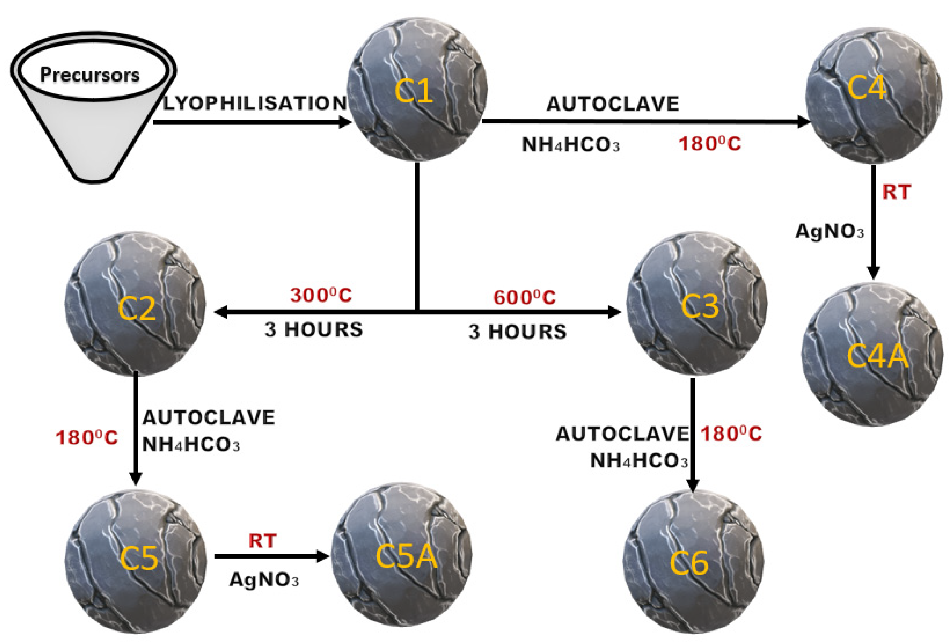
The bacterial species employed was Pseudomonas aeruginosa (P. aeruginosa), chosen for its well-known ability to form biofilms even under nutrient-depleted conditions, such as those found in hospital water systems [22]. The standard strain P. aeruginosa ATCC 27853 was inoculated on Columbia agar with 5% sheep blood (Oxoid Ltd., Hampshire, UK) in 90 mm Petri plates and incubated aerobically at 37 °C for 24 h. A 0.5 MacFarland (1.5 × 10⁸ CFU/mL) bacterial suspension in tryptone soy broth (Oxoid Ltd., UK) was prepared from this culture using a densitometer. Each sample was placed in a polypropylene tube, partially submerged in bacterial suspension, and incubated aerobically at 37 °C for 5 days. The bacterial suspension in the tubes was refreshed every 24 h, and the previous day’s suspension was assessed through light microscopy (Gram stain) as well as inoculated on solid media-Columbia and Brilliance UTI agar (Oxoid Ltd., UK) and incubated aerobically at 37 °C for 24 h. After incubation, the samples were low-frequency vortexed for 1 min in 0.9% sterile saline solution to dislodge any planktonic bacteria from the samples’ pores and rinsed thoroughly resulting in samples C5B and C5AB. Figure 1 depicts a schematic representation of the aerogel manufacturing process. After 5 days, all the culture medium in contact with the aerogel was extracted, the matter in suspension was separated by centrifugation at a speed of 10,000 RPM, it was redispersed three times in 2 mL of distilled water with separation by centrifugation under the same conditions, and then it was redispersed in double-distilled water. Approx. 50 μL of suspensions were placed on aluminum foil and prepared for SEM/EDX analysis by drying with nitrogen (4.6).

2.4. Epifluorescence Microscopy Characterization

Staining was performed with a Live/Dead™ BacLight™ (Thermo Fisher Scientific, Waltham, MA, USA) viability stain kit containing the SYTO 9 and Propidium Iodide nucleic acid stains, targeting bacterial cells. A second protocol was utilized to visualize the biofilm matrix, employing Concanavalin A conjugated with Alexa Fluor 488 (1 mg/mL (w/v)) for matrix polysaccharides, and SYPRO™ Ruby stain for matrix proteins. Staining followed the manufacturer’s instructions.

2.5. Scanning Electron Microscopy Characterization

After being kept for 120 h in the bacterial suspension, parts of the C5B and C5AB samples were extracted and washed twice with 2 mL of distilled water and vortexed for 300 s in distilled water. Samples containing water were extracted and immersed directly in liquid nitrogen. Immediately after this, the samples were freeze-dried for 24 h using a condenser temperature of −50 °C. The freeze-dried samples were cut, mounted with carbon-based double adhesive tape on aluminum supports, metalized by depositing a gold layer by thermal evaporation and then characterized by scanning electron microscopy. The rest of the samples were not metalized before the SEM characterization.

2.6. Equipment Used

X-ray diffraction (XRD) studies were conducted employing an X’Pert PRO MPD apparatus (PANalytical, Almelo, The Netherlands) employing Ni-filtered Cu Kα radiation (λ = 1.54 Å). FTIR spectra were collected using a Vertex 70 FTIR instrument (Bruker, Ettlingen, Germany). Scanning electron microscopy (SEM) analysis was performed utilizing a Quanta FEG 250 (FEI, Hillsboro, OR, USA) equipped with an energy-dispersive X-ray (EDX) spectrometer and Inspect S microscope (FEI, Eindhoven, The Netherlands). Thermogravimetric analysis (TGA) was executed with a TG 209F1 Libra instrument (Netzsch, Selb, Germany), while UV–VIS measurements were conducted using a UV–VIS–NIR Lambda 950 spectrophotometer (PerkinElmer, Waltham, MA, USA) equipped with a 150 mm integrating sphere (Spectralon served as the reflectance reference) using the DRS technique. Bacterial cell concentration was determined using a DEN-1 McFarland Densitometer, (Biosan SIA, Riga, Latvia), Optical microscopic imaging was conducted on a Zeiss Axio Lab A2 microscope using a 100×/1.25 oil objective and a 470 nm LED for excitation, a 515 nm long pass emission filter and an Erc 5s camera for image acquisition.

3. Results

The properties of the materials synthesized in this study are described in parallel with carbon aerogels mentioned in the literature and summarized in Table A1 (Appendix A). These studies share common elements, including the investigation of biofilm growth on aerogels, the cellulosic nature of the precursor, the presence of slightly soluble inorganic compounds in the aerogel, the aerogel’s resistance to temperature and liquid environments, the methods of investigation, and the microorganisms used.

3.1. XRD

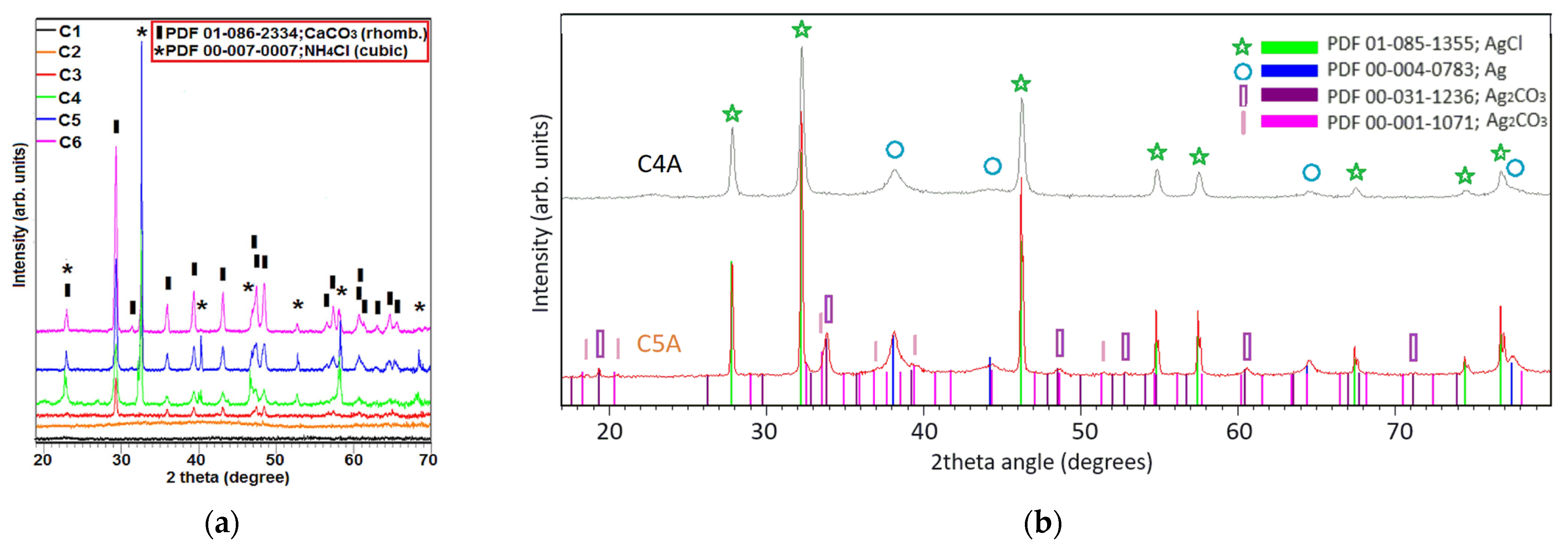
For the C1 and C2 samples, the existence of diffraction maxima (Figure 2a) is not observed although, in the case of sample C1, this was expected. Specific peaks of CaCl2 do not appear because calcium chloride, which is in low concentration, being hygroscopic, can dissolve in the atmospheric absorbed water, which leads to the absence of maxima in the XRD spectrum. In the case of sample C2, this proves that the carbonization of cellulose does not produce significant amounts of CO2 at a temperature of 300 °C, a gas that can react with CaCl2. Heating the aerogel to 600 °C leads to complete decomposition of the cellulose with the formation of CO2 [23], which reacts with CaCl2 to form CaCO3 according to the r1 reaction:
Cellulose → CO2 + H2O + carbonized fibers + by products
(NH4)2CO3 → 2 NH3 + H2O + CO2
2NH3 + H2O + CO2 + CaCl2 → CaCO3 + 2NH4Cl
CaCO3 → CaO + CO2
Another cause of CaCO3 maxima in the temperature range of 300–600 °C may be the crystallization of calcium carbonate formed at temperatures between 100 and 300 °C. The quantities of calcium carbonate formed by this reaction are extremely small. In the TG curve shown in Figure 3 the decomposition of this crystalline phase (see r4) cannot be observed for sample C1.
In the case of the C4 and C5 samples, CaCO3 was formed according to the r3 reaction, especially because of carbon dioxide resulting from the decomposition of ammonium carbonate according to reaction r2. Adding CaCO3 enhances the mechanical strength of the aerogel specifically when carbonized at 300 °C. This allows for subsequent coating with silver salts from water-based solutions. Furthermore, the aerogels synthesized at 300 °C exhibit excellent long-term stability in liquids, as demonstrated by their resilience to repeated washing cycles (Figure 4). The ammonium chloride present in the C4, C5, and C6 samples is very evident in the XRD spectra, being formed in the pressurized reactor by the r3 reaction.
For the C6 sample, CaCO3 present in the sample is due both to the cellulose carbonization process and to the carbon dioxide resulting from the breakdown of the ammonium bicarbonate (r2). For the samples impregnated with silver nitrate, the XRD spectra (Figure 2b) show the appearance of AgCl (cubic) maxima formed according to reaction r5, Ag2CO3 according to reaction r6 (only for C5A sample), and metallic silver formed according to reaction r7.
The reactions that occur when impregnating the aerogel with silver nitrate are:
NH4Cl + AgNO3 → AgCl + NH4NO3
CaCO3 + 2AgNO3 → Ag2CO3 + Ca(NO3)2
AgNO3 + reductant → Ag + by-products

3.2. EDX

The EDX spectra for the C1–C6 samples always indicate the existence of calcium and chlorine as we can observe in Figure 3.
For the samples C1–C3, the C:O ratio increases by about 6 times, which is due to the elimination of water in the carbonization process. For samples C1 and C2, the Ca:Cl atomic ratio is close to the theoretical value of 0.5. For sample C3, it increases slightly by approximately 30%. This shows that the samples do not lose HCl up to the temperature of 300 °C, and above this temperature the loss is small. In samples C4, C5, and C6, the process of carbonization leads to the increase of the C:O atomic ratio, and the process of CaCO3 formation leads to its decrease, both processes taking place simultaneously (Figure 3). After immersing the aerogels in an AgNO3 solution and repeatedly washing, only the C5A sample preserves its shape, while the C4A sample partially decomposes. For this reason, C5 and C5A samples were employed in bacterial biofilm growth assays.

3.3. SEM

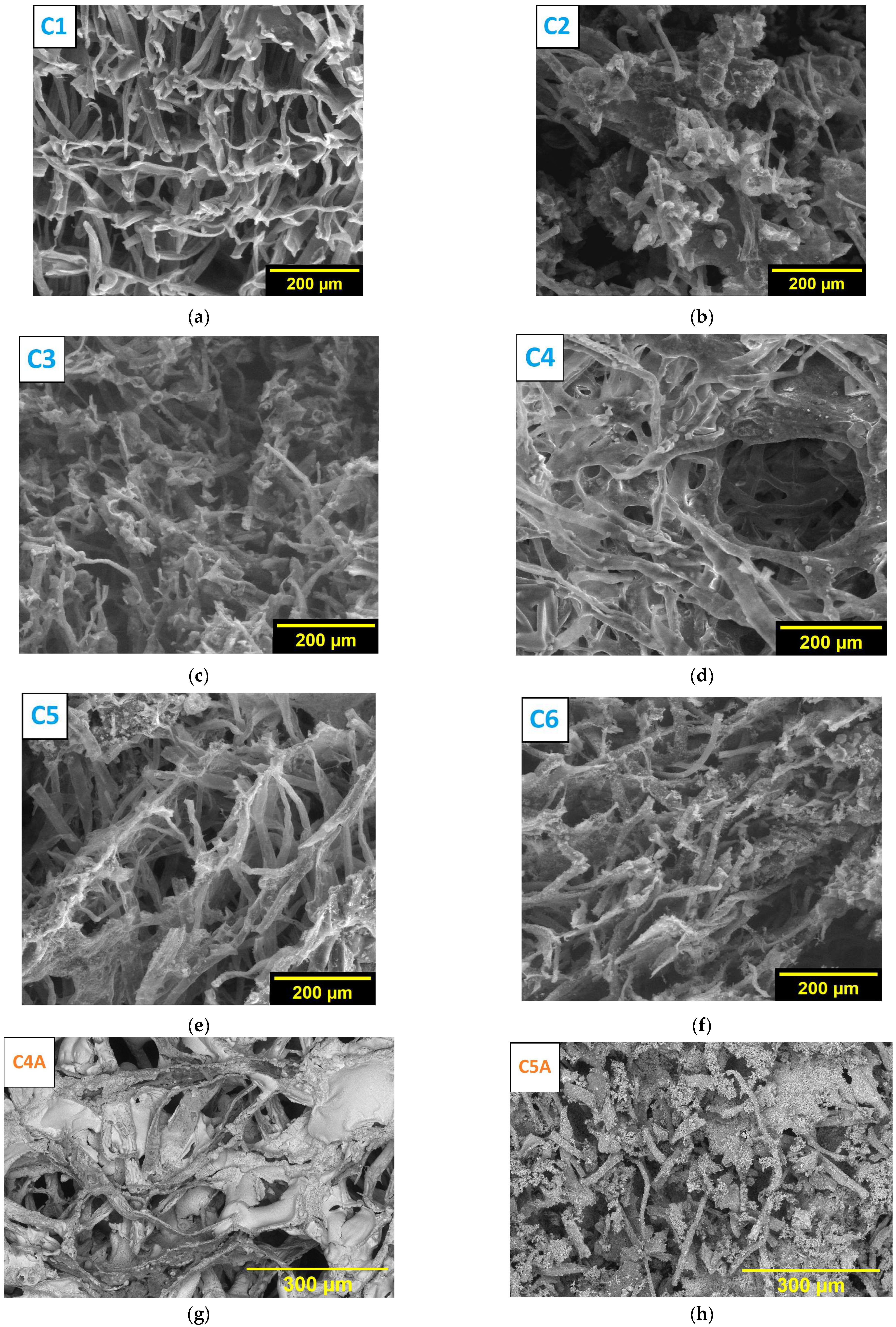
The obtained samples were characterized by analyzing SEM and from Figure 5a–h.
We observed in Figure 5a that the aerogel consists of cellulosic fibers forming pores with a maximum diameter of 100–120 µm. As the temperature increases, it can be seen from the SEM images that the structure becomes more fragile, the fibers having a superficial shell, and the inside presenting a porous structure. This is probably because, as the temperature rises, the calcium chloride dissolves in the hydration water, wetting the fibers. When the temperature rises above 150 °C, the process of carbonization of the fibers begins simultaneously with the loss of water of crystallization of calcium chloride, as we can observe in Figure 5b,c. Since the carbonization takes place with the removal of CO2, it reacts with CaCl2 with the formation of calcium carbonate, as indicated by the XRD spectra in the case of sample C3. Since the formation of CaCO3 is not observed from XRD, in the case of sample C2, we can deduce that the mentioned reaction takes place in the temperature range 300–600 °C. For samples C4 and C5 from Figure 5d,e, a strongly different structure is not observed from that of samples C1 and C2 from which they come, except for the average pore size, which seems smaller due to the contraction of the sample during the cellulose decomposition. However, no distinct crystals are observed, but particles with sizes on the order of a few microns can be observed, suggesting that the samples are heterogeneous.
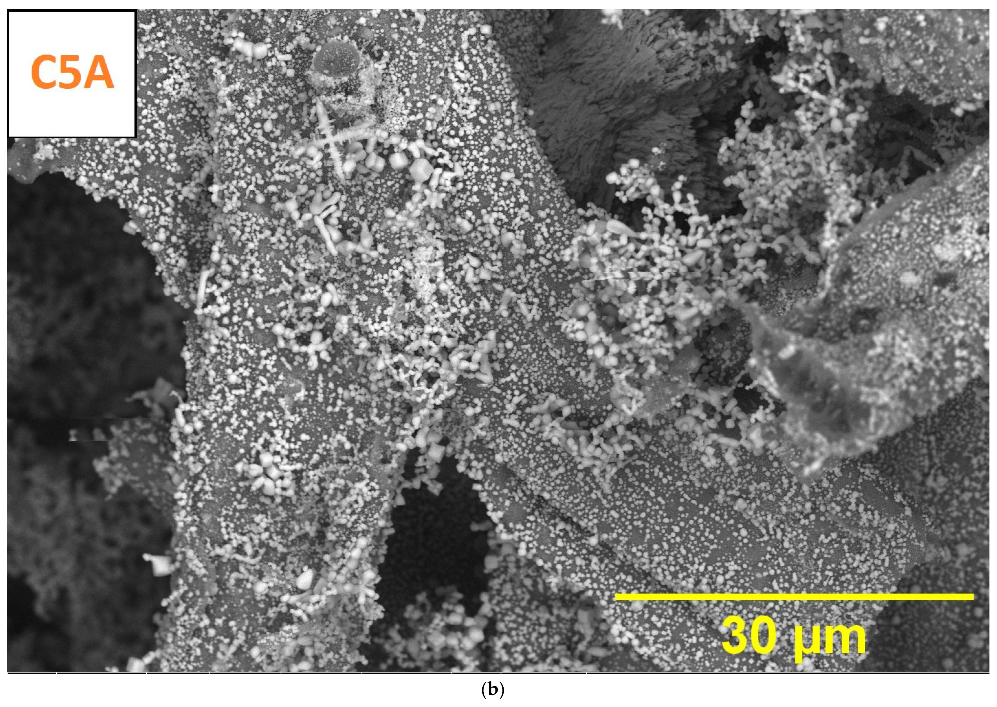
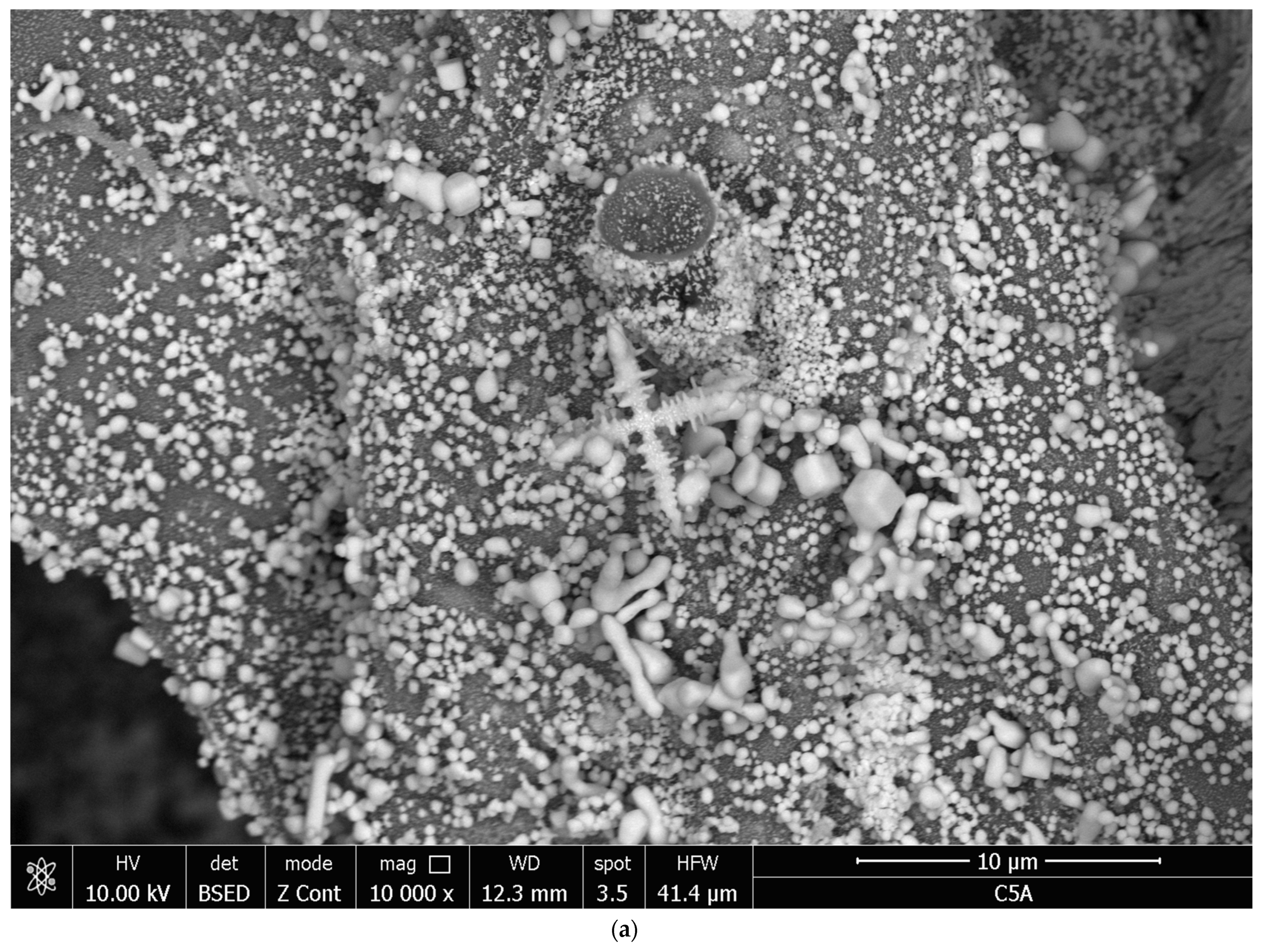
In the case of sample C6 (Figure 5f), a very fragile structure of carbon fibers covered with inorganic particles, some of them agglomerated, can be observed. At higher magnification, particles with a size of 1–2 μm are observed heterogeneously distributed in the aerogel mass. The C4A sample (Figure 5g and Appendix A, Figure A1a) is strongly covered with a thick layer composed of Ag and AgCl nanoparticles, with this coating being uneven. The aspect of the C5A sample (Figure 5h and Appendix A, Figure A2a,b) is markedly different, as the fibers are uniformly covered with submicron particles ranging between 30 and 300 nm, sizes larger than those in the case of the C4A sample.

3.4. TGA and DRS

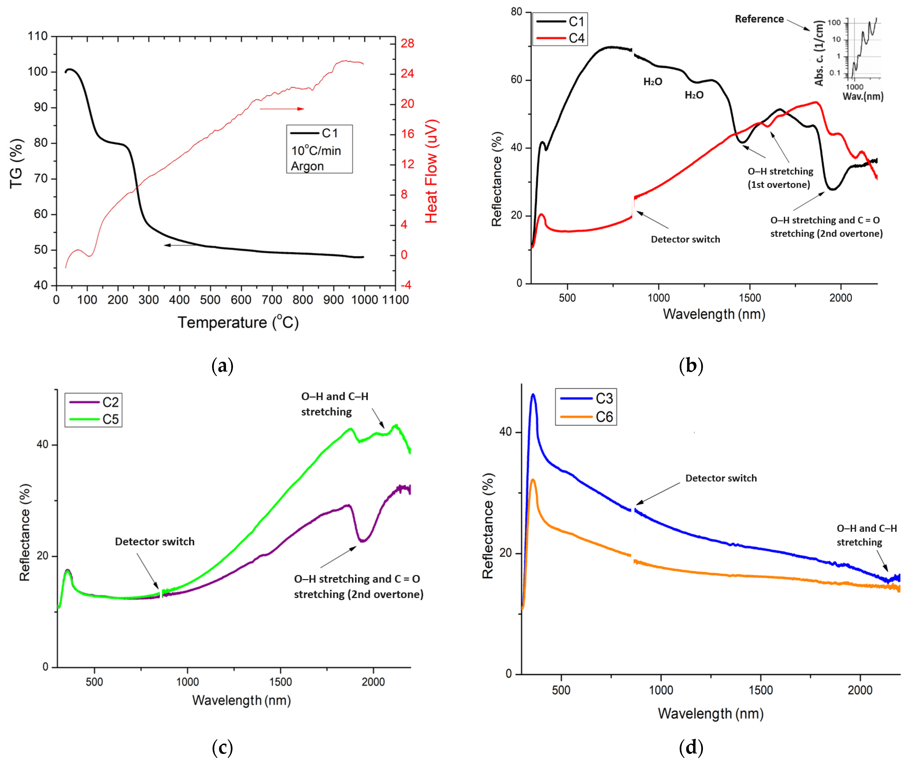
The C1 aerogel sample was also characterized by thermal analysis in an Argon 5.0 environment, at a temperature increase rate of 10 °C/min. The TG analysis (Figure 6a) highlights two stages of mass loss, the first due to water loss from the CaCl2–H2O system, up to the temperature of 150 °C [24] and the second to the cellulose dehydration process, a process that is almost complete at the temperature of 450 °C. The endothermic effect associated with the first process is greater. At a temperature of approximately 850 °C, a broad and small endothermic effect is observed, which is most likely due to the melting of CaCl2 salt that occurs at 772 °C. The DRS spectra presented in Figure 6b–d show the carbonation of the cellulose fibers for the C2–C6 samples. For hemicellulose and cellulose in the C1 sample, the absorption band in the range 1400–1660 nm is linked to O–H stretching (first overtone) [25]. Water absorption coefficients in the NIR region are shown in Figure 6b-inset [26]. Heat-treating the cellulose at 300 °C resulted in incomplete carbonization (Figure 6c), leaving behind C=O and O–H bonds. The band observed at approximately 1920 nm can be attributed to –OH and –C=O groups [27]. The signal at approximately 2100 nm is confidently attributed to the combination of stretching vibrations of O–H and C–H [28]. After heat treatment at 600 °C the FT–IR spectra indicate almost complete carbonization of the cellulose fibers by the almost complete disappearance of the absorption bands (Figure 6d).

3.5. FT–IR

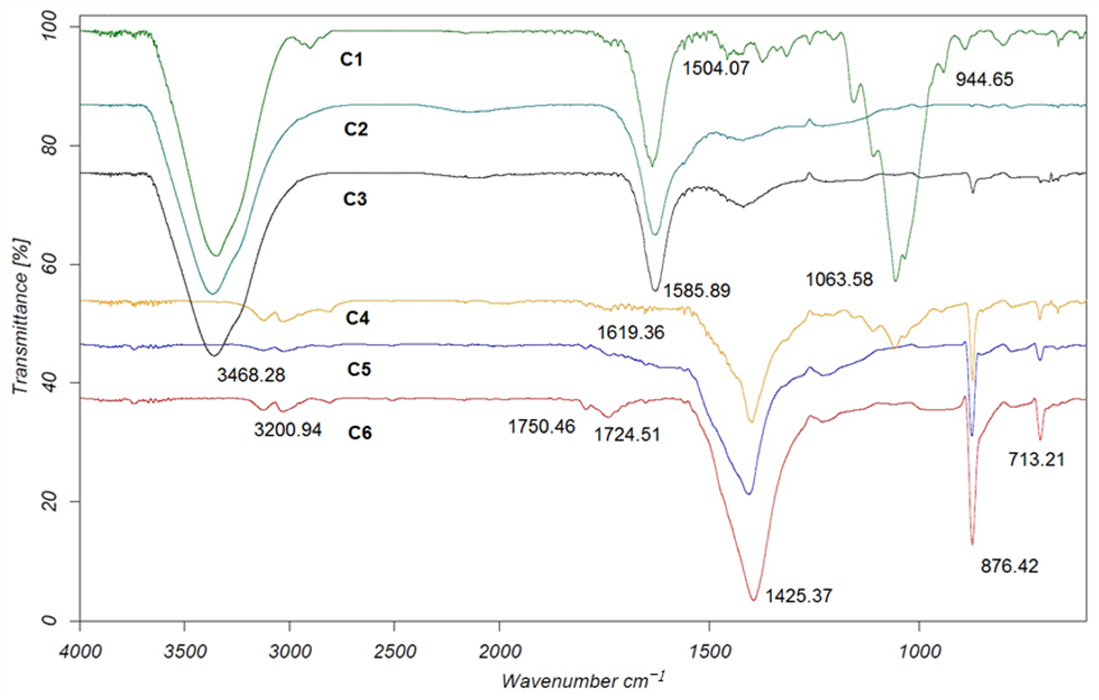
C1–C6 samples were analyzed by FT–IR and represented in Figure 7. The typical bands attributed to cellulose were observed in the region 1530–900 cm−1 [29]. The band at 1063.58 cm−1 in the case of sample C4 is specific to the vibration of the C–O– of secondary alcohols and ethers, functions existing in the cellulose chain. The strong decrease in band intensity is attributed to carbonization of cellulose only by hydrothermal treatment at 180 °C. This carbonization corresponds to the decrease in diffuse reflectance intensity in the visible range (Figure 6b) for the C4 sample. The absorption band at 1585.89 cm−1 and the maximum at 3468.28 cm−1 are due to vibrations of the O–H group in CaCl2 · xH2O [30], because these bands are not split, as in the case of crystalline CaCl2. The absorption bands at 713.21, 876.42 and 1425.37 cm−1 for samples C3–C6 belong to vibration modes υ4, υ2 and υ3 of CaCO3–calcite [31]. The FT–IR spectrum shows the formation of traces of CaCO3 through the absorption band at 1425.37 cm−1 even after heat treatment of the aerogel at 300 °C; the process is strongly intensified with increasing temperature and especially after treatment of the material in CO2. The appearance of vibration bands of N–H groups in the range 3000–3200 cm−1 highlights the formation of NH4Cl [32] after treatment in a CO2 and NH3 atmosphere, confirming XRD analysis.

3.6. Growing of Biofilms on Aerogels

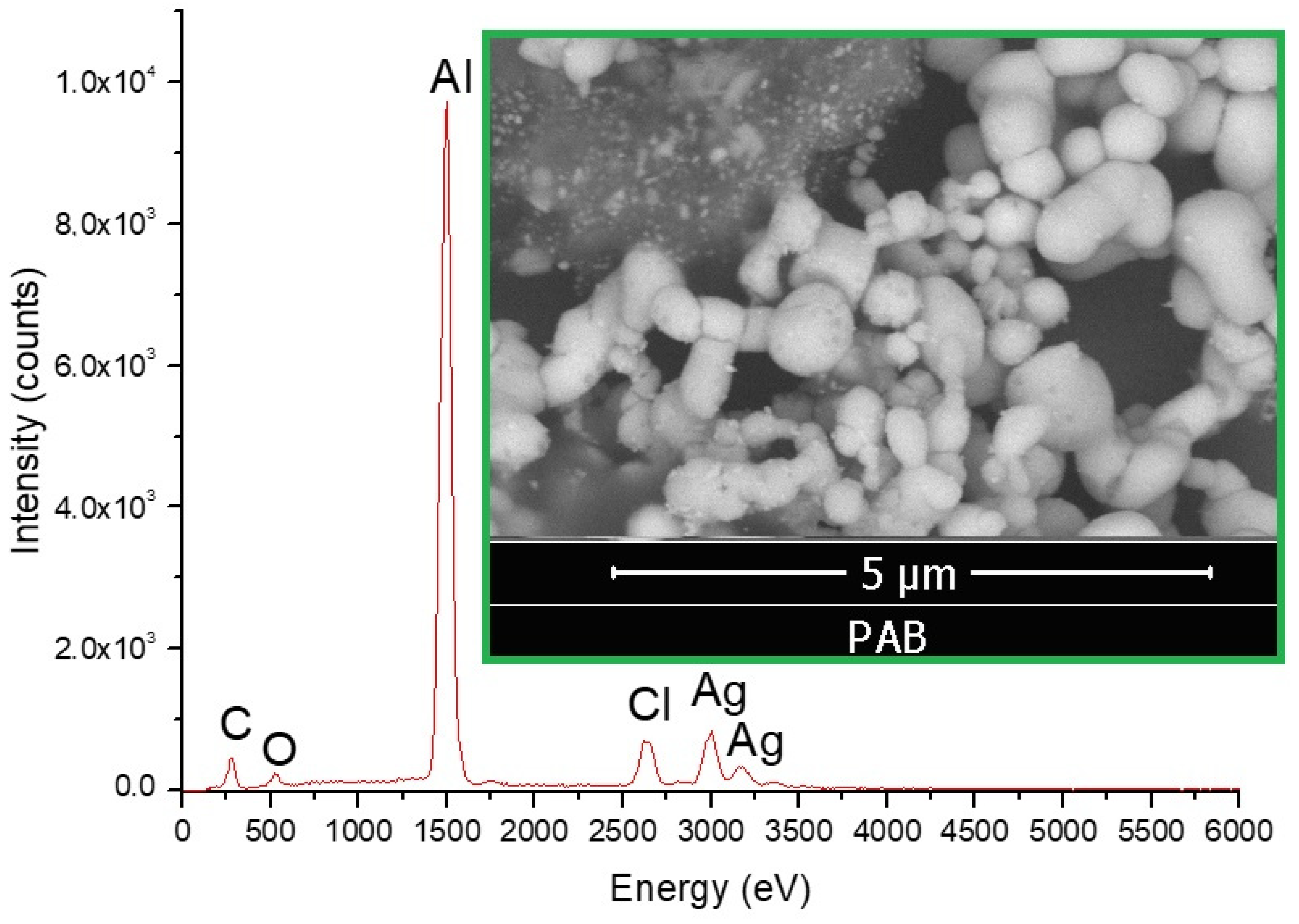
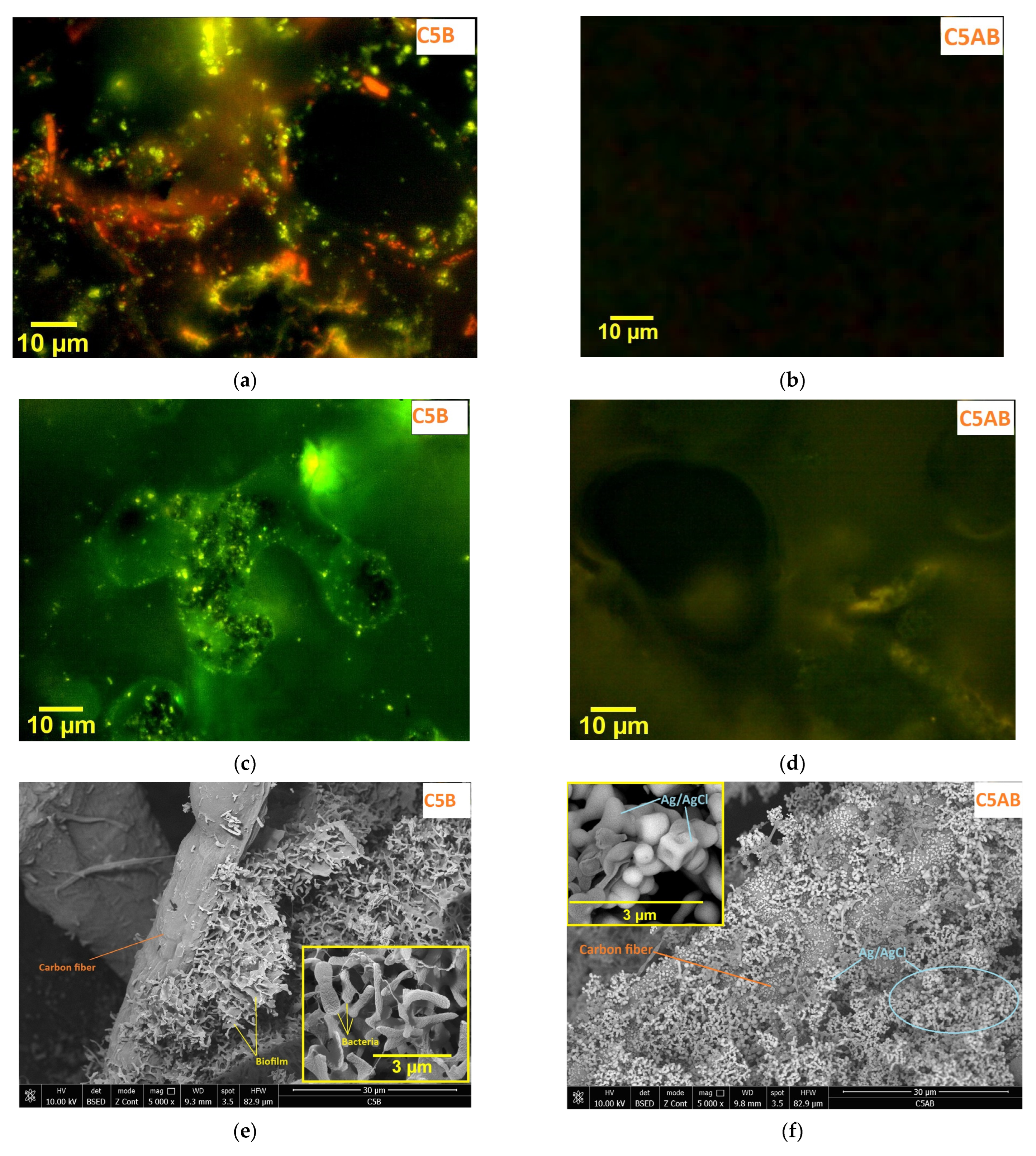
For sample C5B, the biofilm architecture appears heterogeneous, with different degrees and distributions of bacterial viability (Figure 8a). Due to the porous nature of the aerogel, the biofilm may exhibit distinct microenvironments with differential nutrient availability, oxygenation levels, and metabolic activities, influencing bacterial viability and distribution. No viable bacteria can be detected for the Ag and AgCl decorated sample (Figure 8b). The P. aeruginosa biofilm can be visualized in the pores of the C5B sample. There is a clear predominance of polysaccharides over proteins within the biofilm matrix. This is expected given that P.aeruginosa is known for producing alginate-rich biofilms (Figure 8c). No biofilm or similar structure could be visualized on the C5AB sample (Figure 8d). SEM images (Figure 8e,f) confirm the presence of bacterial biofilm in C5B and its absence in C5AB. The presence of AgCl in the sample is indicated by the specific chlorine peaks in EDX spectra (Figure 8g), after maintaining the biofilm in the nutrient medium for over 120 h.
The bacterial suspension was consistently free of bacteria after 24 h of contact with the C5AB sample. The suspension in which C5AB was submerged had 0 UFC/mL after the fifth day of contact with refreshed bacterial suspensions, compared to 5 × 10⁵ UFC/mL for the C5B sample. Taking into account the initial inoculum of 1.5 × 10⁸, this corresponds to a 2.477 log reduction for C5B and complete bacterial elimination for C5AB.
The aerogel serves as a porous and opaque support throughout the UV–VIV–NIR range, preventing the photochemical reduction of AgCl. This results in a high concentration of Ag+ ions in both the aerogel pores and the nutrient medium.

4. Discussion

In the absence of CaCl2, the pressure obtained by the decomposition of ammonium carbonate and the evaporation of water was calculated to be about 11 bar at a temperature of 180 °C. Therefore, this synthesis can be used to create three distinct types of samples that are based on three-dimensional structures of cellulose fibers: partially carbonized cellulose fibers, amorphous carbon, and calcium carbonate in combination with the latter two. Although the decomposition process of CaCO3 is endothermic, in the absence of mass loss, TG–DTA analysis demonstrates the physical rather than chemical nature of the process. Due to the high heating rate, necessary for DTA identification of minor thermal processes, and the low thermal conductivity coefficient of the aerogel, the peak has a rather large width between 780 and 920 °C and a shift of approximately 50 °C compared to the theoretical value. Therefore, the amounts of the important CaCO3 are achieved mainly through reaction with CO2 gaseous in a hydrothermal environment and to a very small extent by reaction between CaCl2 and CO2 produced by the carbonization of cellulose, due to the absence of a basic medium and the low contact time between CaCl2 and CO2 in the case of reaction at ambient pressure. EDX analysis of samples C2–C6 indicates that the higher the amount of carbonate, the lower the C:O ratio. This is the explanation for why the C:O ratio in the C6 sample is lower than that of the C3 sample from which it originates. For the same samples, the Ca:Cl ratio increases strongly with increasing temperature, which demonstrates the fact that the cellulosic fibers retain less ammonium chloride the more advanced the carbonization. The Ca:Cl ratio increases strongly after treatment of C2 and C3 samples in CO2 and NH3 although the XRD spectra indicate the appearance of strongly crystallized ammonium chloride which forms in samples C5 and C6. FT–IR analysis indicates the presence of NH4Cl through bands in the range 3000–3200 cm−1. This phenomenon is unexpected, but it is due to the relatively large crystals of high solubility NH4Cl, uniformly distributed, and the small crystals of CaCO3 with low solubility, uniformly distributed. The heterogeneous nature of the samples therefore affects the EDX analysis, by different total cross-sections of particles belonging to the various crystalline phases with the excitation electron beam.
The FT–IR analysis highlights, as expected, the disappearance of the broad band of water in the range 3000–3700 cm−1 for all samples treated in CO2 medium but not in samples treated at 300 and 600 °C only. Since at 600 °C the carbonization of the cellulose fibers is complete, as shown by both the TG curve and the DRS spectra, the absorption band in the range 3000–3700 cm−1 is due to the CaCl2 hydration water for the samples C1–C3. Due to the highly hygroscopic nature of this substance and its distribution on the surface of the fibers, it rapidly absorbs water from the atmosphere. After treatment in CO2 and NH3 vapor, CaCl2 reacts with the formation of non-hydroscopic CaCO3, the specific O-H vibration bands in the range 3000–3700 cm−1 disappearing.
After immersing aerogels with considerable levels of calcite in an AgNO3 solution, we observed that there was no Ag2CO3 in the C4A sample due to aerogel decomposition in the liquid.
During aerogel decomposition, CaCO3 particles detach from the surface of the fibers and are lost during washing. The size of Ag and AgCl nanoparticles was calculated from Scherrer’s relation. The Debye–Scherrer equation was utilized to estimate the average crystallite sizes (D) of both Ag and AgCl.
D = Kλ/βcosθ
In this equation, K is a constant associated with crystallite shape (assumed to be 0.9 for spherical or cubic crystallites), λ represents the X-ray wavelength, β represents the line broadening of the peak at half of the maximum intensity (FWHM) post-instrumental line broadening subtraction (the instrument’s line broadening was determined using a polycrystalline silicon standard), and θ indicating the Bragg angle. For sample C4A, the average crystallite sizes were calculated to be 9.2 nm for Ag and 46.1 nm for AgCl while, for sample C5A, the respective sizes were 10.7 nm for Ag and 230.3 nm for AgCl. The small size of Ag particles leads to the idea of multiple heterogeneous nucleation centers on the surface of AgCl fibers or nanocrystals. The average size of AgCl nanocrystals increases more than 5-fold when aerogel integrity is preserved after immersion (case of C5A). This is probably due to the slower diffusion of AgNO3 into aerogel pores during impregnation, which increases the concentration gradient between the inside and outside of the aerogel. As the FT–IR spectra show, the surface of the fibers obtained at 300 °C is less hydrophilic than that obtained at 180 °C, the aqueous solution wetting the aerogel cells more slowly. Inside the aerogel, the concentration of AgNO3 is lower than outside, the solution supersaturation is lower and the density of nucleation centers is lower. This leads to the formation of larger AgCl crystals. The density of heterogeneous nucleation centers on the fibers is also higher in the case of the C4A sample due to the higher density of polar groups of type R–OH, C=O, etc. which can stimulate the initial heterogeneous nucleation process. In the case of C4A, the AgCl precipitate sediments, with a large part of the particles not having access to the electrolyte solution. This inhibits the AgCl recrystallization process, as the particle size is smaller. In contrast, in the case of the C5A sample, the particles are distributed three-dimensionally in the aerogel cells in a thin layer, which facilitates the recrystallization process. The strongly rounded edges of the AgCl cubes suggest that the silver reduction process can also take place on the surface of these large AgCl cubes, which become embedded in a mass of very fine silver particles.
Details of the morphology of C4A and C5A samples at higher magnifications are given in Appendix A. A very interesting aspect observed in the SEM images of the C5A sample (see Figure A2a,b) is that most of the particles attached to the fibers are spherical in shape and are generally smaller than 500 nm in size and rarely agglomerated, while the particles not attached to the fibers are between 200 nm and 1.5 μm in size, sometimes having a cubic shape with rounded corners and edges. These particles form extended three-dimensional agglomerations. It is therefore possible that carbon fibers represent heterogeneous nucleation centers for the crystallization of silver nanoparticles. Cubic submicron particles support XRD data showing AgCl crystallization in cubic systems with average crystal sizes larger than 100 nm.
Upon keeping the C5A sample in the culture medium, the faces of the cubic particles have been observed to partially collapse towards the inside of the cubes in several cases. This phenomenon is visible in the C5AB sample (Figure 8f inset) but not in the C5A sample (Figure A2a,b). The formation of a metallic silver shell on the outside of the particle through the reduction of AgCl with organic substances in the culture medium is likely to be the cause of this phenomenon. Another explanation is the dissolving of AgCl inside the cubic particles due to the silver complex with poly-carboxylated ligands in the culture environment.
Due to the dissolution of the inside of the cube, the silver walls of the cubes collapse. Bacteria of the Pseudomonas family are known to concentrate nitrogen and phosphorus in the culture medium [33]. EDX revealed a relatively large amount of N and P in the biofilm-positive ample (C5B). This finding can be explained by the known presence of nitrogen in biofilms, in molecules such as amino acids and nucleic acids which can be found in both bacteria and the extracellular matrix (commonly formed of polysaccharides, proteins, and extracellular DNA) [34]. While P. aeruginosa is not known to fixate nitrogen, this mechanism has been described in the closely related P. stutzeri [35]. P. aeruginosa can uptake phosphorus during biofilm formation [36] and can accumulate phosphorus granules under phosphate-depleted conditions [37]. On the aerogel surface, silver-coated fibers inhibit the development of bacterial biofilms as well as planktonic (free-floating) bacteria.
No colonies were observed on agar plates inoculated with bacterial suspension after 24 h of contact with the C5AB sample, demonstrating a practically infinite log reduction, not only on the initial inoculum but on each of the five subsequent daily replacements of fresh bacterial suspension. EDX analysis also shows no evidence of nitrogen and phosphorus, which supports the SEM observations for C5AB sample. The persistent disinfection capacity and the inhibition of biofilm development demonstrated through fluorescence microscopy suggest a potential for biological water purification using the silver-coated aerogel. The silver-free aerogel, by comparison, inhibited bacterial growth by log 2.477, which is too small of an effect to be considered disinfectant, and allowed the formation of a biofilm on its surface, thus not being suitable for most filtering applications.
The SEM images and EDX analysis of the particles in suspension, separated after 5 days from the liquid culture medium in contact with Ag-decorated surfaces, did not reveal the presence of bacteria, bacterial biofilm, or phosphorus. Instead, only micrometric and sub-micrometer particles containing silver, chloride, and some fragments of carbonized fibers were observed (see Appendix A, Figure A3). Ag particles can be formed according to reaction r7, where organic substances from the culture medium or the cell membrane act as reducing agents. Some of the carbonized materials and Ag particles found in suspension could originate from aerogel fiber dislocation. The colorless suspension indicates a low concentration of particles. These analyses are consistent with the culture assessment, which did not show bacterial colony development. This demonstrates that the microbes in contact with the aerogel were completely inactivated shortly after contact, preventing both planktonic bacterial growth in the liquid culture medium and the formation of bacterial biofilms on the aerogel’s surface.
Further, the use of this type of aerogel in water disinfection processes and the production of composite aerogels containing various metal salts for the adsorption of toxic gases in low-pressure drop gas filters is considered.

5. Conclusions

In this paper, complex aerogel composites based on carbonaceous fibers obtained by carbonization of cellulose fibers in an inert medium have been synthesized and studied. These aerogels were decorated with CaCO3 by a process rarely encountered in the literature of hydrothermal treatment of aerogels in a gas mixture containing CO2, NH3, and H2O under pressure. Aerogels have been characterized by XRD, SEM, EDX, DRS, TG/DTA, FT–IR, and epifluorescence microscopy, to determine the mechanisms, and chemical reactions underlying the materials and the effects of aerogel compositions on the development of the bacterial biofilm of P. aeruginosa.
Using ion exchange reactions, the aerogels were then decorated with Ag, AgCl, and Ag2CO3, proving the possibility of obtaining numerous composite aerogels by this type of reaction. Bacterial biofilm growth tests were performed on silver and silver-salt decorated and undecorated aerogels, proving by SEM, EDX, and Epifluorescence microscopy the ability of silver-free aerogels to support bacterial biofilm formation and the strong bactericidal properties of silver-decorated aerogels, respectively.

Author Contributions

Conceptualization, R.B.; methodology, R.B.; software, C.M. and F.S.R.; validation, R.B. and P.Ș.; formal analysis, F.S.R., C.M. and I.H.; investigation, F.S.R., I.H., C.M., P.Ș. and R.B.; resources, C.M.; data curation, R.B., P.Ș. and F.S.R.; writing—R.B., F.S.R. and C.M.; visualization, F.S.R.; supervision, R.B.; project administration, R.B.; funding acquisition, R.B. All authors have read and agreed to the published version of the manuscript.

Funding

This work was supported by a grant of the Ministry of Research, Innovation and Digitization, CCCDI-UEFISCDI, project number PN-III-P2-2.1-PED-2021-2541, within PNCDI III and PN 40N/2019.

Institutional Review Board Statement

Not applicable.

Informed Consent Statement

Not applicable.

Data Availability Statement

The data presented in this study are available in this article.

Acknowledgments

The authors would like to thank to Cristian Casut, Daniel Ursu, and Corina Orha for the XRD and EDX measurements.

Conflicts of Interest

The authors declare no conflicts of interest.

Appendix A

Table A1. Properties of the materials synthesized described in parallel with carbon aerogels mentioned in the literature.
Reference1M.Bac.eff.det8BGonA9IISinA10HTR11LR12A.TypeOrganism
Henschen et al., 2016 [38]5BCA;
97–99%
nononoyesCellulose/Poly-vinylamine + polyacrylic acidE. coli
Uddin et al., 2017 [39]5BCA;
≈50–> 99%
nononoyesCellulose/CNF, Lysozyme, AgE. coli;
S. aureus.
Zhang et al., 2019 [40]6Impreg;
>99%
noyesnoyesCellulose/NaClOS. aureus;
E. coli
Revin et al., 2020 [14]7Inhib. Z.;
>99% (at 3 mm)
noyesnoNACellulose of bacterial origin/Na fusidate.S. aureus.
Wang et al., 2022 [41]7Inhib.Z
>99% (at 3 mm),
noyesnoNACellulose/AgOE. coli;
S. aureus
Proctor et al. 2023 [42]2EFM
5BCA, ≈99%
yesnononoPVA/furaneol and sotolonP. aeruginosa.
Yue et al., 2018 [43]nononoyesyesCarbon from celluloseno
This work3SEM/4EDX; 2EFM; 5BCA, >99%yesyesyesyes Carbon from cellulose/Ag, Ag+P. aeruginosa
Abbreviations: 1Method for determining the bactericidal effect (M.Bac.eff.det); 2Epi-Fluorescence Microscopy (EFM); 3Scanning electron microscopy (SEM); 4Energy Dispersive Spectroscopy (EDX); 5Bacteria Cultivation on Petrifilm (Agar) (BCA); 6Aerogel impregnation with bacteria suspension (Impreg.); 7Measurement of the inhibition zone on the plates (Inhib. Z.); 8Biofilm growth on aerogel (BGonA); 9Insoluble inorganic salts in aerogel (IISinA); 10High temperature (300 °C) resistance (HTR); 11Long term aerogel structure integrity in liquid media or resistance in liquid (LR); 12Type of aerogel (A.Type).
Figure A1. SEM micrograph of the C4A sample.
Ceramics 07 00051 g0a1
Figure A2. (a) SEM micrograph of the C5A sample at high magnification; (b) SEM micrograph of the C5A sample at lower magnification.
Ceramics 07 00051 g0a2aCeramics 07 00051 g0a2b
Figure A3. SEM images and EDX spectra of particles collected from liquid culture medium of C5AB sample.
Ceramics 07 00051 g0a3

References

  1. Pierre, A.; Pajonk, G. Chemistry of Aerogels and Their Applications. Chem. Rev. 2003, 102, 4243–4265. [Google Scholar] [CrossRef]
  2. Androulidakis, C.; Kotsidi, M.; Gorgolis, G.; Pavlou, C.; Sygellou, L.; Paterakis, G.; Koutroumanis, N.; Galiotis, C. Multi-Functional 2D Hybrid Aerogels for Gas Absorption Applications. Sci. Rep. 2021, 11, 13548. [Google Scholar] [CrossRef]
  3. Kara, I.T.; Kiyak, B.; Gunes, N.C.; Yucel, S. Life Cycle Assessment of Aerogels: A Critical Review. Res. Sq. 3 2024. [Google Scholar] [CrossRef]
  4. Leventis, N.; Sotiriou-Leventis, C.; Chandrasekaran, N.; Mulik, S.; Chidambareswarapattar, C.; Sadekar, A.; Mohite, D.; Mahadik, S.S.; Larimore, Z.J.; Lu, H.; et al. Isocyanate-Derived Organic Aerogels: Polyureas, Polyimides, Polyamides. MRS Online Proc. Libr. 2011, 1306, 301. [Google Scholar] [CrossRef]
  5. Chidambareswarapattar, C.; Xu, L.; Sotiriou-Leventis, C.; Leventis, N. Robust Monolithic Multiscale Nanoporous Polyimides and Conversion to Isomorphic Carbons. RSC Adv. 2013, 3, 26459–26469. [Google Scholar] [CrossRef]
  6. Chang, H.; Wang, G.; Yang, A.; Tao, X.; Liu, X.; Shen, Y.; Zheng, Z. A Transparent, Flexible, Low-Temperature, and Solution-Processible Graphene Composite Electrode. Adv. Funct. Mater. 2010, 20, 2893–2902. [Google Scholar] [CrossRef]
  7. Ibrahim, W.L.; Şar, H.; Ay, F.; Kosku Perkgöz, N. Enhanced Performance of Supercapacitors Based on Rotationally Stacked CVD Graphene. J. Appl. Phys. 2022, 131, 164302. [Google Scholar] [CrossRef]
  8. Kéri, M.; Nyul, D.; László, K.; Novák, L.; Bányai, I. Interaction of Resorcinol-Formaldehyde Carbon Aerogels with Water: A Comprehensive NMR Study. Carbon 2022, 189, 57–70. [Google Scholar] [CrossRef]
  9. Sadjadi, S. Emerging Carbon Materials for Catalysis; Elsevier Science: Amsterdam, The Netherlands, 2020; ISBN 978-0-12-817561-3. [Google Scholar]
  10. Zhang, L.; Lei, Y.; He, P.; Wu, H.; Guo, L.; Wei, G. Carbon Material-Based Aerogels for Gas Adsorption: Fabrication, Structure Design, Functional Tailoring, and Applications. Nanomaterials 2022, 12, 3172. [Google Scholar] [CrossRef] [PubMed]
  11. Aylaz, G.; Okan, M.; Duman, M.; Aydin, H.M. Study on Cost-Efficient Carbon Aerogel to Remove Antibiotics from Water Resources. ACS Omega 2020, 5, 16635–16644. [Google Scholar] [CrossRef]
  12. El Miri, N.; El Achaby, M.; Fihri, A.; Larzek, M.; Zahouily, M.; Abdelouahdi, K.; Barakat, A.; Solhy, A. Synergistic Effect of Cellulose Nanocrystals/Graphene Oxide Nanosheets as Functional Hybrid Nanofiller for Enhancing Properties of PVA Nanocomposites. Carbohydr. Polym. 2016, 137, 239–248. [Google Scholar] [CrossRef] [PubMed]
  13. Shruthi, T.K.; Kumar, M.S.; Arjunan, M.; Pratap, A.; Chandrasekaran, N. Graphene Oxide Aided Structural Tailoring of 3-D N-Doped Amorphous Carbon Network for Enhanced Energy Storage. RSC Adv. 2015, 5, 93423–93432. [Google Scholar] [CrossRef]
  14. Revin, V.V.; Nazarova, N.B.; Tsareva, E.E.; Liyaskina, E.V.; Revin, V.D.; Pestov, N.A. Production of Bacterial Cellulose Aerogels with Improved Physico-Mechanical Properties and Antibacterial Effect. Front. Bioeng. Biotechnol. 2020, 8, 603407. [Google Scholar] [CrossRef] [PubMed]
  15. Gan, G.; Li, X.; Fan, S.; Wang, L.; Qin, M.; Yin, Z.; Chen, G. Carbon Aerogels for Environmental Clean-Up. Eur. J. Inorg. Chem. 2019, 2019, 3126–3141. [Google Scholar] [CrossRef]
  16. Keshavarz, L.; Ghaani, M.R.; MacElroy, J.M.D.; English, N.J. A Comprehensive Review on the Application of Aerogels in CO2-Adsorption: Materials and Characterisation. Chem. Eng. J. 2021, 412, 128604. [Google Scholar] [CrossRef]
  17. Wang, J.; Wan, Y.; Xun, X.; Zheng, L.; Zhang, Q.; Zhang, Z.; Xie, Y.-X.; Luo, H.; Yang, Z. Engineering Bacteria for High-Performance Three-Dimensional Carbon Nanofiber Aerogel. Carbon 2021, 183, 267–276. [Google Scholar] [CrossRef]
  18. Dong, H.; Qiang, Z.; Richardson, S.D. Formation of Iodinated Disinfection Byproducts (I-DBPs) in Drinking Water: Emerging Concerns and Current Issues. Acc. Chem. Res. 2019, 52, 896–905. [Google Scholar] [CrossRef] [PubMed]
  19. Sánchez-Polo, M.; Rivera-Utrilla, J.; Salhi, E.; von Gunten, U. Removal of Bromide and Iodide Anions from Drinking Water by Silver-Activated Carbon Aerogels. J. Colloid Interface Sci. 2006, 300, 437–441. [Google Scholar] [CrossRef] [PubMed]
  20. Banica, R.; Taranu, B.; Ladasiu, C.; Hulka, I.; Linul, P. Three-Dimensional Porous Electrode Based on Silver Nanowires for Hydrogen Sulfide Detection. Mater. Lett. 2021, 304, 130720. [Google Scholar] [CrossRef]
  21. Linul, P.; Bănică, R.; Grad, O.; Linul, E.; Vaszilcsin, N. Highly Electroconductive Metal-Polymer Hybrid Foams Based on Silver Nanowires: Manufacturing and Characterization. Polymers 2024, 16, 608. [Google Scholar] [CrossRef]
  22. Loveday, H.P.; Wilson, J.A.; Kerr, K.; Pitchers, R.; Walker, J.T.; Browne, J. Association between Healthcare Water Systems and Pseudomonas Aeruginosa Infections: A Rapid Systematic Review. J. Hosp. Infect. 2014, 86, 7–15. [Google Scholar] [CrossRef]
  23. Shen, D.; Xiao, R.; Gu, S.; Zhang, H. The Overview of Thermal Decomposition of Cellulose in Lignocellulosic Biomass. In Cellulose—Biomass Conversion; Kadla, J., Ed.; InTech: Houston, TX, USA, 2013; ISBN 978-953-51-1172-6. [Google Scholar]
  24. Van Essen, V.M.; Cot Gores, J.; Bleijendaal, L.P.J.; Zondag, H.A.; Schuitema, R.; Bakker, M.; Van Helden, W.G.J. Characterization of Salt Hydrates for Compact Seasonal Thermochemical Storage. In Proceedings of the ASME 2009 3rd International Conference on Energy Sustainability, San Francisco, CA, USA, 19–23 July 2009; ASMEDC: San Francisco, CA, USA, 2009; Volume 2, pp. 825–830. [Google Scholar]
  25. Huang, A.; Li, G.; Fu, F.; Fei, B. Use of Visible and Near Infrared Spectroscopy to Predict Klason Lignin Content of Bamboo, Chinese Fir, Paulownia, and Poplar. J. Wood Chem. Technol. 2008, 28, 194–206. [Google Scholar] [CrossRef]
  26. Segelstein, D.J. The Complex Refractive Index of Water. Ph.D. Thesis, University of Missouri, Kansas City, MI, USA, 1981. [Google Scholar]
  27. He, W.; Hu, H. Rapid Prediction of Different Wood Species Extractives and Lignin Content Using Near Infrared Spectroscopy. J. Wood Chem. Technol. 2013, 33, 52–64. [Google Scholar] [CrossRef]
  28. Wójciak, A.; Kasprzyk, H.; Sikorska, E.; Krawczyk, A.; Sikorski, M.; Wesełucha-Birczyńska, A. FT-Raman, FT-Infrared and NIR Spectroscopic Characterization of Oxygen-Delignified Kraft Pulp Treated with Hydrogen Peroxide under Acidic and Alkaline Conditions. Vib. Spectrosc. 2014, 71, 62–69. [Google Scholar] [CrossRef]
  29. Abderrahim, B.; Abderrahman, E.; Mohamed, A.; Fatima, T.; Abdesselam, T.; Krim, O. Kinetic Thermal Degradation of Cellulose, Polybutylene Succinate and a Green Composite: Comparative Study. World J. Environ. Eng. 2015, 3, 95–110. [Google Scholar]
  30. Sujith Karunadasa Dehydration of Calcium Chloride as Examined by High-Temperature X-Ray Powder Diffraction. Available online: https://acrobat.adobe.com/id/urn:aaid:sc:EU:5885d871-ee39-43c2-a7e0-f577959ab8cd (accessed on 10 April 2024).
  31. Andersen, F.A.; Brečević, L.; Beuter, G.; Dell’Amico, D.B.; Calderazzo, F.; Bjerrum, N.J.; Underhill, A.E. Infrared Spectra of Amorphous and Crystalline Calcium Carbonate. Acta Chem. Scand. 1991, 45, 1018–1024. [Google Scholar] [CrossRef]
  32. Pajarito, B.; Llorens, C.; Tsuzuki, T. Effects of Ammonium Chloride on the Yield of Carbon Nanofiber Aerogels Derived from Cellulose Nanofibrils. Cellulose 2019, 26, 7727–7740. [Google Scholar] [CrossRef]
  33. Lozano-González, J.M.; Valverde, S.; Montoya, M.; Martín, M.; Rivilla, R.; Lucena, J.J.; López-Rayo, S. Evaluation of Siderophores Generated by Pseudomonas Bacteria and Their Possible Application as Fe Biofertilizers. Plants 2023, 12, 4054. [Google Scholar] [CrossRef]
  34. Shree, P.; Singh, C.K.; Sodhi, K.K.; Surya, J.N.; Singh, D.K. Biofilms: Understanding the Structure and Contribution towards Bacterial Resistance in Antibiotics. Med. Microecol. 2023, 16, 100084. [Google Scholar] [CrossRef]
  35. Babaei, P.; Marashi, S.-A.; Asad, S. Genome-Scale Reconstruction of the Metabolic Network in Pseudomonas Stutzeri A1501. Mol. Biosyst. 2015, 11, 3022–3032. [Google Scholar] [CrossRef]
  36. Neznansky, A.; Blus-Kadosh, I.; Yerushalmi, G.; Banin, E.; Opatowsky, Y. The Pseudomonas Aeruginosa Phosphate Transport Protein PstS Plays a Phosphate-Independent Role in Biofilm Formation. FASEB J. 2014, 28, 5223–5233. [Google Scholar] [CrossRef] [PubMed]
  37. Takade, A.; Umeda, A.; Misumi, T.; Sawae, Y.; Amako, K. Accumulation of Phosphate-Containing Granules in the Nucleoid Area of Pseudomonas Aeruginosa. Microbiol. Immunol. 1991, 35, 367–374. [Google Scholar] [CrossRef] [PubMed]
  38. Henschen, J.; Illergård, J.; Larsson, P.A.; Ek, M.; Wågberg, L. Contact-Active Antibacterial Aerogels from Cellulose Nanofibrils. Colloids Surf. B Biointerfaces 2016, 146, 415–422. [Google Scholar] [CrossRef] [PubMed]
  39. Uddin, K.; Orelma, H.; Mohammadi, P.; Borghei, M.; Laine, J.; Linder, M.; Rojas, O. Retention of Lysozyme Activity by Physical Immobilization in Nanocellulose Aerogels and Antibacterial Effects. Cellulose 2017, 24, 2837–2848. [Google Scholar] [CrossRef]
  40. Zhang, Y.; Yin, M.; Lin, X.; Ren, X.; Huang, T.-S.; Kim, I.S. Functional Nanocomposite Aerogels Based on Nanocrystalline Cellulose for Selective Oil/Water Separation and Antibacterial Applications. Chem. Eng. J. 2019, 371, 306–313. [Google Scholar] [CrossRef]
  41. Wang, C.; Liu, W.; Cao, H.; Jia, L.; Liu, P. Cellulose Nanofibers Aerogels Functionalized with AgO: Preparation, Characterization and Antibacterial Activity. Int. J. Biol. Macromol. 2022, 194, 58–65. [Google Scholar] [CrossRef]
  42. Proctor, C.R.; Taggart, M.G.; O’Hagan, B.M.G.; McCarron, P.A.; McCarthy, R.R.; Ternan, N.G. Furanone Loaded Aerogels Are Effective Antibiofilm Therapeutics in a Model of Chronic Pseudomonas Aeruginosa Wound Infection. Biofilm 2023, 5, 100128. [Google Scholar] [CrossRef]
  43. Yue, X.; Zhang, T.; Yang, D.; Qiu, F.; Li, Z. Hybrid Aerogels Derived from Banana Peel and Waste Paper for Efficient Oil Absorption and Emulsion Separation. J. Clean. Prod. 2018, 199, 411–419. [Google Scholar] [CrossRef]
Figure 1. Workflow representation of sample synthesis.
Ceramics 07 00051 g001
Figure 2. (a) XRD spectrum for samples C1–C6; (b) XRD spectrum for C4A and C5A.
Ceramics 07 00051 g002
Figure 3. EDX spectra and the atomic ratio of C:O and Ca:Cl corresponding to samples C1–C6.
Ceramics 07 00051 g003
Figure 4. Photographic images of C4A and C5A samples before and after washing the Ag-decorated aerogel with distilled water.
Ceramics 07 00051 g004
Figure 5. SEM micrographs for (a) C1, (b) C2, (c) C3, (d) C5, (e) C5, (f) C6, (g) C4A, (h) C5A samples.
Ceramics 07 00051 g005
Figure 6. (a) TG/DTA and comparative DRS spectra (b) C1 and C4; (c) C2 and C5; (d) C3 and C6 samples.
Ceramics 07 00051 g006
Figure 7. Detailed FT−IR spectra of the obtained C1–C6 aerogels.
Ceramics 07 00051 g007
Figure 8. Live/Dead™ stained (a) C5B and (b) C5AB aerogels. Viable bacteria stained with SYTO 9 are represented by green fluorescence, while dead or compromised bacterial are depicted by orange fluorescence (Propidium Iodide); Concanavalin A-stained polysaccharides—green (ConA), matrix proteins—red (SYPROTM RUBY) for samples (c) C5B and (d) C5AB; SEM images of (e) C5B and (f) C5AB samples; (g) EDX spectra of C5B and C5AB samples.
Ceramics 07 00051 g008aCeramics 07 00051 g008b
Disclaimer/Publisher’s Note: The statements, opinions and data contained in all publications are solely those of the individual author(s) and contributor(s) and not of MDPI and/or the editor(s). MDPI and/or the editor(s) disclaim responsibility for any injury to people or property resulting from any ideas, methods, instructions or products referred to in the content.