Article

Carbonate-Hydroxyapatite Cement: The Effect of Composition on Solubility In Vitro and Resorption In Vivo

by
Yulia Lukina
1,2,*,
Leonid Bionyshev-Abramov
1,
Sergey Kotov
3,
Natalya Serejnikova
1,4,
Dmitriiy Smolentsev
1 and
Sergey Sivkov
5
1
National Medical Research Center for Traumatology and Orthopedics Named after N.N. Priorov, Ministry of Health of the Russian Federation, Ul. Priorova 10, 127299 Moscow, Russia
2
Faculty of Digital Technologies and Chemical Engineering, Mendeleev University of Chemical Technology of Russia, Miusskaya Pl. 9, 125047 Moscow, Russia
3
Experimental Tests Preparation and Carry Out Department No. 29, Research Institute of Concrete and Reinforced Concrete (NIIZHB) A.A. Gvozdev, JSC Research Center of Construction, 2nd Institutskaya Str., 6, 109428 Moscow, Russia
4
Institute for Regenerative Medicine Sechenov, First Moscow State Medical University, Ul. Trubetskaya, 8, 119991 Moscow, Russia
5
Faculty of Technology of Inorganic Substances and High-Temperature Materials, Mendeleev University of Chemical Technology of Russia, Miusskaya Pl. 9, 125047 Moscow, Russia
*
Author to whom correspondence should be addressed.
Ceramics 2023, 6(3), 1397-1414; https://doi.org/10.3390/ceramics6030086
Submission received: 29 May 2023 / Revised: 24 June 2023 / Accepted: 27 June 2023 / Published: 3 July 2023

Abstract

:
The rate of resorption of calcium phosphate self-hardening materials for bone regeneration can be changed by changing the phase composition. The Ca3(PO4)2/CaCO3/Ca(H2PO4)2·H2O/Na2HPO4·12H2O system is important for the synthesis of self-curing bioactive materials with variable resorption rates by changing the ratios of the initial components. Cement compositions in twelve figurative points of a four-component composition diagram at a fixed content in the α-Ca3(PO4)2 system were studied with XRD, FTIR, SEM, calorimetric, and volumetric methods to obtain an idea of the effect of composition on solubility in vitro and resorption in vivo. It was found that the presence of the highly resorbable phase of dicalcium phosphate dihydrate in cement and the substitution of phosphate ions with the carbonate ions of hydroxyapatite increased solubility in vitro and resorption in vivo. The obtained results confirm the possibility of changing the solubility of a final product in the Ca3(PO4)2/CaCO3/Ca(H2PO4)2·H2O/Na2HPO4·12H2O system by changing the ratio of the initial components.

1. Introduction

The restoration of bone volume during defect correction is a complex surgical problem. In order to solve it, it is possible to use calcium-phosphate osteoplastic materials, the composition of which is similar to the composition of an inorganic component of bone tissue.
Bone tissue is a carbonate hydroxyapatite with a content of 6-9 wt.% carbonate ions in its structure. Obtaining matrices based on carbonate hydroxyapatite (CO3-HA) using ceramic technology is a complex technological problem due to thermal decomposition during sintering [1,2]. Possible ways to obtain CO3-HA are low-temperature synthesis methods achieved in aqueous solutions with CO2 with the method of dissolution–precipitation, using pressed calcium hydroxide or α-modification of tricalcium phosphate (α-TCP) blocks as a precursor [3,4,5], or with cement technology [6,7,8,9].
Hydroxyapatite cements are relatively insoluble at neutral pH, but their solubility increases at more acidic pH. Therefore, they can be resorbed by osteoclasts during bone remodeling [10]. Calcium phosphate cements based on dicalcium phosphate dihydrate are more soluble than hydroxyapatite cements and, consequently, are resorbed faster in vivo [11,12]. Since hydroxyapatite-based cements are resorbed relatively slow, their wider use in clinical applications is prevented. Thus, increasing the degradation of hydroxyapatite cements is of paramount importance for increasing their clinical use [13].
One of the approaches for increasing the rate of resorption of cement and the formation of new bone involves their chemical modification, in particular, of ion substitutions. CO3-HA resorption increases in direct proportion to the content of carbonate ions in a hydroxyapatite structure and it is more noticeable than during decrease in hydroxyapatite crystallinity [14,15,16,17].
The rate of resorption of a material affects the concentrations of calcium and phosphorus ions in the area surrounding the material, affecting the growth and differentiation of cells involved in bone remodeling. Extracellular stimulation by Ca2+ ions of osteoblasts induces higher cell proliferation depending on the concentration of Ca2+ [18,19,20,21,22,23]. Calcium supplementation is effective for reducing the risk of postoperative hypocalcemia [24,25]. It has a positive effect on the healing of bone fractures [26]. Low-crystalline CO3-HA increases the osteoblastic differentiation of bone marrow cells compared to hydroxyapatite due to higher expressions of genes, including the mRNA of type-I collagen, alkaline phosphatase, osteopontin, osteocalcin [27], and osteoclastogenesis [15]. Low-crystalline CO3-HA shows high adhesion of fibroblasts. This contributes to the restoration of connective tissue [28].
In vivo experiments have found that synthetic CO3-HA is safe and effective. Its application leads to faster bone formation than the use of deproteinized bone tissue (biological hydroxyapatite) [17,29,30].
Another technique to increase the resorption of a bone substitute is to obtain metastable phases in the final product. Materials based on dicalcium phosphate dihydrate (DCPD) were resorbed faster in vivo, unlike materials based on apatite [31]. According to the results of experimental studies, materials based on DCPD have good biocompatibility [32,33,34].
Various researchers have reported the effectiveness of two-phase bone substitutes [35,36,37]. In these cases, various characteristics, such as the solubility and osteoconductivity of various calcium phosphates, caused improvement in the activity of osteoblasts and the formation of new bone. The issues of the formation of cements of combined composition have not been previously considered in the scientific literature.
In our opinion, in the case of a two-phase material based on CO3-HA and DCPD, it is possible to increase the dissolution rate due to the rapid release of Ca2+ and PO43− ions during the decomposition of DCPD. In the Ca3(PO4)2/CaCO3/Ca(H2PO4)2·H2O/Na2HPO4·12H2O system, it is possible to obtain multiphase compositions with different rates of resorption.
The purpose of this work is to study the effect of different concentrations of monocalcium phosphate monohydrate Ca(H2PO4)2·H2O (MCPM), calcium carbonate CaCO3, and sodium hydrogen phosphate 12-aqueous Na2HPO4·12H2O in the Ca3(PO4)2/CaCO3/Ca(H2PO4)2·H2O/Na2HPO4·12H2O system at a fixed content in the system (α-TCP) on the predominant compositions of solid solutions of carbonate-substituted nonstoichiometric hydroxyapatite Ca10- x- y/2(HPO4)x(CO3)y(PO4)6-x-y(OH)2-x and DCPD and their effects on the rate of dissolution and the rate of resorption of the resulting materials.

2. Materials and Methods

Materials. α-Ca3(PO4)2 (α-TCP) was prepared through a solid-state reaction by firing a mixture of CaCO3 (99,9% pure, Vekton, St. Petersburg, Russia) and CaHPO4 in a molar ratio 2:1 at 1400 °C for 1 h. CaHPO4 was obtained through the heat treatment of CaHPO4·2H2O (99% pure, Vekton, St. Petersburg, Russia) at 250 °C for 7 h.
α-Ca3(PO4)2, CaCO3, and Ca(H2PO4)2∙H2O (99% pure, Vekton, St. Petersburg, Russia) were mixed in required quantities (mass%) and homogenized.
The components of the system were mixed in the required quantities (mass%). Na2HPO4·12H2O (99.9% pure, Vekton, St. Petersburg, Russia) was introduced to the liquid phase in the form of a solution.
X-ray diffraction measurements were performed with an ARL Equinox 1000 X-ray diffractometer (Thermo Fisher Scientific INEL SAS, Saint-Herblain, France). Intensity data were collected for reflection geometry using CuKα radiation (angular range: 2θ = 5°–68°; 2θ scan step of 0.03°).
In the qualitative analysis of the resultant X-ray diffraction patterns, we used WinXPOW software and the ICDD PDF-2 database.
In the quantitative analysis, we used the reference intensity ratio method (with the I/Ic intensity ratio of the strongest lines of the substance and corundum (α-Al2O3) in a mixture containing 50 wt.% of both components). The weight fraction was calculated using the following relation:
ω A = I i A / ( I / I c ( A ) ) I i A r e l I i K / ( I / I c ( K ) ) I i K r e l
where IiA is the measured intensity of the ith reflection from phase A; IreljA is the relative intensity of this reflection in the database; I/Ic(A) is the corundum number for the phase A being determined; and IjK, IreljK, and I/Ic(K) are the corresponding quantities for all the components of the mixture (including A).
FTIR spectra were captured with a Perkin Elmer Spectrum One IR Fourier spectrometer in transmission mode in the range of 4000–400 cm−1 with a resolution of 1 cm−1 (5 mg/50 mg of KBr powder).
The volumetric study was based on measuring the carbon dioxide released as a result of the action of a hydrochloric acid solution of 1:1 (p = 1.19) on the cement using a calcimeter.
The CO2 content, mg, was calculated by the following formula:
CO2 = CV
where V is the volume of CO2 released, ml, and C is the mass of 1 mL of CO2 at the temperature and atmospheric pressure of the experiment, mg.
A calorimetric study was carried out with a TAM Air isothermal microcalorimeter (TA Instruments, New Castle, DE, USA). The shooting temperature was 25 °C.
Microstructures were examined with a Carl Zeiss LEO SUPRA 50 VP field emission scanning electron microscope (SEM). Specimens were coated with chromium, which was sputter-deposited using a Quorum Q150T ES automatic sputtering system.
Solubility was assessed by measuring the concentration of calcium ions in a physiological solution (0.9% solution of NaCl). The particle size of the crushed cement was 250–500 microns. Cement was immersed in a physiological solution at a solid/liquid mass ratio of 1 g/10 mL. Samples were kept in an IKA KS 3000 shaker incubator (IKA®—Werke GmbH & Co. KG, Staufen, Germany) at 37 °C for 7 days. When selecting an aliquot, it was replaced with fresh solution. The concentration of calcium ions in the physiological solution was determined with a PE540054005400 HA spectrophotometer (Ekros-Analytics LLC, St. Petersburg, Russia) at a wavelength of 570 nm during the formation of a colored complex of calcium ions with an o-cresolphthalein complex in an alkaline medium.
In order to calculate the stoichiometric formulas of the compositions at figurative points, 1 g of pre-set cement was dissolved in 20 mL of 1n HCl solution, followed by neutralization with 1n NaOH solution. The concentration of calcium ions was evaluated with a PE540054005400 HA spectrophotometer (Ekros-Analytics LLC, St. Petersburg, Russia) at a wavelength of 570 nm during the formation of a colored complex of calcium ions with an o-cresolphthalein complex in an alkaline medium. The concentration of phosphorus ions was estimated with a PE540054005400 HA spectrophotometer (Ekros-Analytics LLC, St. Petersburg, Russia) at a wavelength of 340 nm during the formation of a phosphorous-molybdenum complex of phosphorus ions with ammonium molybdate in an acidic medium.
A preclinical assessment of biocompatibility and resorption of the obtained calcium phosphate matrices was carried out in in vivo studies on Wistar rats using a model of bone perforation of a critical size (2/3 diameter) on the tibia. In all cases, the material was implanted directly into the area of the defect and tamped into the medullary canal with a small excess outside the bone. The study was conducted on five animals. In four animals, two samples were installed (in the right and left lower extremities), while in one animal, one sample was installed. Thus, three cement compositions were studied in three repetitions.
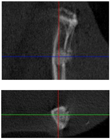
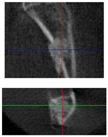
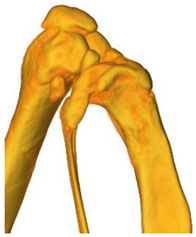
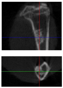
A tomographic examination was carried out in vivo after surgery, as well as posthumously after 3 months of implantation. Microcomputed tomography (micro-CT) was performed with a Bruker SkySkan 1178 scanner (BRUKER BELGIUM, Kontich, Belgium) at a voltage of 65 kV and a current of 615 μA with a 0.5 mm A1 filter. The spatial resolution was 84 microns/pixel. Sections were reconstructed using NRecon v1.6.10.4 software. 3D reconstructions were created in FEI Avizo 9.0.1.
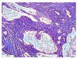
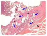
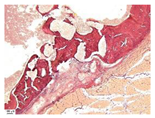
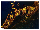
For histological examination, samples were fixed in neutral buffered 10% formalin, decalcificated in a special acid solution (SoftyDek, Biovitrum, St. Petersburg, Russia), subjected to standard alcohol wiring, and embedded into paraffin blocks. Paraffin section of 4 μm thick were stained with hematoxylin and eosin, as well as picrosirius red, according to the standard protocols. The obtained microscopic slides were examined with a universal LEICA DM4000 B LED microscope (Leica Microsystems Wetzlar GmbH, Wetzlar, Germany) equipped with a LEICA DFC7000 T digital video camera using standard light, phase contrast, and polarized microscopy methods.
For statistical analysis, the normality of the distribution was checked using the Shapiro–Wilk test. A one-way ANOVA and a comparative Tukey test were used. The significance level was p ≤ 0.05. Statistics were processed in Origin Pro 2021. The results were presented as average value ± standard deviation. Compact letter display was used to show the statistical difference (p < 0.05) between different materials at one time point.

3. Results

3.1. Composition of Cement at Figurative Points

In order to determine the effect of the ratio of components in the α-Ca3(PO4)2/CaCO3/Ca(H2PO4)2·H2O/Na2HPO4·12H2O system on the final cement composition, twelve figurative points on the diagram were selected in accordance with Figure 1 and Table 1.

3.1.1. X-ray Diffraction (XRD)

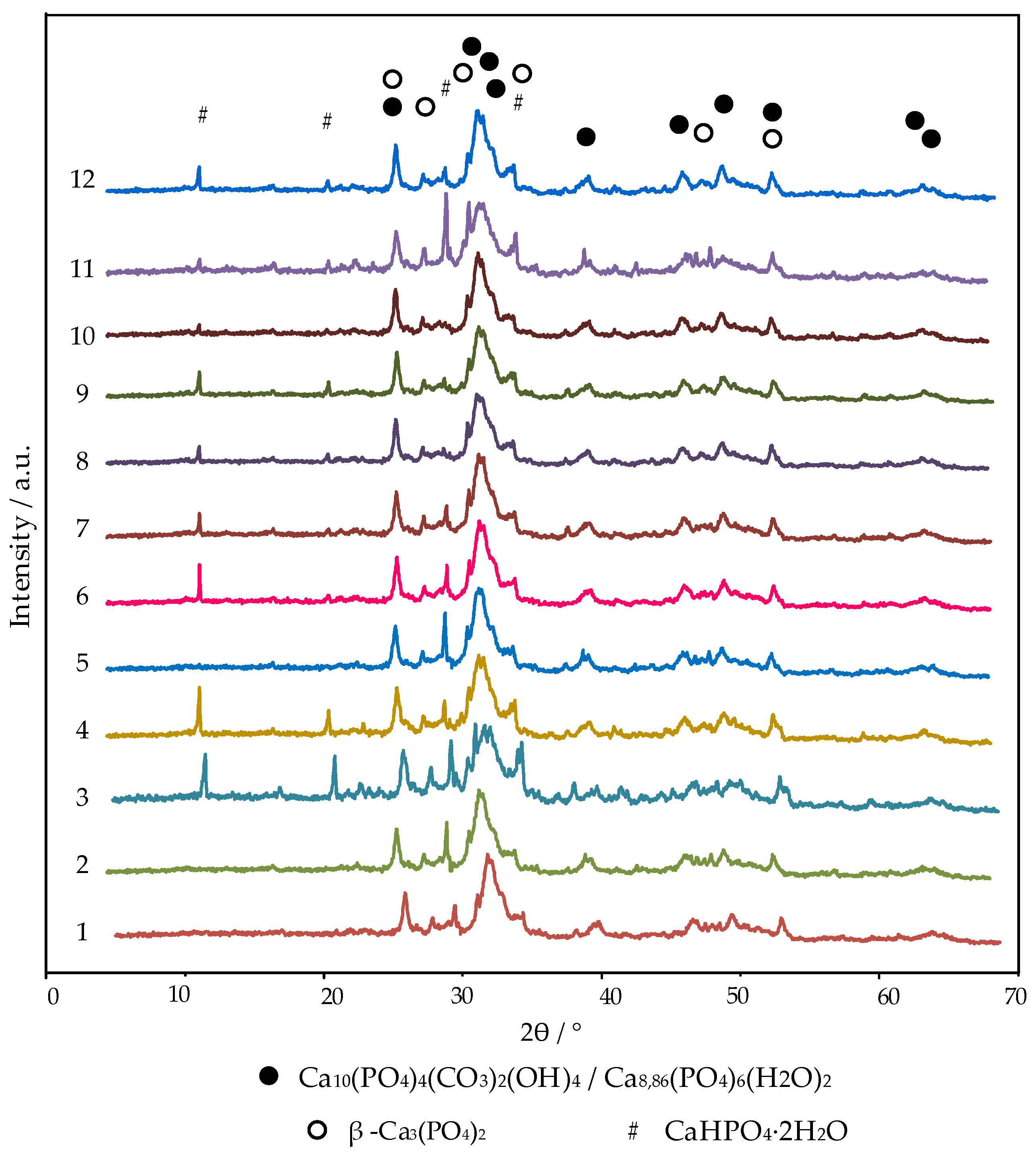
The phase compositions of the cement obtained after 30 days of hardening in wet conditions for all twelve compositions of figurative points were represented by carbonate-substituted hydroxyapatite Ca10(PO4)4(CO3)2(OH)4 [01-085-7370] (CO3-HA), calcium-deficient hydroxyapatite Ca8,86(PO4)6(H2O)2 (CDHA) [01-082-1943], dicalcium phosphate dihydrate CaHPO4·2H2O [01-072-0713] (DCPD), and β-tricalcium phosphate β-Ca3(PO4)2 [01-070-2065] (β-TCP) (Figure 2). Calcium-deficient and carbonate-substituted hydroxyapatites were not marked in separate phases on the diffractogram due to the coincidence of the main peaks. The calculations were performed using the characteristic peaks at interplane distances of 2.7309 and 1.9489 for CDHA and at 2.8066 and 2.7782 for CO3-HA during the quantitative phase analysis. The identification of calcium carbonate through an X-ray phase analysis was impossible since there were no characteristic peaks (not coinciding with the peaks of the present phases) in this system. Calcium carbonate was not taken into account when calculating the quantitative phase composition.
The results of the quantitative determination of the CaHPO4·2H2O phase in the cement at figurative points are presented in Table 2.

3.1.2. Fourier-Transform Infrared Spectroscopy (FTIR)

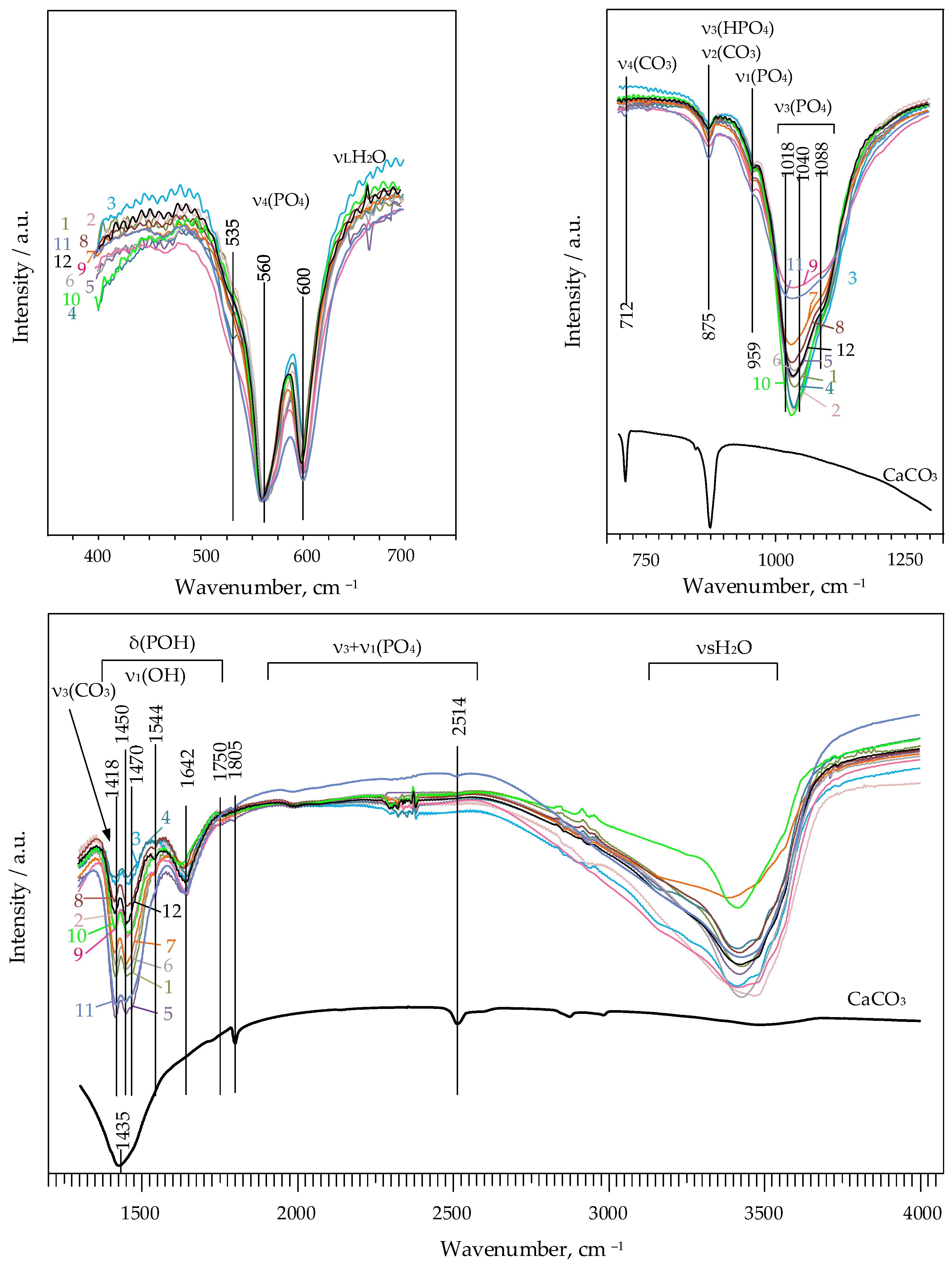
Figure 3 shows the FTIR spectra of the cement compositions at figurative points.
The most intense and high-frequency band of 1435 cm−1 of the calcite spectrum was not a characteristic of the FTIR spectra of the cements. It merged with the doublet at 1409–1418 cm−1 and 1449–1474 cm−1, belonging to mode ν3 of the antisymmetric stretching characteristic of CO32− ions. These bands are characteristic of carbonate-substituted hydroxyapatites. The band at 875 cm−1 was a characteristic of calcite but was hidden by a band of HPO42− ions. Thus, the content of CO32− ions could be inferred only by the band at 712 cm−1. It referred to the asymmetric (ν4) deformation oscillation of CO32– and was absent in the spectra of the cements.
The doublet characteristic of carbonate ions contained peaks characteristic of A (substitution of OH ions) and B types (substitution of PO43− ions) of carbonate hydroxyapatite. In it, the bands at 1418 and 1470 cm−1 were characteristic only for the B type of carbonate hydroxyapatite, and the bands at 1450 and 1544 cm−1 were only for the A type. In the compositions of figurative points 1, 5, 6, and 11, peaks at 1450 and 1470 cm−1 were clearly distinguished, whereas in other compositions, the peaks merged into one wide peak.
Peaks at 600 and 559–560 cm−1 were characteristic of orthophosphoric tetrahedral ions in a hydroxyapatite-type structure. They were wide, merging into a doublet. Peaks at 578 and 631 cm−1 correspond to well-crystallized hydroxyapatite and were absent in the spectra.
Fluctuations in the HPO42− anion in the range of 1400–1750 cm−1 were characteristic. In connection with the superposition of δ (POH) oscillations on the fluctuations of ν3 (CO3), we considered the fluctuations in the range of 1600–1750 cm−1 as decisive. Peaks at 1642 cm−1 were clearly defined on the spectra, which determined the ions of HPO42− calcium-deficient hydroxyapatite and dicalcium phosphate dihydrate.
A semiquantitative analysis based on comparing the concentrations of the same ions in different compositions was carried out using normalized FTIR spectra by finding the integral in the range of wave numbers from 1350 to 1525 cm−1. The dependence of the amount of carbonate ions on the initial compositions of cements was determined with the following formula:
K C O 3 2 = 1350 1525 d I d λ
The intensity integrals from 1350 to 1525 cm−1 are presented in Table 3.

3.1.3. Volumetric Study

The amount of carbonate ions was determined using the volumetric technic with the determination of the volume of released CO2 when the product was exposed to a solution of hydrochloric acid. The concentrations of CO32− ions in the final compositions at figurative points are presented in Table 4.

3.1.4. Calorimetric Study

The formation of different amounts of CaHPO4·2H2O at different figurative points was confirmed by data on the kinetics of heat release (Table 5).
An increase in thermal power in the case of all the compositions was registered in the first 15 min. It indicated an exothermic reaction. Due to the high rate of formation of CaHPO4·2H2O during the acid–base interaction, heat release in the initial periods of hardening was attributed to the formation of this phase.
The main reactions of DCPD formation were dissolution of the monohydrate of monocalcium phosphate, dissolution of α-tricalcium phosphate, dissolution of calcium carbonate, and formation of CaHPO4·2H2O. The dissolution of the initial components occurred mainly with the release of heat:
Ca(H2PO4)2∙H2O → Ca2+ + 2HPO42− + 2H+ + H2O (Q = 0 kJ/mol or 0 J/g);
α-Ca3(PO4)2 + 2H+ → 3Ca2+ + 2HPO42− (Q= −81.2 kJ/mol or −261.94 J/g);
CaCO3 + 2H+ → Ca2+ + CO2↑ + H2O (Q= −14.83 kJ/mol or −14.83 × 1000/100 = −148.3 J/g)
According to the enthalpies of formation, it can be concluded that the formation of CaHPO4·2H2O occurred with the release of heat during the interactions of MCPM with TCP and MCPM with calcium carbonate:
Ca2+ + 2HPO42− + H2O + 3Ca2+ + 2HPO42− + 7H2O → 4 CaHPO4∙2H2O (Q= −6.4 kJ/mol or −37.21 J/g);
Ca2+ + 2HPO42− + 2H+ + 2H2O + Ca2+ + CO2↑ + H2O → 2 CaHPO4∙2H2O (Q= −175.29 kJ/mol or −509.56 J/g).
The total heat release by reaction was as follows:
α-Ca3(PO4)2 + Ca(H2PO4)2∙H2O → 4 CaHPO4∙2H2O (Q = −299.15 J/g);
CaCO3 + Ca(H2PO4)2∙H2O → 2 CaHPO4∙2H2O + CO2↑ (Q = −657.86 J/g).
These calculations of heat release do not reflect the course of the entire set of reactions, but are appropriate for the main ones. They make the greatest contribution to the initial total heat release.
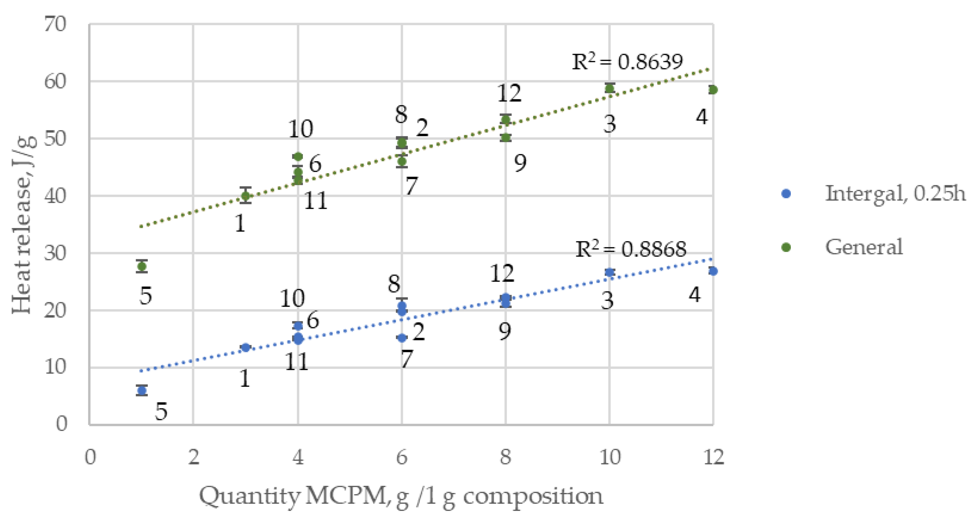
The total heat release by 15 min. consisted of the heat of the formation of CaHPO4·2H2O and the heat of the dissolution of α-TCP. A decrease in the pH of the cement dough due to an increase in the MCPM content affected the dissolution of α-TCP. In this regard, the increase in heat release by 15 min. linearly depended on the increase in the amount of MCPM in the initial composition (Figure 4).
In the initial period of hydration, when α-TCP particles came into contact with water, crystal dissolution reactions began. The liquid phase near the particles was saturated with Ca2+ and PO43− ions. The liquid phase was supersaturated with ions. The final compounds were precipitated.
Heat dissipation was higher in compositions with higher concentrations of Na2HPO4·12H2O with the same amount of MCPM in most cases. When using a solution of Na2HPO4·12H2O as a mixing fluid, an additional source of PO43− ions appeared. The Ca/P ratio shifted toward lower values. Probably, DCPD crystals were formed [38]. They subsequently rearranged into calcium-deficient hydroxyapatite. The formation of DCPD crystals led to an increase in heat release and a new dissolution of α-TCP was provoked, increasing the rate of formation of the hardened cement.
According to the data of integral heat release, the amount of heat released decreased by 1–3 h. Then, it rose slightly until 24 h. This was due to the slowing down of the reaction. On the surface of the α-TCP particles, new crystals of DCPD and hydroxyapatite were formed, which made it difficult to diffuse to the nonhydrated part of the α-TCP particles. The reaction rate and heat release increased when the shells were destroyed by growing crystals.

3.1.5. Stoichiometric Formulas

Based on the data of the volumetric research and the results of the spectrophotometric determination of calcium and phosphorus, the stoichiometric formulas of pre-set cement compositions at figurative points were derived (Table 6). The amounts of (HPO4)2−, (PO4)3− and (OH) ions were determined according to the following formula:
Ca10–x–y/2–z/2Naz(HPO4)x(CO3)y(PO4)6–x–y(OH)2–x
subject to the following conditions:
-
the neutrality of the molecule;
-
the amount of Ca2+ = 10 − (HPO4)2− − (CO3)2v/2 − Na+/2;
-
the amount of OH = 2 − (HPO4)2−.

3.1.6. Scanning Electron Microscopy (SEM)

The microstructures of cement compositions at figurative points were represented mainly by irregular crystals of hydroxyapatite and a small amount of unreacted α-TCP particles (Figure 5). According to SEM, CaHPO4·2H2O was formed both on the surface of the pores and in the bulk of the cement (Figure 5).

3.2. Solubility of Cement Formed in the Ca3(PO4)2/CaCO3/Ca(H2PO4)2·H2O/Na2HPO4·12H2O System at Figurative Points

The solubility of the cement was evaluated at physiological temperature values of 37 °C in a physiological solution. The concentrations of calcium ions in the physiological solution after holding pre-set cement granules formed in the Ca3(PO4)2/CaCO3/Ca(H2PO4)2·H2O/Na2HPO4·12H2O system at figurative points are shown in Figure 6.
The solubility of pre-set cement depended on the content of DCPD and the amount of CO32− ions. In general, the solubility was inversely proportional to the concentration of Na2HPO4·12H2O.

3.3. In Vivo Experiments

Biocompatibility and resorbability were investigated in in vivo experiments on Wistar rats in a model of tibial perforation. Granules of 250–500 microns in size of the compositions at figurative points 3, 5, and 8 were implanted into bone perforations of critical size in the tibia.
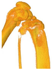
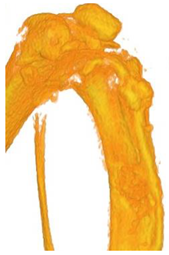
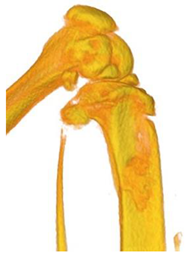
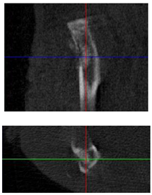
Partial resorption of the matrix and the formation of new bone tissue occurred after 3 months of implantation, according to computer microtomography (Table 7).
The best healing of the defect, consisting of complete closure of the trepanation opening with the formation of a clearly X-ray-distinguishable cortical bone, was observed after three months of implantation in the case of using the cement of composition 3. The granules of the material remained radiopaque in the area of the bone marrow canal. Some decrease in their density was noted. No exostoses were observed.
An increase in the radiopacity of composition 5 on the side facing the defect indicated that the formation of cortical bridges deepened into the bone marrow canal. They were not fully connected to each other and were not yet fully formed. There was also an overgrowth of bone from the borders of the defect toward the implant.
Composition 8 showed the least pronounced degree of closure of the bone defect. There was a bright callus in the projection of the trepanation hole, with bone growth from the defect boundaries by an elongated, thin cortical bridge. The maturity of this regenerate was estimated as minimal compared to other formulations.
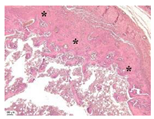
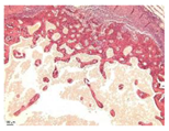
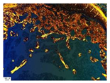
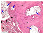
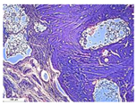
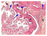
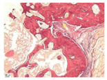
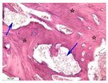
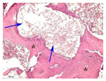
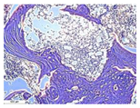
There was good bone regeneration with filling of the defect area with newly formed bone tissue after 3 months of the implantation of granules into the holes of the tibia in almost all cases according to the morphological examination (Table 8). At the same time, inflammatory reaction was absent or minimal. The remains of the implant were detected in bone defects in most cases. The most intensive bone regeneration was characteristic of the compositions at figurative points 3 and 5 compared to composition 8. Moreover, the newly formed bone had a sufficiently mature structure with a large number of osteocytes.
The remains of the implant, in addition to bone, were found in muscles near the defect area when examining samples of different compositions. In this case, they were surrounded by a connective tissue capsule consisting of collagen fibers with fibroblasts, macrophages, and an admixture of giant, multinucleated cells.

4. Discussion

The Ca3(PO4)2/CaCO3/Ca(H2PO4)2·H2O/Na2HPO4·12H2O system is an astringent system; when interacting with water, the product is a pre-set cement of variable composition.
Regulation of the solubility of pre-set cement was formed in the Ca3(PO4)2/CaCO3/Ca(H2PO4)2·H2O/Na2HPO4·12H2O system, possibly due to the presence of a more resorbable phase in the composition compared to hydroxyapatite due to a decrease in energy of the hydroxyapatite crystal lattice. The latter could be achieved by replacing PO43− ions with HPO42− or CO32− and Ca2+ with Na+.
The hydroxyapatite detected through X-ray had variable compositions: the numbers of substituted PO43−, Ca2+, and OH ions varied within the pre-set cement and formed a series of solid solutions. In addition to carbonate-substituted and calcium-deficient hydroxyapatites, more soluble phases of DCPD and β-TCP were found in the compositions of pre-set cement at physiological pH values.
The dissolution of the initial calcium phosphates, saturation with Ca2+ and PO43−- ions of the solution, and precipitation of reaction products from the solution occurred during the interaction of the Ca3(PO4)2/CaCO3/Ca(H2PO4)2·H2O/Na2HPO4·12H2O system with water. As soon as the crystals of the reaction products were formed, calcium ions and phosphate ions saturated the solution again. At the same time, calcium carbonate also decomposed into Ca2+ and CO32− ions in the aqueous solution. There was continuous precipitation–dissolution. In the presence of PO43− ions, the solution became supersaturated with respect to CDHA or DCPD. DCPD could recrystallize into CDHA. The PO43−, CO32−, and Ca2+ ions in the solution were precipitated as CO3-HA since, under such conditions, CO3-HA was the most thermodynamically stable phase [39,40].
Presumably, DCPD was formed during the acid–base interaction of calcium carbonate and α-TCP with MCPM at the early stage of hydration. Acidic MCPM dissolving reduced the pH of the medium, affecting the thermodynamic stability. It increased the solubility of α-TCP and CaCO3 and led to the formation of phases with lower Ca/P ratios. This was confirmed by the straightforward relationship between MCPM and DCPD. The formation of DCPD led to the appearance of a seed for the crystallization of hydroxyapatite and accelerated the dissolution of α-TCP. DCPD crystals were formed on the surface of the initial particles. They gradually dissolved, giving way to newly formed crystals.
The formation of DCPD with a release of carbon dioxide occurred when MCPM interacted with calcium carbonate. The number of embedded CO32 ions in the hydroxyapatite crystal lattice decreased in this regard. Thus, the amount of DCPD increased, and the amount of CO32− decreased when the figurative points were located closer to the stoichiometric amount of calcium carbonate and MCPM. The resulting carbonate-substituted hydroxyapatite was predominantly B-type. No calcium carbonate was found in the final composition of the cement.
Thus, the solubility of pre-set cement formed in the Ca3(PO4)2/CaCO3/Ca(H2PO4)2·H2O/Na2HPO4·12H2O system depended both on the amount of DCPD and carbonate ions in the hydroxyapatite structure. The highest solubility values were characteristic of compositions 3 and 4. Since these compositions contained the largest amounts of DCPD, the Ca/P ratio was 1.4.
The low solubility of the compositions of figurative points 8 and 10 containing high concentrations of Na+ ions may be associated with the formation of Na-containing hydroxyapatites. Their solubility may be lower. Sodium ions of the system could be embedded in the structures of carbonate and calcium-deficient hydroxyapatites and could compensate for the difference in charges when replacing PO43− ions with CO32− or HPO42−.
The compositions of points 8 and 10 contained the least amount of HPO42− and CO32− ions with the highest contents of sodium ions in accordance with the stoichiometric formulas obtained empirically. Na-containing hydroxyapatites lost Na+ and turned into calcium-deficient hydroxyapatite Ca9(HPO4)(PO4)5OH when immersed in aqueous solutions in an open system for about 2 months, according to other authors’ research [41].
All the compositions studied in in vivo experiments showed high biocompatibility, as well as good bone regeneration with filling of the defect area with newly formed bone tissue. In vivo resorption was observed in higher compositions with high contents of DCPD and carbonate groups (points 3 and 5). When using composition 3, the formation of a clearly X-ray-distinguishable cortical bone was observed.
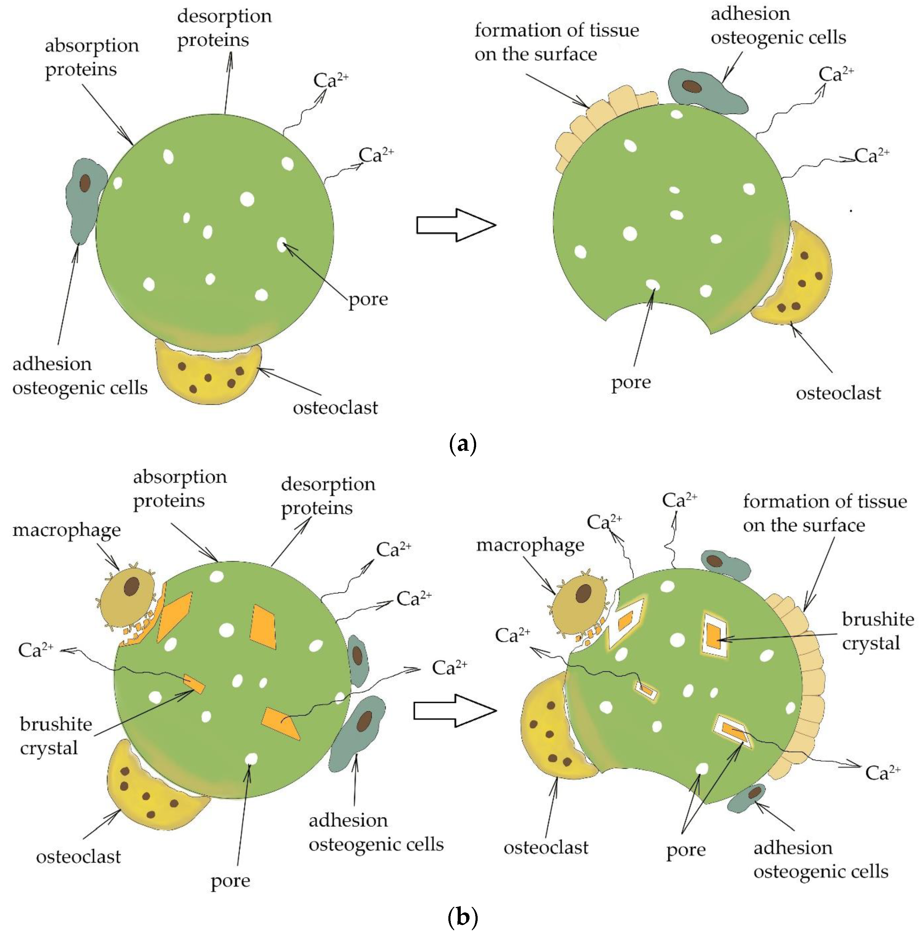
It should be taken into account that in vivo resorption was caused not only by chemical dissolution of the cement in extracellular fluid (passive resorption), but also by active resorption due to cell activity (osteoclasts, giant cells, and macrophages) [42]. Passive resorption (chemical dissolution) depended on the properties of the cement stone, particularly the amounts of DCPD and carbonate ions, and on properties of the environment, particularly pH. The active resorption of the calcium phosphate cements is mediated by giant cells and osteoclasts, with the absorption of fragmented cement particles by macrophages [43,44]. Osteoclasts, which are tightly attached to a mineral surface, decrease the pH locally near a material and dissolve the inorganic calcium phosphate that is located under them [45,46,47]. A local decrease in pH is possible with the dissolution of DCPD. This may slightly affect the solubility of carbonate hydroxyapatite since the pH of DCPD is about 4-5. Figure 7 shows the scheme of the resorption of calcium phosphate cement particles depending on composition.
The concentration of Ca2+ should be higher than the physiological norm in order to increase proliferation and differentiation [16,48]. Consequently, the increased level of Ca2+ as a result of cement dissolution may be one of the key factors in the formation of new bone. The rate of resorption of calcium phosphate cements affected cellular proliferation by binding the released calcium to the extracellular calcium-sensitive receptor that was associated with the G-protein [23]. In this regard, cell proliferation was higher in apatite cements with substituted PO43− on CO32− ions [49] compared to cements without substitutions. The content of the highly resorbable phase of DCPD was also a key factor affecting the formation of new bone tissue.

5. Conclusions

The Ca3(PO4)2/CaCO3/Ca(H2PO4)2·H2O/Na2HPO4·12H2O system is important for the synthesis of self-hardening bioactive materials with variable resorption rates. It allows the adjustment of the characteristics of the final product in wide intervals through changing the ratios of the initial components.
The formation of carbonate-substituted hydroxyapatite and dicalcium phosphate dihydrate in the system increased passive resorption (solubility) due to the substitution of PO43− with CO32− ions and the distortion of the hydroxyapatite crystal lattice, as well as due to the formation of the crystals of a metastable compound in the composition of cement stone under physiological conditions. An increase in active resorption occurred due to the binding of the released calcium to the extracellular calcium-sensitive receptor associated with the G-protein. This increased the proliferation and phenotypic expression of bone cells and led to formation of new bone in direct contact with biomaterial. The high rate of resorption promoted high bone regeneration with filling of the defect area with newly formed bone tissue.
Calcium phosphate cements are bioactive, osteoconductive, and resorbable. They integrate into bone tissue without the formation of a connective tissue capsule. Increasing the rate of resorption of calcium phosphate cements based on hydroxyapatite is an important task for material science and may contribute to its wider use in clinical practice.

Author Contributions

Conceptualization, Y.L.; methodology, Y.L. and S.S.; investigation, Y.L., S.K., L.B.-A., N.S. and D.S.; writing—original draft preparation, Y.L.; writing—review and editing, Y.L.; visualization, Y.L., L.B.-A. and N.S.; supervision, Y.L.; project administration, Y.L. All authors have read and agreed to the published version of the manuscript.

Funding

This research received no external funding.

Institutional Review Board Statement

The protocol of the animal study was approved by the Local Ethical Committee for Medical and Biological Ethics of the N.N. Priorov National Medical Research Center for Traumatology and Orthopedics of the Ministry of Health of the Russian Federation (protocol code 004, approval date 5 May 2021).

Informed Consent Statement

Not applicable.

Data Availability Statement

Not applicable.

Acknowledgments

The authors wish to thank Alina Nesterenko for the illustration in the article.

Conflicts of Interest

The authors declare no conflict of interest. The funders had no role in the design of the study; in the collection, analyses, or interpretation of data; in the writing of the manuscript; or in the decision to publish the results.

References

  1. Rau, J.V.; Cesaro, S.N.; Ferro, D.; Barinov, S.M.; Fadeeva, I.V. FTIR study of carbonate loss from carbonated apatites in the wide temperature range. J. Biomed. Mater. Res. B Appl. Biomater. 2004, 71, 441–447. [Google Scholar] [CrossRef] [PubMed]
  2. Landi, E.; Tampieri, A.; Celotti, G.; Vichi, L.; Sandri, M. Influence of synthesis and sintering parameters on the characteristics of carbonate apatite. Biomaterials 2004, 25, 1763–1770. [Google Scholar] [CrossRef] [PubMed]
  3. Ishikawa, K. Bone Substitute Fabrication Based on Dissolution-Precipitation Reactions. Materials 2010, 3, 1138–1155. [Google Scholar] [CrossRef] [Green Version]
  4. Ishikawa, K.; Matsuya, S.; Lin, X.; Lei, Z.; Yuasa, T.; Miyamoto, Y. Fabrication of low crystalline B-type carbonate apatite block from low crystalline calcite block. J. Ceram. Soc. Jpn. 2010, 118, 341–344. [Google Scholar] [CrossRef] [Green Version]
  5. Ishikawa, K.; Arifta, T.I.; Hayashi, K.; Tsuru, K. Fabrication and evaluation of interconnected porous carbonate apatite from alpha tricalcium phosphate spheres. J. Biomed. Mater. Res. B Appl. Biomater. 2019, 107, 269–277. [Google Scholar] [CrossRef] [PubMed]
  6. Mabroum, H.; Noukrati, H.; Benyoucef, H.; Lefeuvre, B.; Oudadesse, H.; Barroug, A. Physicochemical, setting, rheological, and mechanical properties of a novel bio-composite based on apatite cement, bioactive glass, and alginate hydrogel. Ceram. Int. 2021, 47, 23973–23983. [Google Scholar] [CrossRef]
  7. Ooms, E.M.; Wolke, J.G.C.; van de Heuvel, M.T.; Jeschel, B.; Jansen, J.A. Histological evalution of the bone response to calcium phosphate cement implanted in cortical bone. Biomaterials 2003, 24, 989–1000. [Google Scholar] [CrossRef]
  8. Khairoun, I.; Boltong, M.G.; Driessens, F.C.M.; Planell, J.A. Effect of calcium carbonate on the compliance of an apatitic calcium phosphate bone cement. Biomaterials 1997, 18, 1535–1539. [Google Scholar] [CrossRef] [PubMed]
  9. Boehm, A.V.; Meininger, S.; Tesch, A.; Gbureck, U.; Muller, F.A. The mechanical properties of biocompatible apatite bone cement reinforced with chemically activated carbon fibers. Materials 2018, 11, 192. [Google Scholar] [CrossRef] [Green Version]
  10. Lewis, G. Injectable bone cements for use in vertebroplasty and kyphoplasty: State-of-the-art review. J. Biomed. Mater. Res. B Appl. Biomater. 2006, 76, 456–468. [Google Scholar] [CrossRef]
  11. Apelt, D.; Theiss, F.; El-Warrak, A.O.; Zlinszky, K.; Bettschart-Wolfisberger, R.; Bohner, M.; Matter, S.; Auer, J.; Von Rechenberg, B. In vivo behavior of three different injectable hydraulic calcium phosphate cements. Biomaterials 2004, 25, 1439–1451. [Google Scholar] [CrossRef]
  12. Bohner, M. Calcium orthophosphates in medicine: From ceramics to calcium phosphate cements. Injury 2000, 31, D37–D47. [Google Scholar] [CrossRef]
  13. Schröter, L.; Kaiser, F.; Stein, S.; Gbureck, U.; Ignatius, A. Biological and mechanical performance and degradation characteristics of calcium phosphate cements in large animals and humans. Acta Biomater. 2020, 117, 1–20. [Google Scholar] [CrossRef]
  14. Lukina, Y.; Panov, Y.; Panova, L.; Senyagin, A.; Bionyshev-Abramov, L.; Serejnikova, N.; Kireynov, A.; Sivkov, S.; Gavryushenko, N.; Smolentsev, D.; et al. Chemically Bound Resorbable Ceramics as an Antibiotic Delivery System in the Treatment of Purulent–Septic Inflammation of Bone Tissue. Ceramics 2022, 5, 330–350. [Google Scholar] [CrossRef]
  15. Spence, G.; Pate, N.; Brooks, R.; Bonfield, W.; Rushton, N. Osteoclastogenesis on hydroxyapatite ceramics: The effect of carbonate substitution. J. Biomed. Mater. Res. A 2010, 92, 1292–1300. [Google Scholar] [CrossRef] [PubMed]
  16. Mano, T.; Akita, K.; Fukuda, N.; Kamada, K.; Kurio, N.; Ishikawa, K.; Miyamoto, Y. Histological comparison of three apatitic bone substitutes with different carbonate contents in alveolar bone defects in a beagle mandible with simultaneous implant installation. J. Biomed. Mater. Res. Part B Appl. Biomater. 2020, 108, 1450–1459. [Google Scholar] [CrossRef] [PubMed]
  17. Fujisawa, K.; Akita, K.; Fukuda, N.; Kamada, K.; Kudoh, T.; Ohe, G.; Mano, T.; Tsuru, K.; Ishikawa, K.; Miyamoto, Y. Compositional and histological comparison of carbonate apatite fabricated by dissolution-precipitation reaction and Bio-Oss®. J. Mater. Sci. Mater. Med. 2018, 29, 121. [Google Scholar] [CrossRef]
  18. Hu, F.; Pan, L.; Zhang, K.; Xing, F.; Wang, X.; Lee, I.; Zhang, X.; Xu, J. Elevation of extracellular Ca2+ induces store-operated calcium entry via calcium-sensing receptors: A pathway contributes to the proliferation of osteoblasts. PLoS ONE 2014, 9, e107217. [Google Scholar] [CrossRef]
  19. Fujioka-Kobayashi, M.; Tsuru, K.; Nagai, H.; Fujisawa, K.; Kudoh, T.; Ohe, G.; Ishikawa, K.; Miyamoto, Y. Fabrication and evaluation of carbonate apatite-coated calcium carbonate bone substitutes for bone tissue engineering. J. Tissue Eng. Regen. Med. 2018, 12, 2077–2087. [Google Scholar] [CrossRef]
  20. Doi, Y.; Iwanaga, H.; Shibutani, T.; Moriwaki, Y.; Iwayama, Y. Osteoclastic responses to various calcium phosphates in cell cultures. J. Biomed. Mater. Res. 1999, 47, 424–433. [Google Scholar] [CrossRef]
  21. Ishikawa, K. Carbonate apatite bone replacement: Learn from the bone. J. Ceram. Soc. Jpn. 2019, 127, 595–601. [Google Scholar] [CrossRef] [Green Version]
  22. Hesaraki, S.; Nazarian, H.; Pourbaghi-Masouleh, M.; Borhan, S. Comparative study of mesenchymal stem cells osteogenic differentiation on low-temperature biomineralized nanocrystalline carbonated hydroxyapatite and sintered hydroxyapatite. J. Biomed. Mater. Res. B Appl. Biomater. 2014, 102, 108–118. [Google Scholar] [CrossRef] [PubMed]
  23. Li, W.; Chen, W.; Lin, Y. The efficacy of parathyroid hormone analogues in combination with bisphosphonates for the treatment of osteoporosis: A meta-analysis of randomized controlled trials. Medicine 2015, 94, e1156. [Google Scholar] [CrossRef] [PubMed]
  24. Xing, T.; Hu, Y.; Wang, B.; Zhu, J. Role of oral calcium supplementation alone or with vitamin D in preventing post-thyroidectomy hypocalcaemia: A meta-analysis. Medicine 2019, 98, e14455. [Google Scholar] [CrossRef] [PubMed]
  25. Song, Y.; Xu, L.; Jin, X.; Chen, D.; Jin, X.; Xu, G. Effect of calcium and magnesium on inflammatory cytokines in accidentally multiple fracture adults: A short-term follow-up. Medicine 2022, 101, e28538. [Google Scholar] [CrossRef]
  26. Notomi, T.; Kuno, M.; Hiyama, A.; Nozaki, T.; Ohura, K.; Ezura, Y.; Noda, M. Role of lysosomal channel protein TPC2 in osteoclast differentiation and bone remodeling under normal and low-magnesium conditions. J. Biol. Chem. 2017, 292, 20998–21010. [Google Scholar] [CrossRef] [Green Version]
  27. Nagai, H.; Kobayashi-Fujioka, M.; Fujisawa, K.; Ohe, G.; Takamaru, N.; Hara, K.; Uchida, D.; Tamatani, T.; Ishikawa, K.; Miyamoto, Y. Effects of low crystalline carbonate apatite on proliferation and osteoblastic differentiation of human bone marrow cells. J. Mater. Sci. Mater. Med. 2015, 26, 99. [Google Scholar] [CrossRef]
  28. Egashira, Y.; Atsuta, I.; Narimatsu, I.; Zhang, X.; Takahashi, R.; Koyano, K.; Ayukawa, Y. Effect of carbonate apatite as a bone substitute on oral mucosal healing in a rat extraction socket: In vitro and in vivo analyses using carbonate apatite. Int. J. Implant. Dent. 2022, 8, 11. [Google Scholar] [CrossRef]
  29. Kudoh, K.; Fukuda, N.; Kasugai, S.; Tachikawa, N.; Koyano, K.; Matsushita, Y.; Ogino, Y.; Ishikawa, K.; Miyamoto, Y. Maxillary sinus floor augmentation using low crystalline carbonate apatite granules with simultaneous implant installation: First-in-human clinical trial. J. Oral Maxillofac. Surg. 2019, 77, 985.E1–985.E11. [Google Scholar] [CrossRef]
  30. Nakagawa, T.; Kudoh, K.; Fukuda, N.; Kasugai, S.; Tachikawa, N.; Koyano, K.; Matsushita, Y.; Sasaki, M.; Ishikawa, K.; Miyamoto, Y. Application of low-crystalline carbonate apatite granules in 2-stage sinus floor augmentation: A prospective clinical trial and histomorphometric evaluation. J. Periodontal Implant. Sci. 2019, 49, 382–396. [Google Scholar] [CrossRef]
  31. Chow, L.C.; Markovic, M.; Takagi, S. A dual constant-composition titration system as an in vitro resorption model for comparing dissolution rates of calcium phosphate biomaterials. J. Biomed. Mater. Res. B Appl. Biomater. 2003, 65, 245–251. [Google Scholar] [CrossRef] [PubMed]
  32. Hurle, K.; Oliveira, J.; Reis, R.; Pina, S.; Goetz-Neunhoeffer, F. Ion-doped brushite cements for bone regeneration. Acta Biomater. 2021, 123, 51–71. [Google Scholar] [CrossRef] [PubMed]
  33. Issa, K.; Alanazi, A.; Aldhafeeri, K.A.; Alamer, O.; Alshaaer, M. Brushite: Synthesis, Properties, and Biomedical Applications. In Crystallization and Applications; Smida, Y.B., Marzouki, R., Eds.; IntechOpen: London, UK, 2022. [Google Scholar] [CrossRef]
  34. Morúa, O.C.; Cardoso, M.J.B.; da Silva, H.N.; Carrodeguas, R.G.; Rodríguez, M.A.; Fook, M.V.L. Synthesis of brushite/polyethylene glycol cement for filler in bone tissue injuries. Cerâmica 2021, 67, 289–294. [Google Scholar] [CrossRef]
  35. Tsuchiya, A.; Freitas, P.P.; Nagashima, N.; Ishikawa, K. Influence of pH and ion components in the liquid phase on the setting reaction of carbonate apatite granules. Dent. Mater. J. 2022, 41, 209–213. [Google Scholar] [CrossRef] [PubMed]
  36. Shariff, K.A.; Tsuru, K.; Ishikawa, K. Fabrication of dicalcium phosphate dihydrate-coated β-TCP granules and evaluation of their osteoconductivity using experimental rats. Mater. Sci. Eng. C 2017, 75, 1411–1499. [Google Scholar] [CrossRef] [PubMed]
  37. Tsuchiya, A.; Sato, M.; Takahashi, I.; Ishikawa, K. Fabrication of apatite-coated gypsum granules and histological evaluation using rabbits. Ceram. Int. 2018, 44, 20330–20336. [Google Scholar] [CrossRef]
  38. Fulmer, M.T.; Brown, P.W. Effects of Na2HPO4 and NaH2PO4 on hydroxyapatite formation. J. Biomed. Mater. Res. 1993, 27, 1095–1102. [Google Scholar] [CrossRef]
  39. Ishikawa, K.; Munar, M.L.; Tsuru, K.; Miyamoto, Y. Fabrication of carbonate apatite honeycomb and its tissue response. J. Biomed. Mater. Res. Part A 2019, 107, 1014–1020. [Google Scholar] [CrossRef]
  40. Cahyanto, A.; Maruta, M.; Tsuru, K.; Matsuya, S.; Ishikawa, K. Fabrication of bone cement that fully transforms to carbonate apatite. Dent. Mater. J. 2015, 34, 394–401. [Google Scholar] [CrossRef] [Green Version]
  41. Driessens, F.C.; Boltong, M.; de Maeyer, E.A.; Wenz, R.; Nies, B.; Planell, J. The Ca/P range of nanoapatitic calcium phosphate cements. Biomaterials 2002, 23, 4011–4017. [Google Scholar] [CrossRef]
  42. Lodoso-Torrecilla, I.; van den Beucken, J.J.; Jansen, J.A. Calcium phosphate cements: Optimization toward biodegradability. Acta Biomater. 2021, 119, 1–12. [Google Scholar] [CrossRef]
  43. Grossardt, C.; Ewald, A.; Grover, L.M.; Barralet, J.E.; Gbureck, U. Passive and active in vitro resorption of calcium and magnesium phosphate cements by osteclastic cells. Tissue Eng. Part A 2010, 16, 3687–3695. [Google Scholar] [CrossRef]
  44. Montazerolghaem, M.; Rasmusson, A.; Melhus, H.; Engqvist, H.; Karlsson Ott, M. Simvastatin-doped pre-mixed calcium phosphate cement inhibits osteoclast differentiation and resorption. J. Mater. Sci. Mater. Med. 2016, 27, 5. [Google Scholar] [CrossRef] [PubMed]
  45. Teitelbaum, S.L. Bone resorption by osteoclasts. Science 2000, 289, 1504–1508. [Google Scholar] [CrossRef] [PubMed]
  46. Davison, N.L.; Gamblin, A.-L.; Layrolle, P.; Yuan, H.; de Bruijn, J.D.; Barrère-de Groot, F. Liposomal clodronate inhibition of osteoclastogenesis and osteoinduction by submicrostructured beta-tricalcium phosphate. Biomaterials 2014, 35, 5088–5097. [Google Scholar] [CrossRef] [PubMed]
  47. Odgren, P.R.; Witwicka, H.; Reyes-Gutierrez, P. The cast of clasts: Catabolism and vascular invasion during bone growth, repair, and disease by osteoclasts, chondroclasts, and septoclasts. Connect. Tissue Res. 2016, 57, 161–174. [Google Scholar] [CrossRef] [Green Version]
  48. Maeno, S.; Niki, Y.; Matsumoto, H.; Morioka, H.; Yatabe, T.; Funayama, A.; Toyama, Y.; Taguchi, T.; Tanaka, J. The effect of calcium ion concentration on osteoblast viability, proliferation and differentiation in monolayer and 3D culture. Biomaterials 2005, 26, 4847–4855. [Google Scholar] [CrossRef]
  49. Guo, H.; Su, J.; Wei, J.; Kong, H.; Liu, C. Biocompatibility and osteogenicity of degradable Ca-deficient hydroxyapatite scaffolds from calcium phosphate cement for bone tissue engineering. Acta Biomater. 2009, 5, 268–278. [Google Scholar] [CrossRef]
Figure 1. Figurative points in the cross-section of the Ca3(PO4)2/CaCO3/Ca(H2PO4)2·H2O/Na2HPO4·12H2O composition diagram at a constant α-TCP mass of 80 wt.%.
Ceramics 06 00086 g001
Figure 2. X-ray diffraction patterns of cement at figurative points of compositions.
Ceramics 06 00086 g002
Figure 3. FTIR spectra of cement compositions.
Ceramics 06 00086 g003
Figure 4. Dependence of heat release on the amount of MCPM.
Ceramics 06 00086 g004
Figure 5. SEM images of pre-set cement.
Ceramics 06 00086 g005
Figure 6. The concentrations of calcium ions in the physiological solution after holding the pre-set cement granules formed in the Ca3(PO4)2/CaCO3/Ca(H2PO4)2·H2O/Na2HPO4·12H2O system at figurative points. Letters show the statistical difference (p < 0.05) between different meanings.
Ceramics 06 00086 g006
Figure 7. Scheme of resorption of calcium phosphate cement particles: (a) hydroxyapatite; (b) CO3-HA with DCPD.
Ceramics 06 00086 g007
Table 1. Content of components at figurative points.
Quantity, wt.%
Composition123456789101112
CaCO310845131010668128
Ca(H2PO4)2∙H2O36101214668448
Na2HPO4∙12H2O766366486844
Table 2. The amounts of CaHPO4·2H2O in cement compositions at figurative points.
Phase/CompositionQuantity, wt.%
123456789101112
CaHPO4·2H2O3.0 ± 0.33.4 ± 0.36.4 ± 0.55.7 ± 0.52.0 ± 0.33.7 ± 0.43.8 ± 0.34.2 ± 0.44.6 ± 0.43.3 ± 0.34.3 ± 0.44.4 ± 0.5
Table 3. Integral values of peak intensity in the range of wavenumbers from 1350 to 1525 cm−1.
CompositionArea, Conv. Unit2CompositionArea, Conv. Unit2
1374573282
2238881575
3113992589
41000102439
55158114871
63505121953
Table 4. Concentrations of CO32− ions in the final compositions at figurative points.
CompositionCO32−, mg/1 g CementCompositionCO32−, mg/1 g Cement
156.37750.37
241.00823.06
320.33930.22
414.171029.62
569.181165.48
641.001230.22
Table 5. The kinetics of heat release of cements.
Time, h
Composition
IntegralGeneral
0.250.51361224
Point 113.5 ± 0.12 g4.3 ± 0.101.5 ± 0.151.9 ± 0.193.4 ± 0.104.1 ± 0.125.5 ± 0.0940.1 ± 1.32 cde
Point 219.8 ± 0.05 d6.0 ± 0.322.2 ± 0.122.1 ± 0.093.6 ± 0.113.8 ± 0.184.3 ± 0.1149.2 ± 0.85 cd
Point 326.7 ±0.42 b7.6 ± 0.152.4 ± 0.051.9 ± 0.123.5 ± 0.164.0 ± 0.224.7 ± 0.2358.9 ± 0.79 a
Point 427.0 ± 0.43 b6.5 ± 0.482.0 ± 0.162.8 ± 0.434.7 ± 0.254.2 ± 0.093.7 ± 0.1258.6 ± 0.70 a
Point 56.0 ± 0.78 h3.9 ± 0.111.6 ± 0.091.5 ± 0.222.4 ± 0.122.8 ± 0.244.1 ± 0.1927.7 ± 1.00 g
Point 615.4 ± 0.08 ef5.1 ± 0.091.9 ± 0.202.1 ± 0.083.4 ± 0.273.5 ± 0.704.5 ± 0.2742.7 ± 0.63 f
Point 715.2 ± 0.09 fg7.1 ± 0.202.2 ± 0.182.2 ± 0.103.6 ± 0.193.6 ± 0.174.2 ± 0.4146.0 ± 1.03 e
Point 820.8 ± 1.17 d5.5 ± 0.371.9 ± 0.082.0 ± 0.563.5 ± 0.183.8 ± 0.084.9 ± 0.2949.4 ± 0.77 cd
Point 921.2 ± 0.66 c7.1 ± 0.232.4 ± 0.372.2 ± 0.163.7 ± 0.093.9 ± 0.194.6 ± 0.2353.4 ± 0.78 b
Point 1017.3 ± 0.47 e4.7 ± 0.081.7 ± 0.212.0 ± 0.123.4 ± 0.203.8 ± 0.355.0 ± 0.6544.2 ± 1.01 f
Point 1114.8 ± 0.08 a9.2 ± 0.282.2 ± 0.182.0 ± 0.143.3 ± 0.183.3 ± 0.124.2 ± 0.1546.9 ± 0.18 de
Point 1222.3 ± 0.15 c6.9 ± 0.171.8 ± 0.072.0 ± 0.123.3 ± 0.193.4 ± 0.164.0 ± 0.1250.2 ± 0.50 c
Tukey test; means that do not share a letter are significantly different (p < 0.05).
Table 6. Stoichiometric formulas of compositions at figurative points.
CompositionStoichiometric Formula
1Ca7.9Na0.3(HPO4)1.4(PO4)3.5(CO3)(OH)0.6
2Ca8Na0.3(HPO4)1.6(PO4)3.6(CO3)0.8(OH)0.4
3Ca7.6Na0.3(HPO4)2(PO4)3.4(CO3)0.6
4Ca8Na0.1(HPO4)1.7(PO4)4.0(CO3)0.3(OH)0.3
5Ca8Na0.3(HPO4)1.2(PO4)3.6(CO3)1.2(OH)0.8
6Ca8.2Na0.3(HPO4)1.2(PO4)4(CO3)0.8(OH)0.7
7Ca8Na0.2(HPO4)1.5(PO4)3.7(CO3)0.8(OH)0.5
8Ca8.2Na0.4(HPO4)1.4(PO4)4.1(CO3)0.5(OH)0.6
9Ca7.9Na0.3(HPO4)1.6(PO4)3.8(CO3)0.6(OH)0.3
10Ca8.3Na0.4(HPO4)1.2(PO4)4.2(CO3)0.5(OH)0.8
11Ca7.9Na0.2(HPO4)1.5(PO4)3.4(CO3)1.1(OH)0.5
12Ca8.1Na0.2(HPO4)1.5(PO4)4(CO3)0.5(OH)0.5
Table 7. 3D model and orthogonal projections of the tibia of a laboratory animal created using CT results.
Figurative Point
358
3D model and orthogonal projections

Initial
Ceramics 06 00086 i001Ceramics 06 00086 i002Ceramics 06 00086 i003
Ceramics 06 00086 i004Ceramics 06 00086 i005Ceramics 06 00086 i006
3D model and orthogonal projectionsAfter implantation period of

3 months
Ceramics 06 00086 i007Ceramics 06 00086 i008Ceramics 06 00086 i009
Ceramics 06 00086 i010Ceramics 06 00086 i011Ceramics 06 00086 i012
3D model and orthogonal projections are shown in one example for each composition.
Table 8. Micrographs of morphological examination of cements.
PointType of Microscopy
Standard LightStandard LightPolarizationStandard LightPhase Contrast
Staining with Hematoxylin–EosinStaining with Picrosirius RedStaining with Picrosirius RedStaining with Hematoxylin–EosinStaining with Hematoxylin–Eosin
3Ceramics 06 00086 i013Ceramics 06 00086 i014Ceramics 06 00086 i015Ceramics 06 00086 i016Ceramics 06 00086 i017
5Ceramics 06 00086 i018Ceramics 06 00086 i019Ceramics 06 00086 i020Ceramics 06 00086 i021Ceramics 06 00086 i022
8Ceramics 06 00086 i023Ceramics 06 00086 i024Ceramics 06 00086 i025Ceramics 06 00086 i026Ceramics 06 00086 i027
*—newly formed bone tissue, X—muscles, arrows—remains of the implants, tips of arrows—connective tissue capsule around remains of the implants.
Disclaimer/Publisher’s Note: The statements, opinions and data contained in all publications are solely those of the individual author(s) and contributor(s) and not of MDPI and/or the editor(s). MDPI and/or the editor(s) disclaim responsibility for any injury to people or property resulting from any ideas, methods, instructions or products referred to in the content.