Article

New Material and Revision of the Carnivora, Mammalia from the Lower Pleistocene Locality Apollonia 1, Greece

Aristotle University of Thessaloniki, Department of Geology, Laboratory of Geology and Palaeontology, GR-54124 Thessaloniki, Greece
Quaternary 2018, 1(1), 6; https://doi.org/10.3390/quat1010006
Submission received: 19 March 2018 / Revised: 2 May 2018 / Accepted: 14 May 2018 / Published: 17 May 2018

Abstract

:
During the last field campaigns in the mammal fossiliferous site Apollonia 1 (Macedonia, Greece), new carnivoran material has been discovered. The new collection added two new carnivoran taxa, Homotherium latidens and Panthera gombaszögensis. The new canid material and the revision of the old one (a) suggest the presence of two Canis species, C. etruscus and C. apolloniensis; (b) confirm the presence of the hypercarnivore Lycaon lycaonoides, and (c) allow for re-classifying the vulpine material to Vulpes praeglacialis. The taxonomic status of the species C. apolloniensis and Meles dimitrius is discussed. The composition and diversity of the Apollonia carnivoran assemblage are estimated and compared to those of various Greek and European Villafranchian ones. The results suggest close similarity to the Venta Micena (Spain) and Dmanisi (Georgia) carnivoran assemblages. The biochronological evidence indicates that Apollonia 1 is younger than Venta Micena and older than Untermassfeld (Germany), suggesting an age of 1.3–1.0 Ma. The study of the carnivoran guild structure of Apollonia 1 in comparison to the modern ones from known environments, as well as their functional morphology, suggest an open habitat, agreeing with previous interpretations.

1. Introduction

The mammal fossiliferous site Apollonia 1 (APL) has been known since the beginning of the 1990s, when it was discovered in the Mygdonia Basin (Macedonia, Greece) [1]. The site provided a rich and interesting fauna with several taxa. The first excavations in the locality were carried out during the period 1990–1996 and the collected material was published in a series of articles [2,3,4,5]. Ten taxa were identified in the APL carnivoran assemblage: Ursus etruscus, Canis etruscus, Canis arnensis, Canis apolloniensis, Xenocyon sp., Pachycrocuta brevirostris, Vulpes alopecoides, Meles dimitrius, Megantereon cultridens, Lynx issiodorensis [2,4].
The fossiliferous site APL is located near the village of Nea Apollonia, about 65 km northwest of Thessaloniki (Figure 1). The fossils were found into the Platanochori Formation, which overlies the Gerakarou Fm and crops out in the southeastern part of the Mygdonia Basin; the Platanochori Fm consists of grey-white, grey-green fluvio-lacustrine to lacustrine deposits [1,6,7]. The APL biochronological data suggest an Epivillafranchian age, more precisely ranging from 1.2 to 1.0 Ma. More recently (2011–2016), new field campaigns enriched the APL collection with new material, which includes some interesting carnivoran remains. In this article, the new material is described and compared with the old one and that from Eurasian Villafranchian localities. New taxa are added to the faunal list and the presence of others has been confirmed. The study of the new material and the revision of the old collection improve the knowledge of the systematics of the carnivoran fauna and provide additional evidence on the APL age and palaeoenvironment.

2. Materials and Methods

The studied material is stored in the Laboratory of Geology and Palaeontology, Aristotle University of Thessaloniki (LGPUT), together with the old APL collection. All material (new and old collection) has been measured using a digital caliper with an accuracy of 0.1 mm. All measurements are given in mm; estimated values into brackets. The upper and lower teeth are symbolized with capital and lowercase letters, respectively. The software Excel 10 (Microsoft, Redmond, WA, USA) is used for the scatter diagrams and PAST 3.1 [8] for the box-plots, cluster analysis, and principal component analysis.

Abbreviations

Localities. APL: Apollonia-1, Mygdonia Basin, Greece; DFN: Dafnero 1, Western Macedonia, Greece; DMN: Dmanisi, Georgia; GER: Gerakarou, Mygdonia Basin, Greece; KAL: Kalamoto, Mygdonia Basin, Greece; PIR: Pirro Nord, Italy; SES: Sesklon, Thessaly, Greece; TSR: Tsiotra Vryssi, Mygdonia Basin, Greece; UMF: Untermassfeld, Germany; VMC: Venta Micena, Spain; VOL: Volax, Eastern Macedonia, Greece.
Institutes. LGPUT: Laboratory of Geology and Palaeontology, Aristotle University of Thessaloniki.
Measurements. B: breadth (maximum bucco-lingual diameter of the tooth); DAP: anteroposterior diameter (maximum mesio-distal diameter of the tooth); DT: transverse diameter; dist: distal; epiph: epiphysis; H: height; mid-shaft: middle of the diaphysis; max: maximal; prox: proximal.
Morphological. a.a.c: anterior accessory cusp(-id); p.a.c: posterior accessory cusp(-id).

3. Systematic Palaeontology

3.1. Order Carnivora Bowdich, 1821; Family Ursidae Gray, 1865; Genus Ursus Linnaeus, 1858

Ursus etruscus Cuvier, 1823

Material. New collection: Right radius, APL-759.
Measurements. L = 309.5 mm, DTmax. prox. = 31.2 mm, DAPmax. prox. = 21.4 mm, DTmidshaft. = 27.5 mm, DAPmidshaft. = 16.0 mm, DTmax. dist. = 50.3 mm, DAPmax. dist. = 33.0 mm.
Description. The radius APL-759 (Figure 2a,b) is well preserved, lacking a small part of the proximal diaphysis, which was broken during its discovery; however, plaster was added in the field in order to preserve its original height and the connection with the proximal epiphysis, which is well preserved. The diaphysis is slender, slightly curved and flattened in the dorso-palmar direction. The proximal articular facet is sub-elliptical, with the medio-lateral diameter longer than the dorso-palmar one. The distal epiphysis is wider than the proximal one, bearing a large concave distal articular facet.
Remarks. The genus Ursus was identified in the APL fauna by an upper and lower canine [4,9]. The dimensions of APL-759 are close to those of U. etruscus from Pirro Nord and clearly different from those of U. deningeri, U. spelaeus and U. arctos. In comparison with the latter three taxa, the radius of U. etruscus, though similar in height, is remarkably slenderer, with a more flattened shaft and a reduced distal epiphysis (Figure 3). Based on the morphological and metrical similarities, APL-759 can be attributed to U. etruscus. The upper and lower canines of the APL old collection [4] coincide morphologically and metrically with those of U. etruscus from Tsiotra Vryssi (Mygdonia Basin, Greece) [9], and can also be attributed to this species.

3.2. Family Canidae Fischer von Waldheim, 1817; Genus Canis Linneaeus, 1758

3.2.1. Canis etruscus Forsyth Major, 1877

Synonyms. 1997 Xenocyon sp. Koufos and Kostopoulos, p. 48. (partim)
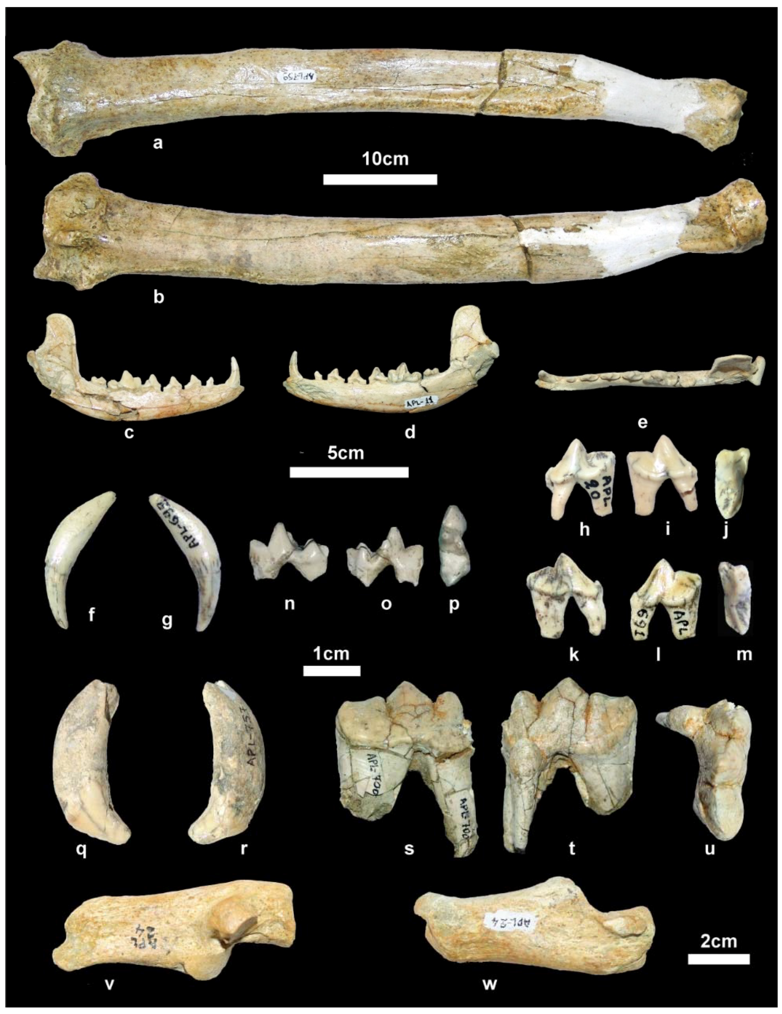
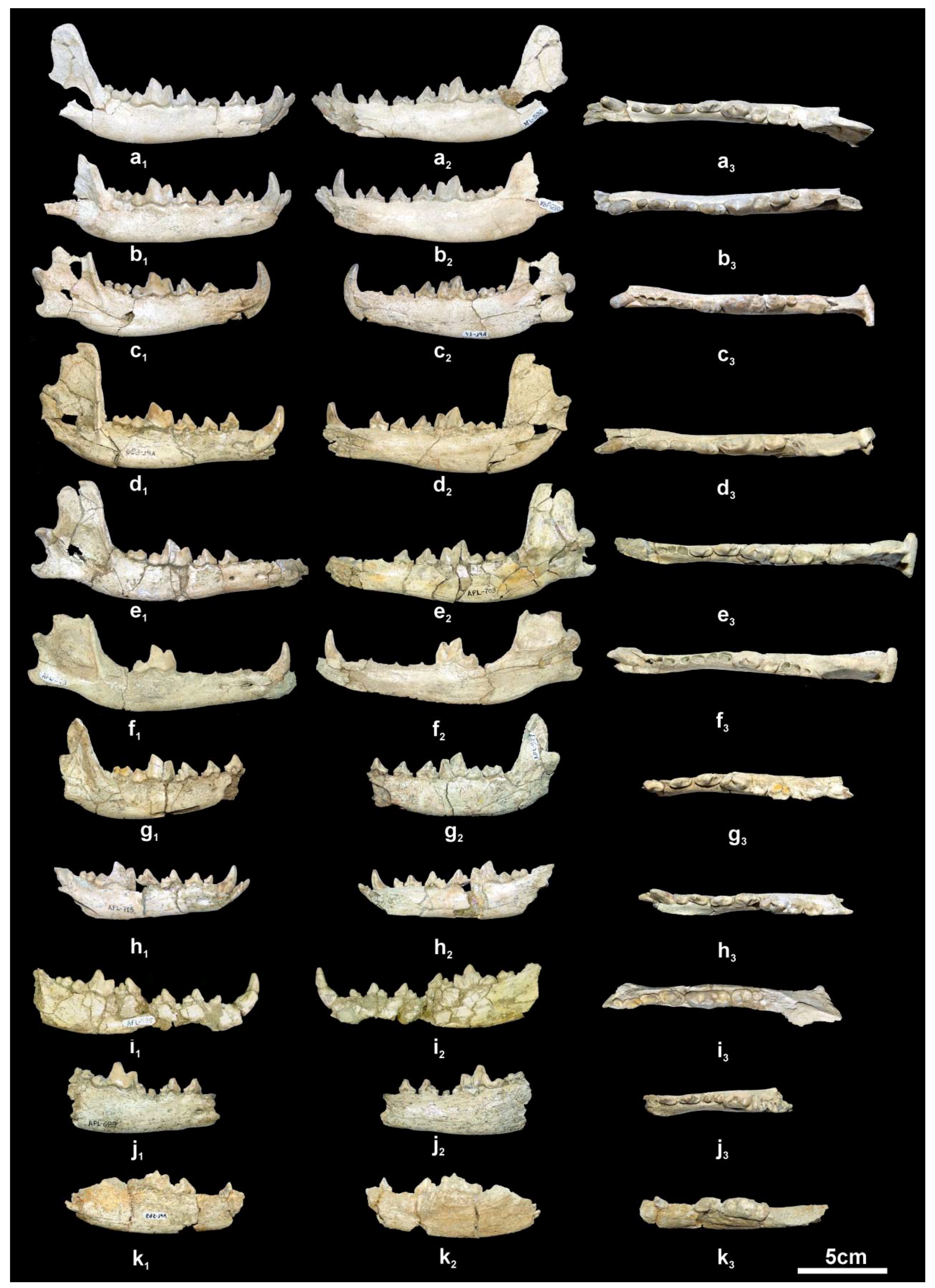
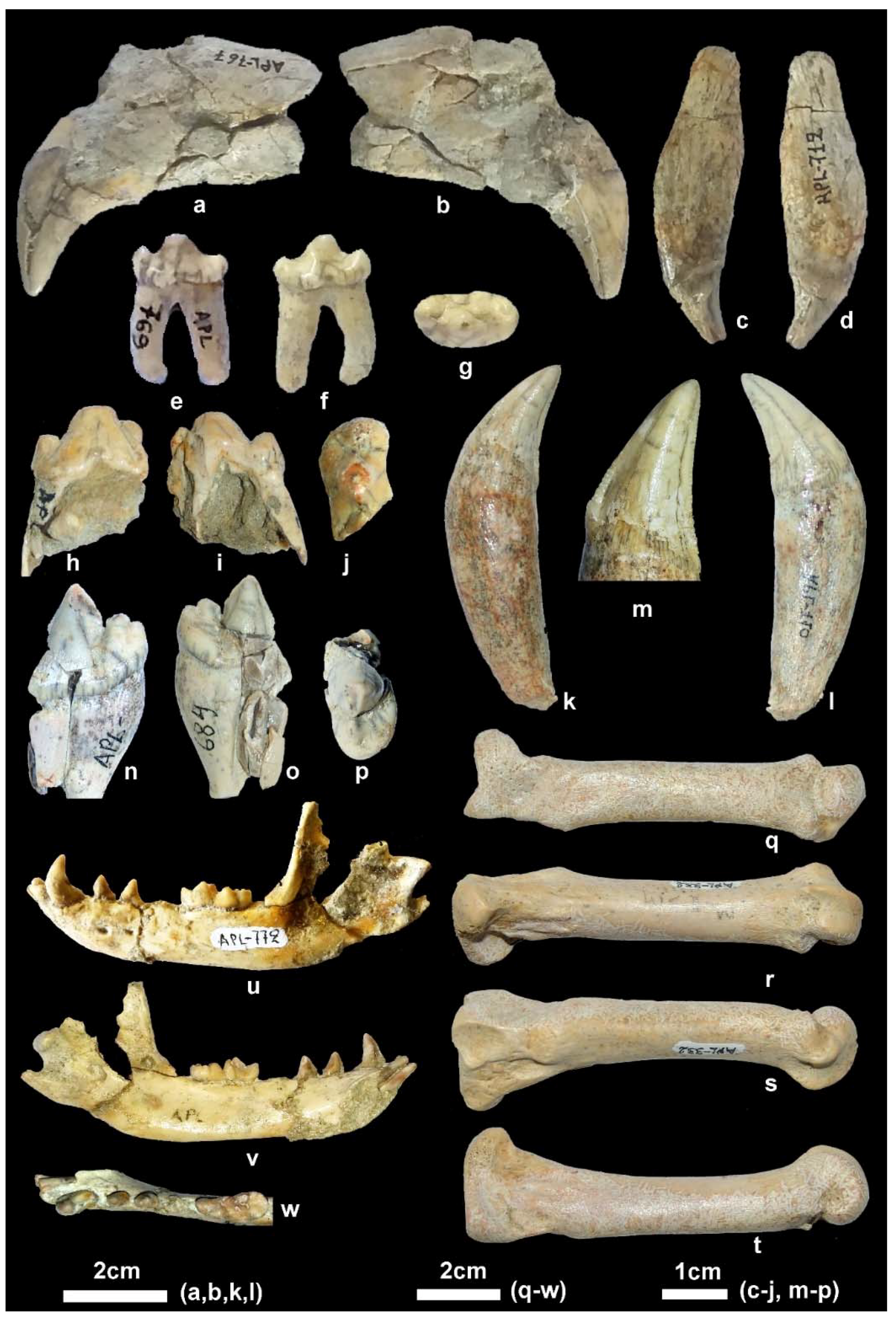
Material. New collection: Right mandibular fragment with p2-m2, APL-689.
Old collection: Skull, APL-522; Right and left mandibular fragments with c-m2, APL-526; left mandibular fragment with p2-m1, APL-569.
Measurements. The measurements are given in Table 1, Table 2, Table 3 and Table 4.
Description. The partial cranium APL-522 (Figure 4f) has been already described [4]. The available mandibular remains are badly preserved or fragmentary (Figure 5i–k) providing limited morphological information. The mandibular corpus is higher than that of C. apolloniensis and its ventral margin is slightly convex below the m1 and m2. It bears a large mental foramen below the mesial part of the p2 and a small one below the middle of the p3 (Figure 5j). The masseteric fossa is partly preserved in APL-526 and its mesial margin (Figure 5i1) is situated below the m3. The dentition of APL-569 and APL-689 is worn but that of APL-526 is less worn. The canine is small relative to the size of the mandible and strongly curved distally; in the right one, a lingual crest starts from its apex and, after the middle of the tooth’s height, is curved distally and connected with the pronounced distal cingulum. The p1 is small, monocuspid and single-rooted as in the other APL canids. The p2 lacks a.a.c. but it bears a p.a.c. on a large distal cingular projection. The p3 lacks a.a.c. but bears a small cingular mesio-lingual prominence; the p.a.c. is larger than that of the p2 and there is an elevation of the distal cingulum. The p4 is like the p3 but it bears a small secondary p.a.c., which originates from the strong elevation of the distal cingulum. The carnassial is relatively short and robust with a small metaconid situated slightly distally to the protoconid; albeit the advanced wear of the talonid, the hypoconid and entoconid are well distinguished and a distal crest, well distinguished in APL-689, connects them. The m2 has an oval occlusal outline with well-developed metaconid and protoconid separated by a shallow furrow; the hypoconid is situated disto-bucally and is smaller than the other two cuspids from which it is separated by a deep valley; the shelf like mesio-buccal cingulum is pronounced.
Remarks. The cranium, APL-522, and a mandibular fragment with badly preserved p4 and m1 (APL-569) of C. etruscus from Apollonia (Figure 4e and Figure 5i) have previously been described [4]. The higher paracone than metacone in the M1, the reduced contact between the M1 and M2, the continuous cingulum in the M1 and M2, the relatively small paraconid and protoconid in the m1, the linked hypoconid and entoconid by a distal sinuous crest in the m1, and the relatively small paraconid and protoconid in comparison with C. lupus of the APL sample agree with the diagnosis for C. etruscus [12]. The dental dimensions of the APL sample are also close to those of
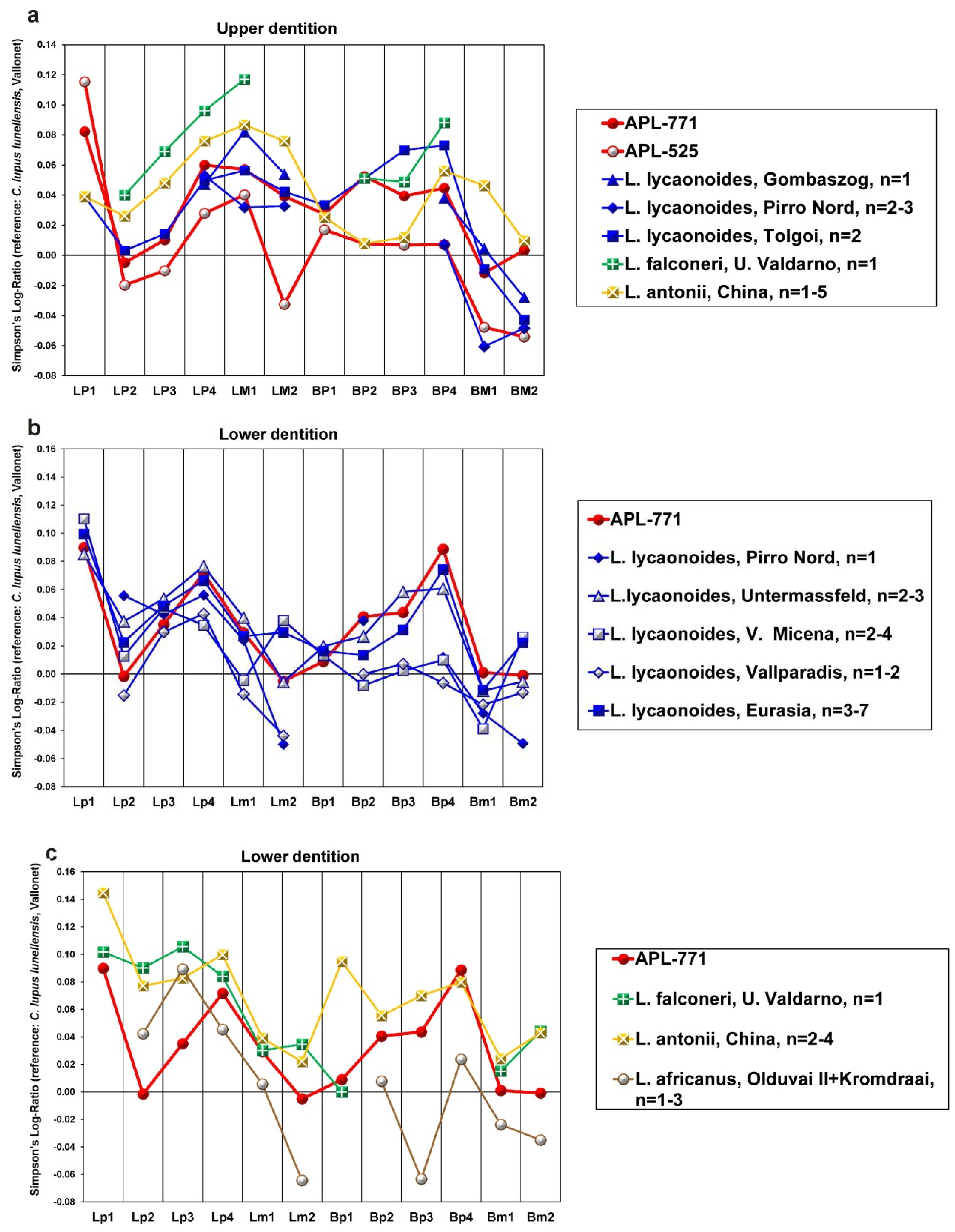
 C. etruscus (
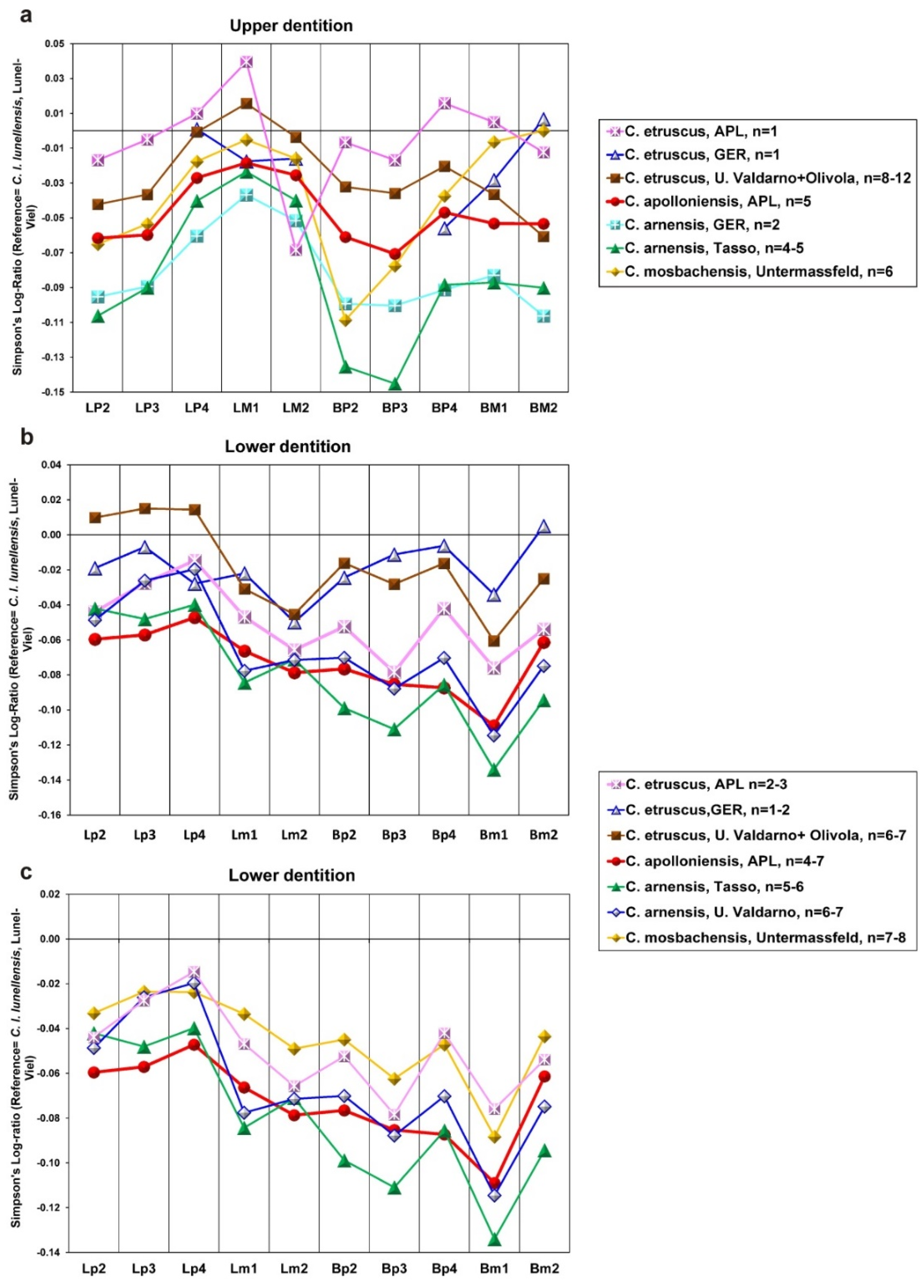
Figure 6). However, the APL upper teeth are larger than those from Upper Valdarno + Olivola (Italy) and Gerakarou (Mygdonia Basin, Greece) except for the M2, which is relatively short (Figure 6a). This might be due to the very limited APL sample, which includes only one specimen. On the other hand, the APL lower teeth have similar proportions to C. etruscus from Gerakarou and U. Valdarno+Olivola, but the lower premolars are narrower than those from the previous localities (Figure 6b). The dental dimensions of the APL C. etruscus are more or less similar to those of C. mosbachensis but larger than those of C. apolloniensis and C. arnensis (Figure 6c). The similar dental morphology and dimensions of the APL sample with C. etruscus from different European localities suggest that it can be attributed to this taxon. Besides APL, C. etruscus is known from two other localities of the Mygdonia Basin, i.e., the early Late Villafranchian locality Gerakarou and the Late Villafranchian locality Tsiotra Vryssi [2,7]. Some dental remains of C. etruscus are also known from the Late Villafranchian locality Alykes (Thessaly, Greece) [13].

3.2.2. Canis apolloniensis Koufos and Kostopoulos, 1997

Synonyms. 1992 Canis arnensis Koufos, p. 7–8 (partim). 1997 Canis arnensis Koufos and Kostopoulos.
Material. New collection: Left maxillary fragment with P4-M3, APL-711; right mandibular corpus with i3-m2, APL-715; left hemimandible with c, p2-m2, APL-690; right hemimandible with p3-m2, APL-703.
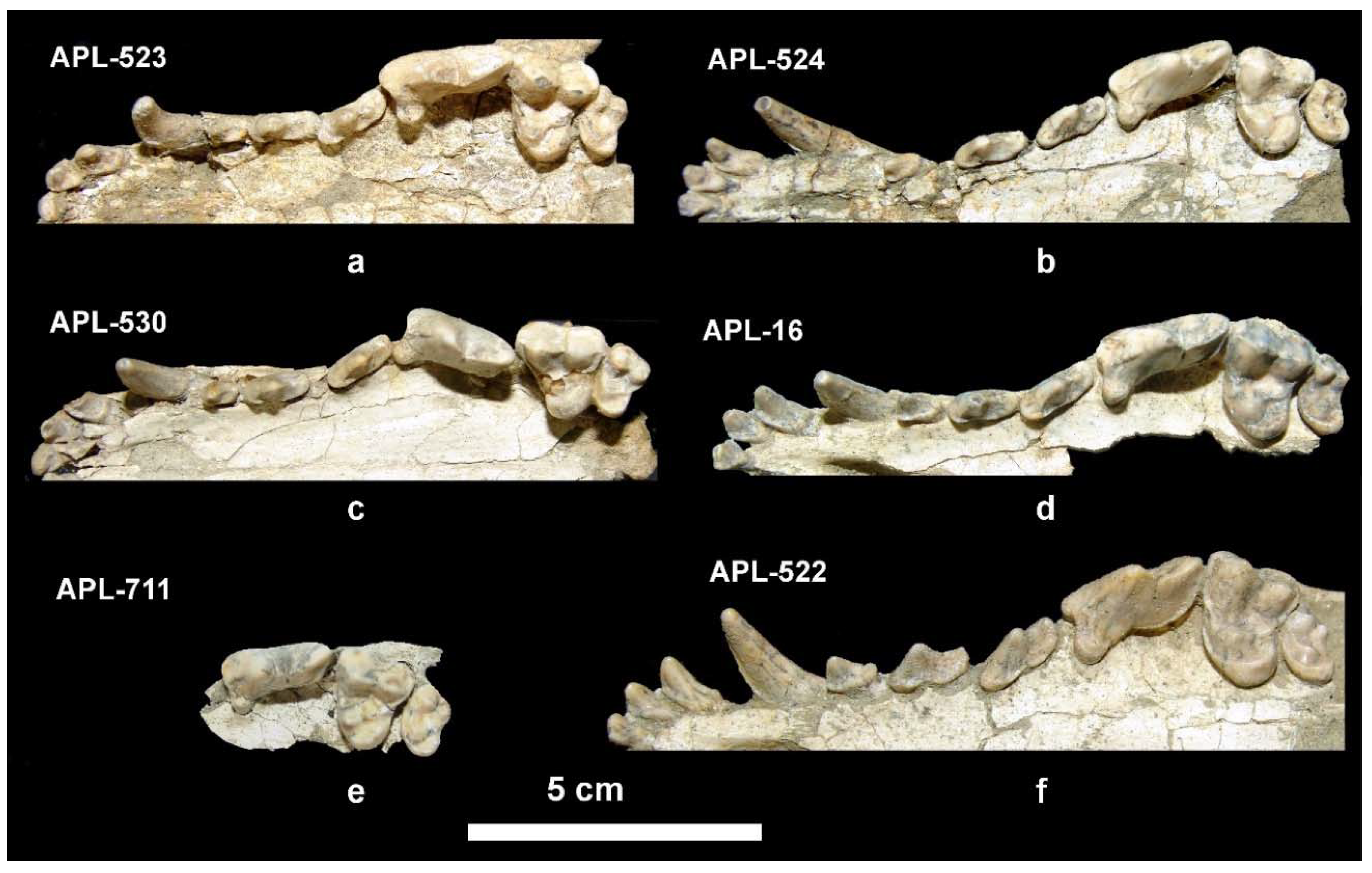
Old collection: Frontal cranial part and mandible, APL-530 (holotype); frontal cranial part, APL-523; maxilla with both I1-M2 tooth rows, APl-524; right maxilla with left I1-M2 and right I1-P2, APL-16; left maxillary fragment with i2-p3, APL-1; left mandibular fragment with c-m3, APL-17; right mandibular fragment with c-m1, APL-528; right mandibular fragment with p2-m2, APL-527.
Measurements. The measurements are given in Table 1, Table 2, Table 3 and Table 4.
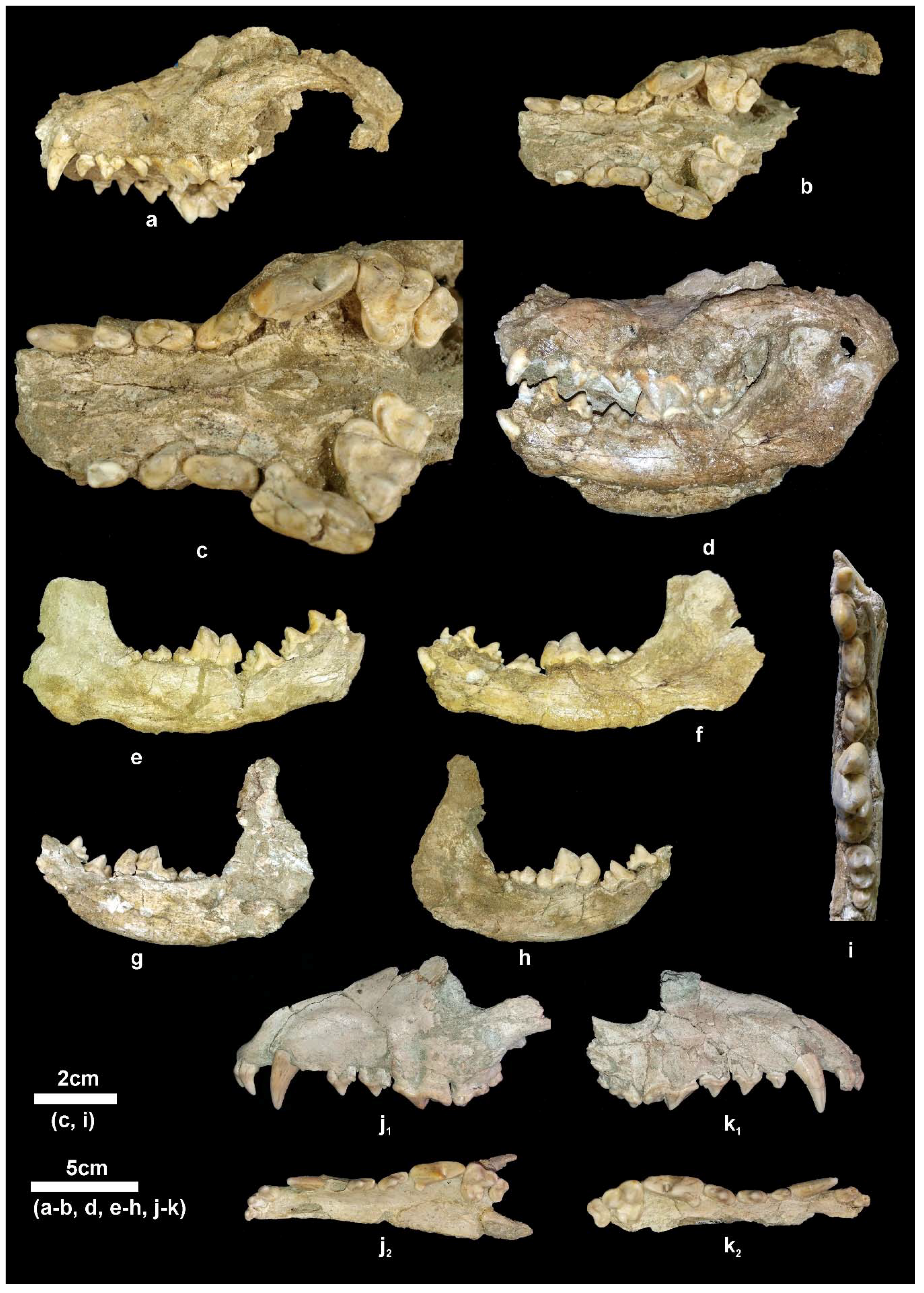
Description. The available cranial remains (APL-523, APL-530) preserve the frontal part of the cranium; they are compressed and the cranial bones are crushed. The nasal bones are better preserved in the holotype APL-530; they are narrow and elongated extending distally behind the anterior border of the orbit. The nasal cavity is small, oval-shaped and slightly inclined distally. The muzzle is relatively short. The well-preserved orbit in APL-523 is rounded with its anterior margin above the distal end of the carnassial. The palate is elongated and narrow. The tooth rows are well preserved in all specimens (Figure 4).
The upper incisors increase in size from I1 to I3 (Figure 4a–f); the last incisor is canine-like with strong lingual and distal cingula and bears a crest across its distal margin. The I1 and I2 have a triangular lingual outline and bear a distal cingular projection; two strong cingula start from this projection and are directed respectively towards the anteromesial and anterodistal extremities of the tooth forming two cusplets. The upper canine is strong, curved distally and strongly flattened bucco-lingually; it bears a weak mesio-lingual and distal crest, running from its apex to the base. The P1 is small, monocuspid, single-rooted with strong buccal and distal cingula; the P1 of APL-523 and APL-530 is slightly smaller than that of the other specimens. The P2 is long and narrow, bearing a p.a.c. situated in the middle of an elongated distal cingular projection; the p.a.c. is large in APL-523 and APL-524 but it is very small in APL-16 and APL-530; the distal margin of the distal cingualar projection is elevated and looks like a small secondary p.a.c.; there is a well-developed lingual cingulum. The P3 is morphologically like the P2, but is larger with clear and strong p.a.c. and with more expressed elevation of the distal cingulum; it bears a small mesio-lingual prominence of the cingulum from which starts a faint mesial crest to the apex of the main cusp. The carnassial is relatively short and wide; it lacks a parastyle and the protocone is small, low, and well separated from the paracone; its mesial margin is aligned with that of the paracone; there is a crest across the mesial border of the paracone from its apex to the base of the crown. The paracone is high and separated from the metacone by a deep valley; the metacone is blade-like and its distal part slightly curves buccally; a well-developed cingulum is observed in the disto-lingual half of the tooth. The M1 is triangular-shaped with slightly convex mesial margin and concave distal one; the paracone is larger and higher than the metacone; the protocone is small and low; the hypocone is crest-like; there is a small protoconule clearly distinguished in the less worn teeth; the entocone is also well distinguished in all specimens and is connected with the protocone by a crest; the main basin is not very deep and that between the protocone and hypocone is small and shallow; the mesial and disto-buccal cingula are well developed but the buccal cingulum is weak. The M2 has elliptical occlusal outline and similar morphology to the M1, but is smaller with faint or absent entocone and stronger buccal cingulum.
The mandibular corpus (Figure 5a–h) is elongated and relatively shallow; the ventral margin of the mandibular corpus bears a faint concavity from below the p2 to the p4 and then it curves gradually till the angular process, where it is strongly concave; the angular process is short, sharp and does not exceed the posterior margin of the condyle. The coronoid process is relatively low and separated from the condyle by a shallow mandibular incision. There are two mental foramina, one below the middle of the p2 and another below the middle of the p3. The masseteric fossa is oval and deep with its anterior margin below the m3.
The lower incisors are well preserved in APL-530 (Figure 5a); they have a triangular shape in lingual view with strong distal projection, like a small cuspid, which is quite large in the i3; the latter is canine-like and stronger than the other two incisors. The lower canine is relatively small, situated bucco-lingually, strongly curved distally and flattened bucco-lingually; it bears a faint mesio-lingual and distal crest. The p1 is small, monocuspid and single-rooted with a weak distal cingular projection. The p2 is elongated and narrow with a strong distal cingular projection, the distal margin of which is elevated and looks like a p.a.c.; a clear crest runs across the mesial and distal margins of the main cuspid. The p3 has similar morphology to the p2 but is larger and has stronger mesial cingulum and distal cingular projection; it lacks a p.a.c. but there is a small one in APL-715 and a vestigial one in APL-527 and APL-703; it bears a weak lingual cingulum. The p4 is more robust than the other premolars, with strong mesial cingulum; the distal cingular projection is large and bears a clear and large p.a.c. situated buccally; in APL-527, APL-530, APL-690 and APL-715 there is a small to faint secondary p.a.c.; the lingual cingulum is well developed. The lower carnassial is relatively short and wide, with a talonid about 1/3 of the tooth’s length. The protoconid is high and separated from the paraconid by a deep valley; the metaconid is small and situated disto-lingually to the protoconid; the talonid bears a large hypoconid and a small entoconid connected distally by a crest. The m2 has an oval occlusal outline with a large protoconid and a relatively small metaconid and hypoconid; it lacks an entoconid, but in some specimens (APL-530, APL-715) there is a crest in its position connected with the hypoconid; the mesial cingulum is strong. The m3 is present in APL-530; it is small, rounded, and single-rooted, with two cuspids; the large protoconid dominates and the paraconid is very small, situated lingually; a strongly developed cingulum is visible around the entire tooth.
Remarks. Canis apolloniensis was originally described from APL based on some maxillary and mandibular remains together with C. etruscus. Some authors consider C. apolloniensis as belonging to the C. etruscus-C. mosbachensis line, or as a southern European form of C. mosbachensis [17,18,19,20]. However, other researchers synonymized it with C. arnensis [14,21]. Although an extensive definition of C. apolloniensis is given in the original description of the taxon [4], the new findings and data published during the last twenty years, allow the knowledge of the taxon.
The inclusion of C. apolloniensis in the evolutionary line of C. etruscus–C. mosbachensis is based on the following distinctive characteristics separating C. etruscus and C. arnensis [17].
  • “In C. arnensis the talonid of […] M1 […] presents two clear different and separated peaks [hypoconid and entoconid] and in C. etruscus both cusplets are linked by a sinuous crest”; the last character is also mentioned in the emended diagnosis of C. etruscus [12].
    The m1 talonid of C. apolloniensis consists of two separated cuspids and bears a small sinuous cristid in its distal part, less pronounced than that of C. etruscus; in APL-690 and APL-703 it is very small, like a vestigial cusplet. In C. mosbachensis the m1 talonid is bicuspid but the two cuspids are connected through a strong transverse crest [15].
  • The m2 in C. etruscus “... shows a very well pronounced cingular antero-external border but it is not present in C. arnensis”.
    The pronounced mesial cingulum is clear in the m2 of C. etruscus from APL and Gerakarou (Mygdonia Basin, Greece) [2]; it looks like a shelf in the mesial and mesio-buccal margin of the tooth. Canis apolloniensis bears a less pronounced mesial cingulum than that of C. etruscus; in some specimens it is faint especially in the lingual half of the tooth. On the contrary, the mesial and mesio-buccal cingula of C. mosbachensis are well developed, like in C. etruscus.
  • “In […] M1 of C. arnensis the protocone is linked to the parastyle and metastyle by a well-defined crest, and the paraconule and metaconule are well marked”.
    In C. apolloniensis we cannot say that there is a crest linking the protocone with the parastyle and metastyle. Additionally, the paraconule of C. apolloniensis is weak and just distinguished, while the metaconule is well defined but relatively smaller than in C. arnensis. In C. mosbachensis both paraconule and metaconule of the m1 are prominent and the buccal cingulum complete and strong [15], distinguishing it from C. apolloniensis.
  • “In […] [M2] of C. arnensis there are 4 cusplets very well isolated [...] C. etruscus has only three peaks, never presents entoconid...”.
    Nevertheless, the presence of accessory cuspids in the disto-lingual portion and sometimes the presence of an entoconid are mentioned for the m2 of C. arnensis [20]. In all available specimens of C. apolloniensis the m2 is tricuspid and in this feature, it is closer to C. etruscus.
  • C. arnensis has diastemes between all the premolars and C. etruscus has no separation between the premolars”.
    This feature was also mentioned earlier [22,23], while a small diastema between the premolars is mentioned in the original description of C. apolloniensis [4]. A more detailed study of the available material indicates that this character varies in the APL material. In APL-524, there are diastemata between the premolars but the specimen is crushed and probably this affected the position of the teeth. In APL-523 there are diastemata, except between P3 and P4 but the specimen is deformed. In APL-530 there is a small diastema between P1 and P2 and a larger one between P2 and P3. In C. mosbachensis there are specimens with diastemata between the premolars (IQW 1982/18052), but the premolars set closely in others (IQW 1984/20177) [15]. Based on the great variation observed in this character, it seems that it cannot be used as diagnostic and rightly was not included in the emended diagnosis of C. arnensis [20].
Based on the abovementioned characteristics, C. apolloniensis has more similarities with C. etruscus than with C. arnensis and for this reason it is considered closer to the C. etruscus-C. mosbachensis evolutionary line [17].
Canis etruscus from the Villafranchian of Italy was recently revised [12]; based on the proposed diagnosis, C. apolloniensis differs from this taxon displaying absence of lingual cingulum in the P1, a vestigial secondary p.a.c. (modified distal cingulum) in the P3 (it is strong in C. etruscus), a weak paraconule in the M1, more constricted medially M1, on average more slender M1 and M2 (Figure 7), absence of a secondary p.a.c. in the p3 (it is strong in C. etruscus, weak mesial cingulum in the m2, and smaller size (Figure 6a–c). Based on the abovementioned comparisons, the APL canid is separated from C. etruscus.
The revision of the Italian sample of C. arnensis and the description of some new material from Poggio Rosso (Italy) provided an emended diagnosis for the taxon [20]. On that ground, C. apolloniensis differs from C. arnensis in having a small p.a.c. in the P2 (it is absent in C. arnensis), larger paracone than metacone in the M1 (they are equal-sized in C. arnensis), deeper protocone than hypocone basin in the M1 (they have the same depth in C. arnensis), remarkably larger hypoconid than entoconid in the m1, absence of p.a.c. in the p3 (there is a variably developed one in C. arnensis, APL-715 bears a small p.a.c. and APL-527 a vestigial one), usually absence of accessory cuspids between the entoconid and metaconid of the m1 (only two specimens present a vestigial cuspid), absence of a well-developed hypoconulid shelf on the distal margin of the m1 (there is a small and variably developed hypoconulid in the m1 of C. arnensis), absence or very weak disto-buccal cingulum in the m1, and larger protoconid than metaconid in the m2 (equal-sized in C. arnensis). In addition to these differences, the comparison with the known Greek material suggests that the two species have more or less the same size, though the upper teeth of C. apolloniensis are larger (Figure 6). Moreover, the M1 and M2 of C. apolloniensis are on average more robust than those of C. arnensis (Figure 7) and the trigonid of the m1 is longer in relation to the talonid (the % ratio Lm1trig./Lm1tal. is on average 268 for C. apolloniensis versus 245 for the Italian C. arnensis). Based on the above comparative data, C. apolloniensis, though it has a similar size to C. arnensis, can be distinguished from it through several morphological and metrical characteristics.
The other opinion concerning the taxonomy of C. apolloniensis is that it represents a southern European form of C. mosbachensis [20]. The latter species was originally described from the Middle Pleistocene locality Mosbach (Germany) [24], while a rich sample was described from the locality Untermassfeld (Germany) [15]. Based on the description and illustrations of C. mosbachensis, C. apolloniensis shows clear differences in displaying a laterally situated P3 in relation to the P4 (their main axes coincide in C. mosbachensis), a vestigial secondary p.a.c. in the P3 (this is an elevation of the distal cingulum which is more pronounced in C. mosbachensis), a small protocone aligned to the paracone, absence of p.a.c. in the p2 (sometimes it exists in C. mosbachensis), a very small secondary p.a.c. in the p4 (it is markedly larger in C. mosbachensis), and a small talonid relatively to the length of the m1 (the % ratio Lm1trig./Lm1tal. is 268 in C. apolloniensis versus 224 in C. mosbachensis). In addition to these differences our comparisons based on the description and illustrations of C. mosbachensis from Untermassfeld [15] indicate that:
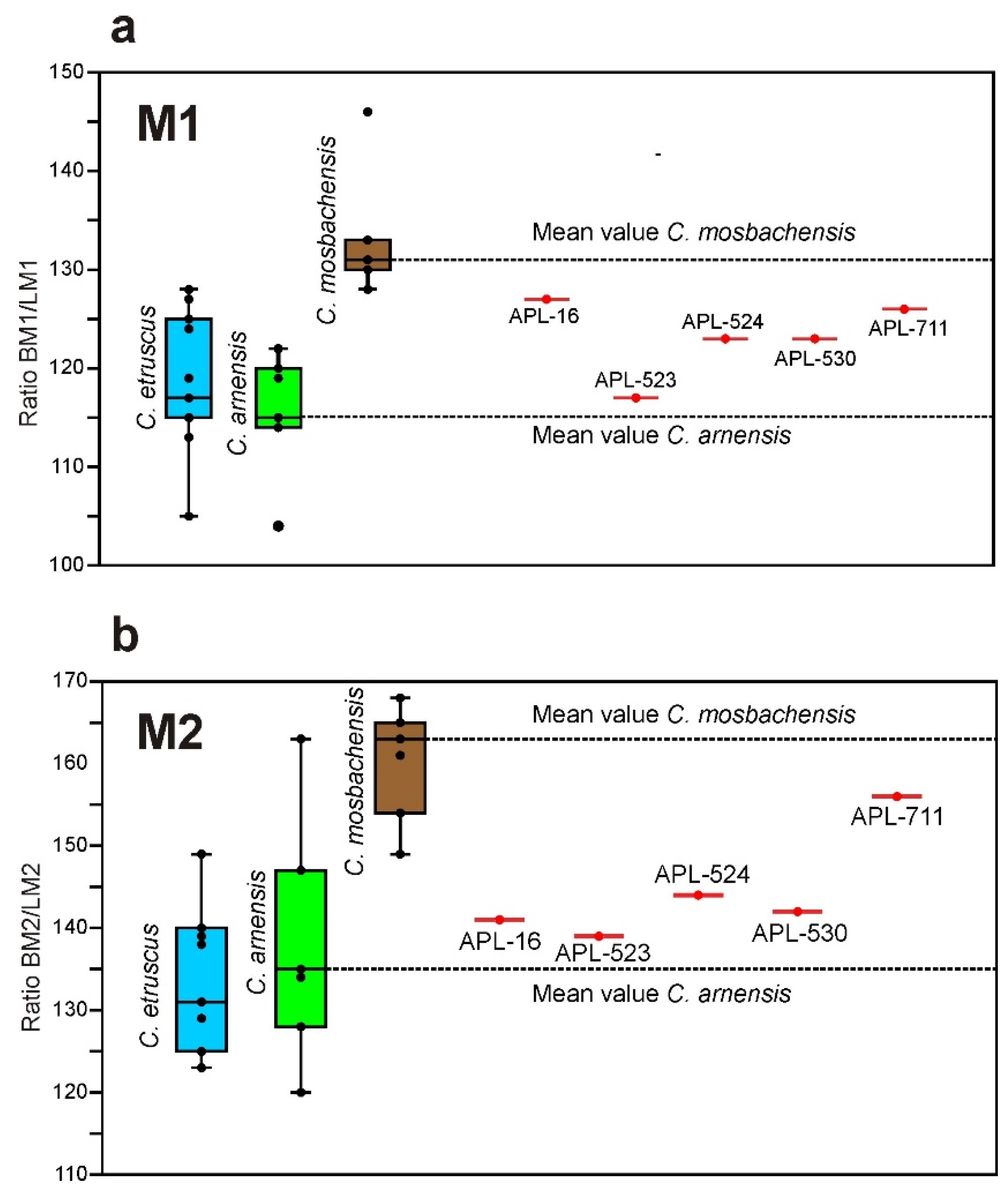
  • The occlusal shape of the M1 differs in the two species: it is sub-triangular and wide in C. mosbachensis, while it is triangular with narrow lingual part in C. apolloniensis. The mean % ratio BM1/LM1 of C. mosbachensis is remarkably larger than that of C. apolloniensis; the mean value of the ratio for C. apolloniensis is between the mean values for C. arnensis (slender M1) and C. mosbachensis (robust M1) (Figure 7a).
  • The M2 of C. mosbachensis is more robust than that of C. apolloniensis; the mean % ratio BM2/LM2 of C. apolloniensis is between those of C. arnensis (slender M2) and C. mosbachensis (robust M2), (Figure 7b).
All the abovementioned comparative data suggest that C. apolloniensis can be separated from the known taxa C. etruscus, C. arnensis and C. mosbachensis; its morphology seems to be closer to C. etruscus and C. mosbachensis and thus could be a potential ancestor of the latter taxon.

3.3. Genus Lycaon Brookes, 1827

Lycaon lycaonoides (Kretzoi, 1938)

Synonyms. 1997 Canis (Xenocyon) sp. Koufos and Kostopoulos, p. 48 (partim).
Material. New collection: Rostral part of the cranium with right P1-M3 and left C-M3 and the two mandibular corpi of the mandible with right p3-m3 and left c-m3, APL-771. Old collection: Right maxillary fragment with I2-M3 and the left one with I1-M3 of the same individual, APL-525.
Measurements. The measurements are given in the Table 1, Table 2, Table 3 and Table 4.
Description. The strongly laterally compressed and crushed cranial fragment lacks most of its morphology; the palate is also deformed and the tooth rows moved downwards. The specimen belongs to a subadult individual; the teeth are unworn and the canines are still erupting, with a large part of their height inside the bone. The teeth are well preserved and only the protocone of the left P4 is broken (Figure 8). The canine is flattened bucco-lingually and bears a distal and mesio-lingual weak crest. The P1 is large in relation to the APL Canis, monocuspid, single-rooted with elliptical crown and strong buccal and distal cingula. The P2 and P3 are elongated and narrow; they lack an a.a.c. but bear a mesial cingular projection; the distal cingular projection is strong and bears a p.a.c. situated in its center; the p.a.c. of the P3 is larger than that of the P2. The upper carnassial is relatively short and robust, with a large paracone; the mesial margin of the paracone inclines distally; the protocone is very small but well separated from the paracone and situated slightly in front of the mesial border of the tooth; a lingual cingulum is present. The M1 is large, triangular-shaped, with a large paracone dominating the crown; the metacone is smaller and lower than the paracone; the protocone is small and the hypocone very small and crest-like; there is a small entocone; the basin of the tooth is deep; there are well-developed buccal and lingual cingula. The M2 is small, like the M1 in the crown morphology, lacks a hypocone and an entocone and bears a strong lingual cingulum.
The left hemimandible (Figure 8e,f) preserves a part of the ascending ramus; the mandibular corpus is high relative to that of C. etruscus and C. apolloniensis (Table 2); the ventral margin of the mandibular corpus is convex, bearing a concavity, which starts below the m2 and continues to the angular process (Figure 8e–h); the masseteric fossa is elliptical and its anterior margin ends below the middle of the m2; there is a small mental foramen below the middle of the p3. The erupting lower canine bears a mesial groove. The p1 is small, monocuspid, single-rooted, with a vestigial distal cingulum. The p2 lacks an a.a.c. and a p.a.c. but bears a large distal cingular projection. The p3 and p4 have similar morphology but the latter is larger; they lack an a.a.c. but bear a large buccal p.a.c. (larger in the p4); there is a distal cingulum that is more pronounced and elevated distally in the p4; no clear cingulum is visible in any premolar; only in the p3 and p4 there is a mesio-lingual cingulum. The lower carnassial is large, with a well-developed paraconid and a large protoconid, separated by a deep lingual valley. The protoconid bears a groove in its mesial surface running from the apex to the valley between it and the paraconid. In the distal margin of the protoconid there is a strong crest running from its apex to that of the metaconid. The metaconid is small and situated behind the protoconid. The trigonid is short in relation to the tooth length and bears a large hypoconid; there are two small cusplets connecting the hypoconid with the lingual margin of the talonid; there is a small distal cingulum. The m2 bears a large protoconid, small and equal-sized metaconid and hypoconid; the disto-lingual margin of the talonid is slightly elevated and consists of a series of vestigial cusplets. The m3 is very small, single-rooted, monocuspid, with rounded crown and bears a variably developed cingulum.
Remarks. The taxonomy of the Plio-Pleistocene hypercarnivore canids is still a matter of debate and various authors, give different generic and specific names and include different samples under these names [25]. There are two main opinions about the taxonomic status of these canids. The first opinion includes all Late Villafranchian to the beginning of Middle Pleistocene forms into the genus Lycaon, in which three different chrono-species are recognized: L. falconeri (Late Villafranchian forms of Eurasia), L. lycaonoides (Epivillafranchian and early Middle Pleistocene Eurasian and African forms), and L. pictus (Middle to Late Pleistocene forms of Eurasia and Africa, as well as the extant African form) [26]; the ages given by the authors have been updated following the new dating of the Plio/Pleistocene boundary. The other opinion accepts the validity of the genus Xenocyon, including the Eurasian species X. lycaonoides and the endemic New World species X. texanus [25]. A number of authors follow the first opinion [11,27,28,29], but others argue that all or most of the Early Pleistocene forms belong to the genus Xenocyon [30,31,32]. Having in mind that the APL material has similarities to those of Pirro Nord (Italy) and Vallparadis (Spain), both attributed to L. lycaonoides (see following paragraphs), as well as that the establishment of a synonymy is beyond the main aim of this article, we shall use the name Lycaon for the APL material.
The type of L. lycaonoides is one M1 from Gombaszög (Hungary) ([33] p. 132; pl. 3, Figure 4). A mandibular fragment with p4-m1, described in the same article as “Canis” gigas ([33] p. 128; pl. 2, Figure 10), has also been included in the type series of the species [25]. The absence of a metaconule, the reduced hypocone and the reduced basin between the protocone and the hypocone of the M1, as well as the strong p.a.c. of the p4, the moderate metaconid in the m1, and the small talonid in the m1 of the APL sample coincide with those of the type material of L. lycaonoides; their dental dimensions are also similar (Figure 9a). Lycaon lycaonoides is characterized by an enlargement of the trigon basin in the M1, a very small or absent basin between the protocone and hypocone, a reduction of the breadth in the upper molars, a reduction of the talonid’s breadth in the m1, entire domination of the hypoconid in the talonid of the m1, and a very small or vestigial entoconid in the m1 [26] (the entoconid is sometimes absent; see the Untermassfeld sample [15]). All these features are present in the APL sample (Figure 8), confirming its attribution to this taxon.
The early species L. falconeri includes the material referred to Canis falconeri from the Upper Valdarno (Italy) and Canis antonii from China [26]. The name C. falconeri was given by Forsyth Major in 1877 to a cranial fragment (IGF 883) from the Upper Valdarno. A mandible (IGF 865) from the same locality was later referred to the same species [35]. These two specimens are considered as the type material of the taxon [22]. Casts of the dentition from these two specimens are housed in LGPUT and have been compared with the APL sample. The dental morphology of the two samples is rather similar, but there are some differences, as the reduced hypocone and the reduced basin between the protocone and hypocone of the M1 in the APL sample. The lower dental dimensions of the APL sample fall into the range of variation of L. lycaonoides from Europe; however, the lower premolars are wider than those of Pirro Nord (Italy), Venta Micena and Vallparadis (Spain), being closer to those of Untermassfeld (Germany) (Figure 9b). Based on the descriptions and illustrations of C. antonii [36], this species differs from the APL sample, displaying a more expressed hypocone a groove between the hypocone and protocone in the upper molars, as well as a larger size (Figure 9c).
The similar dental morphology and dimensions of the APL sample with L. lycaonoides from various Eurasian localities, as well as its morphological and metrical differences from the other taxa, support its attribution to this species. A dental morphology like that of the APL Lycaon is observed in the material of Pirro Nord (Italy), Vallparadis (Spain) and Untermassfeld (Germany), [11,15,29] indicating that the APL L. lycaonoides belongs to the more derived forms of the late Early Pleistocene. The species L. lycaonoides is rare in Greece, being reported only from APL and from the earliest group (early Middle Pleistocene) of the Petralona Cave with a single lower carnassial [28].

3.4. Genus Vulpes Frisch, 1775

Vulpes praeglacialis (Kormos, 1932)

Synonyms. 1992 Vulpes alopecoides Koufos, p. 12.
Material. New collection: Right upper canine, APL-692; left upper carnassial, APL-691; left lower carnassial, APL-770. Old collection: Right P4, APL-20; right hemimandible with c-m2, APL-11;
Measurements. APL-692: 5.4 × 3.5 mm. Measurements are given in Table 5 and Table 6.
Description. The upper canine APL-692 is well preserved and little worn in its apex (Figure 2f,g); it is sharp, and curved distally and slightly buccally; its buccal wall is slightly convex and the lingual one flattened; the crown section at the base is elliptical; the buccal height of the crown is 13.9 mm; the root is strong, robust and flattened bucco-lingually; its buccal height is 16.1 mm. Both upper carnassials are well preserved and scarcely worn (Figure 2h–m); the protocone is small, low, well separated from the paracone and situated in front of the tooth’s mesial margin; the paracone is high, with its mesial margin strongly inclined backwards; the metastyle blade is trenchant and its distal part is slightly buccally directed; it bears a pronounced lingual cingulum; the three roots are well preserved in both specimens except the mesial one in APL-20, which is broken; the distal root is strong and bucco-lingually flattened.
The mandible APL-11 lacks the gonial area and is crushed below the molars (Figure 2c–e); the mandibular corpus is shallow in labial view with convex ventral margin posterior to the m1; it possesses two mental foramina, one small below the mesial root of the p2 and one very small below the middle of the p3; the masseteric fossa is oval, deep and its mesial margin ends below the distal margin of the m3. The lower canine is sharp and strongly curved distally; the buccal wall is convex, while the lingual one is flattened and bears a mesio-lingual and a distal crest; the lingual cingulum is well defined. The p1 is small, monocuspid and single-rooted; it bears a tiny distal cingular projection. The p2 and p3 are elongated and narrow, lacking accessory cuspids but bearing a pronounced distal cingular projection. The p4 is robust in comparison with the p2 and p3 and bears a large p.a.c. The lower carnassial is relatively elongated, with a short talonid; there is a small metaconid situated disto-lingually to the high protoconid; the talonid is bicuspid with a large hypoconid and a smaller entoconid connected distally through a low crest. The m2 has an oval occlusal outline, a large protoconid, a crest-like hypoconid, and a pronounced mesio-buccal cingulum. The m3 is absent but from its alveole it seems to be small and single-rooted. The lower carnassial, APl-770 (Figure 2n–p), is unworn and morphologically like that of the mandible APL-11 from which it differs only in displaying an oblique distal margin.
Remarks. The old material of Vulpes from APL (APL-20, APL-11) was originally ascribed to V. alopecoides [2]. The differences between V. praeglacialis and V. alopecoides are vague and the two taxa can be confused. Some mandibular remains of Vulpes alopecoides were reported from the Greek locality Dafnero 1 (Western Macedonia, Greece). A direct comparison of APL-11 with the Dafnero 1 sample indicated that the former is different in displaying longer distal cingular projection in the p2 and p3, and stouter hypoconid in the m1 [37]. Although some published data [38] refer that no distinct entoconid is present in the m2 of V. alopecoides from La Puebla de Valverde (Spain), there is a small cuspid in that position in the DFN sample, linked with the hypoconid through a crest, and APL-11 shows a clear entoconid. Some mandibular remains from the Petralona cave, housed in LGPUT and referred to V. praeglacialis [], have similar dental morphology to the APL sample. The average dental dimensions of V. praeglacialis are slightly larger than those of V. alopecoides (Figure 10). The premolars of V. praeglacialis are longer than those of V. alopecoides because of the longer distal cingular projection; however, the single specimen of V. alopecoides from St.-Vallier has an elongated p3, like that of V. praeglacialis. The specimen APL-11 has dimensions that proportionally coincide with those of V. praeglacialis from Petralona (Greece), as well as those from L’Escale (France), although the premolars from the latter site are longer (Figure 10). The morphology of the APL upper carnassial fits to that of V. praeglacialis from L’Escale (France), showing a clear protocone less developed than that of the modern V. vulpes [39]. The dimensions of the APL carnassial are close to those of V. praeglacialis from L’Escale (LP4buccal = 12.1 (range: 11.9–12.3), LP4lingual = 12.9 (range: 12.7–13.9), BP4 = 5.8 (range: 5.4–6.5) [39]. Based on all above data, the APL vulpine can be attributed to V. praeglacialis. The taxon is also known from the Villafranchian of Volos (Thessaly) through some undescribed material [40] and from the earliest group (early Middle Pleistocene) of the Petralona cave [28].

3.5. Family Hyaenidae Gray, 1869; Genus Pachycrocuta Kretzoi, 1938

Pachycrocuta brevirostris (Gervais, 1850)

Synonyms. Pliohyaena brevirostris Koufos and Kostopoulos, 1997. Pachycrocuta brevirostris Koufos, 2014.
Material. New collection: Left I3, APL-757; left P4, APL-700; left calcaneum, APL-24. Old collection: Right mandibular fragment with p3 and p4, APL-541; left p2, APL-542.
Measurements. I3: 17.8 × 14.4 mm; P4: 43.5 × 26.1 mm, Lmetastyle =16.7 mm.
Calcaneum: Height = 74.0 mm, DTmax. = 30.5 mm, DAPmax. = 36.1 mm.
Description. The I3, APL-757 (Figure 2q,r), lacks a part of the root and the enamel in the lingual wall of the crown is partially broken. It is worn, robust, and strongly curved distally with elliptical occlusal outline at the crown base; the % ratio B/L is 81. The remains of a worn mesial crest are present in the lingual crown surface; the crest probably continues to the apex of the tooth but is difficult to see because the enamel is locally broken. The root is strong and flattened with oval cross section. The measured buccal height of the root is 42.5 mm.
The upper carnassial (Figure 2s–u) preserves the whole crown, which has minute damage in its mesial border that does not affect the length measurement. The mesial roots, although crushed, are complete but the distal one is crushed and lacks its lower part. The protocone is small in relation with the tooth size; it is well separated from the parastyle by a deep valley, directed mesio-lingually, and its mesial margin is aligned with that of the parastyle. The parastyle is well developed but lower than the paracone, which is higher, having isocline mesial and distal margins; it is well separated from the parastyle and metastyle by deep valleys; the metastyle blade is relatively short and its distal part is directed buccally; a weak buccal cingulum and a pronounced disto-lingual one is observed. The mesial root, corresponding to the protocone, is stronger than that corresponding to the parastyle; both are long with an elliptical cross section and their longitudinal axes are perpendicular to each other; the distal root is large, strong and flattened bucco-lingually.
The calcaneum APL-24 (Figure 2v,w) is relatively elongated and slender. It bears two articular facets for the astragalus well separated by a deep groove. The articular facet for the astragalus situated in the sustentaculum tali is rounded and small. The second articular facet, situated in the coracoid process, is elongated and curved extending to the ventral surface of the bone. The articular facet for the cuboid is sub-elliptical and perpendicular to the longitudinal axis of the calcaneum.
Remarks.Pachycrocuta brevirostris is a well-known and widespread extinct hyaenid distributed from Spain to China. It was originally described from the locality Sainzelles (France) and its taxonomy was discussed for a long time. Recently, the original author and publishing year for the species changed and its valid name is P. brevirostris (Gervais, 1850); for more details see [41]. The upper carnassial of the species is characterized by a well-developed parastyle, a high paracone, a large mesio-lingually directed protocone aligned with the mesial margin of the parastyle, and a weak cingulum [42,43,44]; all these characteristics are present in the upper carnassial APL-700. The dimensions of APL-700 fall into the range of variation of P. brevirostris (Figure 11a) and together with the morphological data suggest its attribution to this taxon. The morphology of APL-757 resembles that of P. brevirostris described from Incarcal (Spain) and its dimensions are like those of the specimen IN-I 956 (17.1 × 14.1 mm) from the latter locality [45] (pl. 1, figs C1, C2.) The calcaneum APL-24 morphologically also resembles that of P. brevirostris from Incarcal (Spain) [45], while its size is close to that of P. brevirostris and Homotherium latidens (Figure 11b). It differs from the latter taxon as it is shorter and more gracile, lacks the small articular facet for the cuneiform and displays narrower and longer sustentaculum tali [45].
Although there are several evidences for the presence of Pachycrocuta brevirostris in the Greek fossil record, the known material is relatively scarce and restricted to some isolated teeth, some maxillary and mandibular fragments with deciduous teeth, and a few postcranial elements. The species co-occurs with P. perrieri in the locality Gerakarou (Mygdonia, Basin, Greece), correlated with the early Late Villafranchian, indicating the Middle to Late Villafranchian transition and marking the Pachycrocuta-event in the Balkan Peninsula [46]. The taxon was also recognized in the Mygdonia Basin Late Villafranchian–Epivillafranchian localities Tsiotra Vryssi and Kalamoto [7,47], in the Late Villafranchian locality Libakos [48] and in the early Middle Pleistocene faunal group of the Petralona Cave [28], representing the latest occurrence of the taxon in Greece.

3.6. Family Felidae (Fischer von Waldheim, 1817) Gray, 1821; Subfamily Felinae Trouessart, 1885; Genus Panthera Oken, 1816

Panthera gombaszögensis (Kretzoi, 1938)

Material. New collection: Right I3, APL-712; left maxillary fragment with the canine, APL-767; left p3, APL-769; mesial fragment of a left p4, APL-758.
Measurements. I3: 10.3 × 9.6 mm; C: 23.5 × 13.0 mm; p3: 14.8 × 8.1 mm; p4: - × 10.40 mm.
Description. The I3 APL-712 (Figure 12c,d) is moderately worn, preserving the entire crown and root. The crown is canine-like and directed buccally. It bears a crest across its mesial and distal margins, running from the apex to the base of the crown. The distal crest, while worn, is stronger than the mesial one and ends in a strong distal cingular process. The root is elongated, bucco-lingually flattened and elongated relatively to the height of the crown.
The specimen APL-767 (Figure 12a,b) preserves the larger portion of the frontal part of the left half of the nasal cavity with the canine; the canine is strongly compressed laterally, crushed, deformed bucco-lingually, so that its length is slightly larger than the original one. The canine is strong at the crown base and relatively low; it bears a slight lingual crest that starts from the crown base and gradually weakens to the apex. The mesial height is 26.0 mm.
The p3 APL-769 (Figure 12e–g) is well preserved retaining both roots; it is little worn with a small dentine pit in the apex of the main cuspid. It bears a large a.a.c. well separated from the main cuspid and placed more lingually; there is a strong distal projection of the distal cingulum, like a talonid; the distal margin of this projection is elevated and two small apices are distinguished at the top of the elevation; in the disto-lingual border of the main cuspid there is a small cuspule; the tooth bears weak lingual and buccal cingulum. The p4, APL-758 (Figure 12h–j), lacks its distal part, of which only the mesial half of the p.a.c. is still preserved. The main cuspid is worn in its apex; the a.a.c. is large and wide, situated mesio-lingually to the main cuspid; it bears a weak buccal cingulum.
Remarks. The species Panthera gombaszögensis was originally described as Leo gombaszögensis [33] (p. 100; Table 1, Figure 1, Figure 2, Figure 3, Figure 4, Figure 5, Figure 6 and Figure 7) several opinions have been proposed on the taxonomy of this species subsequently. Recently, the taxonomy of P. gombaszögensis has been re-discussed and it has been suggested that the taxon appeared in Africa at ~1.90 Ma; the oldest European form is that of U. Valdarno and Olivola, originated from the African P. gombaszögensis at ~1.7 Ma and referred to as P. g. toscana. This subspecies gave origin to two branches, that of P. g. gombaszögensis, which survived in Europe until 0.3 Ma and that of P. g. georgica from which the modern P. onca originates [54]. The available material from APL is scarce and fragmentary, avoiding a subspecific determination and is here referred to P. gombaszögensis.
The I3 has similar morphology to that of P. gombaszögensis [49,54] and its size falls into the range of variation of this species (Figure 13a). The upper canine APL-767 has similar morphology with that of P. gombaszögensis from Gombaszög (Hungary) ([33] Table 1), Figure 4 and to that from Westbury (England) and Chateau Breccia (France) [54,56]. Homotherium latidens and M. cultridens, with their elongated and strongly flattened bucco-lingually upper canines, are well distinguished from APL-767. The dimensions of APL-767 are also close to those of P. gombaszögensis from Europe (Figure 13b); its slightly longer length, as it was referred above, is due to the strong bucco-lingual compression.
The p3 APL-769 is morphologically similar with that of P. gombaszögensis from the type locality ([33] Table 1), Figure 4 and with those from Westbury (England) and Chateau Breccia (France) [54,56]; its dimensions also fall into the range of variation of the European sample of this taxon (Figure 13c). The morphology of the APL-758 resembles that of P. gombaszögensis and their dimensions are similar; the mesial breadth of APL-758 is 9.4 mm, versus 8.8–10.7 for P. gombaszögensis from Untermassfeld (Germany) [55] and 8.8–9.8 mm from Château Breccia (France) [56].
The holotype of the Italian form P. g. toscana is a mandibular fragment with p3-m1. A direct comparison of the APL p3 and p4 with a cast of the holotype, housed in LGPUT, indicates that the p3 differs from this subspecies, displaying a smaller size, a larger a.a.c., a shorter and narrower distal projection of the distal cingulum and a less developed distal cingulum. The APL p4 is smaller and with more pronounced a.a.c. than that of P. g. toscana.
A cranium of P. gombaszögensis is known from the Greek locality Gerakarou (Mygdonia Basin) [2,46]. The absence of the canines and the broken incisors in the Gerakarou cranium make a comparison with the studied APL sample impossible; however, the APL I3 is larger than that of Gerakarou (8.5 × 7.0 mm; [2]). The species is also known from the locality Alykes (Volos, Thessaly) through some cranial and postcranial remains [13]; unfortunately, the dentition of the single mandibular fragment is badly preserved hampering comparison with the APL teeth. Some undescribed remains of P. gombaszögensis are mentioned from the area of Volos, Thessaly. The exact fossiliferous site is unknown and the collected material possibly belongs to a surface collection. The whole assemblage suggests a Villafranchian age [40].

3.7. Subfamily Machairodontinae Gill, 1872; Genus Homotherium Fabrini, 1890

Homotherium latidens (Owen, 1846)

Material. New collection: Left lower canine APL-710; distal fragment of a left p4, APL-684; left second metacarpal, APL-332.
Measurements. c: 16.0 × 11.9 mm; mesial height: 19.2 mm. p4: - × 10.2 mm.
McII: H = 93.8 mm, DTprox. epiph. = 20.9 mm, DAPprox. epiph. = 26.6 mm, DTmidshaft = 14.1 mm, DAPmidshaft = 13.6 mm, DT dist. epiph. = 20.7 mm, DAP dist. epiph. = 19.6 mm.
Description. The slightly worn lower canine APL-710 (Figure 12k–m) preserves the crown and the root. It is relatively small, flattened bucco-lingually; the % ratio B/L is 74. The crown is sharp with serrated mesio-lingual and distal crest (Figure 12m). The mesio-lingual crest is worn in the upper half but the serration is well distinguished in its basal half (Figure 12m). The mesio-lingual crest ends at the crown’s base and forms a small cusplet. The root is lingually flattened and directed distally; its mesial height is 51.8 mm.
The available p4 (Figure 12n–p) lacks the mesial part and the protoconid lacks its lingual basal part. The protoconid is high and slender, bearing a mesial and distal marginal crest; the mesial crest is unworn and finely serrated, but the distal one is worn. The distal part of the p4 has a talonid-like distal projection with a relatively small p.a.c. situated buccally; the distal cingulum is strong and elevated, giving the impression of a second p.a.c. in buccal view; the buccal cingulum is weak.
The McII APL-332 is well preserved (Figure 12q–t). The shaft is straight, except for its proximal part, which is directed laterally; its transverse section is subtriangular. It bears a large, concave almost triangular proximal articular facet, the dorsal part of which is rounded and inclines abruptly distally. There is a large subtriangular medial articular facet, situated in the palmar part of the epiphysis. Below this facet there is a bulging of the bone on the medial surface of the shaft. The distal epiphysis has strong medial and lateral protuberances and the trochlea is well separated from the shaft by a deep palmar groove; the medial part of the trochlea is weakly developed in contrast with the lateral one, which is robust and globular. The keel is well developed dorsally but absent in the palmar surface of the trochlea.
Remarks. Although the genus Homotherium was a widespread machairodontine known from Eurasia, Africa and America, its taxonomical position is still discussed. The current trend recognizes a single polymorphic species, H. latidens (Owen, 1846), for the Plio-Pleistocene of Eurasia [63]. As the main aim of this article is not the taxonomy of the genus, the studied APL material is referred under this name. The small size, the strong lateral flattening and the finely serrated mesial and mesio-lingual borders of the lower canine APL-710 coincide with the known morphology of H. latidens [57,63]. Moreover, the dimensions of the lower canine fall into the range of variation for this species (Figure 14a). The APL-684 is quite fragmentary, but some of its characteristics support its attribution to this species. The presence of the serrated mesial border of the main cuspid and the morphology of the distal part are like those of H. latidens from Domegliara Delvavecchia (Italy) ([57] Figure 6) and fit well with the description of these teeth given for the Incarcal material [58]. The direct comparison of APL-684 with a cast of H. latidens (IGF-824) from Valdarno housed in LGPUT indicates similar morphology, confirming the attribution of the former to H. latidens. Moreover, the breadth of APL-684 is closer to that of H. latidens and larger than that of Megantereon (Figure 14b). The McII APL-332 is morphologically and dimensionally (Figure 14) like that of H. latidens from Pirro Nord (Italy) and Incarcal (Spain) [11,58]. It differs from M. cultridens and P. gombaszögensis in being longer and displaying a remarkably larger proximal epiphysis and a deeper distal one (measurements 1, 2, 3, 7 in Figure 14c).
The APL material of Homotherium is scarce but constitutes the first evidence of its presence in the Epivillafranchian of Greece. The species has been reported from the Pliocene locality Milia (Western Macedonia, Greece) with a few remains [70], as well as from the Middle Villafranchian locality Sesklon [13]; it is possibly present in the Middle Villafranchian locality Tourkovounia 3 [71]. It is also reported from the Villafranchian of Kos Island [72].

3.8. Family Mustelidae Swainson, 1835; Genus Meles Brisson, 1762

Meles dimitrius Koufos, 1992

Material. New collection: Left hemimandible with i1-p3 and m1, APL-772. Old collection: Skull, APL-544; left maxilla with I3-M1, APL-545; left hemimandible, APL-15; right mandibular ramus withc-m1, APL-546.
Measurements. The measurements are given in Table 7.
Description. The hemimandible APL-772 is well preserved but lacks the coronoid process (Figure 12u–w). The mandibular corpus is relatively high with a straight inferior margin, which curves upwards below the m2. It bears a large mental foramen below the p2 and a very small one below the p3. The masseteric fossa is oval and moderately deep with its anterior margin below the m2. The symphysis is oval and roughly inclines backwards. The elongated angular process protrudes beyond the distal margin of the condyle. The tooth row lacks the p1, p2 and m2 but the other teeth are well preserved and little worn. The incisors are small and rather worn; their size increases from i1 to i3. The canine is strongly curved distally and bears a large distal cingulum; there is a clear mesio-lingual crest running from the base to the apex, which is sharp. The p1 is missing but its minute root is clearly distinguished under a stereoscope. The main cuspid of the p2 and p3 is high and both bear a strong distal cingulum. The lower carnassial is long with the talonid wider than the trigonid. The paraconid and the larger protoconid are situated buccally and separated by a lingual valley. The metaconid is robust, situated disto-lingually to the protoconid. The large talonid bears a large hypoconid and a smaller entoconid; a lingual open valley separates the hypoconid from the protoconid; there is a small hypoconulid and another small cuspid behind the entoconid, called “entoconulid” [73]; a distal crest connects the hypoconulid with the “entoconulid,” forming a large talonid basin.
Remarks.Meles dimitrius was erected based on some cranial and mandibular remains from the localities Gerakarou and APL in the Mygdonia Basin [2]; later, a cranium and a hemimandible were described from APL [4]. The mandibular and dental morphology, as well as the size of APL-772 fit well to those of the previously described mandibles of M. dimitrius from APL [2,4] and thus it can be attributed to this species. The taxonomic revision of the Villafranchian badgers of Europe [74] suggests the presence of M. thorali in the Early and Middle Villafranchian and of M. meles atavus in the Late Villafranchian and Epivillafranchian. According to this view, the Greek material from Gerakarou would be included in the former taxon and that from APL in the latter [74].
One of the distinctive characteristics of M. thorali with respect to the extant M. meles is the position of the tympanic bullae, which are situated behind the posterior wall of the postglenoid processes in the former taxon and aligned with them in M. meles [74]. In the partial cranium GER-159 the tympanic bullae are aligned with the posterior wall of the postglenoid processes [2] and therefore the specimen differs from M. thorali. The cranium APL-544 is deformed and crushed but it seems to have similar position of the tympanic bullae with GER-159. However, it is not clear if this character can really be used to distinguish M. thorali from M. meles. In three available crania of the extant M. meles stored in LGPUT the anterior margin of the tympanic bullae is behind the postglenoid processes. The upper carnassial of M. dimitrius is on average smaller than that of M. thorali from Saint-Vallier (Figure 15b); the main difference is the shorter length and thus the P4 of M. dimitrius appears more robust than that of M. thorali. The % ratio LP4/BP4 is smaller in the APL and Gerakarou sample than in M. thorali (Figure 15c). The P4 length relatively to the buccal length of the M1 is shorter than that of M. thorali; the % ratio LP4/LM1buccal is on average 93.5 (range: 93–94) for APL, 94 for Gerakarou versus 108 (range: 101–114) for M. thorali from Saint-Vallier and 113 from Lunel-Viel (data for M. thorali from [39]). The M1 of APL-544 displays a metacone smaller than the paracone instead of equal-sized in M. thorali; its buccal length is smaller in comparison to that of M. thorali. The m1 of the APL and Gerakarou sample is smaller on average than that of M. thorali from the type locality of Saint-Vallier and its talonid is relatively longer than the trigonid. The % ratio Lm1trig./Lm1tal. of the APL m1 is small than that of M. thorali (Figure 15c), indicating that its trigonid is longer relatively to the talonid.
Meles atavus was erected based on a mandibular fragment with m1 from Püspökfürdő (Hungary) ([77] p. 241; taf. 8, Figure 9); the correct name of the type locality of M. atavus is Betfia 5 [76]. Later, the species was synonymized with M. meles and a left mandibular fragment with i2-c and p2-m2 from Gombaszög (Hungary) was described as M. m. atavus ([33] p. 126; taf. 2, Figure 8 and Figure 9). Recently M. m. atavus was traced in the locality of Vallparadís (Spain) [74]. The m1 of M. atavus displays a small cuspid between the protoconid and hypoconid [33]. The Gerakarou m1 is worn, hampering the observation of this small cuspid. The latter is absent in the m1 of the APL sample. Both Gerakarou and APL m1 are quite smaller than the type of M. atavus (Figure 15e).
The Vallparadis sample is richer including both cranial and mandibular remains allowing a detailed comparison with the material from the Mygdonia Basin. The APL-544 differs from the Vallparadis sample in displaying:
  • Wider and more robust upper carnassial; the % ratio LP4/BP4 is quite smaller than that for Vallparadis (Figure 15c), indicating a more robust P4 for the APL sample.
  • A more triangular occlusal outline of the upper carnassial with an angular lingual margin instead of the curved one observed in the Vallparadis sample (Figure 15a).
  • A relatively large cusp in the lingual angular margin of the P4; in the Vallparadis P4 the lingual margin bears a crest-like elevation the end of which continues to the paracone’s apex (Figure 15a).
  • A small cusplet in the mesial margin between the paracone and the cusp of the lingual margin. a cusplet is mentioned as “cuspule on the precingulum at the base of the paracone” [78], which is absent from the Vallparadis P4 [74].
  • A labial incision between the metacone and metaconule and a well-developed postprotocrista reaching the lingual crown margin of the M1 (B1 morphotype [78]); the Vallparadis M1 lacks a labial incision and has a short postprotocrista (B3 morphotype [78]).
  • Short buccal length of M1 in relation to the carnassial length; the % ratio LP4/LM1buccal is on average 93.5 (range: 93–94) for APL versus 79 (range: 77–81) for the Vallparadis sample.
The P4 and M1 from Gerakarou are badly preserved, making any comparisons difficult. The P4 is smaller than that of Vallparadis, and closer in size to that from APL (Figure 15b). It is more robust than the Vallparadis P4; the % ratio LP4/BP4 is smaller than that fromVallparadis and closer to the APL premolar (Figure 15c). The Gerakarou upper carnassial, while worn, bears a large dentine pit at the lingual margin (Figure 15a) corresponding to a cusplet and reinforcing its similarity with the APL one. The single Gerakarou M1 is very worn but shows the same morphotype as the APL one and is thus different from the Vallparadis M1.
A new badger species named Meles hollitzeri has been described from the locality Deutsch-Altemburg (Austria) [75]; besides the type locality, the species has also been recorded from the Cave Treugolnaã in Caucasus Mountains (Russia) and from Untermassfeld (Germany) [76]. Meles hollitzeri was consequently synonymized with M. meles and it included in the fossil subspecies M. m. atavus [74]. The APL badger differs from the Altenburg 2 and Untermassfeld material in displaying small and robust P4 (Figure 15b,c), a cusp in the mesial margin of the P4, a cusplet in the lingual margin of the P4, different M1 morphotype (the APL M1 belongs to the B1 morphotype instead of B3 for the Untermassfeld and B4 for the Altenburg ones [78]), and small m1 with long talonid relatively to the trigonid (Figure 15c,e).
The abovementioned comparisons indicate that the P4, M1 and m1 play an important role for the distinction of fossil badger species. The morphology of the P4 clearly separates the APL and Gerakarou material from European ones referred to M. m. atavus, and M. hollitzeri, which are close chronologically to the APL material. Additionally, the dimensions and the shape of the P4, as well as the trigonid length in relation to that of the talonid in m1 allow the distinction of the APL sample from the abovementioned two species. To check all metrical differences, all dimensions of the M1 (Lbuccal, Llingual, B) and m1 (L, B, Ltrig., Ltal.) have been analyzed in comparison with various European samples of Meles using principal component analysis (PCA); only complete and specimens lacking only one measurement were included in the analysis. The results for the M1 suggest that the APL specimens are larger than all the others except one from Vallparadis, which has a quite long M1 (Figure 15d). The single Gerakarou M1 falls into the convex hull of M. thorali but its morphology, as well as that of the P4, are closer to the APL teeth. On the contrary, the APL and Gerakarou m1 are on average smaller than all other samples and are thusseparated from them (Figure 15e). I shall agree with M. Wolsan [76], that it is better to leave the European species of Meles unsynonymized until more material is found, to better understand their morphological and metrical variation, as well as to resolve the conspecifity of European and Asian recent badgers. Therefore, based on the abovementioned morphological and metrical differences both APL and Gerakarou badgers are attributed to Meles dimitrius.

4. Discussion and Conclusions

4.1. Faunal Composition and Similarity

The APL carnivoran assemblage is quite rich, including 11 taxa, two of which, H. latidens and P. gombaszögensis, are reported here for the first time. The canids and felids are represented by four taxa and account for 36.4% of the assemblage. The ursids, mustelids and hyaenids are represented by a single taxon each (Figure 16). Several Villafranchian mammal faunas are known from Greece, but most of them are older than that from APL, and those with similar age include limited number of species or doubtful attributions. The composition of the APL carnivoran assemblage at family level seems to be different from the Middle Villafranchian carnivoran assemblages of Volax, Dafnero 1 and Sesklon and the early Late Villafranchian one of Gerakarou (Figure 16). In fact, the Volax and Gerakarou assemblages lack ursids, Sesklon and Volax lack mustelids, and Dafnero lacks felids. However, this observation is probably artificial as the collection of more material can provide representatives of these families, e.g., remains of a felid have been discovered in Dafnero this year, while bone remains of an ursid have been found in Gerakarou. Thus, we can say that the composition at family level of the Greek Villafranchian carnivoran assemblages is more or less similar. However, the similarity and diversity at species level are different. The canids account for 30–40% of the fauna in all Greek carnivoran assemblages, but in APL the canids are more diversified, including four taxa versus two in the other assemblages.
Similarly, felids are present with four taxa in APL versus one or two in the other carnivoran assemblages (Figure 16). In contrast to the ursids, which are represented by U. etruscus in all assemblages, hyaenids are represented by different taxa (P. brevirostris in APL, Chasmaprthetes lunensis in Dafnero, and P. brevirostris together with P. perrieri in Gerakarou).
The APL carnivoran assemblage is also compared with some Eurasian ones covering the time span from the Late Villafranchian to the Epivillafranchian: (a) Dmanisi (Georgia) dated at 1.77 Ma, early Late Villafranchian [80]; (b) Pirro Nord (Italy) dated between 1.5 and 1.2 Ma, late Late Villafranchian [81]; (c) Venta Micena (Spain) dated at ~1.3 ± 0.1 Ma, late Late Villafranchian–Epivillafranchian [82]; (d) Untermassfeld (Germany) dated at ~1.0 Ma, Epivillafranchian [83]. The high abundance of felids and canids observed in the abovementioned carnivoran assemblages agrees to that from the APL one (Figure 16). The taxonomic composition (presence/absence matrix) of these carnivoran assemblages is analyzed at specific and generic level by hierarchical cluster analysis, using the Raup–Crick index, to compare their similarity. The carnivoran assemblages are separated in two main clusters at specific level (Figure 17a). The Cluster A includes Pirro Nord and Untermassfeld carnivoran assemblages which show high similarity (79.7%). The APL, Venta Micena and Dmanisi constitute the Cluster B, which is separated in two subclusters: the subcluster-B1, including APL and Venta Micena with a high similarity (99.9%) and the subcluster-B2, including only Dmanisi; the latter has a similarity of ~87% with the sub-cluster B1 (see table of Figure 17a). The similarity between the Cluster A and Cluster B is low (~35.0%). The clustering at generic level distinguishes the Pirro Nord carnivoran assemblage as a separate Cluster A, whereas the others (APL, Venta Micena, Dmanisi, Untermassfeld) match together in the Cluster B; the similarity between the two clusters is moderate (~57%), (Figure 17b). The Cluster B is separated in three subclusters: the subcluster B1 including APL and Venta Micena (similarity of 99.5%), the subcluster B2 including only Untermassfeld with a similarity of ~80% with the subcluster B1 and the subcluster B3 including only Dmanisi and displaying a similarity of ~72% with the subcluster B2 (Figure 17b). The separation of the Pirro Nord carnivoran assemblage from the others is possibly due to the presence of some small carnivoran taxa, such as Pannonictis nestii and Mustela palaerminea, which are absent from the other assemblages. The Untermassfeld carnivoran assemblage is separated from the others because it includes some younger taxa such as C. mosbachensis, M. hollitzeri, and Ursus dolinensis (= U. rodei).

4.2. Biochronology

The APL faunal assemblage correlated with the Latest Villafranchian or Epivillafranchian, with an estimated age of 1.2–1.0 Ma [46] and references therein. The term Epivillafranchian has been proposed as a biochronological or sub-biochronological unit, expressing the time span from 1.5–1.3 to 0.9–0.8 Ma [84,85,86]. According to recent publications, the Epivillafranchian is considered as a biochron included within the Praemegaceros verticornis-Bison menneri first occurrences and Crocuta crocuta first occurrence, corresponding to the time span between 1.2–0.9 Ma [87]. However, the validity of the Epivillafranchian is disputed, as there are several factors (taxonomical, palaeoecological, diachrony/asynchrony) that do not allow for accepting it as a typical biochronological unit [88]. The problem is under discussion and needs more fossil material and additional studies to be resolved, but this is beyond the main aim of this work.
We shall try to check if the APL carnivoran assemblage fits well with the previously published dating. The APL carnivoran assemblage is a mixture of archaic and modern elements. The canids are well represented with four different species. The dispersal of the Canini from Asia was rather rapid [89]. The occurrence of Canis gives an archaic feature to the APL fauna as the genus apparently reached Europe at the beginning of the Late Villafranchian; its arrival is known as “wolf-event”, dated at ~1.95 Ma [90,91,92]. This event was re-named as the “Pachycrocuta-event”, as this hyaenid is well recognized and widespread in Eurasia [93]. Pachycrocuta brevirostris and P. gombaszögensis appeared for the first time in Olivola (Italy). The Olivola Faunal Unit is considered the first Late Villafranchian one and is followed by the Tasso Faunal Unit. The Olivola/Tasso transition has been magnetostratigraphically calibrated at ~1.8 Ma [94] and thus the Olivola faunal assemblage is older than 1.8 Ma. The “Pachycrocuta-event” has been recognized in Greece in the Gerakarou assemblage, where P. perrieri co-exists with its replacer Pachycrocuta brevirostris, as well as with P. gombaszögensis and C. etruscus [46]. The presence of P. perrieri gives a more archaic feature to the Gerakarou fauna, suggesting an age closer to the Middle/Late Villafranchian transition. Therefore, the Gerakarou assemblage has an age closer to that of the Olivola Faunal Unit, near the Middle/Late Villafranchian transition. The absence of P. perrieri in the APL carnivoran assemblage suggests an age younger than that of Gerakarou.
The machairodontine Megantereon was a felid widely distributed in the Old World. Although its origin and taxonomy are still debated [95,96]. The APL Megantereon is like that from Venta Micena (Spain) and both belong to the small-sized forms that appeared at the end of Early Pleistocene. Moreover, the APL and Venta Micena carnivoran assemblages are similar both at generic and specific levels (Figure 17) and thus a similar age is quite possible for them. The Venta Micena fauna has been dated at 1.3 ± 0.1 Ma [82] and thus the APL fauna must have at least a similar or a younger age, in other words an Epivillafranchian age. This age is supported by the presence of V. praeglacialis, which is reported from the Epivillafranchian of France [97], Spain (Venta Micena) [98]) and Austria (Deutsch-Altenburg 2 [75]). The morphological characteristics of the hypercarnivore L. lycaonoides suggest similarities to the Epivillafranchian forms of the taxon; additionally, L. lycaonoides is considered as a chronospecies characterizing the Epivillafranchian and early Middle Pleistocene [26]. The canid C. apolloniensis has more primitive characteristics than C. mosbachensis from Untermassfeld, taking an intermediate position between that and the earlier C. etruscus. Therefore, an age older than that of Untermassfeld is quite possible for APL. The palaeomagnetic record supports an estimated age of ~1.0 Ma for the Untermassfeld assemblage [83]. Consequently, the APL carnivoran assemblage can be correlated with the early Epivillafranchian and dated more precisely to 1.3–1.0 Ma, confirming the previous estimations.

4.3. Palaeoenvironment

The carnivorans are rarely used for palaeoenvironmental reconstructions, as they normally have a large geographic distribution and are easily adaptable. However, their guild structure and locomotor behavior can be indicative for adaptation to a specific habitat [99,100,101]. The guild structure of a carnivoran assemblage in comparison with modern ones from known environments can provide information about the palaeoenvironment [99,102,103,104]. Three variables (body mass, locomotion and diet) are used for the construction of a guild diagram. The guild structure of the modern assemblages from Serengeti and Guyana have been used as comparative ones for open and closed conditions respectively [99]; their guild diagrams have been reported in the literature [104] and used for the comparison with APL. The body mass of Serengeti taxa belongs mainly to the classes 4–6 (10–100 kg); the Serengeti carnivoran assemblage lacks arboreal taxa with most taxa being terrestrial and cursorial; most of the taxa are carnivorous to bone/meat eaters (classes 3–5). The APL guild diagram (Figure 18a) includes taxa belonging to the body mass classes 4 to 6. The majority (five taxa) belong to the medium size class-4 (10–30 kg), two taxa to the large size class-5 (30–100 kg) and three to the very large size class-6 (>100 kg), (Figure 18b). Such body mass carnivoran association fits better with the Serengeti guild diagram. The presence of mainly terrestrial and cursorial taxa and the absence of arboreal and scansorial ones in the APL carnivoran assemblage, indicate that the APL guild diagram is closer to the Serengeti one. Most of the APL carnivorans have carnivorous-hypercarnivorous diet, two are omnivores (Meles, Ursus) and one bone/meat eater (Pachycrocuta), (Figure 18b); this diet feature corresponds better to that of the Serengeti guild. The close similarity of the APL guild structure to that of Serengeti suggests an open landscape.
The long bone functional morphology of the Villafranchian large-sized carnivoran taxa provided some information about their environment (i.e., ecomorphological data); the various carnivoran taxa are classified as “adapted” or “non-adapted” to “tropical” or “grassland” conditions depending on the morphology of the selected skeletal elements [100,101]. Following the latter author, the APL canids C. apolloniensis and L. lycaonoides are considered as “grassland” specialists, like their relative-forms C. etruscus and L. falconeri. Other three APL taxa i.e., P. gombaszögensis, H. latidens and P. brevirostris are predicted, as “grassland” specialists. M. cultridens and L. issiodorensis are adapted to “tropical” environment, while U. etruscus is predicted as “non-adapted” to both “grassland” and “tropical” biome. The “grassland” large carnivoran taxa dominate in the APL assemblage, indicating a grassy landscape; the APL “tropical” specialists give a more closed character to the palaeoenvironment, which could have been like the modern open forests or woodland savannah. This palaeoenvironment agrees with the results obtained from the guild structure of the APL assemblage.
Previous environmental studies for the Villafranchian, using different analyses (taxonomic, habitat, and dietary diversity; cenograms), allowed the reconstruction of the environment in the Eastern Mediterranean region. During the Late Villafranchian the palaeoenvironment of the region was open like the modern woodland savannah. A faunal reorganization, which started at that time with the arrival of several taxa, marked a change in the environmental conditions. This faunal renewal was completed during the Epivillafranchian, with the arrival of several new taxa and the disappearance of others. The analysis and comparison of the Epivillafranchian faunas indicate an open grassy landscape and mild climatic conditions ([46] and references therein), which agree with those obtained from the analysis of the APL carnivoran assemblage. The dental mesowear of the Apollonia Bison suggests grazing feeding behavior indicative of an arid habitat with developed grassy cover; the morphometric analysis of the metapodials suggests open and arid conditions [111], agreeing to the ecomorphological data reported above. Although the various palaeoenvironmental proxies agree on a relatively open, dry environment, the dental microwear analysis of the Apollonia cervids suggests a different habitat. Two large cervids have been recognized in Apollonia, Praemegaceros pliotarandoides and Arvernoceros cf. verestchagini. Their dental wear pattern (six specimens) indicates a relatively cold environment comparable to the modern taigas (closed habitat) [112], agreeing with the presence of some carnivorans (M. cultridens, L. issiodorensis) characterized as “adapted to tropical” conditions. Combining all the abovementioned data, a relatively open landscape with grassy floor and with more forest areas (patchy or mosaic landscape) is possible for Apollonia 1. However, more material and research (e.g., study of dental meso- and micro-wear of the dominant taxa Bison and Equus, study of the functional morphology of the equid metapodials, etc.) are necessary for a more precise determination of the habitat. The investigations in the Mygdonia Basin are in progress and we hope to have more data soon.

Acknowledgments

Many thanks are due to several colleagues and students for participating in the excavations in the Apollonia locality since the beginning of the 1990s. I wish to thank J. Madurell Malapeira for providing me with photos of Meles from Vallparadis. I also thank M.R. Palombo for her useful comments on the draft manuscript. Many thanks to the three anonymous reviewers for their useful comments that improved the text remarkably. I also thank K. Vasileiadou for the linguistic improvement of the text and J. Llorca for his work on the manuscripts.

Conflicts of Interest

The author declares no conflict of interest.

References

  1. Koufos, G.D.; Syrides, G.E.; Kostopoulos, D.S.; Koliadimou, K.K. Apollonia, a new vertebrate site in the Pleistocene of the Mygdonia basin (Macedonia, Greece); the first fossil fresh-water mollusks in the area. C. R. Acad. Sci. Paris 1992, 315, 1041–1046. [Google Scholar]
  2. Koufos, G.D. The Pleistocene carnivores of the Mygdonia basin (Macedonia, Greece). Ann. Paléontol. 1992, 78, 205–257. [Google Scholar]
  3. Kostopoulos, D.S. The Plio-Pleistocene artiodactyls (Vertebrata, Mammalia) of Macedonia, 1. The fossiliferous site “Apollonia 1”, Mygdonia Basin of Greece. Geodiversitas 1997, 19, 845–875. [Google Scholar]
  4. Koufos, G.D.; Kostopoulos, D.S. New Carnivore material from the Plio-Pleistocene of Macedonia, Greece with the description of a new canid. München Geowiss. Abh. 1997, 34, 33–63. [Google Scholar]
  5. Koufos, G.D.; Kostopoulos, D.S.; Sylvestrou, I. Equus apolloniensis n. sp. (Mammalia, Equidae) from the latest Villafranchian locality of Apollonia, Macedonia, Greece. Palaeontol. Evol. 1997, 30–31, 49–76. [Google Scholar]
  6. Koufos, G.D.; Syrides, G.E.; Kostopoulos, D.S.; Koliadimou, K.K. Preliminary results about the stratigraphy and the palaeoenvironment of Mygdonia basin, Macedonia, Greece. Geobios 1995, 18, 243–249. [Google Scholar] [CrossRef]
  7. Konidaris, G.E.; Tourloukis, V.; Kostopoulos, D.S.; Thompson, N.; Giusti, D.; Michailidis, D.; Koufos, G.D.; Harvati, K. Two new vertebrate localities from the Early Pleistocene of Mygdonia Basin (Macedonia, Greece): Preliminary results. C. R. Palevol 2015, 14, 353–362. [Google Scholar] [CrossRef]
  8. Hammer, Ø.; Harper, D.A.T.; Ryan, P.D. PAST: Paleontological statistics software package for education and data analysis. Palaeontol. Electr. 2001, 4, 1–9. [Google Scholar]
  9. Koufos, G.D.; Konidaris, G.E.; Harvati, K. Revisiting Ursus etruscus (Carnivora, Mammalia) from the Early Pleistocene of Greece with description of new material. Quat. Int. 2017, in press. [Google Scholar] [CrossRef]
  10. Quilles, J. Les Ursidae du Pléistocène Moyen et Supérieur en Midi Méditerranéen: Apports Paléontologiques, Biochronologiques et Archéozoologiques. Ph.D. Thesis, Museum National d’Histoire Naturelle, Paris, France, 2003. [Google Scholar]
  11. Petrucci, M.; Cipullo, A.; Martínez-Navarro, B.; Rook, L.; Sardella, R. The Late Villafranchian (Early Pleistocene) carnivores (Carnivora, Mammalia) from Pirro Nord (Italy). Palaeontographica A 2013, 298, 113–145. [Google Scholar] [CrossRef]
  12. Cherin, M.; Bertè, D.F.; Rook, L.; Sardella, R. Re-defining Canis etruscus (Canidae, Mammalia): A new look into the evolutionary history of Early Pleistocene dogs resulting from the outstanding fossil record from Pantalla (Italy). J. Mamm. Evol. 2013, 21, 95–110. [Google Scholar] [CrossRef]
  13. Athanassiou, A. Contribution to the study of the fossil mammals of Thessaly; National and Kapodistrian University of Athens, Editions of the Department of Geology: Athens, Greece, 1998; Volume 5, pp. 1–354, (In Greek with English Summary). [Google Scholar]
  14. Boudadi-Maligne, M. Les Canis Pleistocenes du Sud de La France: Approche Biosystematique, Evolutive et Biochronologique. Ph.D. Thesis, Université Bordeaux 1, Bordeaux, France, 2011. [Google Scholar]
  15. Sotnikova, M. Remains of Canidae from the lower Pleistocene site of Untermassfeld. In Das Pleistozän von Untermassfeld bei Meiningen (Thüringen); Monographie Romisch-Germanisches Zentramuseum; Kahlke, R.D., Ed.; Dr Rudolf Habelt: Bonn, Germany, 2001; pp. 607–632. ISBN 978-3-7749-3080-3. [Google Scholar]
  16. Rook, L. I Cani dell’Eurasia dal Miocene Superiore al Pleistocene Medio. Ph.D. Thesis, Modena-Bologna-Firenze-Roma “La Sapienza” Universities, Rome, Italy, 1993. [Google Scholar]
  17. Martínez-Navarro, B. Presence of African large mammals (primates, carnivores and ungulates) in the lower Pleistocene of the Middle East and Europe. Ann. Géol. Pays Hell. 2002, 39, 337–351. [Google Scholar]
  18. García, N. Los Carnívoros de Los Yacimientos Pleistocenos de La Sierra de Atapuerca. Ph.D. Thesis, Universidad Complutense de Madrid, Madrid, Spain, 2002. [Google Scholar]
  19. Garrido, G.; Arribas, A. Canis accitanus nov. sp., a new small dog (Canidae, Carnivora, Mammalia) from the Fonelas P-1 PlioPleistocene site (Guadix basin, Granada, Spain). Geobios 2008, 41, 751–761. [Google Scholar] [CrossRef]
  20. Bartolini Lucenti, S.; Rook, L. A review on the Late Villafranchian medium-sized canid Canis arnensis based on the evidence from Poggio Rosso (Tuscany, Italy). Quat. Sci. Rev. 2016, 151, 58–71. [Google Scholar] [CrossRef]
  21. Brugal, J.P.; Boudadi-Maligne, M. Quaternary small to large canids in Europe: Taxonomic status and biochronological contribution. Quat. Int. 2011, 243, 171–182. [Google Scholar] [CrossRef]
  22. Torre, D. I cani Villafranchiani della Toscana. Paleontogr. Ital. 1967, 33, 112–138. [Google Scholar]
  23. Koufos, G.D. Canis arnensis Del Campana, 1913 from the Villafranchian (Villanyian) of Macedonia (Greece). Paleontol. Evol. 1987, 21, 3–10. [Google Scholar]
  24. Soergel, W. Die Säugetierfauna des altdiluvialen Tonlagers von Jockgrim in der Pfalz. Z. Dtsch. Geol. Gesell. 1925, 77, 405–438. [Google Scholar]
  25. Tedford, R.H.; Wang, X.; Taylor, B.E. Phylogenetic systematics of the North American fossil Caninae (Carnivora: Canidae). Bull. Am. Mus. Nat. Hist. 2009, 325, 1–218. [Google Scholar] [CrossRef]
  26. Martínez-Navarro, B.; Rook, L. Gradual evolution in the African hunting dog lineage: Systematic implications. C. R. Palevol 2003, 2, 695–702. [Google Scholar] [CrossRef]
  27. Harstone-Rose, A.; Werdelin, L.; de Ruiter, D.J.; Berger, L.; Churchill, S.E. The Plio-Pleistocene ancestor of wild dogs, Lycaon sekowei n. sp. J. Paleontol. 2010, 84, 299–308. [Google Scholar] [CrossRef]
  28. Baryshnikov, G.F.; Tsoukala, E. New analysis of the Pleistocene carnivores from Petralona Cave (Macedonia, Greece) based on the Collection of the Thessaloniki Aristotle University. Geobios 2010, 43, 389–402. [Google Scholar] [CrossRef]
  29. Madurell-Malapeira, J.; Rook, L.; Martínez-Navarro, B.; Alba, D.M.; Garrido, J.A.; Moyà-Solà, S. The latest European painted dog. J. Vertebr. Paleontol. 2013, 33, 1244–1249. [Google Scholar] [CrossRef]
  30. Moullé, P.-E.; Lacombat, F.; Echassoux, A. Apport des grands mammifères de la grotte du Vallonnet (Roquebrune-Cap-Martin, Alpes-Maritimes, France) à la connaissance du cadre biochronologique de la seconde moitié du Pléistocène inférieur d’Europe. L’Anthropologie 2006, 110, 837–849. [Google Scholar]
  31. Tong, H.W.; Hu, N.; Wang, X. New remains of Canis chihliensis (Mammalia, Carnivora) from Shanshenmiaozui, a lower Pleistocene site in Yangyuan, Hebei. Vertebrata. PalAsiatica 2012, 50, 335–360. [Google Scholar]
  32. Echassoux, A.; Moigne, A.M.; Moullé, P.E.; Li, T.Y.; Feng, X.B.; Li, W.S.; Wu, Z.Z. Les faunes de grands mammifères du site de l’Homme de Yunxian. In Le Site de l’Homme de Yunxian; HAL-00472960; De Lumley, H., Li, T.Y., Eds.; CNRS Editions: Paris, France, 2008; pp. 253–364. [Google Scholar]
  33. Kretzoi, M. Die Raubtiere von Gombaszög nebst einer Übersicht der Gesamtfauna (Ein Beitrag zur Stratigraphie des Altquartärs). Ann. Mus. Nat. Hung. 1938, 31, 88–157. [Google Scholar]
  34. Rook, L. The Plio-Pleistocene Old World Canis (Xenocyon) ex gr. falconeri. Boll. Soc. Paleontol. Ital. 1994, 33, 71–82. [Google Scholar]
  35. Del Campana, D. I cani pliocenici di Toscana. Palaeontogr. Ital. 1913, 19, 189–254. [Google Scholar]
  36. Zdansky, O. Jungteriäre Carnivoren Chinas. Paleontol. Sin. 1924, C2, 1–149. [Google Scholar]
  37. Koufos, G.D. Late Pliocene carnivores from western Macedonia (Greece). Paläontol. Z. 1993, 67, 357–376. [Google Scholar] [CrossRef]
  38. Kurtén, B.; Crusafont Paíro, M. Villafranchian carnivores (Mammalia) from La Puebla de Valverde (Teruel, Spain). Comment. Biol. 1977, 85, 1–39. [Google Scholar]
  39. Bonifay, M.F. Carnivores quaternaries du Sud-Est de la France. Mém. Mus. Nat. Hist. Nat. 1971, C21, 1–377. [Google Scholar]
  40. Van der Meulen, A.; van Kolfschoten, T. Review of the late Turolian to early biharian mammal faunas from Greece and Turkey. Mem. Soc. Geol. Ital. 1986, 31, 201–211. [Google Scholar]
  41. Alba, D.M.; Vinuesa, V.; Madurell-Malapeira, J. On the original author and year of description of the extinct hyaenid Pachycrocuta brevirostris. Acta Palaeontol. Pol. 2015, 60, 573–576. [Google Scholar] [CrossRef]
  42. Qiu, Z. Die Hyaeniden aus dem Ruscinium und Villafranchium Chinas. München Geowiss. Abh. A 1987, 9, 1–108. [Google Scholar]
  43. Turner, A. Remains of Pachycrocuta brevirostris (Carnivora, Hyaenidae) from the Lower Pleistocene site of Untermassfeld. In Das Pleistozän von Untermassfeld bei Meiningen (Thüringen); Monographie Romisch-Germanisches Zentramuseum; Kahlke, R.D., Ed.; Dr Rudolf Habel: Bonn, Germany, 2001; pp. 673–690. ISBN 978-3-7749-3080-3. [Google Scholar]
  44. Madurell-Malapeira, J.; Alba, D.M.; Moyà-Solà, S. Carnivora from the late Early Pleistocene of Cal Guardiola [Terrassa, Vallés Penedés Basin, Catalonia, Spain]. J. Palaeontol. 2009, 83, 969–974. [Google Scholar] [CrossRef]
  45. Galobart, A.; Antón, M.; Maroto, J. Hienidos y canidos de los yacimientos de Incarcal (Girona, NE de la Peninsula Iberica). Una aproximacion a la paleobiologia del hienido del Pleistoceno inferior. Paleontol. Evol. 2003, 34, 79–98. [Google Scholar]
  46. Koufos, G.D.; Kostopoulos, D.S. The Plio-Pleistocene Large Mammal Record of Greece: Implications for Early Human Dispersals into Europe. In Palaeoanthropology of the Balkans and Anatolia: Human Evolution and Its Context; Harvati, K., Roksanic, M., Eds.; Springer: Dordrecht, Germany, 2016; pp. 269–280. ISBN 978-94-024-087-4. [Google Scholar]
  47. Tsoukala, E.; Chatzopoulou, K. A new Early Pleistocene (Latest Villafranchian) site with mammals in Kalamotó (Mygdonia Basin, Macedonia, Greece)—Preliminary report. Mitt. Komm. Quartärforsch. 2005, 14, 213–233. [Google Scholar]
  48. Steensma, K.J. Plio-/Pleistozäne Großäugetiere (Mammalia) aus dem Becken von Kastoria/Grevena, südlich von Neapolis-NW Griechenland. Ph.D. Thesis, Technische Universität Clausthal, Clausthal-Zellerfeld, Germany, 1988. [Google Scholar]
  49. Madurell-Malapeira, J.; Morales, J.; Vinuesa, V.; Boscaini, A. Úrsidos, hiénidos y fèlidos del Pleistoceno inferior de Cueva Victoria (Cartagena, Murcia). Mastia 2015, 11–13, 401–432. [Google Scholar]
  50. Martínez Navarro, B. Revisión Sistemática y Estudio Cuantitativo de la Fauna de Macromamíferos del Yacimiento de Venta Micena (Orce, Granada). Ph.D. Thesis, Autonomous University of Barcelona, Bellaterra, Spain, 1999. [Google Scholar]
  51. Turner, A.; Antón, M. The giant hyaena Pachycrocuta brevirostris (Mammalia, Carnivora, Hyaenidae). Geobios 1996, 29, 455–468. [Google Scholar] [CrossRef]
  52. Kostopoulos, D.S.; Sen, S. Late Pliocene (Villafranchian) mammals from Saricol Tepe, Ankara, Turkey. Mitt. Bayer. Staatssamml. Paläontol. Hist. Geol. 1999, 39, 165–202. [Google Scholar]
  53. Vinuesa, V.; Madurell-Malapeira, J.; Ansón, M.; Alba, D.M. New cranial remains of Pliocrocuta perrieri (Carnivora, Hyaenidae) from the Villafranchian of the Iberian Peninsula. Boll. Soc. Paleontol. Ital. 2014, 53, 39–47. [Google Scholar]
  54. Argant, A.; Argant, J. The Panthera gombaszögensis story: The contribution of the Château breccia (Saône-et-Loire, Burgundy, France). Quaternaire 2011, Hors-série 4, 247–269. [Google Scholar]
  55. Hemmer, H. Die Feliden aus dem Epivillafranchium von Untermassfeld. In Das Pleistozän von Untermassfeld bei Meiningen (Thüringen); Monographie Romisch-Germanisches Zentramuseum; Kahlke, R.D., Ed.; Dr Rudolf Habelt: Bonn, Germany, 2001; pp. 679–782. ISBN 978-3-7749-3080-3. [Google Scholar]
  56. Bishop, M.J. The mammal fauna of the Early Pleistocene cavern infill site of Westbury-Dub-Mendip, Somerset. Spec. Pap. Palaeontol. 1982, 28, 1–108. [Google Scholar]
  57. Sardella, R.; Iurino, D.A. The latest early pleistocene sabertoothed cat Homotherium (Felidae, Mammalia) from Monte Peglia (Umbria, central Italy). Boll. Soc. Paleontol. Ital. 2012, 51, 15–22. [Google Scholar]
  58. Galobart, A.; Pons-Moya, J.; Antón, M.; Maroto, J. Descripcion del material de Homotherium latidens (Owen) de los yacimentos del Pleistoceno inferior de Incarcal (Girona, NE de la Penísula Ibérica). Paleontol. Evol. 2003, 34, 99–141. [Google Scholar]
  59. Del Campana, D. Nuove ricerche sui feline del Pliocene italiano. Palaeontol. Ital. 1915, 21, 223–291. [Google Scholar]
  60. Ficarrelli, G. The Villafranchian machairodonts of Tuscany. Palaeontol. Ital. 1979, 71, 17–26. [Google Scholar]
  61. Garcia, N.; Arsuaga, J.L. Carnivores from the Early Pleistocene hominid-bearing Trinchera Dolina 6 (Sierra de Atapuerca, Spain). J. Hum. Evol. 1999, 37, 415–430. [Google Scholar] [CrossRef] [PubMed]
  62. Langlois, A. Présence de Panthera gombaszoegensis Kretzoï, 1938 à la grotte XIV (Cénac-et-Saint-Julien, Dordogne). Paleo 2002, 14, 1–14. [Google Scholar]
  63. Antón, M.; Salesa, M.J.; Galobart, A.; Tseng, Z.J. The Plio-Pleistocene scimitar-toothed felid genus Homotherium Fabrini, 1890 (Machairodontinae, Homotheriini): Diversity, palaeogeography and taxonomic implications. Quat. Sci. Rev. 2014, 96, 259–268. [Google Scholar] [CrossRef]
  64. Baryshnikov, G.F. Pleistocene Felidae (Mammalia, Carnivora) from the Kudaro paleolithic cave sites in the Caucasus. Proc. Zool. Inst. RAS 2011, 315, 197–226. [Google Scholar]
  65. Thenius, E. Die Feliden (Carnivora) aus dem Pleistozän von Stránská skála. Stud. Mus. Morav. Anthrop. 1972, 20, 121–135. [Google Scholar]
  66. De Bonis, L. Un Félidé à Longues Canines de la Colline de Perrier (Puy-de-Dôme). Ann. Paléontol. 1976, 62, 159–198. [Google Scholar]
  67. Hemmer, H.; Kahlke, R.D.; Vekua, A.K. The jaguar Panthera onca gombaszoegensis (Kretzoi, 1938) (Carnivora: Felidae) in the late Lower Pleistocene of Akhalkalaki (South Georgia; Transcaucasia) and its evolutionary and ecological significance. Geobios 2001, 34, 475–486. [Google Scholar] [CrossRef]
  68. Tsoukala, E.; Bonifay, M.F. The Early Pleistocene carnivores (Mammalia) from Ceyssaguet (Haute-Loire). Paléo 2004, 16, 193–242. [Google Scholar]
  69. Baryshnikov, G.F. Late Pleistocene Felidae remains (Mammalia, Carnivora from geographical society cave in the Russian far East. Proc. Zool. Inst. RAS 2016, 320, 84–120. [Google Scholar]
  70. Tsoukala, E.; van Loghem, W.; Lazaridis, G.; Mol, D. Carnivores of the early Villafranchian site of Milia, (Grevena, Macedonia, Greece). In Abstracts Book, Proceedings of the 6th International Conference on Mammoths and Their Relatives, Siatista, Greece, 5–12 May 2014; Scientific Annals; School of Geology, Aristotle University of Thessaloniki: Thessaloniki, Greece, 2014; Volume 102, pp. 6–7. [Google Scholar]
  71. Symeonidis, N.; de Vos, J. Grosssäuger-Funde aus den Pleistozänen spatenfülungen von Turkovunia in Athen. Ann. Géol. Pays Hell. 1977, 28, 135–144. [Google Scholar]
  72. Desio, A. Le isole Italiane dell’Egeo. Mem. Descr. Carta Geol. Ital. 1931, 24, 1–534. [Google Scholar]
  73. Viret, J. Meles thorali n. sp. du loess Villafranchien de Sait-Vallier. Ecl. Geol. Helv. 1950, 43, 274–287. [Google Scholar]
  74. Madurell-Malapeira, J.; Alba, D.M.; Marmi, M.; Aurell, J.; Moyà-Solà, S. The taxonomic status of European Plio-Pleistocene badgers. J. Vertebr. Paleontol. 2011, 31, 885–894. [Google Scholar] [CrossRef]
  75. Rabeder, G. Die Carnivoren (Mammalia) aus dem Altpleistozän von Deutsch-Altenburg 2. Mit Beiträgen zur Systematik einiger Musteliden und Caniden. Beitr. Paläontol. Österr. 1976, 1, 5–119. [Google Scholar]
  76. Wolsan, M. Remains of Meles hollitzeri (Carnivora, Mustelidae) from the Lower Pleistocene site of Untermassfeld. In Das Pleistozän von Untermassfeld bei Meiningen (Thüringen); Monographie Romisch-Germanisches Zentramuseum; Kahlke, R.D., Ed.; Dr Rudolf Habelt: Bonn, Germany, 2001; pp. 659–671. ISBN 978-3-7749-3080-3. [Google Scholar]
  77. Kormos, T. Drei neue Raubtiere aun den Präglazial-Schichten des Somlyóhegy bei Püspökfürdő. Mitt. Jahr. König. Ungar. Geol. Reich. 1914, 22, 223–247. [Google Scholar]
  78. Baryshnikov, G.F.; Puzachenco, A.Y.; Abramov, A.V. New analysis of variability of cheek teeth in Eurasian badgers (Carnivora, Mustelidae, Meles). Russ. J. Theriol. 2003, 1, 133–149. [Google Scholar] [CrossRef]
  79. Musil, R. Die Ursiden-Reste aus dem Unterpleistozän von Untermassfeld. In Das Pleistozän von Untermassfeld bei Meiningen (Thüringen), teil 1; Monographie Romisch-Germanisches Zentramuseum, Mainz, Kahlke, R.D., Eds.; Dr Rudolf Habelt: Bonn, Germany, 1997; pp. 633–658. ISBN 978-3-7749-2847-3. [Google Scholar]
  80. Lordkipanidze, D.; Jashashvili1, T.; Vekua, A.; Ponce de León, M.S.; Zollikofer, C.P.E.; Rightmire, G.P.; Pontzer, H.; Ferring, R.; Oms, O.; Tappen, M.; et al. Postcranial evidence from early Homo from Dmanisi, Georgia. Nature 2007, 449, 305–309. [Google Scholar] [CrossRef] [PubMed]
  81. Arzarello, M.; De Weyer, L.; Peretto, C. The first European peopling and the Italian case: Peculiarities and “opportunism”. Quat. Int. 2016, 393, 41–50. [Google Scholar] [CrossRef]
  82. Palmqvist, P.; Gröcke, D.R.; Arribas, A.; Farina, R.A. Paleoecological reconstruction of a lower Pleistocene large mammal community using biogeochemical (d13C, d15N, d18O, Sr: Zn) and ecomorphological approaches. Paleobiology 2003, 29, 205–229. [Google Scholar] [CrossRef]
  83. Wiegank, F. Paläomagnetische charakteristik des Unterpleistozäns von Untermassfeld. In Das Pleistozän von Untermassfeld bei Meiningen (Thüringen), teil 1; Kahlke, R.D., Ed.; Monographie Romisch-Germanisches Zentramuseum, Mainz; Dr Rudolf Habelt: Bonn, Germany, 1997; pp. 63–69. ISBN 978-3-7749-2847-3. [Google Scholar]
  84. Bourdier, F.L. Bassin du Rhône au Quaternaire: Géologie et Préhistoire; CNRS Editions: Paris, France, 1961. [Google Scholar]
  85. Kahlke, R.D. (Ed.) Das Pleistozän von Untermassfeld bei Meiningen [Thüringen]; Römisch-Germanisches Zentralmuseum, Mainz; Dr Rudolf Habelt: Bonn, Germnay, 2001; Volume 2, pp. 699–1030. [Google Scholar]
  86. Kahlke, R.-D. Les communautés de grands mammiféres du Pléistocéne inférieur terminal et le concept d'un biochrone Épivillafranchien. Quaternaire 2009, 20, 415–427. [Google Scholar] [CrossRef]
  87. Bellucci, L.; Sardella, R.; Rook, L. Large mammal biochronology framework in Europe at Jaramillo: The Epivillafranchian as a formal biochron. Quat. Int. 2015, 389, 84–89. [Google Scholar] [CrossRef]
  88. Palombo, M.R. Large mammals faunal dynamics in southwest-ern Europe during the late early Pleistocene: Implication for thebiochronological assessment and correlation of mammalian faunas. Alpine Mediterr. Quat. 2016, 29, 143–168. [Google Scholar]
  89. Palombo, M.R. Discrete dispersal bioevents of large mammals in Southern Europe in the post-Olduvai Early Pleistocene: A critical review. Quat. Int. 2017, 431, 3–19. [Google Scholar] [CrossRef]
  90. Azzaroli, A. Quaternary mammals and the “end Villafranchian” dispersal event: A turning point in the history of Eurasia. Palaeogeogr. Palaeoclimatol. Palaeoecol. 1983, 44, 117–139. [Google Scholar] [CrossRef]
  91. Sotnikova, M.; Rook, L. Dispersal of the Canini (Mammalia, Canidae: Caninae) across Eurasia during the late Miocene to early Pleistocene. Quat. Int. 2010, 212, 86–97. [Google Scholar] [CrossRef]
  92. Kahlke, R.D.; García, N.; Kostopoulos, D.S.; Lacombat, F.; Lister, A.M.; Mazza, P.P.; Spassov, N.; Titov, V.V. Western Palaearctic palaeoenvironmental conditions during the Early and early Middle Pleistocene inferred from large mammal communities, and implications for hominin dispersal in Europe. Quat. Sci. Rev. 2011, 30, 1368–1395. [Google Scholar] [CrossRef]
  93. Rook, L.; Martínez-Navarro, B. Villafranchian: The long story of a Plio-Pleistocene European large mammal biochronologic unit. Quat. Int. 2010, 219, 134–144. [Google Scholar] [CrossRef]
  94. Napoleone, G.; Albianelli, A.; Azzarolli, A.; Bertini, A.; Magi, M.; Mazzini, M. Calibration of the Upper Valdarno Basin to the Plio-Pleistocene for correlating the Apenine continental sequences. Ital. J. Quat. Sci. 2003, 16, 131–166. [Google Scholar]
  95. Martínez Navarro, B.; Palmqvist, P. Presence of the African saber-toothed felid Megantereon whitei (Broom, 1937) (Mammalia, Carnivora, Machairodontinae) in Apollonia-1 (Mygdonia Basin, Macedonia, Greece). J. Archaeol. Sci. 1996, 23, 869–872. [Google Scholar] [CrossRef]
  96. Lewis, M.E.; Werdelin, L. Carnivoran dispersal out of Africa during the Early Pleistocene: Relevance for hominins? In Out of Africa I: The First Hominin Colonization of Eurasia; Fleagle, J.G., Shea, J.J., Grine, F.E., Baden, A.L., Leakey, R.E., Eds.; Springer Press: Dordrecht, The Netherlands, 2010; pp. 13–26. ISBN 978-90-481-9035-5. [Google Scholar]
  97. Palombo, M.R.; Valli, A.M. Similarities between large mammal faunas of the Italian Peninsula and France from the Pliocene to the Middle Pleistocene. N. Jb. Geol. Paläontol. Abh. 2004, 233, 69–102. [Google Scholar] [CrossRef]
  98. Pons-Moyà, J. Los carnívores (Mammalia) de Venta Micena (Granada, España). Paleontol. Evol. 1987, 1, 109–127. [Google Scholar]
  99. Morlo, M.; Gunnell, G.F.; Nagel, D. Ecomorphological analysis of carnivore guilds in the Eocene through Miocene of Laurasia. In Carnivore Evolution. New Views on Phylogeny, Form, and Function; Goswami, A., Friscia, A., Eds.; Cambridge University Press: Cambridge, UK, 2010; pp. 269–310. ISBN 978-0-521-51529-0. [Google Scholar]
  100. Meloro, C. Plio-Pleistocene Large Carnivores from the Italian Peninsula: Functional Morphology and Macroecology. Ph.D. Thesis, University of Napoli “Federico II”, Naples, Italy, 2007. [Google Scholar]
  101. Meloro, C. Locomotor adaptations in Plio-Pleistocene large carnivores from the Italian Peninsula: Palaeoecological implications. Curr. Zool. 2010, 57, 269–283. [Google Scholar] [CrossRef]
  102. Morlo, M.; Gunnell, G.F. Small Limnocyoninae (Hyaenodontidae, Mammalia) from the Bridgerian, middle Eocene of Wyoming: Thinocyon, Prolimnocyon, and Iridodon, new genus. Contr. Mus. Paleontol. Univ. Michigan 2003, 31, 43–78. [Google Scholar]
  103. Nagel, D.; Koufos, G.D. 15. Carnivore Guild Structure. Beitr. Paläont. 2009, 31, 391–396. [Google Scholar]
  104. Koufos, G.D.; Konidaris, G.E. Late Miocene carnivores of the Greco-Iranian Province: Composition, guild structure and palaeoecology. Palaeogeogr. Palaeoclimatol. Palaeoecol. 2011, 305, 215–226. [Google Scholar] [CrossRef]
  105. Van Valkenburgh, B. Skeletal and dental predictors of body mass in carnivores. In Body Size in Mammalian Paleobiology. Estimation and Biological Implications; Damuth, J., MacFadden, B.J., Eds.; Cambridge University Press: Cambridge, UK, 1990; pp. 181–205. ISBN 0-521-36099-4. [Google Scholar]
  106. MacLeod, N.; Rose, K.D. Inferring locomotor behavior in Paleogene mammals via Eigenshape analysis. Am. J. Sci. 1993, 293, 300–355. [Google Scholar] [CrossRef]
  107. Morlo, M. Niche structure and evolution in creodont (Mammalia) faunas of the European and North American Eocene. Geobios 1999, 32, 297–305. [Google Scholar] [CrossRef]
  108. NOW. Neogene of the Old World Database of Fossil Mammals; University of Helsinki: Helsinki, Finland, 2017. [Google Scholar]
  109. Van Valkenburgh, B. Trophic diversity in past and present guilds of large predatory mammals. Paleobiology 1988, 14, 155–173. [Google Scholar] [CrossRef]
  110. Van Valkenburgh, B. Carnivore dental adaptations and diet: A study of trophic diversity within guilds. In Carnivore Behavior, Ecology, and Evolution; Cornell University Press: Ithaca, NY, USA, 1989; Volume 1, pp. 410–436. ISBN1 080142190X. ISBN2 9780801421907. [Google Scholar]
  111. Maniakas, J.; Kostopoulos, D.S. Morphometric-palaeoecological discrimination between Bison populations of the western Palaearctic. Geobios 2017, 50, 155–171. [Google Scholar] [CrossRef]
  112. Berlioz, E. Ecologie Alimentaire et Paleoenvironment des Cervides Europeens du Pleistocene Inferieur: Le Message des Textures de Micro-Usure Dentaire. Ph.D. Thesis, Universite de Poitiers, Poitiers, France, 2017. Unpublished. [Google Scholar]
Figure 1. Map indicating the Neogene–Quaternary deposits and the fossiliferous sites in the Mygdonia Basin (Macedonia, Greece). APL: Apollonia 1; GER: Gerakarou 1; KAL: Kalamoto; TSR: Tsiotra Vryssi. Map provided by Dr. D. Mountrakis (Aristotle University of Thessaloniki, Department of Geology).
Quaternary 01 00006 g001
Figure 2. (a,b) Ursus etruscus, right radius, APL-759; (a) palmar, and (b) dorsal view. (c,e) Vulpes praeglacialis, right hemimandible with c-m2, APL-11; (c) buccal, (d) lingual, and (e) occlusal view. (f,g) V. praeglacialis, right upper canine, APL-692; (f) lingual, and (g) buccal view. (hj) V. praeglacialis, right upper carnassial, APL-20; (h) buccal, (i) lingual, and (j) occlusal view. (km) V. praeglacialis left upper carnassial, APL-691; (k) buccal, (l) lingual, and (m) occlusal view. (np) V. praeglacialis, left lower carnassial, APL-770; (n) buccal, (o) lingual, and (p) occlusal view. (q,r) Pachycrocuta brevirostris left I3, APL-757; (q) buccal, and (r) lingual view. (su) P. brevirostris left upper carnassial, APL-757; (s) buccal, (t) lingual, and (u) occlusal view. (v,w) P. brevirostris, left calcaneum, APL-24; (v) dorsal, and (w) ventral view.
Quaternary 01 00006 g002
Figure 3. Simpson’s Log-ratio diagram comparing the radius of the APL ursid with others from various species and localities. Reference: U. arctos, Fâte, France, n = 7 [10]. Data taken from [10,11].
Quaternary 01 00006 g003
Figure 4. Upper tooth row of the APL Canis; (ae) Canis apolloniensis, and (f) Canis etruscus.
Quaternary 01 00006 g004
Figure 5. Lower tooth row of the APL Canis; (ah) Canis apolloniensis, (a) LGPUT-APL-530 right, (b) LGPUT-APL-530 left (reversed), (c) LGPUT-APL-17 (reversed), (d) LGPUT-APL-690 (reversed); (e) LGPUT-APL-703, (f) LGPUT-APL-528, (g) LGPUT-APL-527, (h) LGPUT-APL-715. (ik) Canis etruscus, (i) LGPUT-APL-689, (k) LGPUT-APL-569 (reversed). 1. buccal, 2. lingual, and 3. occlusal view.
Quaternary 01 00006 g005
Figure 6. Simpson’s log ratio diagram comparing the APL upper (a) and lower (b,c) teeth of Canis with those from various European localities. Reference: C. lupus lunellensis, Lunel-Viel; for upper dentition n = 5–22, and for the lower one n = 16–24. Data taken from [14,15,16].
Quaternary 01 00006 g006
Figure 7. Box-plot diagrams comparing the % ratio B/L for the M1 (a) and M2 (b) of the APL Canis apolloniensis with other European species. Data taken from [15,16].
Quaternary 01 00006 g007
Figure 8. Lycaon lycaonoides, APL-771. (ad) Rostral cranial part with upper dentition; (a) left lateral view, (b) ventral view, (c) upper tooth rows, (d) cranial and mandibular parts of APL-771 before their separation. (e,f) Left mandibular ramus hemimandible, APL-771; € lingual, and (f) buccal view. (g,h) Right hemimandible, APL-771; (g) lingual, and (h) buccal view. (i) Left lower tooth row of the mandible APL-771; occlusal view. (j,k) L. lycaonoides, APL-525; (j) left maxillary fragment with I1-M2, and (k) right maxillary fragment with I3-M2 of the same individual; 1. lateral and 2. ventral view.
Quaternary 01 00006 g008
Figure 9. Simpson’s log ratio diagrams comparing the APL upper (a) and lower (b,c) teeth of Lycaon lycaonoides with those from various Eurasian localities. Reference: C. lupus lunellensis, Lunel-Viel, n = 5–22 for the upper, and n = 16–24 for the lower dentition. Data taken from [14,15,16,29,34].
Quaternary 01 00006 g009
Figure 10. Simpson’s Log-ratio diagram comparing the lower teeth of the APL Vulpes with those from various European localities. Reference: Vulpes vulpes, recent [38].
Quaternary 01 00006 g010
Figure 11. (a) Bivariate plot (length/breadth) comparing the hyaenid upper carnassial from APL with P. brevirostris and P. perrieri from various European localities (Sainzelles, Gerakarou, Petralona, Upper Valdarno, Cueva Victoria, Gombaszog, Venta Micena, Cal Guardiola, Untermassfeld). Data taken from [28,44,49,50,51,52,53]. (b) Bivariate plot (height/DTmax.) comparing the hyaenid calcaneum from APL with that of different Villafranchian carnivorans. Data taken from [45,54,55].
Quaternary 01 00006 g011
Figure 12. (a,b) Panthera gombaszögensis, left maxillary fragment with C, APL-767; (a) buccal, and (b) lingual view. (c,d) P. gombaszögensis, right I3, APL-712; (c) lingual, and (d) buccal view. (eg) P. gombaszögensis, left p3, APL-769; € buccal, (f) lingual, and (g) occlusal view. (hj) P. gombaszögensis, mesial fragment of the left p4, APL-758; (h) buccal, (i) lingual, and (j) occlusal view. (km) Homotherium latidens, left lower canine, APL-710; (k) lingual, and (l) buccal view, (m) lingual crown surface, indicating the serrated mesial and distal margins. (np) H. latidens, distal fragment of the left p4, APL-684; (n) buccal, (o) lingual, and (p) occlusal view. (qt) H. latidens, left second metacarpal, APL-332; (q) dorsal, (r) palmar, (s) medial, and (t) lateral view. (uw) Meles dimitrius, left hemimandible with i1-p3 and m1, APL-772; (u) buccal, (v) lingual, and (w) occlusal view.
Quaternary 01 00006 g012
Figure 13. Bivariate plots (length/breadth) comparing the I3 (a), upper canine (b) and p3 (c) of the APL Panthera with P. gombaszögensis, Homotherium latidens and Megantereon cultridens. Data taken from [2,11,38,49,54,55,56,57,58,59,60,61,62,63,64].
Quaternary 01 00006 g013
Figure 14. (a) Bivariate plot (length/breadth) comparing the lower canine of the APL Homotherium with H. latidens and Meganteron cultridens from various European localities. Data taken from [58,60,65,66,67,68]. (b) Box-plot diagram comparing the p4 breadth of the APL Homotherium with H. latidens, M. cultridens, and M. whitei. Data taken from [49,57]. (c) Simpson’s Log-ratio diagram comparing the second metacarpal of APL Homotherium with those of various Villafranchian carnivores. Reference: Panthera leo, recent, n = 2 [69].
Quaternary 01 00006 g014
Figure 15. (a) Comparison of the P4 morphology of the Apollonia (APL-544), Gerakarou (GER-159) and Vallparadis (EV 24058) Meles. (b) Scatter plot comparing the P4 dimensions of Meles from the various European localities. (c) Boxplot diagrams comparing the %ratio LP4/BP4 and Lm1trig./Lm1tal. of Meles from various European localities; data references as in (b). (d,e). Principal component analysis of the M1 and m1 dimensions from Apollonia 1 and Gerakarou, Greece in comparison to those of Meles from various European localities; data references as in (b). Red star: Apollonia 1, Mygdonia Basin, Greece; blue star: Gerakarou, Mygdonia, Basin, Greece; green dot: M. thorali, Saint-Vallier and Lunel-Viel, France [39]; brown square: Vallparadis, Spain [74]; brown triangle: Deutsch-Altenburg 2, Austria [75]; brown diamond: Untermassfeld, Germany [76]; brown pentagon: Püspökfürdő (= Betfia 5), Hungary, holotype of M. atavus [77]; brown square with cross: Pirro Nord, Italy [11].
Quaternary 01 00006 g015
Figure 16. Composition (= number of species per family) and diversity of the APL carnivoran assemblage in comparison with other Villafranchian ones. The top row of numbers in the bars shows the % abundance of the corresponding family, while the numbers in the bars show the number of species in each family. Data taken from [11,15,55,69,79]; those for Greek localities are from the author’s dataset.
Quaternary 01 00006 g016
Figure 17. Hierarchical cluster analysis of the taxonomic composition (presence/absence matrix), using the Raup–Crick index at specific (a) and generic (b) level of the APL carnivoran assemblage with some late Late Villafranchian and Epivillafranchian ones. Similarity values are given in the tables. Data are as in Figure 16.
Quaternary 01 00006 g017
Figure 18. (a) Guild structure of the APL carnivoran assemblage. The table (b) gives the carnivoran taxa with their numbering and their classification in the various body mass, locomotion, and diet classes. Body mass classes: 1. 0–1 kg; 2. 1–3 kg; 3. 3–10 kg; 4. 10–30 kg; 5. 30–100 kg; and 6. >100 kg; species without body mass estimates are referred to as “unknown.” The estimated body mass of the carnivore species is calculated according to [105]. Locomotion classes: 1. arboreal, 2. scansorial, 3. generalized terrestrial, 4. ambulatorial terrestrial, 5. cursorial, 6. semifossorial, and 7. semiaquatic; those without data about their locomotor pattern are included as “unknown” [106,107]. The locomotor data were taken from [108]. Diet classes: 0. unknown, 1. insectivorous, 2. hypocarnivorous, 3. carnivorous, 4. hypercarnivorous, and 5. bone/meat [99,109,110]. The diet data are taken from [108].
Quaternary 01 00006 g018
Table 1. Cranial dimensions (mm) of canids from Apollonia 1 (APL), Mygdonia Basin, Greece.
CRANIUMAPL-522APL-16APL-523APL-524APL-530APL-711APL-525
1. Prosthion-Acrocranion-------
2. Prosthion-Basion186.1------
3. Prosthion-Choanae104.7------
4. Prosthion-middle of the line connected the posterior borders of P486.1------
5. Prosthion-Mandibular fossa153.5------
6. Prosthion-middle of the line connected the anterior borders of bullae-------
7. Prosthion-anterior border of the orbit95.2------
8. Basion-anterior border of choanae83.7------
9. Basion-anterior border of the orbit(101)------
10. Basion-middle of the line connected the posterior borders of P4101.5------
11. Breadth at the base of the zygomatic arcs-------
12. Maximal breadth at the zygomatic arcs-------
13. Breadth at the posterior borders of the orbits (in projection)-------
14. Breadth of the occipital condyles (external)38.8------
15. Breadth of foramen magnum-------
16. Height of foramen magnum-------
17. Height: occipital condyles-occipital protuberance-------
18. Maximal height: posterior end of choanae-frontal-------
19. Length of bulla-------
20. Breadth of bulla-------
21. Breadth of maxilla between C (in the middle)-21.119.719.222.9--
22. Idem in P2-23.621.213.624.6--
23. Idem in P3--25.032.233.0--
24. Idem in the posterior ends of P456.6-47.8 49.0--
25. Idem in M225.9-20.927.4 --
26. Incisor’s breadth26.925.824.324.226.5--
27. Diastema C-I35.53.84.45.85.1--
28. Idem C-P23.210.010.616.79.6--
29. Idem P2-I318.923.324.433.423.9--
Length I1-M2113.6100.798.1115.4101.9-116.6
Length P2-P436.645.246.650.446.7-62.3
Length M1-M222.622.222.021.622.222.223.5
Table 2. Mandibular dimensions (mm) of canids from Apollonia 1 (APL), Mygdonia Basin, Greece.
MANDIBLEAPL-569APL-689APL-526APL-17APL-527APL-528APL-530APL-690APL-703APL-715APL-771
1. Length anterior c-coronoid process------127.4----
2. Length anterior c-condyle---126.0-139.0(130)132.0---
3. Height inferior border at angle-coronoid-------64.660.4-(63+)
4. Height inferior border at angle-condyle---25.5-28.0--27.6--
5. Height condyle-coronoid--------32.79--
6. Symphysis length------36.3----
7. Height in front of p2--19.8--17.818.317.016.915.7-
8. Idem in the middle of p3 (lingual)-22.0 17.1-16.718.717.316.816.521.0
9. Idem in p4 (lingual)23.121.621.919.420.319.721.419.819.4-22.5
10. Idem in m1 (lingual)25.126.724.521.622.624.123.222.520.920.028.4
11. Idem behind m2--24.624.824.025.523.923.924.8-30.6
12. Diastema p2-c------10.313.5-7.3-
12. Diastema p2-i3------21.8 -18.5-
13. Diastema i3-c------0.00.0-2.00-
19. Length c-m2---82.8--90.591.0-83.4-
16. Length p2-p4-35.2+(42.6)-37.3-39.136.0-(37)40.5
17. Length m1-m3-- -------47.0
Table 3. Upper dental dimensions (mm) of canids from Apollonia 1 (APL), Mygdonia Basin, Greece.
Upper TeethAPL-522APL-16APL-523APL-524APL-530APL-711APL-771APL-525
dexsindexsindexsindexsindexsinsindexsindexsin
LI15.66.1-5.24.84.85.05.25.35.1----6.0
BI15.15.6-5.24.64.64.64.95.05.3----5.8
LI26.97.1-6.25.85.86.76.46.05.7---7.06.9
BI27.16.5-6.24.95.65.75.66.05.5---7.07.1
LI38.08.07.67.87.08.07.67.37.46.5---9.08.6
BI36.56.36.16.24.95.15.75.45.55.1---7.77.5
LC-11.29.89.89.19.110.410.59.79.7---11.7+12.0 +
BC-8.16.26.25.85.35.87.05.45.6---7.2+8.0+
LP18.38.47.47.4 6.96.66.96.16.3-8.58.29.0-
BP15.14.74.74.7 4.34.24.14.04.2-5.45.35.2-
LP212.913.111.811.411.911.812.412.811.511.2-13.713.112.813.0
BP25.45.65.04.84.84.64.94.95.04.6-6.36.35.75.7
LP315.014.8-13.113.212.913.713.313.212.7-15.615.414.515.0
BP35.86.3-5.35.15.05.25.25.14.8-6.96.96.46.4
LP423.223.1-20.721.522.2-21.8-20.521.326.313.324.024.2
BP412.612.3-10.411.011.1-10.6-10.611.213.3 12.012.4
Bblade8.08.0- 7.613.4-7.47.26.97.611.010.28.28.2
LM115.915.6-13.414.414.513.913.713.814.013.516.316.615.815.8
BM119.219.6-17.016.717.016.916.917.516.817.018.119.317.217.2
LM27.17.1-8.17.67.67.47.8-7.98.09.38.97.77.8
BM212.412.4-11.410.710.511.010.8-11.312.412.912.911.411.2
Table 4. Lower dental dimensions (mm) of canids from Apollonia 1 (APL), Mygdonia Basin, Greece.
Lower TeethAPL-526APL-569APL-689APL-17APL-527APL-528APL-530APL-690APL-703APL-715
dexsinsindexsindexdexdexsinsindexsin
Li1-------4.24.0---
Bi1-------2.92.7---
Li2-------5.04.8---
Bi2-------4.44.3---
Li3-------5.65.4--5.5
Bi3-------5.45.8--4.9
Lc9.39.0--9.5-9.9-9.29.6-9.6
Bc6.56.0--6.3-6.5-6.37.2-6.6
Lp15.76.0-----5.04.8--5.4
Bp14.03.8-----3.43.4--3.8
Lp211.111.6-9.6-10.1-10.610.510.5-9.7
Bp25.04.8-4.9-4.5-4.74.64.9-4.4
Lp312.5--11.311.210.6-11.711.611.111.011.3
Bp35.04.7-5.04.84.5-4.84.85.05.05.0
Lp414.514.313.6-13.013.1-13.113.312.912.8-
Bp46.46.06.7-5.96.0-5.75.75.35.96.0
Lm122.222.524.424.421.823.223.622.322.422.523.222.0
Bm18.59.39.99.78.79.09.08.79.18.88.88.4
LM1trig.16.316.5-17.516.415.815.816.115.916.617.115.7
Lm29.79.4-11.19.311.1-9.89.610.410.59.0
Bm27.07.1-7.86.97.4-7.27.47.57.86.9
Lm3----5.0--5.15.2---
Bm3----4.1--4.24.4---
Table 5. Upper dental dimensions (mm) of V. praeglacialis from Apollonia 1 (APL), Mygdonia Basin, Greece.
LP4 buccalLP4 lingualBP4 mesialBP4 distal
APL-69112.513.15.14.6
APL-2011.912.55.44.4
Table 6. Lower dental dimensions (mm) of V. praeglacialis from Apollonia 1 (APL), Mygdonia Basin, Greece.
cp1p2p3p4m1m1trig.m2
APL-11L5.73.17.37.68.013.59.26.3
B3.61.92.72.83.65.6-4.9
APL-770L-----13.68.6-
B-----5.2--
Table 7. Upper and lower dental dimensions of Meles dimitrius from Apollonia 1 (APL), Mygdonia Basin, Greece.
Upper TeethAPL-544APL-545GER-159GER-160GER-163Lower TeethAPL-15APL-546APL-772GER-161GER-162
dexsinsinsindexsinsindexsinsindex
LI3-5.1----Lc6.87.97.27.0-
BI3-4.2----Bc4.65.74.84.7-
LC7.88.27.0--6.9Lp21.15.44.84.7-
BC5.75.85.1--5.0Bp2-3.13.13.1-
LP24.5-----Lp35.46.05.9--
BP23.3---5.9-Bp33.23.63.4--
LP36.06.25.3-3.9-Lp46.36.5-7.0-
BP34.14.14.48.08.3-Bp43.64.0-3.9-
LP49.09.08.87.88.2-Lm115.216.316.015.414.9
BP48.28.08.18.5--Bm16.47.16.57.17.3
LM1buccal-9.69.513.313.0-Lm1trig.8.08.98.58.38.4
LM1lingual14.113.714.512.1--Lm1tal.7.17.37.47.07.1
BM112.011.511.9---Lm2----6.6
------Bm2----5.8

