Article

Renal Decompression for Malignant Ureteric Obstruction: A Tertiary Hospital Cohort Analysis

Division of Surgery, Lyell McEwin Hospital, Northern Adelaide Local Health Network, Haydown Road, Elizabeth Vale, Adelaide, SA 5112, Australia
*
Author to whom correspondence should be addressed.
Soc. Int. Urol. J. 2025, 6(5), 62; https://doi.org/10.3390/siuj6050062
Submission received: 17 June 2025 / Revised: 28 September 2025 / Accepted: 16 October 2025 / Published: 21 October 2025

Abstract

Background/Objectives: Malignant ureteric obstruction is an increasingly common problem; however, its treatment remains challenging due to associated poor survival and quality of life outcomes. There is a lack of consensus on how to best manage these patients. We provide a description of the survival outcomes, renal function outcomes, complications, and prognostic factors associated with the treatment of malignant ureteric obstruction in a cohort of patients at our tertiary Urology unit. Methods: A retrospective review of prospectively identified patients treated for malignant ureteric obstruction at our tertiary Urology unit was performed. Obstruction was relieved with either retrograde insertion of a ureteric stent or percutaneous nephrostomy between the 1st of January 2018, and 31st of December 2023. Renal function, complications, and survival data were recorded. Subgroup analysis and survival analysis were performed to determine prognostic factors. Results: Eighty-four patients underwent treatment for malignant ureteric obstruction with a median survival of 197 days (3–1549 days). A total of 51% percent of patients had a stent-related complication requiring hospitalisation, resulting in a total of 966 additional days in hospital. A total of 78% of patients had improved renal function at 12 months. Factors associated with worse survival included emergency treatment of malignant ureteric obstruction, having no further oncological treatment, receiving no oncological-specific treatment for malignancy, bilateral obstruction, female gender, and poor Primary site, Laterality, serum Creatinine level, and Treatment for primary site (PLaCT) prognosis group (p =< 0.01). Conclusions: Patients with malignant ureteric obstruction have a poor prognosis despite intervention. Treatment is often futile and associated with a significant burden of complications related to ureteric stents and percutaneous nephrostomies.

1. Introduction

Malignant Ureteric Obstruction (MUO) is an increasingly common problem encountered by Urologists [1]. The mechanisms of obstruction include intrinsic/intraluminal obstruction from urological malignancies, external compression by primary or metastatic tumours, and lymph nodes of the pelvis and retroperitoneum [2], as a consequence of retroperitoneal fibrosis caused by the malignancy, or by treatments such as chemotherapy, radiation therapy, and surgery [3].
The two methods of renal decompression include cystoscopic, retrograde insertion of a ureteric stent, or percutaneous insertion of a nephrostomy with or without additional antegrade ureteric stent insertion [4]. Each of these methods has relative advantages and disadvantages. Some studies have suggested that the possible insertion of ureteric stents is preferred, although this is not always technically feasible [5].
The median survival of patients with MUO is known to be short; a recent systematic review has demonstrated the median survival of less than 12 months, even when renal decompression takes place [6].
The greatest challenge for Urologists becomes predicting and counselling patients who are likely to benefit from MUO decompression. Treatment may help to relieve symptoms of urinary tract obstruction and allow patients to continue systemic treatment for their malignancy by preventing loss of renal function [6]. There is limited support from guidelines to assist with approaching these patients, a recent American Urological Association (AUA) update [1] has recommended an individualised approach to care, whilst the European Association of Urology (EAU) only discusses management in the setting of Urothelial cancer, sepsis, and symptomatic obstruction [7].
Several researchers have attempted to develop clinical scores that can help predict the survival of patients in this setting. The Primary site, Laterality, serum Creatinine level, and Treatment for primary site (PLaCT) score, developed in Japan by Izumi et al. [8], uses simple, readily available clinical information about MUO to classify patients into good, intermediate, and poor prognostic groups, which have associated survival estimates (385 days, 183 days, and 57 days, respectively). We elected to apply this to our own cohort to see whether it correlates well with Australian patients. The method of calculating the score is available in Table 1.
Our study aimed to describe the characteristics, survival outcomes, renal function outcomes, complications, and the application of the novel PLaCT prognostic score in our patient cohort, to highlight the clinical challenges encountered in managing patients with malignant ureteric obstruction.

2. Materials and Methods

A retrospective study of prospectively identified patients treated for MUO between 1 January 2018 and 31 December 2023 was performed.

2.1. Inclusion Criteria

The study’s population includes all consecutive patients referred to our tertiary urology unit with malignant ureteric obstruction who underwent either retrograde insertion of a ureteric stent or percutaneous nephrostomy insertion with or without an antegrade stent insertion. Sources of referral included the emergency department, Oncologists, general practitioners, and other inpatient specialty teams. Patients who did not undergo surgical decompression were excluded, and this decision was made by the on-call consultant Urologist. The diagnosis of malignant ureteric obstruction was made based on the patient’s history of known, or retrospectively confirmed, malignancy and radiological findings from computed tomography (CT) and intra-operative retrograde pyelogram imaging demonstrating ureteric obstruction.

2.2. Exclusion Criteria

Patients were excluded if the stent or nephrostomy was inserted for an alternative (non-malignant) indication.

2.3. Data Collection

Patients were prospectively identified upon presentation, and further data were obtained by requesting the operative records of all patients who underwent ureteric stenting or percutaneous nephrostomy insertion at the Northern Adelaide Local Health Network (NALHN). Outcome data was collected through a combination of physical case notes and electronic records. Information collected included patients’ demographics data, past medical history, details of their malignancy, including type, and treatment prior to and after urinary tract decompression.
Details on MUO were collected, including the cause and location of obstruction, the laterality, and the methods of urinary tract decompression.
To compare the success of renal function recovery, the measurements of serum creatinine and estimated glomerular filtration rate (eGFR) were recorded prior to a cancer diagnosis (pre-morbid), at the time of MUO diagnosis (lowest renal function pre-MUO treatment), following MUO treatment (end of admission), and at regular time intervals post-treatment.
To describe complications, we collected data on the length of stay and complications resulting in hospital admission or presentation to an emergency department. Presentations to community services with complications were not captured.
To describe survival outcomes, we collected data on mortality and used time to treatment from MUO treatment as the primary measure of comparison.
The PLaCT prognostic score was applied to each patient, and the three subgroups were compared using survival analysis.
Finally, to help provide a description of factors associated with better or worse survival, we recorded clinical factors such as primary method of MUO treatment, failure of the primary MUO procedure, elective or emergency primary MUO treatment, and treatment intent of the primary malignancy.

2.4. Statistical Analysis

Categorical and continuous variables were compared using the Fischer Exact Test and Pearson’s chi-squared test, or the Wilcoxon Rank Sum test, respectively. A p-value of <0.05 was considered statistically significant. Subgroup analysis of a number of variables was performed; due to the large amount of data, it has not been included in the manuscript, but is available on request. Univariate survival analysis was performed and visualised using Kaplan–Meier curves, and log-rank tests were used for statistical comparison. A multivariate analysis was performed following univariate analysis using a cox-proportional hazards model. Covariates included in the model included age, gender, charlson comorbidity index score, tumour stage, whether patients had further cancer treatment, treatment intent, whether patients were planned for further cancer treatment, laterality of hydronephrosis, PLaCT score, emergency vs. elective renal tract decompression, and gynaecological malignancy (included due to significant univariate gender findings). The proportional hazards assumption held true for all covariates and globally for the model using Schoenfield residuals. A concordance index score (Harrell’s C Statistic) was calculated to assess the model’s performance. Statistical analysis was performed using the R language, Version 4.4.1 (R Foundation for Statistical Computing, Vienna, Austria).
Ethics Approval for this project was provided by the Central Adelaide Local Health Network Human Research Ethics Committee, with reference number—18242.

3. Results

A total of 84 patients underwent intervention for MUO; the median age was 65 (27–95) years, and 47 were female (56%). The most common primary malignancy was colorectal cancer (n = 23, 27%), followed by bladder cancer (n = 13, 15%), then cervical cancer (n = 12, 13%). Most patients had stage 4 cancer (n = 59, 70%), and palliative intent cancer treatment (n = 62, 74%). Further patient demographic characteristics are outlined in Table 2.
The most common sites and causes of obstruction were the distal ureter (n = 58, 69%) and direct extrinsic tumoral compression (n = 51, 61%), respectively. For Gynaecological cancers, 15 (88%) were caused by obstruction from the primary tumour, and 2 (12%) were caused by obstructing lymph nodes. For Urological cancers, the distribution was 16 (67%) by the tumour, 6 (25%) by an obstructing lymph node, 1 each (4%) for retroperitoneal fibrosis and secondary to radiation treatment. For all pelvic malignancies, 45 (71%) were caused by tumour obstruction, 11 (18%) by an obstructing lymph node, 3 (5%) by radiation, and 2 each (3%) by retroperitoneal fibrosis and ischaemia from prior surgery. Unilateral obstruction (n = 45, 54%) was more common. Sixty-three (75%) patients had their first MUO intervention performed in an emergency setting. Retrograde insertion of ureteric stents was attempted in 78 (93%) patients and was successful in 60 (77%). All patients who required a nephrostomy initially had a successful antegrade stent insertion. Table 3 summarises the patient clinical presentation and initial treatment outcomes.

3.1. Survival Outcomes

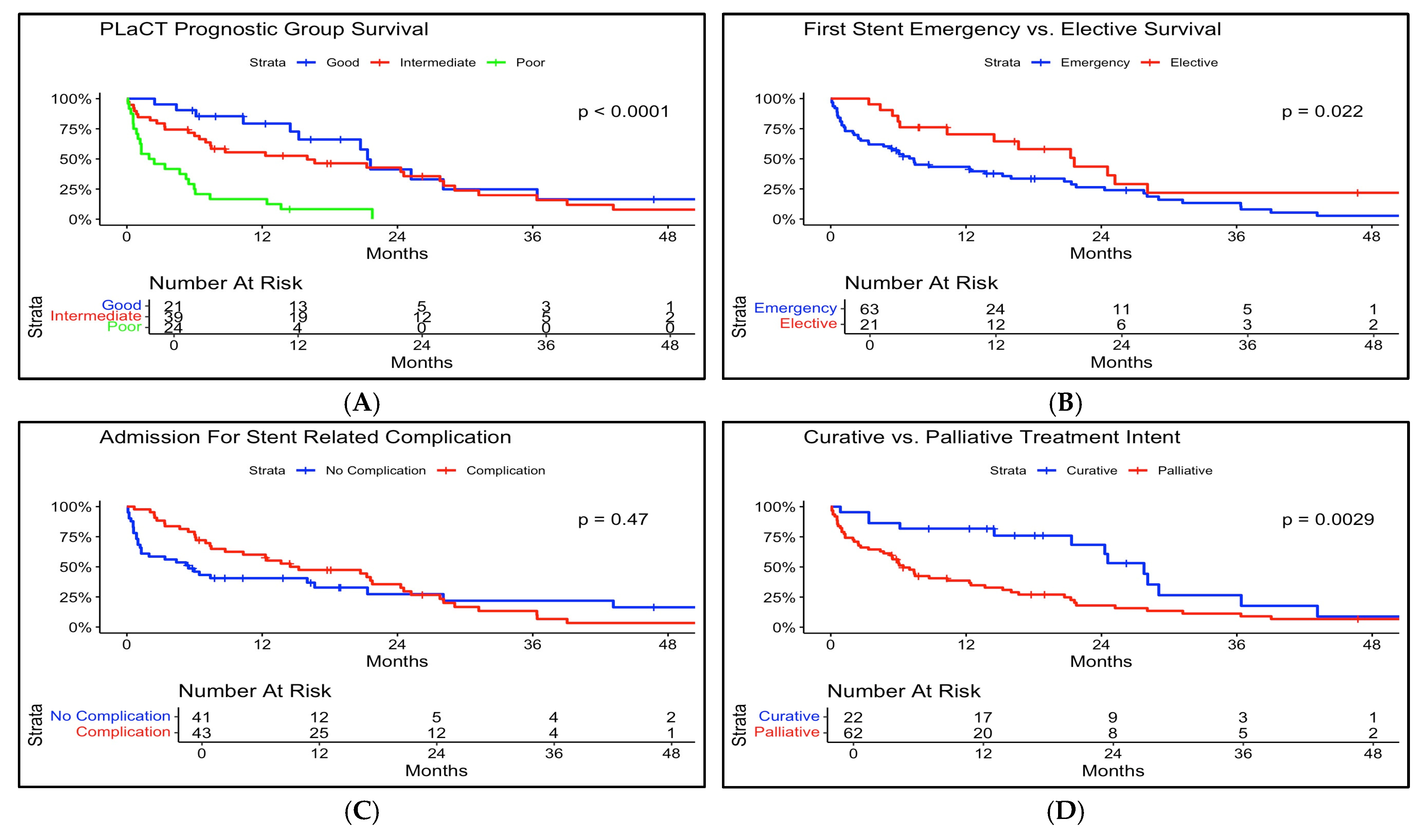
Sixty-six patients (79%) died, with a median survival of 197 days (6.5 months) from the time of renal tract decompression. Factors associated with poorer survival on univariate analysis included; receiving no treatment for primary malignancy (p = 0.07), no further oncological treatment planned after MUO intervention (p < 0.01), palliative treatment intent (p = 0.03), bilateral hydronephrosis (p =< 0.01), poor PLaCT prognostic group (p < 0.01), emergency first stent insertion (p = 0.02) and female patients (p = 0.03). See Figure 1, Figure 2 and Figure 3, and mortality data in Table 4. Multivariate analysis was performed following univariate analysis, which demonstrated that only gender (Hazard Ratio [HR] = 0.53, p = 0.05) and having further cancer treatment planned (HR = 0.24, p = 0.02) remained statistically significant (Table 5).

3.2. Renal Function Outcomes

Median pre-morbid serum creatinine was 73 μmol/L (range 38–231 μmol/L) and 149 μmol/L (range 50–1049 μmol/L) pre-decompression. At 6 months and 12 months post renal decompression, median serum creatinine was 103 μmol/L (range 58–672 μmol/L) and 105 μmol/L (range 58–762 μmol/L). A total of 84% and 72% of patients had reduced renal function compared to pre-morbid levels at 6 and 12 months, and 84% and 78% improved compared to pre-intervention levels at 6 and 12 months. (Table 4—mortality and renal function outcomes data).

3.3. Complications

There were 143 total complications recorded. Urinary tract infection (n = 67, 47%) was the most common. Forty-three patients (51%) had at least one complication. Complications resulted in 966 additional days spent in the hospital. There were four patients who underwent ureteric reconstruction (5%) during the follow-up period as a means of maintaining urinary tract drainage due to poor tolerance of their ureteric stents. There were twenty occasions of stents being removed and not replaced during the follow-up period due to a combination of poor stent tolerance (removed at patient request), successful treatment of malignancy, post-ureteric reconstruction, failed stent exchange due to progressive disease, and development of renal failure. One hundred and twenty-one stent exchanges took place, mostly electively (n = 77, 64%). However, there were 44 unplanned stent exchanges (36%) due to complications and occurred in an emergency setting for reasons which have been summarised in Table 6. Of note, all ureteric stents used in our institution were standard polyurethane stents.

3.4. PLaCT Prognostic Score

The PLaCT score was applied to our patient cohort. Patients were classified as good (n = 21), intermediate (n = 39), or poor (n = 24) risk (see Figure 1) [8]. There were significant differences in median survival amongst patients within these risk categories (good = 555 days, intermediate = 235 days, poor = 67 days, p < 0.01). The PLaCT score also showed differences in median survival across emergency vs. elective patients (Elective—174.5 days, 749 days, 659 days, Emergency—60 days, 266 days, 632 days for poor, intermediate, and good, respectively). Multivariate analysis, as described above, did not show a statistically significant difference in PLaCT groups once other variables were accounted for (Table 5). It must be noted that this is not a formal analysis for validation, but appears to apply well to our Australian cohort.

4. Discussion

Managing MUO presents a significant challenge for urologists, who are tasked with balancing restoration of renal function and unproven survival benefits [4,6,9,10,11] against reduced quality of life or prolonging hospital stays. This complexity is exacerbated by the diverse presentations of MUO. Some patients are diagnosed incidentally with hydronephrosis on staging imaging [12,13], or with symptoms of uncomplicated ureteric obstruction [2], allowing for a more planned approach to management through nuanced discussions among urologists, oncologists, and other specialists. In contrast, others present with life-threatening conditions such as sepsis or obstructive renal failure [13,14] necessitating intervention to avoid imminent death, despite a poor oncological prognosis. In our cohort, 75% of patients were diagnosed and treated in an emergency setting, with many cases representing the index presentation and diagnosis of their malignancy, further complicating decision making.
Our cohort of patients had a median survival time of just 197 days (6.5 months), highlighting the poor oncological prognosis in patients with MUO. Without a control group, we could not compare whether the intervention improved survival. This is consistent with other studies, where median survival times are almost universally less than 12 months [10,15,16,17]. There is limited literature comparing survival outcomes for treated versus untreated MUO patients. Artiles Medina et al. [3] presented a series of 188 patients with MUO in which 48 of the patients underwent urinary diversion with percutaneous nephrostomy or retrograde ureteric stent. In the Artiles Medina cohort, the median survival of the cohort was 6.43, identifying a slight survival benefit (8.7 months) for those undergoing urinary diversion. Similarly, a study by Lapitan et al. [18] reported a series of 205 patients with MUO from cervical cancer, which included patients who required MUO intervention with percutaneous nephrostomy or ureteric stenting, and patients who required the intervention, but it was not performed. There was a short-term survival benefit to MUO intervention when comparing survival rates at 3 months, 6 months, and 9 months, but by 12 months, the survival rates equalised (16% for both cohorts). Tatenuma et al. [19] reported on 151 patients with MUO, 87 of the patients underwent immediate palliative diversion based on expert opinion that they would have a better prognosis or benefit from future treatment, whilst 64 patients were just observed. Despite this obvious bias in the selection of patients, the median survival between the groups was not statistically significant, including in 19 patients from the observation group who later underwent urinary diversion. A 2024 systematic review identified only five papers that reported survival data on a total of 149 patients who did not receive MUO intervention; the median survival was only 4.2 months [6]. Selection bias is a concern in comparing treated and untreated patients, and prospective studies that account for markers of prognosis (such as age, stage of malignancy, plans for further treatment), are needed to clarify survival benefits.
Loss of renal function is another key consideration when considering renal decompression for MUO. In our cohort, renal function improved compared to the time of referral at 6 and 12 months in 72% and 78% of patients, respectively; however, compared to pre-morbid levels, 83% of patients at 6 months and 84% of patients at 12 months had reduced renal function. Patients with bilateral hydronephrosis had a greater degree of improvement compared to unilateral hydronephrosis, an expected outcome given the higher mean creatinine at presentation. These results compare to other studies [5,6,14,20]. The urinary obstruction appears to lead to an insult that renal function does not fully recover from, a conclusion made by other researchers [10,14]. The improvement in renal function itself has not been found to not correlate to survival outcomes in some studies [20,21], but it did in others [22]. Aside from a possible survival benefit, the main reason to maintain renal function outside of life-threatening renal failure would be to facilitate further oncological treatment. Heo et al. [23] conducted a study on 778 patients with MUO treated with ureteric stent or percutaneous nephrostomy, in which 447 patients were able to receive chemotherapy after MUO decompression, and it provided a survival advantage of over 7 months to patients who did not receive chemotherapy. Folkard et al. [20] concluded on their series of 105 patients treated for MUO with percutaneous nephrostomy that treatment of MUO did not facilitate further systematic treatment. Only 30% of the patients received treatment post-nephrostomy insertion, and there was no correlation between the degree of renal failure pre-nephrostomy and those who received chemotherapy. Blackmur et al. [4] presented a cohort of 852 patients, and 43% of them were able to receive oncological treatment of all modalities post-decompression. Cartapatti et al. [21], reported 420 patients with MUO, 300 patients (71%) were able to receive anti-neoplastic treatment after MUO intervention, but the majority were with palliative intent. A 2024 systematic review by Shat et al. [6] examined the receipt of additional anti-cancer treatment, and found that 64% of 1234 patients received anti-cancer treatment (of all modalities) after MUO intervention, but could not differentiate whether this would have occurred regardless of whether MUO was treated or not. In this systematic review, only 3 papers of 186 patients had data on the intent of this anti-cancer treatment, and for the majority, it was palliative [6]. Data comparing anti-cancer treatment in patients who did not receive MUO intervention is sparse.
MUO intervention is not without significant complications. In our cohort, 23% of patients had a failed primary insertion of their stent, and over 50% required admission to the hospital for a complication, most commonly urinary tract infection. The complications resulted in 966 additional days in hospital, and 44 unplanned surgeries to exchange ureteric stents or nephrostomies. Rates of complications in our cohort were similar to several other cohorts [6,10]. The retrospective design of this study did not allow for a formal quality of life assessment, and of greater interest would be comparing the quality of life and complication rates between MUO patients who did and did not have intervention. A systematic review on this topic [6] had reported that complications and quality of life reporting in previous studies have been limited and of low quality. A narrative review by Prentice et al. [10] also identified a few papers that did a formal quality of life analysis, and the ones that did had significant flaws, including not measuring baseline symptoms and not having control groups. Neither review identified any papers that reported a significant quality of life improvement after MUO intervention.
In our cohort, we found a number of variables that were associated with poor survival with univariate analysis; these included lack of further treatment planned for their primary malignancy, patients who received no treatment at any stage for their cancer, palliative intent treatment, bilateral obstruction, emergency presentation of MUO, and, surprisingly, female sex. However, following multivariate analysis, only gender and having further cancer treatment planned remained statistically significant variables once the other variables were accounted for. Gynaecological malignancies did not show any significant difference in survival on univariate or multivariate analysis. In our cohort, it is unclear as to why females would have a worse prognosis for MUO.
Prognostic tools based on clinical and functional variables aim to identify patients who would benefit most from intervention. Commonly utilised variables include low serum albumin, malignancy-related events, ECOG (European Cooperative Cancer Group) status, hyponatraemia, and the presence of metastatic disease [10], severely raised serum creatinine levels, and the degree of hydronephrosis [6]. Blackmur et al. [4] have recently validated a Scottish MUO score based on a number of clinical factors, which provides a ‘bespoke’ prediction of how likely a patient will survive to different time points (30 days, 90 days, 6 months, and 12 months). In our study, we opted to utilise the PLaCT prognostic tool produced by Izumi et al. [8], for its ease of calculation and application to our cohort. In our cohort, it accurately predicted survival (median follow-up 67, 266, and 498 days for poor, intermediate, and good prognostic groups); however, as described above in our multivariate analysis, it did not independently explain any difference in survival outcomes. Using a tool like PLaCT could provide useful guidance for patient counselling on the expected benefits and risks of MUO treatment when used in a prospective manner. It must be noted that tools like PLaCT may be less useful in emergency settings, where intervention is necessary despite a poor long-term prognosis. A prospective study using prognostic tools to aid decision making in such cases would be valuable.
In our cohort, there was no difference in survival between patients who received a ureteric stent vs. those who received a nephrostomy up front. Previously, literature has been summarised by a systematic review [5] concluded that stents are preferred for their easier maintenance, and no significant difference in complications, renal function improvement, and survival.
Our study has several limitations. Its retrospective design means there are inherent sources of bias, including selection bias, measurement bias, the effects of uncontrolled founding and incomplete patient records. Patients who were not treated for MUO was at the discretion of the on-call Urologist; the reasons for not treating were not explored in this paper, and would have provided some insight into how this selection bias would have affected the interpretation of the results. This paper did not include a control group, which, when appropriately stratified, would provide a more robust comparison of any survival and renal function benefits and the complications of treating MUO. The cohort size was small, which limited subgroup analysis, particularly when it comes to individual malignancy types. We did not perform a formal quality of life assessment due to the retrospective design; any future studies on this topic would greatly benefit from a quality of life assessment. Finally, our survival analyses were only univariate, which means confounding factors such as stage of malignancy and age have not been taken into account in the individual comparisons.

5. Conclusions

The findings in our cohort of Australian patients with Malignant Ureteric Obstruction corroborate findings from other research, demonstrating that patients with MUO generally have a poor prognosis due to their underlying malignancy. Renal decompression can alleviate life-threatening sepsis, renal failure and restore renal function, although not usually back to its baseline. Due to the lack of a control group, we cannot make any conclusions about the survival benefits or ability to resume oncological treatment. Treatment of MUO carries the risk of significant complications, contributing to extended hospital stays and additional healthcare burden. Several prognostic tools, including the PLaCT score, show promise in predicting survival outcomes and helping guide conversations about treatment decisions. Like many other researchers, we acknowledge that large, well designed, prospective trials are required to determine the optimum management and to examine quality of life outcomes. At present, the complex decision to intervene on MUO still comes down to a thorough discussion between the multidisciplinary treating teams and the patients.

Author Contributions

Conceptualization, R.D. and A.K.; methodology, A.B., R.D. and A.K.; software, A.B.; validation, A.B. and A.K.; formal analysis, A.B.; investigation, A.B.; resources, A.B.; data curation, A.B.; writing—original draft preparation, A.B.; writing—review and editing, A.B., R.D. and A.K.; visualisation, A.B. and R.D.; supervision, A.K.; project administration, A.B.; All authors have read and agreed to the published version of the manuscript.

Funding

This research received no external funding.

Institutional Review Board Statement

The study was conducted in accordance with the Declaration of Helsinki, and approved by the Institutional Review Board (or Ethics Committee) of the Central Adelaide Local Health Network Human Research Ethics Committee (protocol code 18242, approval date 9 October 2023).

Informed Consent Statement

Patient consent was waived due to the retrospective methodology and inability to gain consent from the majority of patients due to their death prior to the commencement of the study, the waiver of consent was approved by the Central Adelaide Local Health Network human research ethics committee. The CALHN HREC determined that the waiver request meets the requirements of section 2.3.10 of the NHMRC National Statement.

Data Availability Statement

The data that support the findings of this study are available from the corresponding author upon reasonable request. Due to the sensitivity of patient information and to comply with institutional and ethical guidelines, the raw data are not publicly available. De-identified summary data may be shared following approval by the institutional review board and data-sharing agreements.

Conflicts of Interest

The authors declare no conflicts of interest.

Abbreviations

The following abbreviations are used in this manuscript:
AUA American Urological Association
EAUEuropean Association of Urology
ECOGEuropean Cooperative Oncology Group
HRHazard Ratio
MUOMalignant Ureteric Obstruction
NALHNNorthern Adelaide Local Health Network
PLaCTPrimary site, Laterality, serum Creatinine level, and Treatment
RPFRetroperitoneal Fibrosis

References

  1. Lomas, D.J.; Ziegelmann, M.J. Update on the Management of Malignant Ureteral Obstruction. Available online: https://auanews.net/issues/articles/2023/may-2023/update-on-the-management-of-malignant-ureteral-obstruction (accessed on 1 October 2023).
  2. Kouba, E.; Wallen, E.M.; Pruthi, R.S. Management of ureteral obstruction due to advanced malignancy: Optimizing therapeutic and palliative outcomes. J. Urol. 2008, 180, 444–450. [Google Scholar] [CrossRef] [PubMed]
  3. Artiles Medina, A.; Laso García, I.; González Tello, F.; Álvarez Rodríguez, S.; Hevia Palacios, M.; Mata Alcaraz, M.; Mínguez Ojeda, C.; Arias Funez, F.; Gómez Dos Santos, V.; Burgos Revilla, F.J. The challenging management of malignant ureteral obstruction: Analysis of a series of 188 cases. Curr. Urol. 2024, 18, 34–42. [Google Scholar] [CrossRef]
  4. Blackmur, J. Management of malignant ureteric obstruction with ureteric stenting or percutaneous nephrostomy. Br. J. Surg 2024, 111, znae035. [Google Scholar] [CrossRef]
  5. Gauhar, V.; Pirola, G.M.; Scarcella, S.; De Angelis, M.V.; Giulioni, C.; Rubilotta, E.; Gubbiotti, M.; Lim, E.J.; Law, Y.X.T.; Wroclawski, M.L.; et al. Nephrostomy tube versus double J ureteral stent in patients with malignant ureteric obstruction. A systematic review and meta-analysis of comparative studies. Int. Braz. J. Urol. 2022, 48, 903–914. [Google Scholar] [CrossRef] [PubMed] [PubMed Central]
  6. Shah, M.; Blest, F.; Blackmur, J.; Laird, A.; Dawson, S.; Aning, J. Malignant upper urinary tract obstruction in cancer patients: A systematic review. BJUI Compass 2024, 5, 405–416. [Google Scholar] [CrossRef]
  7. van der Heijden, A.G.; Rink, M.; Bruins, H.M.; Carrion, A.; Cathomas, R.; Compérat, E.M.; Dimitropoulos, K.; Efstathiou, J.A.; Fietkau, R.; Lorch, A.; et al. Muscle-Invasive and Metastatic Bladder Cancer. In EAU Guidelines. Edn. Presented at the EAU Annual Congress Paris 2024; EAU Guidelines Office: Arnhem, The Netherlands, 2024; ISBN 978-94-92671-23-3. [Google Scholar]
  8. Izumi, K.; Shima, T.; Shigehara, K.; Sawada, K.; Naito, R.; Kato, Y.; Ofude, M.; Kano, H.; Iwamoto, H.; Yaegashi, H.; et al. A novel risk classification score for malignant ureteral obstruction: A multicenter prospective validation study. Sci. Rep. 2021, 11, 4455. [Google Scholar] [CrossRef]
  9. Sountoulides, P.; Mykoniatis, I.; Dimasis, N. Palliative management of malignant upper urinary tract obstruction. Hippokratia 2014, 18, 292–297. [Google Scholar]
  10. Prentice, J.; Amer, T.; Tasleem, A.; Aboumarzouk, O. Malignant ureteric obstruction decompression: How much gain for how much pain? A narrative review. J. R. Soc. Med. 2018, 111, 125–135. [Google Scholar] [CrossRef]
  11. Hyams, E.S.; Shah, O. Malignant extrinsic ureteral obstruction: A survey of urologists and medical oncologists regarding treatment patterns and preferences. Urology 2008, 72, 51–56. [Google Scholar] [CrossRef]
  12. Hsu, L.; Li, H.; Pucheril, D.; Hansen, M.; Littleton, R.; Peabody, J.; Sammon, J. Use of percutaneous nephrostomy and ureteral stenting in management of ureteral obstruction. World J. Nephrol. 2016, 5, 172–181. [Google Scholar] [CrossRef]
  13. De Lorenzis, E.; Lievore, E.; Turetti, M.; Gallioli, A.; Galassi, B.; Boeri, L.; Montanari, E. Ureteral Stent and Percutaneous Nephrostomy in Managing Malignant Ureteric Obstruction of Gastrointestinal Origin: A 10 Years’ Experience. Gastrointest. Disord. 2020, 2, 456–468. [Google Scholar] [CrossRef]
  14. Ghannam, E.; Musleh, H.; Ahmad, T.; Mustafa, M.; Odeh, R.; Shawahna, R. Outcomes of nephrostomy and double J stent in malignant ureteral obstruction in the Palestinian practice. BMC Urol. 2024, 24, 245. [Google Scholar] [CrossRef]
  15. Izumi, K.; Mizokami, A.; Maeda, Y.; Koh, E.; Namiki, M. Current outcome of patients with ureteral stents for the management of malignant ureteral obstruction. J. Urol. 2011, 185, 556–561. [Google Scholar] [CrossRef]
  16. Ishioka, J.; Kageyama, Y.; Inoue, M.; Higashi, Y.; Kihara, K. Prognostic model for predicting survival after palliative urinary diversion for ureteral obstruction: Analysis of 140 cases. J. Urol. 2008, 180, 618–621; discussion 621. [Google Scholar] [CrossRef]
  17. Cordeiro, M.D.; Coelho, R.F.; Chade, D.C.; Pessoa, R.R.; Chaib, M.S.; Colombo-Júnior, J.R.; Pontes-Júnior, J.; Guglielmetti, G.B.; Srougi, M. A prognostic model for survival after palliative urinary diversion for malignant ureteric obstruction: A prospective study of 208 patients. BJU Int. 2016, 117, 266–271. [Google Scholar] [CrossRef]
  18. Lapitan, M.C.; Buckley, B.S. Impact of palliative urinary diversion by percutaneous nephrostomy drainage and ureteral stenting among patients with advanced cervical cancer and obstructive uropathy: A prospective cohort. J. Obs. Gynaecol. Res. 2011, 37, 1061–1070. [Google Scholar] [CrossRef]
  19. Tatenuma, T.; Tsutsumi, S.; Yasui, M.; Noguchi, G.; Umemoto, S.; Kishida, T. Outcome of Palliative Urinary Diversion and Observation for Malignant Extrinsic Ureteral Obstruction. J. Palliat. Med. 2019, 23, 254–258. [Google Scholar] [CrossRef]
  20. Folkard, S.S.; Banerjee, S.; Menzies-Wilson, R.; Reason, J.; Psallidas, E.; Clissold, E.; Al-Mushatat, A.; Chaudhri, S.; Green, J.S.A. Percutaneous nephrostomy in obstructing pelvic malignancy does not facilitate further oncological treatment. Int. Urol. Nephrol. 2020, 52, 1625–1628. [Google Scholar] [CrossRef]
  21. Cartapatti, M.; Machado, R.D.; Mesquita, J.C.; Freua, R.; Cáceres, D.; dos Reis, R.B. Urinary Diversion Can Improve the Chance of Implementing New Therapeutic Lines in Patients with Malignant Ureteral Obstruction: A Multicenter Study. Curr. Oncol. 2024, 31, 7107–7116. [Google Scholar] [CrossRef]
  22. Yoon, J.H.; Park, S.; Park, S.; Moon, K.H.; Cheon, S.H.; Kwon, T. Renal function is associated with prognosis in stent-change therapy for malignant ureteral obstruction. Investig. Clin. Urol. 2018, 59, 376–382. [Google Scholar]
  23. Heo, J.E.; Jeon, D.Y.; Lee, J.; Ham, W.S.; Choi, Y.D.; Jang, W.S. Clinical Outcomes After Urinary Diversion for Malignant Ureteral Obstruction Secondary to Non-urologic Cancer: An Analysis of 778 Cases. Ann. Surg. Oncol. 2021, 28, 2367–2373. [Google Scholar] [CrossRef] [PubMed]
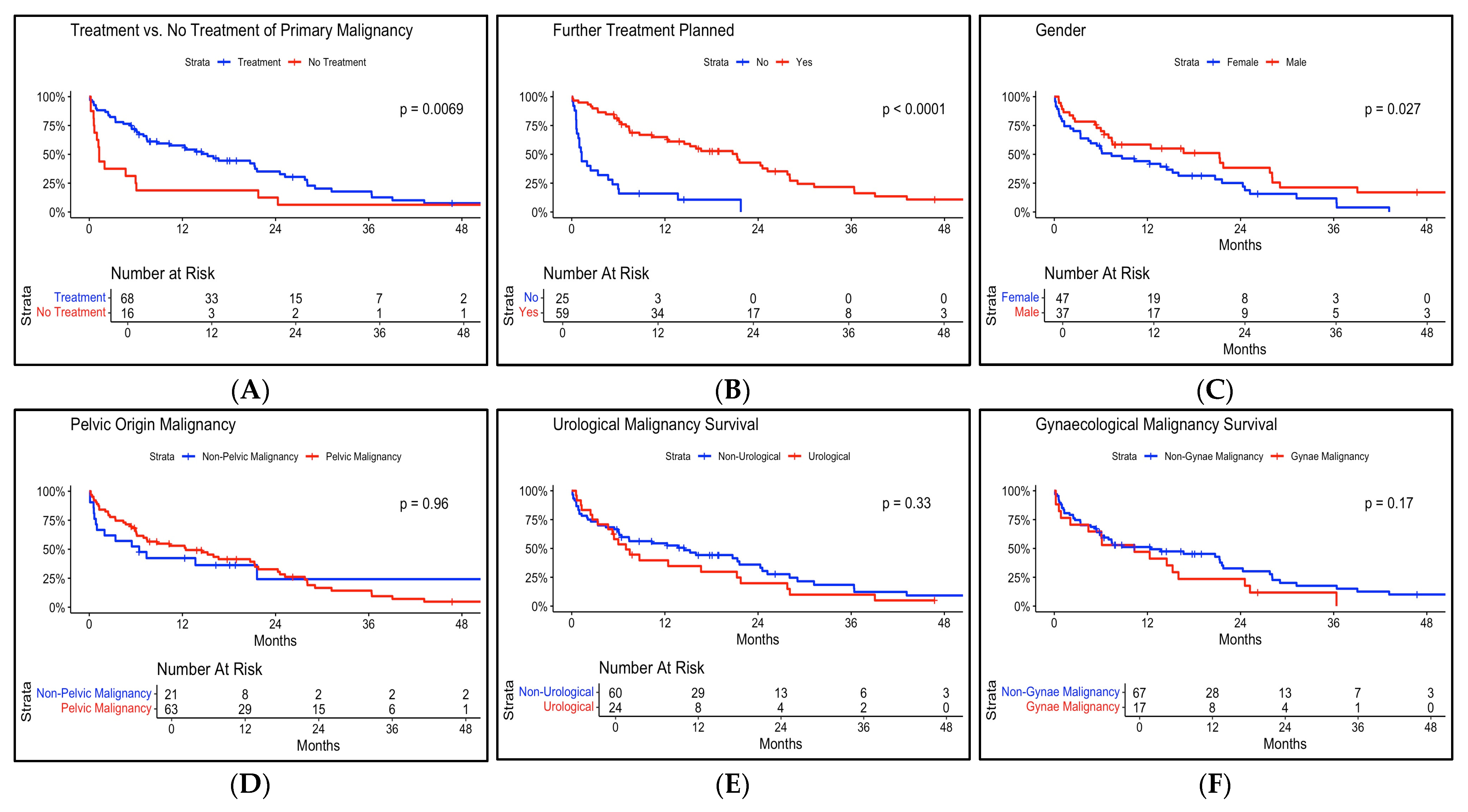
Figure 1. Kaplan–Meier survival curves by subgroups Part 1. (A): Treatment vs. no treatment for malignancy. (B): Treatment vs. no further treatment planned. (C): Gender. (D): Pelvic origin malignancy. (E): Primary urological malignancy. (F): Primary gynaecological malignancy.
Siuj 06 00062 g001
Figure 2. Kaplan–Meier curves by subgroups part 2. (A): Age over/under 65 years old. (B): Serum creatinine over/under 105 umol/L. (C): Site of obstruction. (D): Unilateral vs. bilateral obstruction. (E): Approach to first stent. (F): First stent insertion failure survival.
Siuj 06 00062 g002
Figure 3. Kaplan–Meier curves by subgroups part 3. (A): poor Primary site, Laterality, serum Creatinine level, and Treatment for primary site (PLaCT) prognostic group. (B): Emergency vs. elective survival. (C): Admitted with stent stent-related complication. (D): Curative vs. palliative treatment intent survival.
Siuj 06 00062 g003
Table 1. PLaCT risk classification score [8].
CharacteristicPoints
Primary Cancer Site (P)
Gynaecological0
Lower Digestive Tract or Urinary Tract1
Others2
Laterality (La)
Unilateral0
Bilateral1
Serum Creatinine (C)
<1.2 mg/mL0
>1.2 mg/mL2
Treatment of Primary Cancer (T)
In process or on schedule0
No intention2
PLaCT Risk Group
Good0–2
Intermediate3–4
Poor5–7
PLaCT: Primary site, Laterality, serum Creatinine level, and Treatment for primary site.
Table 2. Patient Demographic Characteristics.
CharacteristicN = 84 1
Age
   Median (Q1, Q3)64.50 (55, 76)
   Min, Max27, 95
Gender
   Female47/84 (56%)
   Male37/84 (44%)
Charleston Comorbidity Score
   1. <46/84 (7%)
   2. 4–624/84 (29%)
   3. 7–933/84 (39%)
   4. >1021/84 (25%)
Primary Cancer
   Appendiceal1/84 (1%)
   Bladder13/84 (15%)
   Breast4/84 (5%)
   Cervical12/84 (14%)
   Colorectal23/84 (27%)
   Endometrial1/84 (1%)
   Gastric5/84 (6.0%)
   Jejunal1/84 (1%)
   Lymphoma6/84 (7%)
   Oesophageal1/84 (1%)
   Ovarian4/84 (5%)
   Prostate10/84 (12%)
   Renal Cell1/84 (1%)
   Testicular2/84 (2%)
Cancer Stage
   27/84 (8%)
   318/84 (21%)
   459/84 (70%)
Treatment Intent
   Curative22/84 (26%)
   Palliative62/84 (74%)
Further Oncological Treatment Planned
   No25/84 (30%)
   Yes59/84 (70%)
Never had Treatment For Malignancy
   No68/84 (81%)
   Yes16/84 (19%)
Total Length of Follow-up (Days)
   Median (Q1, Q3)233 (78, 641)
   Min, Max3, 2077
1 n/N (%).
Table 3. MUO characteristic data.
CharacteristicN = 84 1
Emergency vs. Elective Initial Stent Insertion
   Emergency63/84 (75%)
   Elective21/84 (25%)
Method of First Stent Insertion
   Antegrade14/84 (17%)
   Retrograde64/84 (76%)
   Retrograde and Antegrade6/84 (7%)
Level of Ureteric Obstruction
   Distal58/84 (69%)
   Mid16/84 (19%)
   Proximal10/84 (12%)
Cause of Obstruction
   Ischaemic2/84 (2%)
   Nodal25/84 (30%)
   Radiation3/84 (4%)
   RPF5/84 (6.0%)
   Tumour49/84 (58%)
Unilateral vs. Bilateral Obstruction
   Bilateral39/84 (46%)
   Unilateral45/84 (54%)
First Stent Insertion Failure
   No66/84 (79%)
   Yes18/84 (21%)
MUO: Malignant Ureteric Obstruction, RPF: Retroperitoneal Fibrosis. 1 n/N (%).
Table 4. Mortality and renal function outcomes data.
CharacteristicN = 84 1
Mortality
   No18/84 (21%)
   Yes66/84 (79%)
Time to Death from MUO Treatment (Days)
   Median (Q1, Q3)197 (67, 651)
   Min, Max3, 1549
Total Follow-up Days
   Median (Q1, Q3)233 (78, 641)
   Min, Max3, 2077
Survived 30 Days Post-MUO Treatment
   No12/84 (14%)
   Yes72/84 (86%)
Survived 90 Days Post-MUO Treatment
   No22/84 (26%)
   Yes62/84 (74%)
Survived 6 Months Post MUO Treatment
   No33/84 (39%)
   Yes51/84 (61%)
Survived 12 Months Post-MUO Treatment
   No47/84 (56%)
   Yes37/84 (44%)
PLaCT Prognostic Group Survival (Days)
   Poor (Med) (Min, Max)67 (6, 765)
   Intermediate (Med) (Min, Max)235 (3, 1549)
   Good (Med) (Min, Max)555 (75, 1162)
Baseline (Pre-Morbid) Serum Creatinine (umol/L)
   Median (Q1, Q3)73 (64, 95)
   Min, Max38, 231
Pre-Stent Serum Creatinine (umol/L)
   Median (Q1, Q3)149 (98, 333)
   Min, Max50, 1049
Serum Creatinine 6 Months Post MUO Treatment (umol/L)
   Median (Q1, Q3)103 (82, 146)
   Min, Max58, 672
Change in Serum Creatinine from Pre-Stent at 6 Months (umol/L)
   Median (Q1, Q3)−16 (−72, 4)
   Min, Max−446, 262
Improvement in Serum Creatinine at 6 Months from Pre-Stent
   No13/47 (28%)
   Yes34/47 (72%)
   Missing data4
Worsening Serum Creatinine at 6 Months from Pre-Morbid Baseline
   No8/48 (17%)
   Yes40/48 (83%)
   Missing data3
Serum Creatinine 12 Months Post MUO Treatment (umol/L)
   Median (Q1, Q3)105 (80, 145)
   Min, Max58, 762
Change in Serum Creatinine from Pre-Stent at 12 Months (umol/L)
   Median (Q1, Q3)−27 (−55, −1)
   Min, Max−264, 53
Improvement in Serum Creatinine at 12 Months from Pre-Stent
   No8/36 (22%)
   Yes28/36 (78%)
   Missing data1
Worsening Serum Creatinine at 12 Months from Pre-Morbid Baseline
   No6/37 (16%)
   Yes31/37 (84%)
Malignant Ureteric Obstruction (MUO), poor Primary site, Laterality, serum Creatinine level, and Treatment for primary site (PLaCT). 1 n/N (%).
Table 5. Cox-proportional hazards regression model showing hazard ratios (HR) and 95% confidence intervals, p-value, and Harrell’s C Statistic, for predictors of overall survival in patients with malignant ureteric obstruction.
VariableHazard Ratio (95% CI)p-Value
Age1.01 (0.98–1.05)0.38
Gender (Male)0.53 (0.28–1.00)0.05
Charlson Comorbidity Index1.04 (0.86–1.26)0.70
Stage (3)1.08 (0.31–3.76)0.90
Stage (4)1.46 (0.45–4.79)0.53
No cancer treatment (Yes)0.84 (0.36–1.94)0.68
Treatment Intent (Palliative)1.81 (0.87–3.74)0.11
Hydronephrosis (Unilateral)0.92 (0.44–1.89)0.81
Cancer Treatment Planned (Yes)0.24 (0.07–0.80)0.02
PLaCT Group (Intermediate)1.2 (0.55–2.6)0.65
PLaCT Group (Poor)1.25 (0.3–5.23)0.76
Elective MUO Treatment0.69 (0.32–1.5)0.35
Gynaecological Malignancy (Yes)1.18 (0.55–2.52)0.68
Harrell’s C Statistic: 0.76 (0.71–0.82)
Proportional Hazards Assumption: Global p-value = 0.13
Malignant Ureteric Obstruction (MUO), poor Primary site, Laterality, serum Creatinine level, and Treatment for primary site (PLaCT), Confidence Interval (CI).
Table 6. Complications Data.
CharacteristicN = 84 1
Admitted with Stent Complication
   No41/84 (49%)
   Yes43/84 (51%)
Total Number of Stent-Related Complications143
   Urinary Tract Infections67
   Migrated Stents4
   Occluded Stents31
   Encrusted Stents2
   Stent-Related Pain15
   Urinary Frequency and Urgency2
   Haematuria22
Length of Stay for Stent Complications (Days)966
Failed Stent Procedures34
Planned Stent Exchanges77
Unplanned Stent Exchanges44
Stents Removed20
Ureteric Reconstructions Performed4
1 n/N (%); Sum.
Disclaimer/Publisher’s Note: The statements, opinions and data contained in all publications are solely those of the individual author(s) and contributor(s) and not of MDPI and/or the editor(s). MDPI and/or the editor(s) disclaim responsibility for any injury to people or property resulting from any ideas, methods, instructions or products referred to in the content.