Article

The “Footprint” Sign in Voiding Cystourethrography Indicates Poor Renal Function in Vesicoureteral Reflux: Is It a Pop-Off Mechanism?

by
Hooman Kamran
1,2,
Negar Mohammadi Ganjaroudi
1,
Nooshin Tafazoli
1,3,
Mehrzad Mehdizadeh
4 and
Abdol-Mohammad Kajbafzadeh
1,*
1
Pediatric Urology and Regenerative Medicine Research Center, Children’s Medical Center, Gene, Cell & Tissue Research Institute, Tehran University of Medical Sciences, Tehran 1419733151, Iran
2
Laparoscopy Research Center, Shiraz University of Medical Sciences, Shiraz 7144995377, Iran
3
Department of Urology, Stanford University School of Medicine, Palo Alto, CA 94304, USA
4
Department of Radiology, Children’s Medical Center, Tehran University of Medical Sciences, Tehran 1419733151, Iran
*
Author to whom correspondence should be addressed.
Soc. Int. Urol. J. 2025, 6(4), 55; https://doi.org/10.3390/siuj6040055
Submission received: 19 May 2025 / Revised: 9 August 2025 / Accepted: 11 August 2025 / Published: 19 August 2025

Abstract

Background/Objectives: To assess the “footprint” sign in voiding cystourethrogram (VCUG) for its diagnostic and prognostic significance in indicating a “pop-off” mechanism that may reduce pressure on the contralateral kidney. Methods: A retrospective analysis included patients with a “footprint” sign or high-grade vesicoureteral reflux (VUR) in VCUG. They were categorized into two groups: those with (Group A) and those without (Group B) the “footprint” sign. Results: Among 55 patients (median age 18 months), 18 exhibited the “footprint” sign. Group A had predominantly male patients. Bilateral VUR was less common in Group A (38.9%) compared to Group B (51.4%), though not statistically significant (p-value: 0.385). The “footprint” sign was predominantly associated with the left side (77.8% in Group A). The median differential renal function (DRF) was significantly lower in kidneys with the “footprint” sign (11.5%) compared to those without (44.5%, p-value < 0.001). All patients with a DRF under 20% exhibited the “footprint” sign. Conclusions: The “footprint” sign correlates with poor renal function, suggesting a potential pop-off mechanism protecting the contralateral kidney. Management should prioritize the contralateral kidney with normal function. The “footprint” sign alone in VCUG may suffice to indicate compromised renal function, negating the need for further ipsilateral kidney assessment.

1. Introduction

Vesicoureteral reflux (VUR) is a prevalent congenital disorder of the urinary tract in children who have had a previous urinary tract infection (UTI) or in asymptomatic infants with a prenatal sonographic diagnosis of hydronephrosis or hydroureter [1,2]. The currently available imaging techniques used to determine VUR include radiographic voiding cystourethrogram (VCUG), direct radionuclide cystography, and the recently introduced contrast-enhanced voiding ultrasonography [3,4,5]. VCUG is the most commonly used technique and employs a well-defined international grading system (I to V) based on the level of reflux and the morphology of the pelvicalyceal system, bladder, ureters, and urethra, but it does not infer the function of the affected kidney [6]. However, in some cases of moderate to severe reflux, the morphology of the refluxing pelvicalyceal system and ureters may not conform to the criteria described in the international grading system.
In 2015, Martin et al. [7] introduced the “flowerpot” sign as an indicator of poor kidney function in high-grade VUR. This sign was characterized by the cephalic configuration of calyces during maximal reflux. Their study included children with grade IV or V VUR and categorized the patients based on the presence of the “flowerpot” sign. The authors found decreased renal function in units with the “flowerpot” sign, with a median differential renal function (DRF) of 23%, compared to 48% and 40% in grade IV and V VUR units without the sign, respectively. In this paper, we refer to this sign as the “footprint” sign, as we believe it resembles an infant’s footprint.
In the current study, we report our observations in VCUG of a distinct pattern in the refluxing pelvicalyceal system that resembles an infant’s “footprint”, characterized as dilated calyces atop a dilated, elongated renal pelvis. We conducted this study to re-evaluate the sign reported by Martin et al. [7], which appears to be a reliable indicator of poor renal unit function, and to introduce a possible hypothesis. We propose that this sign may indicate a “pop-off” mechanism, potentially decreasing the pressure on the contralateral kidney.

2. Materials and Methods

2.1. Study Design and Participants

Medical records of individuals diagnosed with VUR from 2007 to 2019 were retrospectively reviewed. For each patient, demographic and clinical information, VCUG and Tc99m dimercaptosuccinic acid (Tc99m-DMSA) scintigraphy results, and VUR grading based on the standards set by the International Reflux Study in Children were extracted. Inclusion criteria included the presence of either a “footprint” sign or high-grade VUR (grade IV or V) in VCUG, as indicated in radiology reports and pediatric urologist notes. The “footprint” sign was characterized as dysmorphic calyces connected to a dilated renal pelvis, forming a distinct “footprint” shadow shape, depicted in Figure 1. While no quantitative measurements (e.g., calyceal angle or pelvis ratios) were performed in this study, we based our visual identification on previously published radiological definitions. Martin et al. [7] described the sign as a superior migration of the calyceal axis with an inferior displacement and increased filling of the renal pelvis, producing a “flowerpot” configuration. They also reported that during maximal reflux, calyces are located largely cephalad to the renal pelvis rather than lateral, with a significantly greater median calyceal-spine angle (52° [range 37–66]) in affected kidneys compared to high-grade controls (13° [range 2–37]). These quantitative measures support the visual criteria used to identify the footprint sign. VCUG images were first reviewed by two experienced pediatric radiologists, and the presence of the “footprint” sign was subsequently confirmed by a senior pediatric urologist. Exclusion criteria encompassed individuals with a single kidney, multicystic dysplastic kidney, prune-belly syndrome, duplex collecting system, renal ectopia, posterior urethral valve (PUV), or those lacking a Tc99m-DMSA scan.
Patients were categorized into two groups based on the presence of a “footprint” sign: Group A (with a “footprint”) and Group B (without a “footprint” sign). The DRF of the affected kidneys in Group A (Figure 2) was compared with the DRF of the refluxing kidneys in patients with unilateral reflux or the kidneys with a higher grade of reflux in patients with bilateral reflux in Group B.

2.2. Statistical Analysis

The Statistical Package for the Social Sciences (SPSS) version 26 was employed for statistical analysis. The Shapiro–Wilk test assessed data normality. Data were presented as median (interquartile range [IQR]) for continuous variables and as numbers and percentages for categorical variables. The Mann–Whitney U test was used to compare serum creatinine and DRF between patients with and without a “footprint” sign. Additionally, Fisher’s exact test/Chi-square test was utilized for comparing the bilaterality of VUR and gender between groups. Multivariable linear regression was performed to compare serum creatinine levels between the groups, adjusting for age and gender. A p-value of less than 0.05 was considered significant.

2.3. Ethical Considerations

The research protocol was in accordance with the ethical standards of the local ethics committee of the Tehran University of Medical Sciences and the Helsinki Declaration of the World Medical Association [8].

3. Results

3.1. Patients’ Characteristics

A total of 55 patients, comprising 28 males and 27 females, met the inclusion criteria. The median age was 18.0 [50.5] months. Bilateral VUR was present in 26 patients (47.3%). The higher grade of VUR was on the right side in 22 patients (40.0%) and on the left in 33 patients (60.0%). The VCUGs revealed the “footprint” sign in 18 of the 55 patients (Group A) and high-grade reflux without the “footprint” sign in the remaining 37 patients (Group B). Table 1 summarizes the clinical and imaging findings for Groups A and B. Notably, only 2 patients (11.1%) in Group A were female, in contrast to the majority in Group B, where 25 out of 37 patients were female (p-value < 0.001).
The presentation of VUR was antenatal hydronephrosis in 19 patients (34.5%), recurrent UTIs in 32 patients (58.2%), and other/incidental in 4 patients (7.3%). Additionally, 7 patients (12.7%) had spina bifida occulta, 6 patients (10.9%) had sacral abnormalities, and 4 patients (7.3%) had a history of ureterovesical junction obstruction.

3.2. VUR Grade

In Group A, 7 patients (38.9%) had bilateral VUR, which was less than the 19 patients (51.4%) in Group B; however, this difference was not statistically significant (p-value: 0.385). Additionally, the side with the higher grade was the left in 77.8% of patients (14/18) in Group A, compared to 51.4% (19/37) in Group B. In Group B, the grade of VUR in unilateral reflux and the higher grade in cases of bilateral VUR was IV in 26 patients (70.3%) and V in 11 patients (29.7%). Table 2 displays the VUR grades for all renal units, classified based on the patient groups.
In Group A, 11 patients (61.1%) had no VUR in the contralateral kidney, and the distribution of VUR grades I, II, III, and IV in the kidney with the lower grade was 1 (5.6%), 2 (11.1%), 2 (11.1%), and 2 (11.1%), respectively. In Group B, 18 patients (48.6%) had no VUR in the contralateral kidney, and the distribution of VUR grades I, II, III, and IV in the kidney with the lower grade was 2 (5.4%), 4 (10.8%), 12 (32.4%), and 1 (2.7%), respectively.

3.3. Tc99m-DMSA

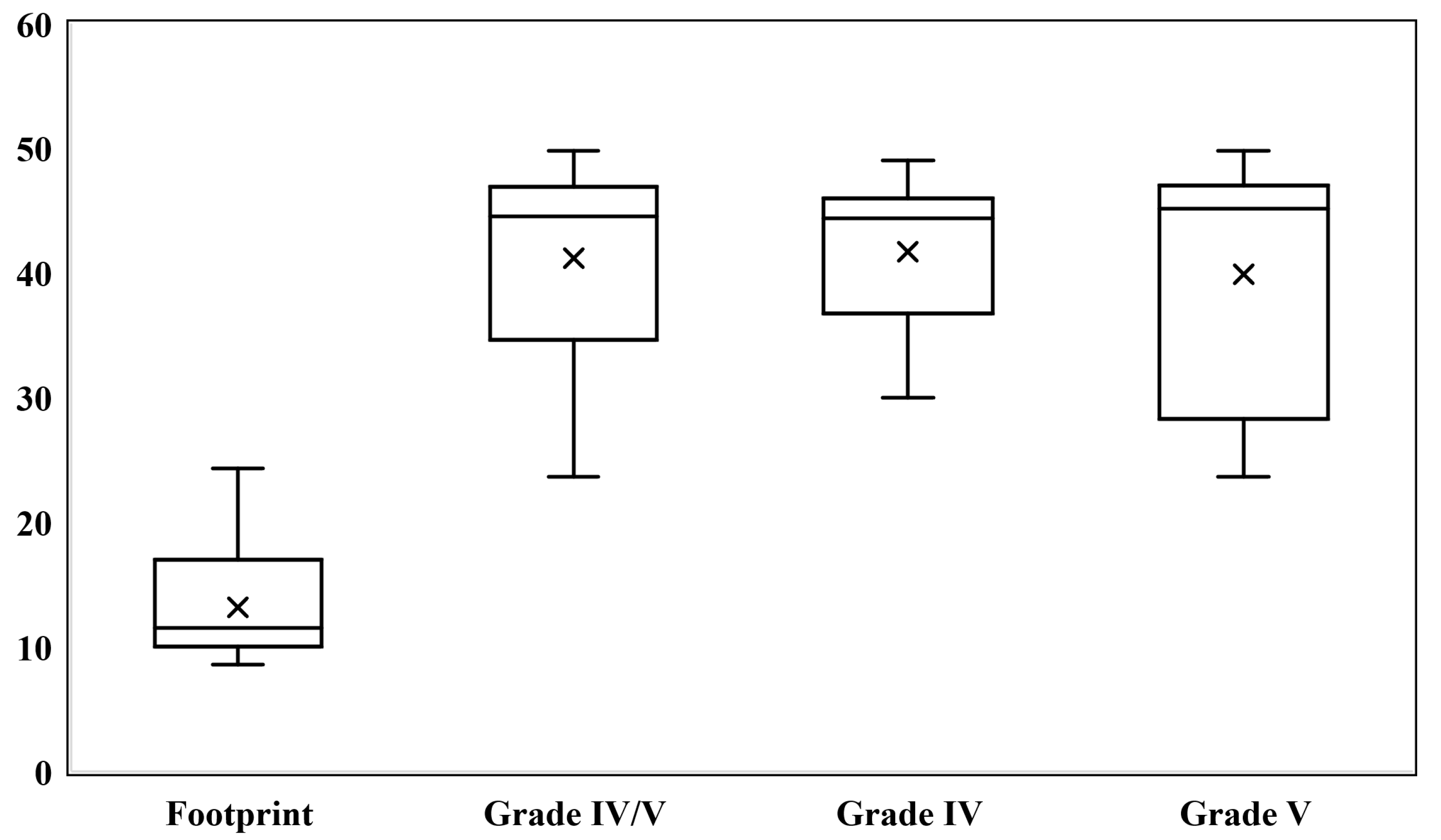
The median differential Tc99m-DMSA renal function was 11.5% [7.0] for renal units with the “footprint” shadow, in contrast to 44.5% [12.3] for high-grade renal units without this sign (p-value < 0.001) (Figure 3). All 16 out of 55 patients (29.1%) with a DRF of less than 20% exhibited the “footprint” sign. Consequently, the sensitivity, specificity, positive predictive value, and negative predictive value of the “footprint” sign for detecting refluxing units with a DRF of less than 20% were 100%, 94.9%, 88.9%, and 100%, respectively.

3.4. Clinical Information at Follow-Up

At the last follow-up, none of the patients had undergone nephrectomy, and none had developed hypertension. Among the patients in Group B, two had trace proteinuria detected by urinalysis, while the remaining 35 showed no proteinuria. Notably, none of the patients with the “footprint” sign had proteinuria. Median serum creatinine levels were similar between the “footprint” group (0.6 mg/dL; IQR: 0.18) and the comparison group (0.6 mg/dL; IQR: 0.21) (p-value: 0.139) (Table 1). Considering the influence of age and gender on creatinine, multiple linear regression adjusting for age and gender was performed. The difference remained non-significant between groups (p-value: 0.163).

4. Discussion

Children with VUR typically undergo evaluation using multiple imaging techniques. Weitz et al. [9] hypothesized that renal volume adjusted for body size, measured by ultrasonography, provides a simpler and less invasive alternative to renal scintigraphy for assessing renal function in individuals with primary VUR. A statistically significant correlation was found between kidney volume measured by ultrasonography and split renal function determined by renal scintigraphy at initial examination and follow-up. Nevertheless, VCUG remains the primary imaging modality for initial assessment, detection, and grading of VUR in many cases [10]. Numerous studies have been conducted to identify factors contributing to renal damage, a complex multifactorial process not yet fully understood [1,11,12,13,14,15,16].
In this study, we demonstrated that VCUG, which illustrates the anatomical features of the refluxing system, can also provide insights into the functional status of the kidneys. In refluxing units with impaired function, dilated calyces connected to an enlarged and elongated renal pelvis form a distinct pattern, which we refer to as the “footprint” sign, as it aptly describes the observed shape. Our findings corroborate those of Martin et al. [7], who identified a similar pattern termed the “flowerpot” sign. They examined 57 patients with high-grade VUR, 11 of whom exhibited the “flowerpot” sign on their VCUG. “Flowerpot” renal units had a median DRF of 23%, while renal units with grade IV or V reflux had a median DRF of 45%. These results align with our study, where the median relative uptake of Tc99m-DMSA was 11.5% in “footprint” units, significantly lower than that of the comparison group.
The median relative uptake of Tc99m-DMSA was 11.5% for “footprint” units, indicating severely impaired renal function typical of high-grade VUR. Hunziker et al. [12] demonstrated an increased risk of renal functional disorders in children with grade V VUR. Similarly, Arapović et al. [1] reported that abnormal Tc99m-DMSA scans (indicating small kidneys or scar development) are more likely in high-grade VUR. They found that the incidence of small kidneys was 13.4% in patients with non-dilated VUR, 12.5% in those with grade III VUR, and 34% in the group with grade IV or V VUR. Additionally, as the VUR grade increased, renal scarring became more frequent, with a significant difference between the groups. The prevalence of scarring was 3.8 times higher in the grade IV-V VUR group compared to those with non-dilated VUR [1].
The clinical outcome of VUR depends on kidney function severity, which determines the risk of persistent reflux, permanent renal injury, and renal insufficiency. Besides high-grade VUR, dysfunctional voiding, recurrent pyelonephritis, and delayed antibiotic therapy are potential causes of kidney scarring in individuals with VUR. Therefore, assessing kidney function and implementing timely interventions are crucial in cases of high-grade reflux to prevent potential long-term consequences [13].
To our knowledge, the “footprint” sign is the only known pattern in VCUG that can provide a clinical interpretation of renal function prior to further assessment. We believe that “footprint” kidneys represent a distinct category of high-grade refluxes not classifiable by the international grading system. However, radiologists need to recognize this pattern so they can inform the referring physician.
The etiology of the “footprint” pattern is unknown. One hypothesis is that it results from developmental dysregulation, forming a pop-off mechanism in a refluxing unit with severely compromised renal function. This pop-off mechanism reduces pressure on the contralateral kidney, functioning as a pressure venting system, as initially described by Hoover and Duckett in 1982 in patients with PUV concurrent with vesicoureteral reflux and unilateral renal dysplasia [17]. Although not statistically significant, the incidence of bilateral VUR was lower in the “footprint” group, potentially supporting this hypothesis. The lack of significance might be attributable to the small sample size. Additionally, we observed a higher incidence of left-side involvement in patients with the “footprint” sign compared to controls, with 14 out of 18 patients exhibiting the “footprint” on the left side. This finding aligns with previous studies on the pop-off mechanism in PUV [17,18], where Hoover and Duckett noted that the left side is more susceptible to abnormalities during embryogenesis [17]. However, 33.3% of our patients with the “footprint” pattern presented with antenatal hydronephrosis, compared to 50.0% who presented with recurrent UTIs. Therefore, this pattern may also develop after birth rather than being due to abnormalities during embryogenesis. We suggest further investigation to answer this question. Assuming either of the two hypotheses—where the “footprint” pattern occurs during renal development or after birth—we hypothesize that the pop-off mechanism may be the underlying etiology in both cases.
Furthermore, 16 out of 18 patients (88.9%) with the “footprint” renal unit were male, contrasting with the female predominance in the comparison group. This male predominance mirrors the findings of Martin et al. [7], who described the “flowerpot” sign, thus affirming the higher incidence of renal abnormalities in males and supporting the hypothesis of congenital developmental anomalies associated with this sign [7,12].
We also posit that treating reflux on the side with the “footprint” sign could lead to dysregulation of urinary system pressures, potentially initiating or exacerbating reflux in the better-functioning kidney. Therefore, prioritizing the management of reflux in the contralateral normally functioning kidney and postponing nephrectomy decisions for “footprint” kidneys may be more advantageous for patients. Moreover, the presence of the “footprint” sign negates the need for endoscopic dextranomer/hyaluronic acid copolymer injection in the ipsilateral kidney. Consequently, VCUG alone may suffice when a “footprint” sign is present, eliminating the need for additional assessments or Tc99m-DMSA scans on the affected kidney, as dysfunction is already confirmed. This approach facilitates the management and monitoring of these patients, particularly in low- to middle-income countries lacking facilities for renal scans for every patient and also reduces costs and radiation exposure for children in all countries. While the “footprint” sign may mitigate damage to the contralateral kidney, Tc99m-DMSA may still be required if ultrasonography of the contralateral kidney is abnormal.
It is noteworthy that at our center, endoscopic dextranomer/hyaluronic acid copolymer injection is the preferred method for VUR correction in the contralateral “footprint” kidney when surgery is indicated. Indications for this procedure include breakthrough febrile infections despite continuous prophylactic antibiotics, high grades of VUR (grades IV and V), and kidney scarring. None of our patients underwent nephrectomy or ureteral reimplantation for contralateral VUR correction.

Limitations and Suggestions

We acknowledge that this study has certain limitations, as it is a retrospective observational study with a limited dataset and relies exclusively on imaging. This retrospective, single-center study is inherently limited by potential selection bias, lack of standardization in imaging interpretation, and possible confounding variables such as a history of previous febrile UTIs, which may influence renal scan findings. The absence of formal interobserver validation may introduce bias, as the “footprint” sign was identified by two pediatric radiologists and subsequently confirmed by a pediatric urologist, rather than being assessed independently, precluding calculation of a kappa statistic. The small sample size (55 patients, of whom only 18 had the footprint sign) limits generalizability and may overestimate the predictive value of the “footprint” sign.
Additionally, while we propose the “footprint” sign as a potential protective “pop-off” mechanism, this remains a hypothesis. A definitive evaluation would ideally require comparison between patients with the “footprint” sign who underwent ipsilateral nephrectomy or endoscopic correction by bulking agent and those who did not. As our approach was not to intervene on the ipsilateral kidney during follow-up, we are currently unable to perform such an analysis. Future studies including these patient groups are necessary to validate this hypothesis.
Nonetheless, the proposed concept could serve as a basis for future research. Future research should focus on prospective, multicenter studies to validate the “footprint” sign across diverse populations and settings. Moreover, independent image reviews with interobserver agreement analysis are warranted to improve reliability. The use of emerging techniques such as machine learning for automated image analysis may further enhance detection accuracy and support clinical implementation.

5. Conclusions

We contend that “footprint” kidneys represent a distinct subset of high-grade reflux that does not fit within the international VUR grading system and should be acknowledged as a unique category. The “footprint” sign is a strong indicator of compromised renal function and warrants prompt reporting by radiologists to the referring physician. We propose that the pop-off mechanism may serve as a protective response, preventing the progression of reflux in the contralateral kidney. Consequently, it may be more advantageous to manage reflux in the contralateral kidney with normal function and postpone nephrectomy decisions for kidneys with the “footprint” sign. Furthermore, VCUG alone may suffice when a “footprint” sign is present, obviating the need for additional assessments or Tc99m-DMSA scans for the ipsilateral kidney, as impaired renal function is already established.

Author Contributions

Conceptualization, A.-M.K.; validation, A.-M.K.; methodology, A.-M.K., N.T., H.K., N.M.G. and M.M.; investigation, A.-M.K., N.M.G. and M.M.; supervision, A.-M.K.; data curation, N.M.G.; formal analysis, H.K.; writing—original draft, N.T. and H.K.; writing–review and editing, A.-M.K., H.K., N.M.G. and M.M. All authors have read and agreed to the published version of the manuscript.

Funding

This research received no external funding.

Institutional Review Board Statement

This study was conducted in accordance with the Decla-ration of Helsinki and was approved by the research ethics committee of Tehran University of Medical Sciences on 29 January 2019 (part of the study with approval ID IR.TUMS.VCR.REC.1397.878).

Informed Consent Statement

Informed consent for participation was not required as per the Tehran University of Medical Sciences research ethics committee. Since prior consent was obtained from patients at the time of admission for the use of medical record data, it was determined that there were no ethical issues.

Data Availability Statement

The data presented in this study are available on request from the corresponding author due to ethical reasons.

Conflicts of Interest

Abdol-Mohammad Kajbafzadeh is an Editorial Board member of SIUJ. He was not involved in the selection of peer reviewers for the manuscript nor any of the subsequent editorial decisions. The remaining authors have nothing to disclose.

Abbreviations

DRF, differential renal function; IQR, interquartile range; PUV, posterior urethral valve; SPSS, Statistical Package for the Social Sciences; Tc99m-DMSA, Tc99m dimercaptosuccinic acid; UTI, urinary tract infection; VCUG, voiding cystourethrogram; VUR, vesicoureteral reflux.

References

  1. Arapović, A.; Punda, A.; Brdar, D.; Čapkun, V.; Bajo, D.; Veljačić, D.; Punda, H.; Simičić-Majce, A.; Saraga-Babić, M.; Vukojević, K.; et al. Types of Parenchymal Changes Diagnosed on DMSA Scans of Kidneys Affected by Different Grades of Vesicoureteral Reflux. Med. Sci. Monit. Int. Med. J. Exp. Clin. Res. 2021, 27, e929617. [Google Scholar] [CrossRef] [PubMed]
  2. Mattoo, T.K. Vesicoureteral reflux and reflux nephropathy. Adv. Chronic Kidney Dis. 2011, 18, 348–354. [Google Scholar] [CrossRef] [PubMed]
  3. Darge, K. Voiding urosonography with US contrast agents for the diagnosis of vesicoureteric reflux in children. II. Comparison with radiological examinations. Pediatr. Radiol. 2008, 38, 54–63, quiz 126-127. [Google Scholar] [CrossRef] [PubMed]
  4. Koyle, M.A.; Elder, J.S.; Skoog, S.J.; Mattoo, T.K.; Pohl, H.G.; Reddy, P.P.; Abidari, J.M.; Snodgrass, W.T. Febrile urinary tract infection, vesicoureteral reflux, and renal scarring: Current controversies in approach to evaluation. Pediatr. Surg. Int. 2011, 27, 337–346. [Google Scholar] [CrossRef] [PubMed]
  5. Unver, T.; Alpay, H.; Biyikli, N.K.; Ones, T. Comparison of direct radionuclide cystography and voiding cystourethrography in detecting vesicoureteral reflux. Pediatr. Int. Off. J. Jpn. Pediatr. Soc. 2006, 48, 287–291. [Google Scholar] [CrossRef] [PubMed]
  6. Lebowitz, R.L.; Olbing, H.; Parkkulainen, K.V.; Smellie, J.M.; Tamminen-Möbius, T.E. International system of radiographic grading of vesicoureteric reflux. International Reflux Study in Children. Pediatr. Radiol. 1985, 15, 105–109. [Google Scholar] [CrossRef] [PubMed]
  7. Martin, A.D.; Gupta, K.; Swords, K.A.; Belman, A.B.; Majd, M.; Rushton, H.G.; Pohl, H.G. The “flowerpot” sign: Inference of poor renal function in high grade vesicoureteral reflux by calyceal orientation. J. Pediatr. Urol. 2015, 11, 31.e1–31.e4. [Google Scholar] [CrossRef] [PubMed]
  8. World Medical Association Declaration of Helsinki: Ethical principles for medical research involving human subjects. JAMA 2013, 310, 2191–2194. [CrossRef] [PubMed]
  9. Weitz, M.; Licht, C.; Müller, M.; Haber, P. Renal ultrasound volume in children with primary vesicoureteral reflux allows functional assessment. J. Pediatr. Urol. 2013, 9 Pt B, 1077–1083. [Google Scholar] [CrossRef] [PubMed]
  10. Rossleigh, M.A. Renal infection and vesico-ureteric reflux. Semin. Nucl. Med. 2007, 37, 261–268. [Google Scholar] [CrossRef] [PubMed]
  11. Peters, C.A.; Skoog, S.J.; Arant, B.S., Jr.; Copp, H.L.; Elder, J.S.; Hudson, R.G.; Khoury, A.E.; Lorenzo, A.J.; Pohl, H.G.; Shapiro, E.; et al. Summary of the AUA Guideline on Management of Primary Vesicoureteral Reflux in Children. J. Urol. 2010, 184, 1134–1144. [Google Scholar] [CrossRef] [PubMed]
  12. Hunziker, M.; Colhoun, E.; Puri, P. Prevalence and predictors of renal functional abnormalities of high grade vesicoureteral reflux. J. Urol. 2013, 190, 1490–1494. [Google Scholar] [CrossRef] [PubMed]
  13. Peters, C.; Rushton, H.G. Vesicoureteral reflux associated renal damage: Congenital reflux nephropathy and acquired renal scarring. J. Urol. 2010, 184, 265–273. [Google Scholar] [CrossRef] [PubMed]
  14. Silva, J.M.; Diniz, J.S.; Silva, A.C.; Azevedo, M.V.; Pimenta, M.R.; Oliveira, E.A. Predictive factors of chronic kidney disease in severe vesicoureteral reflux. Pediatr. Nephrol. 2006, 21, 1285–1292. [Google Scholar] [CrossRef] [PubMed]
  15. Yeung, C.K.; Sreedhar, B.; Sihoe, J.D.; Sit, F.K. Renal and bladder functional status at diagnosis as predictive factors for the outcome of primary vesicoureteral reflux in children. J. Urol. 2006, 176, 1152–1156, discussion 1156–1157. [Google Scholar] [CrossRef] [PubMed]
  16. Lin, Q.; Ni, J.; Li, Y.; Jin, J.; Zhu, Y. Renal function damage in children with duplex kidneys. Int. Urol. Nephrol. 2023, 55, 1–8. [Google Scholar] [CrossRef] [PubMed]
  17. Hoover, D.L.; Duckett, J.W., Jr. Posterior urethral valves, unilateral reflux and renal dysplasia: A syndrome. J. Urol. 1982, 128, 994–997. [Google Scholar] [CrossRef] [PubMed]
  18. Greenfield, S.P.; Hensle, T.W.; Berdon, W.E.; Wigger, H.J. Unilateral vesicoureteral reflux and unilateral nonfunctioning kidney associated with posterior urethral valves--a syndrome? J. Urol. 1983, 130, 733–738. [Google Scholar] [CrossRef] [PubMed]
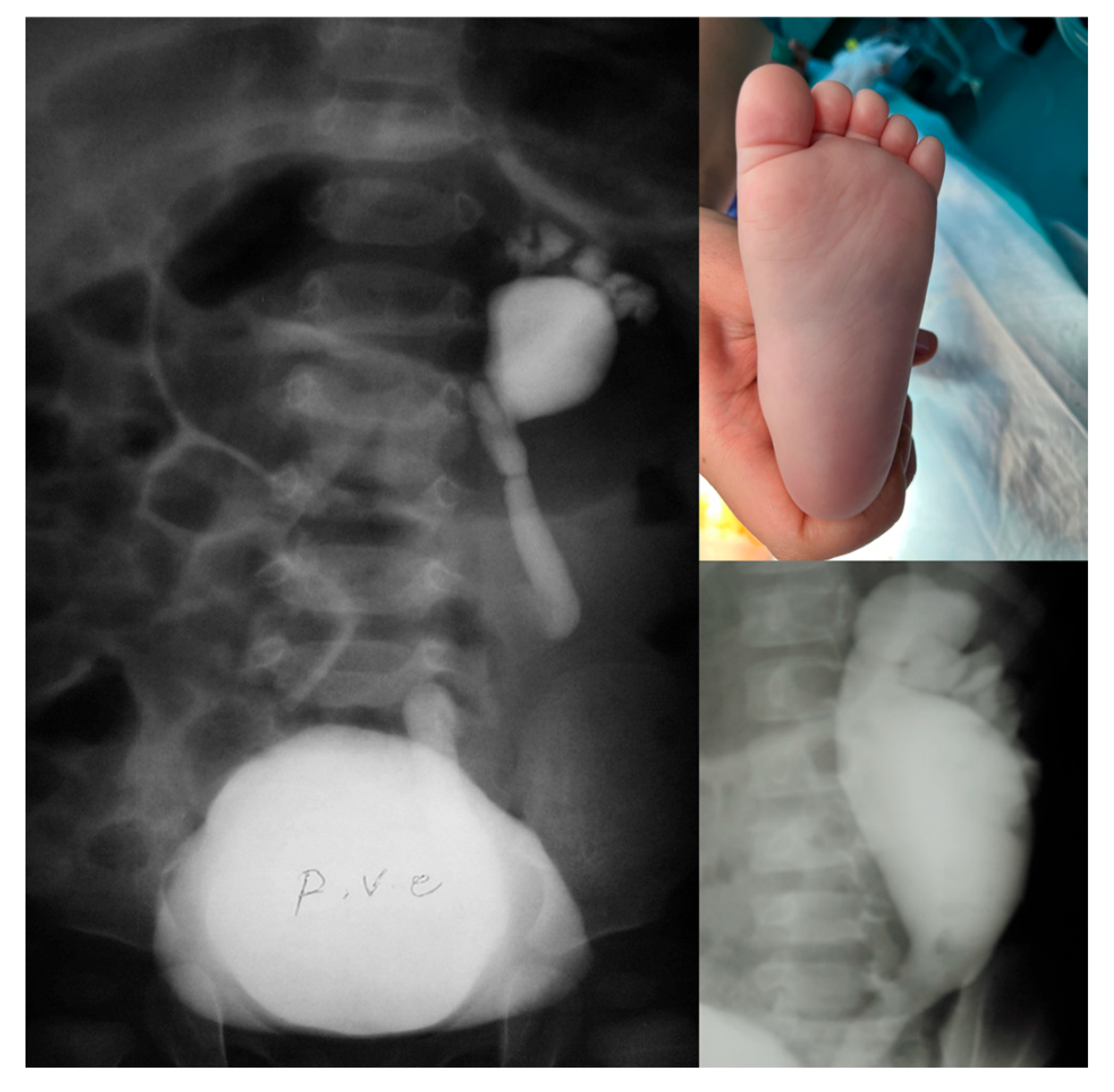
Figure 1. Dysmorphic calyces attached to a dilated renal pelvis form a distinct “footprint” shadow shape.
Siuj 06 00055 g001
Figure 2. Different views of Tc99m dimercaptosuccinic acid (Tc99m-DMSA) scan uptake in a refluxing unit with “footprint” shadow. Relative Tc99m-DMSA uptake of the right (footprint) kidney: 8%, the left kidney: 92%. The coloured spectrum in the DMSA scan represents the differential uptake of radiotracer by functioning renal cortical tissue. This spectrum reflects relative perfusion and metabolic activity of kidney function, with colour intensity corresponding to tracer uptake based on count density. Red, orange, yellow: Higher tracer uptake, interpreted as normal or hyper-functioning cortical tissue. Green, blue: Moderate to lower uptake, interpreted as reduced function. Black or absent colour: No uptake, interpreted as non-functioning areas. ANT, anterior; LPO, left posterior oblique; POST, posterior; RPO, right posterior oblique.
Siuj 06 00055 g002
Figure 3. Relative Tc99m dimercaptosuccinic acid (Tc99m-DMSA) scan uptake percentage comparison between high-grade renal units with and without the “footprint” sign.
Siuj 06 00055 g003
Table 1. Pertinent clinical and imaging findings of patients with the “footprint” sign (group A) and those with grade IV–V reflux but without the “footprint” sign (group B).
VariablePatients
Group A (n = 18)Group B (n = 37)
Gender; male/female16:212:25
Age, months; median [IQR]28.2 [66.5]13.0 [42.0]
Bilateral VUR; n (%)7 (38.9%)19 (51.4%)
Side of “footprint” or high-grade VUR; right: left4: 1418: 19
DRF; median [IQR]11.5 [7.0]44.5 [12.3]
Presentation; n (%)
      Antenatal hydronephrosis6 (33.3%)13 (35.1%)
      Recurrent UTIs9 (50.0%)23 (62.2%)
      Other/Incidental3 (16.7%)1 (2.7%)
Neurological and other related disorders; n (%)
      Spina bifida occulta4 (22.2%)3 (8.1%)
      Sacral abnormality1 (5.6%)5 (13.5%)
      History of ureterovesical junction obstruction0 (0.0%)4 (10.8%)
Clinical information at follow-up
      Hypertension; n (%)0 (0.0%)0 (0.0%)
      Significant proteinuria in urinalysis; n (%)0 (0.0%)0 (0.0%)
      Nephrectomy; n (%)0 (0.0%)0 (0.0%)
      Creatinine, mg/dL; median [IQR]0.6 [0.18]0.6 [0.21]
DRF differential renal function, IQR interquartile range, n number, UTI urinary tract infection, VUR vesicoureteral reflux.
Table 2. The grade of vesicoureteral reflux in renal units of all patients.
VUR GradeRenal Units
Group A (n = 25)Group B (n = 56)
I12
II24
III212
IV227
V-11
Footprint18-
VUR, vesicoureteral reflux.
Disclaimer/Publisher’s Note: The statements, opinions and data contained in all publications are solely those of the individual author(s) and contributor(s) and not of MDPI and/or the editor(s). MDPI and/or the editor(s) disclaim responsibility for any injury to people or property resulting from any ideas, methods, instructions or products referred to in the content.