Article

Is the Bulbar Urethral Stricture a Single and Uniform Disease?

1
Department of Urology, Fujisawa Shounandai Hospital, Fujisawa 252-0802, Japan
2
Department of Urology, National Defense Medical College, Tokorozawa 359-8513, Japan
3
Department of Plastic Surgery, National Defense Medical College, Tokorozawa 359-8513, Japan
*
Author to whom correspondence should be addressed.
Soc. Int. Urol. J. 2024, 5(2), 85-92; https://doi.org/10.3390/siuj5020014
Submission received: 27 July 2023 / Revised: 27 September 2023 / Accepted: 21 October 2023 / Published: 4 April 2024

Abstract

:
Objectives: Proximal and distal bulbar urethral strictures (BUS) have different disease characteristics and require different treatment strategies despite being regarded as a single condition. To clarify the differences, we analyzed our database by distinguishing the two types of BUS. Methods: We retrospectively reviewed the data of 196 patients with BUS who underwent urethroplasty at the National Defense Medical College (Japan) between August 2004 and March 2022. We divided patients into proximal (group 1) or distal (group 2) groups based on the stricture segment and compared patient background and surgical techniques for each group. We assessed whether the stricture segment was an independent predictive factor for substitution urethroplasty selection using multivariate logistic regression analysis. The recurrence rates were calculated and compared using the Kaplan–Meier method and log-rank test, respectively. Results: Patients in group 1 had a less frequent non-obliterated lumen (73% vs. 94%, p = 0.020) and significantly shorter strictures (10 mm vs. 23 mm, p < 0.001) more frequently caused by external traumas (47% vs. 26%, p = 0.010) than those in group 2. Logistic regression analysis revealed that the stricture segment (distal) (p < 0.001), stricture length (≥20 mm) (p < 0.001), ≥2 prior transurethral procedures (p = 0.030), and a non-obliterated lumen (p = 0.020) were independent predictive factors for substitution urethroplasty. However, the recurrence rate (p = 0.18) did not significantly differ between the two groups. Conclusions: Proximal and distal BUS have substantially different anatomical characteristics and etiologies and require different reconstructive techniques.

1. Introduction

Urethral stricture is typically caused by fibrosis or epithelial tissue and corpus spongiosum inflammation, causing urethral lumen narrowing. The bulbar urethra is the most frequent segment affected in male anterior urethral strictures [1,2,3,4].
Current guidelines for male urethral strictures recommend transurethral surgery for short (<2 cm) and treatment-naive bulbar urethral strictures (BUS) with little spongiofibrosis or urethroplasty; however, there are minor variations [5,6,7,8]. Although these guidelines describe BUS as a single condition, some reconstruction experts have noticed a certain variability in the disease characteristics between proximal and distal BUS [9]. The evidence on this issue is scarce; thus, to fill the gap between clinical practice and evidence, we identified and investigated the disease characteristics of the proximal/distal BUS. Additionally, we investigated whether this distinction influenced the selection of surgical technique through a retrospective analysis of a database at a tertiary referral reconstructive urological center in Japan.

2. Materials and Methods

2.1. Patient Selection

We retrospectively reviewed the data of 556 patients who were diagnosed with BUS by urethrography and subsequently underwent urethroplasty at our institution between August 2004 and March 2022. After classifying the patients according to the LSE (length, segment and etiology) classification [10] and excluding those with non-bulbar urethral strictures, multiple strictures, and incomplete data, a total of 196 were selected for further analysis. The patient selection process is illustrated in the supplementary file.

2.2. Patient Classification

T.T. conducted the evaluations of the stricture segment and length using the original images of retro- and antegrade urethrography in the proper position [1,2]. The bulbar urethra was defined as the urethral segment from a line between the inferior margins of the bilateral obturator foramina to the penoscrotal junction (PSJ). Strictures were categorized into proximal (group 1) and distal (group 2) BUS based on the stricture site relative to the midpoint of the bulbar urethra as evaluated in preoperative retrograde urethrography (RUG). Cases in which the stricture involved the distal bulbar urethra were categorized as distal BUS.

2.3. Surgical Technique

All urethroplasties were performed by HA or under the supervision of HA via the perineal approach in the lithotomy position under general anesthesia. The urethroplasty technique was determined by combining the preoperative RUG findings, and the stricture site was confirmed cystoscopically at the bulbar urethra exposure time. Theoretically, anastomotic urethroplasty, including non-transecting anastomotic urethroplasty (NTAU) [11] and excision and primary anastomosis (EPA) [12], was exclusively selected whenever tension-free anastomosis was feasible. Therefore, the bulbar urethra was fully mobilized from the bulbomembranous junction to the PSJ. The bilateral corpus cavernosum was separated in the midline when performing anastomotic urethroplasty to reduce tension at the anastomosis [13], if required. Until 2020, all cases eligible for anastomotic repair were treated with EPA. However, from 2020 onwards, the use of NTAU was prioritized for cases with less fibrosis, and EPA was chosen for traumatic strictures with dense spongiofibrosis. However, in cases in which tension-free anastomosis was not possible, an onlay augmentation urethroplasty (OA), such as dorsal, ventral, or dorsolateral onlay urethroplasty, was performed using an oral mucosa or penile skin graft. The final decision between anastomotic urethroplasty and substitution urethroplasty was determined by assessing the feasibility of tension-free anastomosis between the intended sites of anastomosis before urethral transection or release. This evaluation occurred after sufficient urethral mobilization and, if necessary, following separation of the corpus cavernosum. In contrast, staged urethroplasty (SU) was performed for patients with long and obliterated bulbar strictures and dense spongiofibrosis.

2.4. Patient Follow-Up and Definition of Recurrence

All patients were followed up 3, 6, and 12 months after urethroplasty and annually thereafter; at every visit, they provided a validated patient-reported outcome measure for urethral stricture surgery [14] and were examined using uroflowmetry and postvoid residual urine volume estimation. Cystoscopy was performed when re-stricture was suspected owing to decreased urinary flow and/or worsening symptoms. Recurrence was defined as the requirement for additional treatments.

2.5. Statistical Analysis

For all statistical tests, p < 0.05 was considered significant. We compared patients’ age, prior urethroplasty, prior transurethral procedure, stricture length, patency of urethral lumen, body mass index (BMI), diabetes mellitus, disease etiology, and smoking habits between the stricture segment groups. Continuous and categorical variables were compared using the Mann–Whitney U and chi-squared tests, respectively. Continuous variables are expressed as medians (interquartile range: IQR), whereas categorical variables are expressed as numbers (percentages).
To assess whether the stricture segment affected the selection of substitution urethroplasty, a multivariate logistic regression model with stepwise selection using Akaike’s information criteria was implemented. Based on clinical practice, the cut-off values of stricture length and BMI were 20 mm and 25 kg/m2, respectively, whereas 53 years was the cut-off value for age, or the median age of this cohort. The recurrence rate was calculated using the Kaplan–Meier method and compared using the log-rank test. The database was constructed in Excel 2016 (Microsoft, Redmond, WA, USA), and analyses were performed using R version 4.1.2.

3. Results

3.1. Comparison of Stricture Characteristics

The patient characteristics are listed in Table 1. Patients were categorized as group 1 (165, 84%) and group 2 (31, 16%). There were no statistically significant differences between groups in terms of age, prior transurethral procedures, and prior urethroplasty. The median stricture length of group 1 (10 mm, 7–15) was significantly shorter than that of group 2 (23 mm, 10–32) (p < 0.001), and stricture etiology was significantly different between groups (p = 0.01). The most common stricture etiologies were perineal trauma in group 1 (47%) and idiopathic etiologies in group 2 (35%). The urethral lumen at the stricture site was obliterated in 43 (27%) patients in group 1, which was significantly higher than that in group 2 (6.0%, p = 0.02).

3.2. Comparison of Required Surgical Techniques and Outcomes

Table 2 presents the selected surgical procedures for groups 1 and 2. The ratio of patients who underwent substitution urethroplasty (OA and SU) (OA: 12%, SU 2.4%) was significantly lower in group 1 than in group 2 (OA 58%, SU 23%, p < 0.0001). Corporal splitting was carried out for 62% of group 1 patients and 3% of group 2 patients (<0.001).
No significant difference in the operation time (p = 0.19) or amount of blood loss (p = 0.67) was observed between groups. A total of 179 (91%) patients were stricture-free at the median postoperative period of 69 months, and 17 patients (8.7%) experienced recurrence. The Kaplan–Meier curves of the stricture segments did not show any significant differences in stricture-free survival rates between groups (p = 0.18) (Figure 1).

3.3. Comparison of Stricture Lengths in Patients Treated with EPA or NTAU

The median stricture lengths of patients in groups 1 and 2 treated by EPA or NTAU were 10 mm (7.0–15.0) and 7.0 mm (6.25–9.25), respectively, which were not significantly different (p = 0.098). The maximum stricture lengths were 33 and 11 mm, respectively.

3.4. Predictive Factors for Substitution Urethroplasty

We further investigated whether the stricture segment was a predictor of the requirement for substitution urethroplasty (Table 3). In the multivariate logistic regression model, group 2 (OR: 19, 95% CI: 5.6–66, p < 0.001), stricture length longer than 20 mm (OR: 18, 95% CI: 6.1–54, p < 0.001), a prior history of transurethral procedure (OR 3.1, 95% CI 1.1–8.3, p = 0.03), and non-obliterated urethral lumen (OR: 13, 95% CI: 1.5–100, p = 0.02) were independent predictors of the need for substitution urethroplasty.

4. Discussion

In this retrospective study, we successfully confirmed the variability in disease characteristics between stricture segments in the bulbar urethra. Patients in group 1 had shorter and obliterated strictures, mostly caused by perineal traumas, than those in group 2. Furthermore, we demonstrated that group 2 was an independent factor for deciding surgical techniques, as well as stricture length. The proximal bulbar urethra has a thick blood-rich spongiosum [15], whereas the distal bulbar urethra is less mobile and has a limited blood supply. Therefore, the proximal bulbar urethra is more amenable to long excisional and anastomotic repair in clinical practice [16,17], owing to its mobility. Nevertheless, despite apparently different anatomical or surgical characteristics, the definitions of the proximal or distal bulbar urethra remain unclear. Terlecki et al. [18] defined the proximal bulbar urethra as the segment within 5 cm of the membranous urethra and the distal bulbar urethra as the adjacent segment extending to the penoscrotal junction. In the recently published LSE classification system, the bulbar urethra was divided into two parts; however, the definition was not disclosed, whereas our definition of dividing the bulbar urethra at the mid-point was straightforward and could account for individual differences.
The differences in characteristics between groups 1 and 2 can be partially explained by different etiologies, as approximately half of group 1 cases were caused by external traumas, which accounted for only 26% in group 2, in which idiopathic and iatrogenic etiologies were more common. These outcomes are consistent with those of a previous study [10]. For example, in straddle injuries, the bulbous urethra is aggressively pushed against the pubic bone, a mechanism that more frequently affects the proximal bulbar urethra, which may be explained by differences in etiology. Prior to the analysis, we hypothesized that traumatic strictures might influence the choice of treatment. However, traumatic strictures were eliminated in the middle step of the multivariate analysis and obliteration of the lumen was observed to be the significant factor. While these two factors appeared related, the variance inflation factor was not substantial. In this analysis, we believe that the risk of multicollinearity is low.
The most critical factor defining the urethroplasty technique for bulbar urethral stricture is the stricture length. Substitution urethroplasty is generally recommended for bulbar strictures longer than 20 mm because of the inevitable tension in the urethral anastomosis [5,6,7,8]. However, based on experience, reconstructive urologists acknowledge that stricture length is just one of the factors to determine whether substitution is required. The eligibility of group 1 for anastomotic urethroplasty is broader than that of group 2, and anastomotic urethroplasty can be used even for strictures longer than 20 mm in the proximal BUS. For example, Morey et al. reported that strictures of up to 5 cm in the proximal BUS might be treated with EPA in young men [9]. In contrast, those in the distal BUS reportedly require substitution procedures more frequently, even when adjusting the stricture length to 2–5 cm [18], and are more vulnerable to recurrence. After classifying 2162 urethral strictures, Erickson et al. [10]. observed that distal BUS was more likely to be treated by an OA compared with proximal BUS. Despite the lack of significant differences in the median stricture length between the groups in the present study, stricture length up to 33 mm in group 1 was treated using EPA, whereas 11 mm was the longest stricture length in group 2. In group 1, adequate urethral mobilization extended the available urethra, and corporal splitting further reduced the tension at the anastomotic site. Therefore, we speculate that the length limit for anastomotic repair in group 1 cannot be absolute and may depend on the individual anatomical condition. Additionally, to our knowledge, previous reports suggesting group 2 as an independent predictor of substitution urethroplasty using multivariate analysis are lacking, which highlights the novelty of our findings.
Nevertheless, the present study has some limitations. First, the study was retrospective in nature, and since our institution is the largest center in Japan by volume, some selection bias may exist. Additionally, patients in group 2, who were possible candidates for EPA, might have been treated elsewhere, whereas patients requiring challenging procedures were referred to our department. Furthermore, some techniques rarely performed by us can change the frequency of the surgical strategy. To reduce tension at the anastomotic site, corporal spitting was conducted in 62.42% of cases in group 1, whereas it was only performed in 3.23% of the cases in group 2, as this technique was unsuitable for group 2. Performing tunica albuginea plication (TAP) could have resulted in more patients from group 2 being treated using EPA [19]. Nevertheless, we avoid performing TAP routinely owing to its disadvantages, such as shortening the penile length and curving the penis upon erection. TAP is used only in rare patients who have lost erectile functions and are not concerned with aesthetics. However, this does not impact our conclusion as the requirements for such specialized techniques differed between groups 1 and 2.
Second, the effects of the stricture segment on prognosis remain unelucidated. As recurrence occurred only in 8.67% of patients, the statistical power was insufficient for analysis to detect the difference. The small sample size and the imbalance in the number of cases between groups 1 and 2 might impact the statistical power. However, it is worth noting that even in the original study of the LSE classification, a similar imbalance is observed (S1a: 1223, S1b: 282). This aspect itself could reflect the inherent differences between these two groups [10].
Third, the recurrence rate must be interpreted with caution. Given the significantly different backgrounds of groups 1 and 2, the observed differences in prognosis may stem from their distinct characteristics. This study has not provided evidence to establish that the location of the bulbar urethral stricture itself is an independent prognostic factor.
Consequently, studies with larger sample sizes must be conducted to determine whether the stricture segment independently affects the recurrence rate.

5. Conclusions

Groups 1 and 2 had substantially different disease characteristics in terms of anatomy, etiology, surgical technique. Distal BUS tend to have more complex underlying factors and often require the use of substitute tissues in treatment. Due to these considerations, a more cautious treatment approach is warranted.

Supplementary Materials

The following supporting information can be downloaded at: https://www.mdpi.com/article/10.3390/siuj5020014/s1, Figure S1: Patient selection process.

Author Contributions

Conceptualization, T.T. and AH.; methodology, T.T. and A.H.; software, T.T.; validation, M.S. and K.O.; formal analysis, T.T.; investigation, Y.H.; resources, A.H.; data curation, A.H., M.S., K.O. and Y.H.; writing—original draft preparation, T.T.; writing—review and editing, A.H.; visualization, A.H.; supervision, K.I. and R.A.; project administration, A.H.; funding acquisition, A.H. All authors have read and agreed to the published version of the manuscript.

Funding

This study was supported by a Grant-in-Aid for Scientific Research from the Ministry of Education, Science, Sports, and Culture (20K09534).

Institutional Review Board Statement

This retrospective study was approved by the institutional review board (approval number: 4154).

Informed Consent Statement

Informed consent was obtained from all subjects involved in the study.

Data Availability Statement

The datasets analyzed in the current study are available from the corresponding author upon request.

Conflicts of Interest

The authors declare no conflict of interest.

Abbreviations

LSELength, segment, and etiology
BUSbulbar urethral strictures
NTAUnon-transecting anastomotic urethroplasty
EPAexcision and primary anastomosis
OAonlay augmentation urethroplasty
SUstaged urethroplasty
BMIbody mass index
IQRinterquartile range
ORodds ratio
95% CI95% confidence interval
TAPtunica albuginea plication

References

  1. Palminteri, E.; Berdondini, E.; Verze, P.; De Nunzio, C.; Vitarelli, A.; Carmignani, L. Contemporary urethral stricture characteristics in the developed world. Urology 2013, 81, 191–196. [Google Scholar] [CrossRef]
  2. Lumen, N.; Hoebeke, P.; Willemsen, P.; De Troyer, B.; Pieters, R.; Oosterlinck, W. Etiology of urethral stricture disease in the 21st century. J. Urol. 2009, 182, 983–987. [Google Scholar] [CrossRef]
  3. Cotter, K.J.; Hahn, A.E.; Voelzke, B.B.; Myers, J.B.; Smith, T.G., 3rd; Elliott, S.P.; Alsikafi, N.F.; Breyer, B.N.; Vanni, A.J.; Buckley, J.C.; et al. Trends in Urethral Stricture Disease Etiology and Urethroplasty Technique from a Multi-institutional Surgical Outcomes Research Group. Urology 2019, 130, 167–174. [Google Scholar] [CrossRef]
  4. Stein, D.M.; Thum, D.J.; Barbagli, G.; Kulkarni, S.; Sansalone, S.; Pardeshi, A.; Gonzalez, C.M. A geographic analysis of male urethral stricture aetiology and location. BJU Int. 2013, 112, 830–834. [Google Scholar] [CrossRef] [PubMed]
  5. Gómez, R.G.; Mundy, T.; Dubey, D.; El-Kassaby, A.W.; Firdaoessaleh; Kodama, R.; Santucci, R. SIU/ICUD consultation on urethral strictures: Pelvic fracture urethral injuries. Urology 2014, 83 (Suppl. S3), S48–S58. [Google Scholar] [CrossRef] [PubMed]
  6. Wessells, H.; Angermeier, K.W.; Elliott, S.; Gonzalez, C.M.; Kodama, R.; Peterson, A.C.; Reston, J.; Rourke, K.; Stoffel, J.T.; Vanni, A.J.; et al. Male Urethral Stricture: American Uro-logical Association Guideline. J. Urol. 2017, 197, 182–190. [Google Scholar] [CrossRef] [PubMed]
  7. Lumen, N.; Campos-Juanatey, F.; Greenwell, T.; Martins, F.E.; Osman, N.I.; Riechardt, S.; Waterloos, M.; Barratt, R.; Chan, G.; Esperto, F.; et al. European Association of Urology Guidelines on Urethral Stricture Disease (Part 1): Management of Male Urethral Stricture Disease. Eur. Urol. 2021, 80, 190–200. [Google Scholar] [CrossRef] [PubMed]
  8. Rourke, K.F.; Welk, B.; Kodama, R.; Bailly, G.; Davies, T.; Santesso, N.; Violette, P.D. Canadian Urological Association guideline on male urethral stricture. Can. Urol. Assoc. J. 2020, 14, 305–316. [Google Scholar] [CrossRef] [PubMed]
  9. Morey, A.F.; Kizer, W.S. Proximal bulbar urethroplasty via extended anastomotic approach—What are the limits? J. Urol. 2006, 175, 2145–2149; discussion 2149. [Google Scholar] [CrossRef] [PubMed]
  10. Erickson, B.A.; Flynn, K.J.; Hahn, A.E.; Cotter, K.; Alsikafi, N.F.; Breyer, B.N.; Broghammer, J.A.; Buckley, J.C.; Elliott, S.P.; Myers, J.B.; et al. Development and Validation of A Male Anterior Urethral Stricture Classification System. Urology 2020, 143, 241–247. [Google Scholar] [CrossRef] [PubMed]
  11. Andrich, D.E.; Mundy, A.R. Non-transecting anastomotic bulbar urethroplasty: A preliminary report. BJU Int. 2012, 109, 1090–1094. [Google Scholar] [CrossRef] [PubMed]
  12. Horiguchi, A.; Ojima, K.; Shinchi, M.; Hirano, Y.; Hamamoto, K.; Ito, K.; Asano, T.; Takahashi, E.; Kimura, F.; Azuma, R. Single-surgeon experience of excision and primary anastomosis for bulbar urethral stricture: Analysis of surgical and patient-reported outcomes. World J. Urol. 2021, 39, 3063–3069. [Google Scholar] [CrossRef] [PubMed]
  13. Mundy, A.R. Anastomotic urethroplasty. BJU Int. 2005, 96, 921–944. [Google Scholar] [CrossRef] [PubMed]
  14. Horiguchi, A.; Shinchi, M.; Ojima, K.; Masunaga, A.; Ito, K.; Asano, T.; Takahashi, E.; Kimura, F.; Azuma, R. Evaluation of the effect of urethroplasty for anterior urethral strictures by a validated disease-specific patient-reported outcome measure. World J. Urol. 2019, 37, 601–606. [Google Scholar] [CrossRef] [PubMed]
  15. Heinke, T.; Gerharz, E.W.; Bonfig, R.; Riedmiller, H. Ventral onlay urethroplasty using buccal mucosa for complex stricture repair. Urology 2003, 61, 1004–1007. [Google Scholar] [CrossRef] [PubMed]
  16. Jezior, J.R.; Schlossberg, S.M. Excision and primary anastomosis for anterior urethral stricture. Urol. Clin. N. Am. 2002, 29, 373–380. [Google Scholar] [CrossRef] [PubMed]
  17. Iselin, C.E.; Webster, G.D. Dorsal onlay urethroplasty for urethral stricture repair. World J. Urol. 1998, 16, 181–185. [Google Scholar] [CrossRef]
  18. Terlecki, R.P.; Steele, M.C.; Valadez, C.; Morey, A.F. Grafts are Unnecessary for Proximal Bulbar Reconstruction. J. Urol. 2010, 184, 2395–2399. [Google Scholar] [CrossRef]
  19. VanDyke, M.E.; Baumgarten, A.S.; Ortiz, N.M.; Dropkin, B.M.; Joice, G.A.; Khouri, R.K.; Filho, J.E.P.; Ward, E.E.; Hudak, S.J.; Morey, A.F. Extended Primary Anastomosis with Penile Plication (EPAPP): A Promising New Alternative to Perineal Urethrostomy for Reconstruction of Long Urethral Strictures. Urology 2021, 149, 245–250. [Google Scholar] [CrossRef]
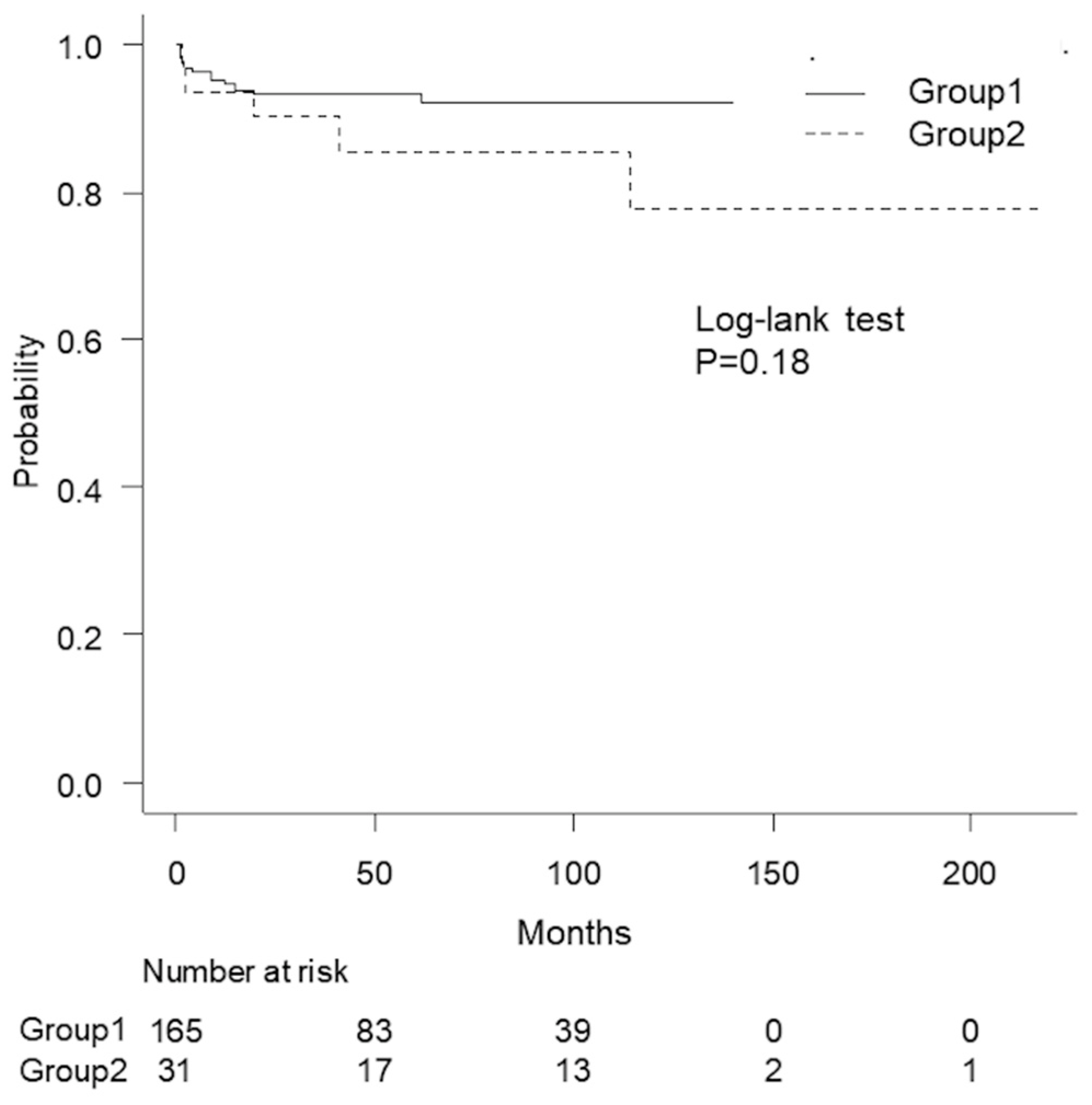
Figure 1. Kaplan–Meier curves of group 1 (solid line) and group 2 (dashed line). No significant differences were observed in the recurrence-free survival values (p = 0.18).
Siuj 05 00014 g001
Table 1. Patient characteristics of the two groups.
Segment Group 1Group 2p
FactorsUnit/CategoryMedian/NumberIQR/%Median/NumberIQR/%
N (%) 165 (84)31 (16)
Agey.o.5339–645543–700.05
Smokenone603611350.99
DMpresent106.113.20.83
BMIkg/m22322–252321–260.53
Prior urethroplasty≥119127230.16
Stricture lengthmm107–152310–32<0.001
Lumennon-obliterated1217329940.020
Prior transurethral procedure≥2714318580.17
Etiologytrauma78478260.010
idiopathic51311135
iatrogenic3421929
others21.0310
Abbreviations: IQR: interquartile range; DM: diabetes mellitus; BMI: body mass index.
Table 2. Comparison of surgical techniques, surgical duration, and bleeding.
Group 1Group 2p
FactorUnit/CategoryMedian/NumberIQR/%Median/NumberIQR/%
Surgical techniqueEPA12576619<0.001 a
NTAU161000
OA20121858
SU42.4723
Corporal splittingPresent626213.2<0.001
Operation timeminute157138–187186135–2130.19
Blood lossmL5932–1156821–1560.67
Abbreviations: IQR: interquartile range; EPA: excision and primary anastomosis; NTAU: non-transecting anastomotic urethroplasty; OA: onlay augmentation; SU: staged urethroplasty. a Comparison between the groups in terms of anastomotic (EPA/NTAU) or substitution urethroplasty (OA/SU).
Table 3. Results of the multivariate logistic regression analysis with stepwise selection using Akaike’s information criteria.
Initial ResultFinal Result
VIFOR95% CIpOR95% CIp
Segmentgroup 21.1185.1–62<0.001195.6–66<0.001
Prior urethroplasty≥11.02.00.58–7.00.27
Lumennon-obliterated1.1131.2–1000.030131.5–1000.020
Prior transurethral procedure≥21.22.50.85–7.20.103.11.1–8.30.030
Smoking present1.21.70.57–5.00.35
Age≥53 y.o.1.10.980.36–2.70.96
Stricture length>20 mm1.1165.3–49<0.001186.1–54<0.001
BMI>251.21.9 0.65–5.40.24
Etiologynon-traumatic1.31.5 0.45–5.20.49
Abbreviations: VIF: variance inflation factor; OR: odds ratio; CI: confidence interval; BMI: body mass index.
Disclaimer/Publisher’s Note: The statements, opinions and data contained in all publications are solely those of the individual author(s) and contributor(s) and not of MDPI and/or the editor(s). MDPI and/or the editor(s) disclaim responsibility for any injury to people or property resulting from any ideas, methods, instructions or products referred to in the content.