Next Article in Journal
Calling All Urologists Around the Globe to Tell Their Stories
Previous Article in Journal
A Rare Case of Hypersensitivity Reaction Associated With Sacral Neuromodulation Hardware
 
 
Société Internationale d’Urologie Journal is published by MDPI from Volume 5 Issue 1 (2024). Previous articles were published by another publisher in Open Access under a CC-BY (or CC-BY-NC-ND) licence, and they are hosted by MDPI on mdpi.com as a courtesy and upon agreement with Société Internationale d’Urologie (SIU).
Interesting Images

Gadolinium Contrast in the Bladder: A Malignant Mimic

In affiliation with the Division of Surgery and Anaesthesia, Department of Urology, Gosford District Hospital, Gosford, Australia
*
Author to whom correspondence should be addressed.
Soc. Int. Urol. J. 2022, 3(1), 48; https://doi.org/10.48083/OFWX4645
Submission received: 20 September 2021 / Accepted: 27 September 2021 / Published: 10 January 2022
A 50-year-old female presented to hospital with diplopia and unsteady gait. The patient was identified to have a disconjugate gaze palsy involving the right medial rectus muscle. Investigation for an ischaemic event proved negative, with an unremarkable MRI of the brain. A paraneoplastic screen, including a CT of the chest, abdomen, and pelvis, was undertaken to identify an alternative cause for her symptoms.
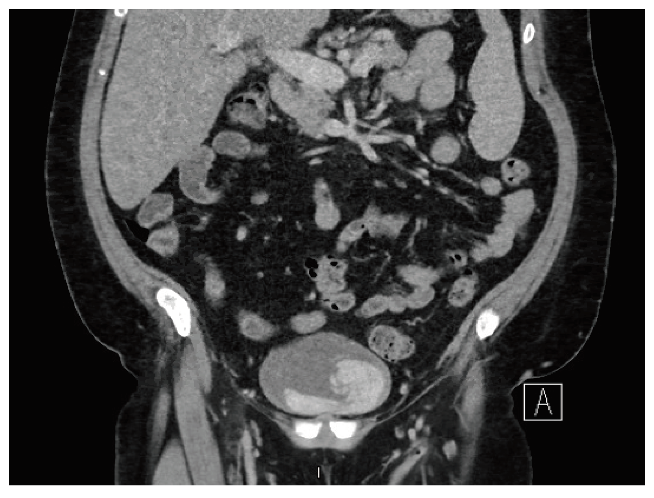
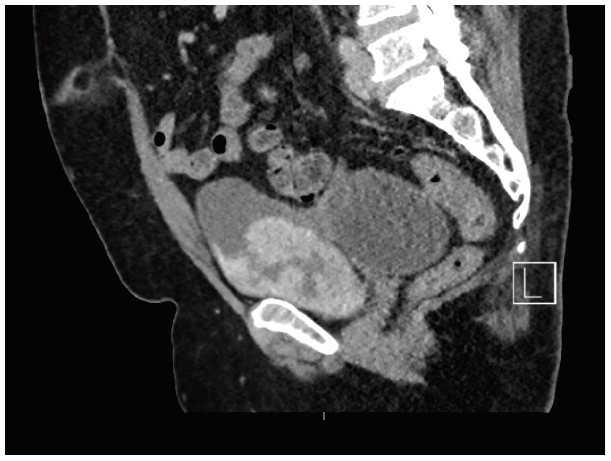
An incidental finding of a large, enhancing bladder mass was identified on CT imaging (Figure 1 and Figure 2). The bladder mass was hyperdense arising from the anterior bladder wall and had an atypical serpiginous appearance. There was no hydroureteronephrosis. Differentials included a bladder lesion or, less likely, an organised haematoma. The patient had a significant smoking history but no prior haematuria. Urine cytology was negative, and a renal tract ultrasound failed to identify the lesion.
Flexible cystoscopy was performed, demonstrating a normal appearing bladder. Further discussion with the radiol- ogists identified that the gadolinium contrast given for her MRI scan 2 hours before the CT scan was likely layering within the bladder, masquerading as a bladder lesion.

Acknowledgments

The authors wish to thank the Gosford District Hospital staff involved with this case.

Conflicts of Interest

None declared. Patient Consent: Obtained.
Figure 1.
Figure 1.
Siuj 03 00048 g001
Figure 2.
Figure 2.
Siuj 03 00048 g002