Next Article in Journal
Biomarker Evaluation and Clinical Development
Previous Article in Journal
Personalized Medicine in Urologic Oncology
 
 
Société Internationale d’Urologie Journal is published by MDPI from Volume 5 Issue 1 (2024). Previous articles were published by another publisher in Open Access under a CC-BY (or CC-BY-NC-ND) licence, and they are hosted by MDPI on mdpi.com as a courtesy and upon agreement with Société Internationale d’Urologie (SIU).
Review

Classification of Molecular Biomarkers

by
Ankeet Shah
,
Dominic C. Grimberg
and
Brant A. Inman
*
Duke Cancer Institute, Division of Urology, Duke University Medical Center, Durham, USA
*
Author to whom correspondence should be addressed.
Soc. Int. Urol. J. 2020, 1(1), 8-15; https://doi.org/10.48083/AKUI6936
Submission received: 30 June 2020 / Revised: 30 June 2020 / Accepted: 1 August 2020 / Published: 13 October 2020

Abstract

:
A “biomarker” is any measurable characteristic that indicates the presence or absence of disease or the biological response to a stimulus, typically an exposure or intervention. The FDA-NIH Biomarker Working Group has produced a document called Biomarkers, EndpointS and other Tools (BEST), which defines 7 categories of biomarkers according to their clinical usage: susceptibility and risk, diagnostic, monitoring, prognostic, predictive, pharmacodynamic and treatment response, and safety. We approach the classification of biomarkers in 2 additional ways: their bodily source and their measurement type. In the context of their use in genitourinary malignancy, we also consider factors that influence their use and reliability in clinical and research applications.

Introduction

A “biomarker” is any measurable characteristic that indicates the presence or absence of disease or the biological response to a stimulus, typically an exposure or intervention. The FDA-NIH Biomarker Working Group has defined 7 categories of biomarkers according to their clinical usage: susceptibility and risk, diagnostic, monitoring, prognostic, predictive, pharmacodynamic and treatment response, and safety. We approach the classification of biomarkers in 2 additional ways: their bodily source and their measurement type. In the context of their use in genitourinary malignancy, we also consider factors that influence their use and reliability in clinical and research applications.

Biomarkers by Source

Blood

Blood and its various components represent a valuable source for a wide variety of molecular biomarkers. Although direct sampling of cells in solid tumours of urologic oncology is not accomplished with peripheral blood draws, circulating tumour cells, as well as cell-free circulating DNA, can be used for genomic biomarkers [1,2]. Proteomics, lipidomics, and metabolomics in oncology are growing fields that can also be applied to blood samples for additional biomarker evaluation [3].
The means used to obtain blood are less invasive than those used to obtain tissue and some biofluids, and many patients with urologic malignancies are likely to undergo blood draws for standard care. Blood is largely composed of water but also contains erythrocytes, leukocytes, platelets, fibrinogen and other clotting factors, proteins including albumins and globulins, glucose, and electrolytes. Importantly, these components may limit the assessment of a given analyte if the blood is not processed appropriately [4,5]. It is also challenging to control the variation of individual components that make up blood that can occur in disease states such as dehydration, infection, or malignancy [3,4,6].
To prevent degradation, blood and blood fractions have traditionally been cryopreserved in aliquots to limit the damage to target analytes caused by thawing and re-freezing within the specimen. A major critique of this approach is that the cost associated with cryopreservation can be significant [7,8]. Alternative methods of storage that aim to decrease costs tied to cryopreservation include drying with newer methods such as lyophilization and isothermal vitrification; however, these methods are not yet standardized [9,10]. For low molecular-weight protein, drying on silica chips is feasible but does not protect specimens at higher temperatures. Dried blood spots using a paper system to evaporate water and contain blood components are useful in settings where access to cooling is limited for initial specimen handling. However, DBS requires controlled storage conditions, and certain analytes are more susceptible to oxidative damage. Novel techniques for safeguarding blood components remain an area of exploration [10].

Serum and plasma

Although whole blood has many uses for biomarker assessment, certain measurement modalities require sample refinement to optimize detection of a particular analyte. To this end, separating the cellular fraction out from the liquid portion of blood facilitates spectroscopy-based analysis with less interference from blood cells. The liquid fraction of blood can be isolated as either serum or plasma. Plasma is stored in a way that prevents coagulation and clot formation. Various clotting factors, fibrinogen, and platelets are maintained in suspension in plasma. Serum, on the other hand, is allowed to clot over 30 minutes before use and can give a cleaner sample when interference from platelets and other contaminants is undesirable. There are trade-offs of the 2 forms [4,11], and the liquid fraction used should be individualized to the analyte of interest [12].

Cellular fractions

Cellular components of blood are also used in a variety of biomarkers. For example, a high neutrophil to lymphocyte ratio has been found to be a poor prognostic marker of systemic inflammation and to correspond to worse outcomes in a variety of malignancies [13,14], while anemia and thrombocytopenia are used in risk stratification for renal cell carcinoma [15] and may broadly correlate with late stage tumours [16]. Isolation of cellular fractions may be achieved by centrifugation and separation by size or using advanced spectroscopy [17,18]. Cellular fractions are less subject to coagulation when blood is stored as plasma. Reassessment of cellular biomarkers from blood samples may be facilitated with such specimens, although the anticoagulant or freezing technique used may affect the viability of cells [19,20]. Flow cytometry and other immunological techniques can be used to characterize the cellular components of blood to a high degree of precision using fluorescent antibody labelling [21].

Urine

Among the least invasive liquid biomarkers to obtain, urine also has the advantage of a simpler constituent matrix than other biofluids. Urine is more thermodynamically stable than other biofluids and generally requires less processing for preservation. Also, in the case of urinary tract facing malignancies, an opportunity exists to capture tumour cells and their biochemical by-products. Urinary extracellular vesicles containing a wide variety of molecular biomarker classes have also been discovered. A vast majority of the molecular biomarker classes are identifiable in urine. Not all patients are able to supply urine for analysis, depending on their renal function or disease state. When urine can be provided, it is subject to variations in composition and pH, which can have varying effects on any given class of biomarker. Uniquely, urine is also subject to contamination by the urinary microbiome, which can make interpretation of the source of particular analytes challenging [22,23,24,25].

Ejaculate and Prostatic Secretions

Of particular relevance to prostate cancer are prostatic biofluids, which capture analytes more effectively than other sources [26]. Of course, an intact prostate and ejaculatory pathway is required for procuring these specimens. The post-prostatic massage urine is a proxy for capturing prostatic secretions, and so this particular biofluid is also subject to the constraints of urinary specimens noted above. There are different social acceptability thresholds for semen and prostatic secretions, compared to other biofluids, making these secretions more procedurally intensive to collect. Recent efforts have shown the ability to collect RNA, DNA, proteins, and other molecular biomarkers from these biofluids [26,27,28,29,30]. Few data exist on storage considerations of prostatic secretions, although cryopreservation of seminal ejaculate is a standard practice in fertility scenarios [2,27,30].

Tissue

Arguably, tissue is the most invasive specimen type to obtain, and using tissue has additional costs for procurement, processing, and storage. In urologic oncology, though, tissue samples are often already obtained during routine clinical practice and may be used to identify biomarkers that guide treatment or provide prognostic information [31,32]. The full range of molecular biomarkers can be obtained from tissue samples, including more direct measurement of immune parameters at the tumour site (eg, tumour-infiltrating leukocytes), which influences endogenous immune response to tumour as well as chemotherapy and immunotherapy efficacy [33,34].
A major advantage of tissue specimens is the inherent ease with which the signal-to-noise ratio can be optimized in evaluating molecular biomarkers derived from tumours or tumour microenvironments. Depending on the biomarker of interest, a sample may be “enriched” to exclude normal tissue and prioritize tumour tissue for analysis (eg, laser capture microdissection). Recently, efforts have been made to standardize the manner in which tissue samples for various types of tumours, are delineated from surrounding stroma on histopathologic analysis with the intent of decreasing inter-observer variability of certain biomarker assessments [35].
Like other sources of biomarkers, tissue-based biomarkers are subject to degradation and contamination. This is particularly true in fresh frozen tissue samples, in which tissue will be subject to predictable ischemic changes in the ex vivo state, such as apoptosis and in situ coagulation until freezing occurs. The timeliness of such processing would affect the accuracy and quality of biomarker analysis across a range of analytes, including more sensitive proteins [36].
Formalin-fixed, paraffin-embedded (FFPE) samples increase the longevity of the specimen regardless of storage temperature. However, residual paraffin (even after appropriate treatment) can contaminate the analysis of such a preserved sample [36]. There are trade-offs of additional processing considerations for FFPE samples obtained for clinical evaluation. These may be associated with different contaminants or constraints in methodology for evaluation, and are discussed in more detail below[37,38].

Biomarkers by Type

Genomic biomarkers

The European Medicines Agency, in concert with the International Council for Harmonisation of Technical Requirements for Pharmaceuticals for Human Use, has defined a genomic biomarker as “a measurable DNA and/or RNA characteristic that is an indicator of normal biologic processes, pathogenic processes, and/or response to therapeutic or other interventions” [39].

Factors affecting genomic biomarkers

Although DNA and RNA are generally reliable biomarkers, there are some commonly encountered situations in biospecimen collection that occur in clinical medicine that can affect nucleic acid quantity and quality and impact their accuracy as biomarkers. A few of these conditions are described here.
Pre-fixation time: Pre-fixation time is the duration of time between obtaining the biopsy or surgical specimen and its preservation. As the tissue samples removed are ischemic during this interval, several important biologic processes occur in the tissue that can affect nucleic acids. RNA, in particular, is susceptible to the effects of this “cold” (ie, <37 °C) ischemia. Changes that are seen during cold ischemia include increased expression (quantity) of RNA molecules from hypoxia response genes (eg, hypoxia-inducible factor 1α [HIF-1α]); digestion and loss of RNA molecules with short half-lives; and broad RNA degradation and reduction in quality, starting at about 5 to 6 hours at room temperature [40]. In general, the shorter the time from patient to preservation (preservative or freezing), the better.
Formalin: Formalin fixation is a common method used to preserve biological tissue samples that have been obtained surgically or by biopsy, and subsequent paraffin-embedding allows for the cutting of thin slices for histological examination. FFPE samples are abundant and represent the standard method of clinical tissue preservation in most hospitals. Formalin has several effects on DNA that affect DNA quality, including DNA denaturation and cross-linking with cytosine residues [41]. As a result of these and other effects on DNA, formalin induces artificial mutations at a rate of approximately 1 mutation per 500 base pairs. RNA shares these formalin effects, but it is also affected by formalin in other ways which impede reverse transcription [41,42]. Factors that increase the formalin-induced artificial mutation rate include increasing formaldehyde concentration, increasing temperature, increasing duration of fixation, and decreasing pH [41].
Tissue nucleases: Deoxyribonucleases (DNases) and ribonucleases (RNases) are tissue nucleases that digest DNA and RNA, respectively. RNA molecules are particularly susceptible to degradation by RNases, and for this reason, RNase inhibition is part of most RNA extraction protocols. DNase is felt to be an important contributor to DNA degradation in FFPE tissue samples [43].
Storage conditions: The age of the FFPE sample and storage temperature can have an impact on nucleic acid quality [44]. In general, storage at −20°C is better than room temperature, and shorter duration of storage is better.

DNA

DNA has many attributes that make it an excellent biomarker. First, DNA tends to be a very stable molecule—a biological requirement, as it directs the replication of all human cells—and is consequently affected less by environmental conditions than many other molecules. Second, many characteristics are measurable in DNA, including single-nucleotide variants (formerly single-nucleotide polymorphisms), variability of short repeated segments (eg, microsatellites), epigenetic modifications (eg, methylation), haplotypes, deletion mutations, insertion mutations, copy number variations, and cytogenetic variations (eg, translocations, duplications, deletions, or inversions).
One important distinction with DNA is the difference between germline changes and somatic changes. Germline DNA is the complement of genes that an individual is born with and can pass on to future progeny. Generally, blood leukocytes are used as the source for germline DNA, but there are scenarios (eg, leukemia) where this is not ideal, and buccal swabs, saliva, or other normal tissue are used. Most evidence suggests that buccal swabs and saliva yield similar DNA quality to blood leukocytes, although quantity is usually less [45,46]. Germline DNA alterations can inform the presence of an inherited tumour syndrome (eg, von Hippel-Lindau disease), a susceptibility to exposures (eg, glutathione-S-transferase [GSTM1] null and N-acetyltransferase 2 [NAT2] slow acetylator increase the risk for bladder cancer), an ability to metabolize drugs, and a susceptibility to developing certain diseases or adverse events associated with treatment.
Somatic DNA refers to DNA collected from an affected tissue or organ, usually a tumour, and reflects a change that occurred in the DNA after conception. Somatic alterations are not passed on to children. Somatic alterations are useful for predicting responsiveness to treatment (eg, microsatellite instability and programmed death 1 ligand 1 [PD-L1] response), determining prognosis, and diagnosing the presence or absence of disease.

RNA

RNA is the transmitter of genetic information coded in the DNA and is therefore a significantly more dynamic molecule than DNA. RNA quantity and composition change significantly from tissue to tissue under normal physiologic conditions. Characteristics that are measured in RNA include sequences, splicing, expression levels, and subtype (eg, miRNA). As alluded to above, while RNA is a more responsive molecule and, perhaps, a better reflector of genetic activity within a particular tissue, it is also substantially less stable and is affected by a larger number of environmental conditions than DNA.
There are numerous types of RNA molecules and they are generally classified as the following: (a) those involved in protein synthesis, (b) those involved in RNA modification, and (c) those whose function is mainly regulatory [47]. A non-exhaustive summary of the main types of RNA is shown in Table 1.

Protein

Proteins are the workhorses of the cell and are often highly dysregulated in disease states. Proteins can be isolated from nearly all biofluids but, like all analytes, they are also subject to degradation and alteration. Human blood and urine contain proteases that cleave proteins into smaller peptides, which can be cleaved by peptidases into even smaller pieces [48]. Interestingly, the pattern of cleavage can be used as a signature to identify certain cancers [49]. Adding protease inhibitors to biospecimens can help reduce artifactual changes in proteins caused by enzymatic degradation, although these additions can also affect downstream applications.
Urine can be a particularly challenging source for protein biomarkers because of dramatic changes in pH (ranges from 4 to 8), the influence of hydration status on protein concentration, and proteolysis that occurs during storage in the bladder [50]. About 30% of urinary proteins are derived from glomerular filtration and 70% from the renal tubules and urothelium, so the urine protein pool is a mix of systemic and local–regional sources [51].
Protein-based biomarkers have generally been focused on the quantification of a particular protein or isoform. However, assessment of post-translational modifications is also important. Post-translational modifications that can important to biomarkers include phosphorylation, methylation, glycosylation, ubiquitination, acetylation, and lipidation [52].

Glycans

The attachment of carbohydrates to molecules, such as proteins and lipids—a process known as glycosylation— is common, occurring in > 50% of human proteins [53]. Several important glycoproteins have been found to be good biomarkers in urology, including α-fetoprotein, prostate-specific antigen, and human chorionic gonadotropin. There are different forms of protein glycosylation, including N-linked (glycan attached to the nitrogen of asparagine) and O-linked (glycan attached to the oxygen of threonine and serine). Tumours may show differences in the amount, size, and type of glycosylation when compared with normal tissue. For example, N-linked glycans tend to become larger and more branched, whereas O-linked glycans tend to be truncated and expose underlying peptide epitopes. Other glycans can be important biomarkers, too. For example, glycolipids (glycans bound to lipid molecules) and glycosaminoglycans (mucopolysaccharides) have been studied as biomarkers.

Lipids

Lipids are key molecules in cellular metabolism and are a critical structural component in the biological membranes that wrap all human cells. Lipids are different from other biomolecules in that they are soluble in organic solvents, which is an important processing step in lipid analysis and characterization [54]. Lipids are subdivided into 8 classes, each of which has had some biological role described in cancer biology: fatty acyls, glycerophospholipids, glycerolipids, sphingolipids, sterol lipids, prenol lipids, saccharolipids, and polyketides [55]. Mass spectroscopy and related techniques are the main tools used for profiling biological lipids.

Imaging

Although it may not seem intuitive, imaging can also serve as a biomarker [56,57]. Examples of widely available imaging-based biomarkers include basic radiological lesion characteristics (eg, size, shape, location), lesion density (computed tomography), lesion echogenicity (ultrasound), lesion signal intensity (magnetic resonance imaging), and contrast enhancement. The Response Evaluation Criteria In Solid Tumors (RECIST) criteria for evaluating tumour response to therapy is a radiological biomarker that is commonly used in clinical trials [58,59]. Functional molecular imaging has been further developed, whereby specific molecular features are studied using novel radiological ligands. For example, in positron emission tomography (PET) imaging, functional biomarkers are being explored to improve the detection of cancer, including, 18F-fluorodeoxyglucose (18F-FDG), carbon 11 choline (11C-choline), 68Gallium prostate-specific membrane antigen (68Ga-PSMA), and numerous others.
In other cases, theranostic imaging is being pursued whereby a molecular target is imaged in a patient in vivo before the administration of a targeted agent against that molecular target [60].

Pathology

The histological evaluation of tissue samples (or blood smears) is not only a routine clinical component of cancer care but also an important source of clinical biomarkers. Many standard descriptors of tissue morphology can be quantified and used as biomarkers. Common examples in genitourinary oncology include tumour grade, presence of lymphovascular invasion, presence of mitoses, and histological tumour type and subtype. More recently, digital imaging has allowed for a new era of digital pathology, in which pattern recognition and artificial intelligence software tools can be used to characterize tissue sections with increasingly precise and reproducible methods [61,62]. It is highly likely that in the future digital pathology tools will form the backbone of the analysis of most tissue sections.

Conclusions

Biomarkers can be obtained and characterized from a highly diverse set of biological sources of measurement. There is no clear optimal biomarker, and each has inherent strengths and flaws. The future will likely consist of a collation of large networks of biomarkers that are merged computationally to provide a consensus picture of the pathological process that is occurring in the patient. This will undoubtedly require new informatic and artificial intelligence tools but will also lead to a new era of precision medicine.

Conflicts of Interest

None declared.

Abbreviations

DNAdeoxyribonucleic acid
DNasedeoxyribonuclease
FFPEformalin-fixed, paraffin-embedded
RNAribonucleic acid
RNaseribonuclease

References

  1. Vandekerkhove, G.; Struss, W.J.; Annala, M.; et al. Circulating Tumor DNA Abundance and Potential Utility in De Novo Metastatic Prostate Cancer. Eur. Urol. 2019, 75, 667–675. [Google Scholar] [CrossRef]
  2. How Kit, A.; Nielsen, H.M.; Tost, J. DNA methylation based biomarkers: Practical considerations and applications. Biochimie 2012, 94, 2314–2337. [Google Scholar] [CrossRef] [PubMed]
  3. Loke, S.Y.; Lee, A.S.G. The future of blood-based biomarkers for the early detection of breast cancer. Eur. J. Cancer 2018, 92, 54–68. [Google Scholar] [CrossRef] [PubMed]
  4. Pietrowska, M.; Wlosowicz, A.; Gawin, M.; Widlak, P. MS-Based Proteomic Analysis of serum and plasma: Problem of high abundant components and lights and shadows of albumin removal. Adv. Exp. Med. Biol. 2019, 1073, 57–76. [Google Scholar] [CrossRef]
  5. O’Connell, G.C.; Treadway, M.B.; Petrone, A.B.; et al. Leukocyte dynamics influence reference gene stability in whole blood: Data-driven qRT-PCR normalization is a robust alternative for measurement of transcriptional biomarkers. Lab. Med. 2017, 48, 346–356. [Google Scholar] [CrossRef]
  6. Matomaki, P.; Kainulainen, H.; Kyrolainen, H. Corrected whole blood biomarkers—The equation of Dill and Costill revisited. Physiol. Rep. 2018, 6, e13749. [Google Scholar] [CrossRef] [PubMed]
  7. Mitchell, B.L.; Yasui, Y.; Li, C.I.; Fitzpatrick, A.L.; Lampe, P.D. Impact of freeze-thaw cycles and storage time on plasma samples used in mass spectrometry based biomarker discovery projects. Cancer Inform. 2005, 1, 98–104. [Google Scholar] [CrossRef]
  8. Scaramuzzino, D.A.; Schulte, K.; Mack, B.N.; Soriano, T.F.; Fritsche, H.A. Five-year stability study of free and total prostate-specific antigen concentrations in serum specimens collected and stored at −70 degrees C or less. Int. J. Biol. Markers 2007, 22, 206–213. [Google Scholar] [CrossRef] [PubMed]
  9. Elliot, G. Preservation of biologics in a dry state: Advances in isothermal vitrification technology. Cryobiology 2013, 67, 428. [Google Scholar] [CrossRef]
  10. Kluge, J.A.; Li, A.B.; Kahn, B.T.; Michaud, D.S.; Omenetto, F.G.; Kaplan, D.L. Silk-based blood stabilization for diagnostics. Proc. Natl. Acad. Sci. USA 2016, 113, 5892–5897. [Google Scholar] [CrossRef]
  11. Jackson, D.H.; Banks, R.E. Banking of clinical samples for proteomic biomarker studies: A consideration of logistical issues with a focus on pre-analytical variation. Proteom. Clin. Appl. 2010, 4, 250–270. [Google Scholar] [CrossRef] [PubMed]
  12. Dittadi, R.; Fabricio, A.S.C.; Rainato, G.; et al. Preanalytical stability of [-2]proPSA in whole blood stored at room temperature before separation of serum and plasma: Implications to Phi determination. Clin. Chem. Lab. Med. 2019, 57, 521–531. [Google Scholar] [CrossRef]
  13. Cantiello, F.; Russo, G.I.; Vartolomei, M.D.; et al. Systemic inflammatory markers and oncologic outcomes in patients with high-risk non-muscle-invasive urothelial bladder cancer. Eur. Urol. Oncol. 2018, 1, 403–410. [Google Scholar] [CrossRef]
  14. Bartlett, E.K.; Flynn, J.R.; Panageas, K.S.; et al. High neutrophil-to-lymphocyte ratio (NLR) is associated with treatment failure and death in patients who have melanoma treated with PD-1 inhibitor monotherapy. Cancer 2020, 126, 76–85. [Google Scholar] [CrossRef]
  15. Okita, K.; Hatakeyama, S.; Tanaka, T.; et al. Impact of disagreement between two risk group models on prognosis in patients with metastatic renal-cell carcinoma. Clin. Genitourin. Cancer 2019, 17, e440–e6. [Google Scholar] [CrossRef]
  16. Zhao, L.; He, R.; Long, H.; et al. Late-stage tumors induce anemia and immunosuppressive extramedullary erythroid progenitor cells. Nat. Med. 2018, 24, 1536–1544. [Google Scholar] [CrossRef]
  17. Atkins, C.G.; Buckley, K.; Blades, M.W.; Turner, R.F.B. Raman Spectroscopy of blood and blood components. Appl. Spectrosc. 2017, 71, 767–793. [Google Scholar] [CrossRef]
  18. Riedhammer, C.; Halbritter, D.; Weissert, R. Peripheral blood mononuclear cells: Isolation, freezing, thawing, and culture. Methods Mol. Biol. 2016, 1304, 53–61. [Google Scholar] [CrossRef] [PubMed]
  19. Klein, A.; Ramcharitar, S.; Christeff, N.; Nisbett-Brown, E.; Nunez, E.; Malkin, A. Effect of anticoagulants in vitro on the viability of lymphocytes and content of free fatty acids in plasma. In Vitro Cell Dev. Biol. 1991, 27, 307–311. [Google Scholar] [CrossRef] [PubMed]
  20. Buhl, T.; Legler, T.J.; Rosenberger, A.; Schardt, A.; Schon, M.P.; Haenssle, H.A. Controlled-rate freezer cryopreservation of highly concentrated peripheral blood mononuclear cells results in higher cell yields and superior autologous T-cell stimulation for dendritic cell-based immunotherapy. Cancer Immunol. Immunother. 2012, 61, 2021–2031. [Google Scholar] [CrossRef]
  21. McKinnon, K.M. Flow cytometry: An overview. Curr. Protoc. Immunol. 2018, 120, 5–1. [Google Scholar] [CrossRef] [PubMed]
  22. Rodrigues, D.; Jeronimo, C.; Henrique, R.; et al. Biomarkers in bladder cancer: A metabolomic approach using in vitro and ex vivo model systems. Int. J. Cancer 2016, 139, 256–268. [Google Scholar] [CrossRef] [PubMed]
  23. Wang, X.; Gu, H.; Palma-Duran, S.A.; et al. Influence of storage conditions and preservatives on metabolite fingerprints in urine. Metabolites 2019, 9, 203. [Google Scholar] [CrossRef] [PubMed]
  24. Merchant, M.L.; Rood, I.M.; Deegens, J.K.J.; Klein, J.B. Isolation and characterization of urinary extracellular vesicles: Implications for biomarker discovery. Nat. Rev. Nephrol. 2017, 13, 731–749. [Google Scholar] [CrossRef] [PubMed]
  25. De Palma, G.; Di Lorenzo, V.F.; Krol, S.; Paradiso, A.V. Urinary exosomal shuttle RNA: Promising cancer diagnosis biomarkers of lower urinary tract. Int. J. Biol. Markers 2019, 34, 101–107. [Google Scholar] [CrossRef]
  26. Roberts, M.J.; Richards, R.S.; Gardiner, R.A.; Selth, L.A. Seminal fluid: A useful source of prostate cancer biomarkers? Biomark Med. 2015, 9, 77–80. [Google Scholar] [CrossRef] [PubMed]
  27. Etheridge, T.; Straus, J.; Ritter, M.A.; Jarrard, D.F.; Huang, W. Semen AMACR protein as a novel method for detecting prostate cancer. Urol. Oncol. 2018, 36, 532.e1–532.e7. [Google Scholar] [CrossRef] [PubMed]
  28. Ponti, G.; Maccaferri, M.; Mandrioli, M.; et al. Seminal cell-free DNA assessment as a novel prostate cancer biomarker. Pathol. Oncol. Res. 2018, 24, 941–945. [Google Scholar] [CrossRef] [PubMed]
  29. Ploussard, G.; de la Taille, A. The role of prostate cancer antigen 3 (PCA3) in prostate cancer detection. Expert. Rev. Anticancer Ther. 2018, 18, 1013–1020. [Google Scholar] [CrossRef]
  30. Goessl, C.; Muller, M.; Heicappell, R.; Krause, H.; Miller, K. DNA-based detection of prostate cancer in blood, urine, and ejaculates. Ann. N. Y. Acad. Sci. 2001, 945, 51–8. [Google Scholar] [CrossRef]
  31. Moschini, M.; Spahn, M.; Mattei, A.; Cheville, J.; Karnes, R.J. Incorporation of tissue-based genomic biomarkers into localized prostate cancer clinics. BMC Med. 2016, 14, 67. [Google Scholar] [CrossRef] [PubMed]
  32. Tao, D.L.; Bailey, S.; Beer, T.M.; et al. Molecular Testing in Patients With Castration-Resistant Prostate Cancer and Its Impact on Clinical Decision Making. JCO Precis Oncol. 2017, 1. [Google Scholar] [CrossRef] [PubMed]
  33. Wahlin, S.; Nodin, B.; Leandersson, K.; Boman, K.; Jirstrom, K. Clinical impact of T cells, B cells and the PD-1/PD-L1 pathway in muscle invasive bladder cancer: A comparative study of transurethral resection and cystectomy specimens. Oncoimmunology 2019, 8, e1644108. [Google Scholar] [CrossRef] [PubMed]
  34. Xiong, Y.; Liu, L.; Xia, Y.; et al. Tumor infiltrating mast cells determine oncogenic HIF-2alpha-conferred immune evasion in clear cell renal cell carcinoma. Cancer Immunol. Immunother. 2019, 68, 731–741. [Google Scholar] [CrossRef] [PubMed]
  35. Hendry, S.; Salgado, R.; Gevaert, T.; et al. Assessing tumor-infiltrating lymphocytes in solid tumors: A practical review for pathologists and proposal for a standardized method from the International Immuno-Oncology Biomarkers Working Group: Part 2: TILs in melanoma, gastrointestinal tract carcinomas, non-small cell lung carcinoma and mesothelioma, endometrial and ovarian carcinomas, Squamous Cell Carcinoma of the Head and Neck, Genitourinary Carcinomas, and Primary Brain Tumors. Adv. Anat. Pathol. 2017, 24, 311–335. [Google Scholar] [CrossRef]
  36. Cole, L.M.; Clench, M.R.; Francese, S. Sample treatment for tissue proteomics in cancer, toxicology, and forensics. Adv. Exp. Med. Biol. 2019, 1073, 77–123. [Google Scholar] [CrossRef] [PubMed]
  37. Jacobs, S. Sample processing considerations for detecting copy number changes in formalin-fixed, paraffin-embedded tissues. Cold Spring Harb. Protoc. 2012, 2012, 1195–1202. [Google Scholar] [CrossRef]
  38. Jacobs, S. Data analysis considerations for detecting copy number changes in formalin-fixed, paraffin-embedded tissues. Cold Spring Harb Protoc. 2012, 2012, 1203–1209. [Google Scholar] [CrossRef]
  39. European Medicines Agency. ICH Topic E15. Definitions for Genomic Biomarkers, Pharmacogenomics, Pharmacogenetics, Genomic Data and Sample Coding Categories. 2007. Available online: https://ema.europa.eu/en/documents/scientific-guideline/ich-e-15-establish-definitions-genomic-biomarkers-pharmacogenomics pharmacogenetics-genomic-data_en.pdf (accessed on 10 August 2020).
  40. Grizzle, W.E.; Otali, D.; Sexton, K.C.; Atherton, D.S. Effects of cold ischemia on gene expression: A review and commentary. Biopreserv. Biobank. 2016, 14, 548–558. [Google Scholar] [CrossRef]
  41. Srinivasan, M.; Sedmak, D.; Jewell, S. Effect of fixatives and tissue processing on the content and integrity of nucleic acids. Am. J. Pathol. 2002, 161, 1961–1971. [Google Scholar] [CrossRef]
  42. Evers, D.L.; Fowler, C.B.; Cunningham, B.R.; Mason, J.T.; O’Leary, T.J. The effect of formaldehyde fixation on RNA: Optimization of formaldehyde adduct removal. J. Mol. Diagn. 2011, 13, 282–288. [Google Scholar] [CrossRef] [PubMed]
  43. Tokuda, Y.; Nakamura, T.; Satonaka, K.; et al. Fundamental study on the mechanism of DNA degradation in tissues fixed in formaldehyde. J. Clin. Pathol. 1990, 43, 748–751. [Google Scholar] [CrossRef] [PubMed]
  44. Groelz, D.; Viertler, C.; Pabst, D.; Dettmann, N.; Zatloukal, K. Impact of storage conditions on the quality of nucleic acids in paraffin embedded tissues. PLoS ONE. 2018, 13, e0203608. [Google Scholar] [CrossRef] [PubMed]
  45. King, I.B.; Satia-Abouta, J.; Thornquist, M.D.; et al. Buccal cell DNA yield, quality, and collection costs: Comparison of methods for large-scale studies. Cancer Epidemiol. Biomarkers Prev. 2002, 11, 1130–1133. [Google Scholar]
  46. Hansen, T.V.; Simonsen, M.K.; Nielsen, F.C.; Hundrup, Y.A. Collection of blood, saliva, and buccal cell samples in a pilot study on the Danish nurse cohort: Comparison of the response rate and quality of genomic DNA. Cancer Epidemiol. Biomarkers Prev. 2007, 16, 2072–6. [Google Scholar] [CrossRef] [PubMed]
  47. Xi, X.; Li, T.; Huang, Y.; et al. RNA biomarkers: Frontier of precision medicine for cancer. Noncoding RNA 2017, 3, 9. [Google Scholar] [CrossRef] [PubMed]
  48. Kushnir, M.M. Are samples in your freezer still good for biomarker discovery? Am. J. Clin. Pathol. 2013, 140, 287–288. [Google Scholar] [CrossRef] [PubMed]
  49. Villanueva, J.; Shaffer, D.R.; Philip, J.; Chaparro, C.A.; Erdjument-Bromage, H.; Olshen, A.B.; et al. Differential exoprotease activities confer tumor-specific serum peptidome patterns. J Clin Investig. 2006, 116, 271–284. [Google Scholar] [CrossRef]
  50. Thomas, C.E.; Sexton, W.; Benson, K.; Sutphen, R.; Koomen, J. Urine collection and processing for protein biomarker discovery and quantification. Cancer Epidemiol. Biomarkers Prev. 2010, 19, 953–9. [Google Scholar] [CrossRef]
  51. Harpole, M.; Davis, J.; Espina, V. Current state of the art for enhancing urine biomarker discovery. Expert. Rev. Proteom. 2016, 13, 609–626. [Google Scholar] [CrossRef]
  52. Khoury, G.A.; Baliban, R.C.; Floudas, C.A. Proteome-wide post-translational modification statistics: Frequency analysis and curation of the swiss-prot database. Sci. Rep. 2011, 1. [Google Scholar] [CrossRef]
  53. Kailemia, M.J.; Park, D.; Lebrilla, C.B. Glycans and glycoproteins as specific biomarkers for cancer. Anal. Bioanal. Chem. 2017, 409, 395–410. [Google Scholar] [CrossRef]
  54. Zhao, Y.Y.; Cheng, X.L.; Lin, R.C. Lipidomics applications for discovering biomarkers of diseases in clinical chemistry. Int. Rev. Cell Mol. Biol. 2014, 313, 1–26. [Google Scholar] [CrossRef]
  55. Stephenson, D.J.; Hoeferlin, L.A.; Chalfant, C.E. Lipidomics in translational research and the clinical significance of lipid-based biomarkers. Transl. Res. 2017, 189, 13–29. [Google Scholar] [CrossRef] [PubMed]
  56. White paper on imaging biomarkers. Insights Imaging 2010, 1, 42–45. [CrossRef]
  57. Medical imaging in personalised medicine: A white paper of the research committee of the European Society of Radiology (ESR). Insights Imaging 2011, 2, 621–630. [CrossRef] [PubMed]
  58. Schwartz, L.H.; Litiere, S.; de Vries, E.; et al. RECIST 1.1-Update and clarification: From the RECIST committee. Eur. J. Cancer 2016, 62, 132–7. [Google Scholar] [CrossRef]
  59. Seymour, L.; Bogaerts, J.; Perrone, A.; et al. iRECIST: Guidelines for response criteria for use in trials testing immunotherapeutics. Lancet Oncol. 2017, 18, e143–e152. [Google Scholar] [CrossRef] [PubMed]
  60. Turner, J.H. An introduction to the clinical practice of theranostics in oncology. Br. J. Radiol. 2018, 91, 20180440. [Google Scholar] [CrossRef]
  61. Janowczyk, A.; Madabhushi, A. Deep learning for digital pathology image analysis: A comprehensive tutorial with selected use cases. J. Pathol. Inform. 2016;7, 7, 29. [Google Scholar] [CrossRef]
  62. Janowczyk, A.; Zuo, R.; Gilmore, H.; Feldman, M.; Madabhushi, A. HistoQC: An open-source quality control tool for digital pathology slides. JCO Clin. Cancer Inform. 2019, 3, 1–7. [Google Scholar] [CrossRef] [PubMed]
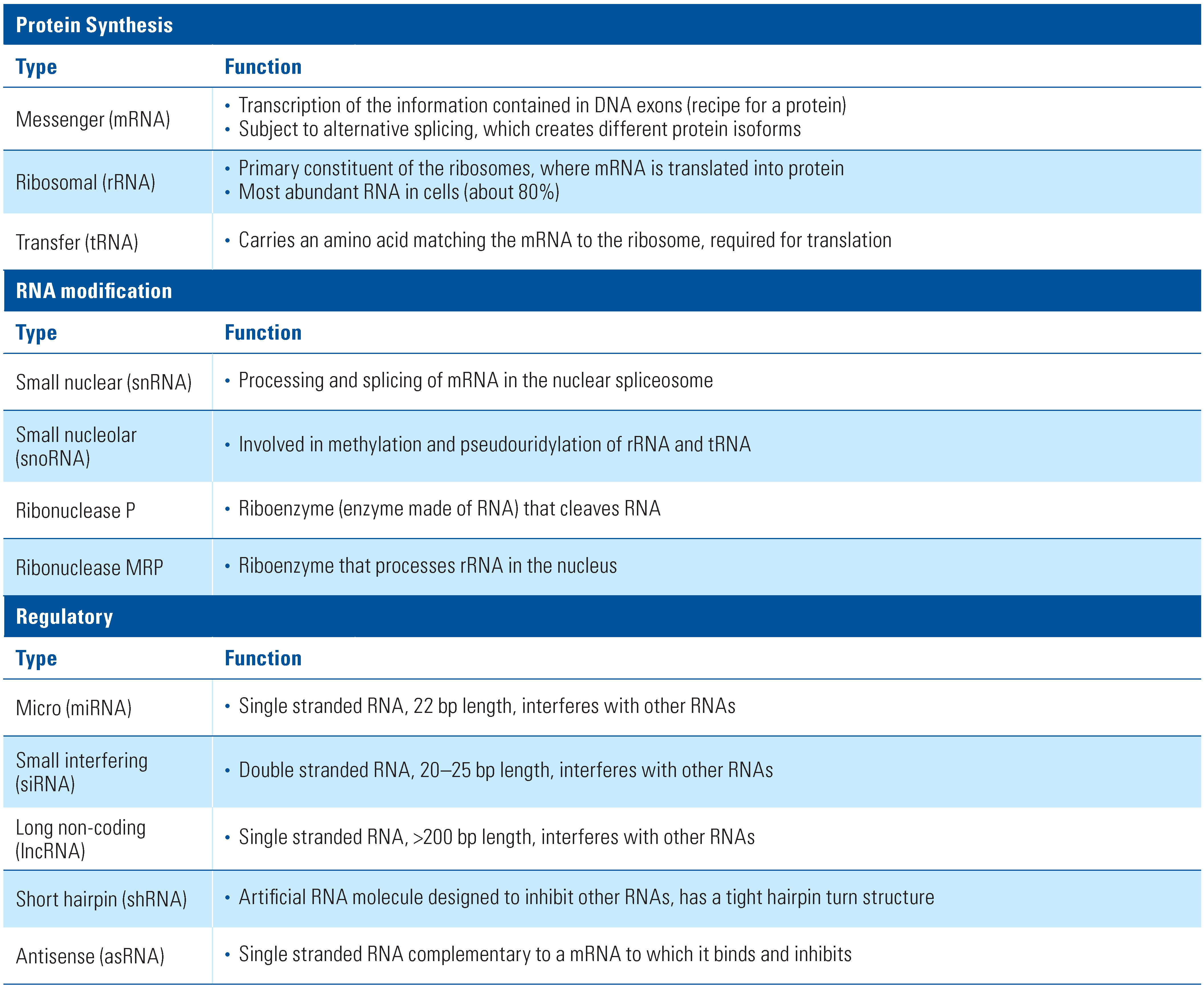
Table 1. Main Types of RNA.
Siuj 01 00008 i001