Perspective

Bridging Bench to Bedside for Brain Health: Non-Invasive Brain Stimulation for Neurodegenerative Diseases

by
Umberto Nencha
1,2,* and
Friedhelm C. Hummel
1,3,4
1
Neuro-X Institute (INX), École Polytechnique Fédérale de Lausanne (EPFL), 1202 Geneva, Switzerland
2
Geneva Memory Clinic, Department of Geriatrics and Rehabilitation, Geneva University Hospitals, 1205 Geneva, Switzerland
3
Neuro-X Institute (INX), EPFL Valais, Clinique Romande de Réadaptation, 1950 Sion, Switzerland
4
Clinical Neuroscience, Medical School, University of Geneva, 1205 Geneva, Switzerland
*
Author to whom correspondence should be addressed.
Clin. Transl. Neurosci. 2025, 9(3), 43; https://doi.org/10.3390/ctn9030043
Submission received: 14 July 2025 / Revised: 23 August 2025 / Accepted: 9 September 2025 / Published: 18 September 2025
(This article belongs to the Special Issue Brain Health)

Abstract

The prevalence and the burden of neurodegenerative diseases is projected to increase in the future, but therapeutic options remain limited, relatively invasive, and not readily accessible. In this context, non-invasive brain stimulation (NIBS) techniques, mainly transcranial magnetic stimulation (TMS) and transcranial electrical stimulation (tES), are emerging as safe and reliable instruments to enhance cognitive performance in asymptomatic individuals and patients with cognitive decline. Nevertheless, these techniques face some limitations that delay their deployment in clinical practice. Here, we describe the current status in the development of these technologies for the treatment of neurodegenerative diseases, and we present a novel promising approach for focally and non-invasively target deep brain regions. In light of these technological advances, we then propose a stepwise research roadmap to achieve an effective clinical translation of these techniques. Firstly, the constitution of open-access multimodal databases will allow to inform future interventions and design a new generation of clinical trials. Secondly, research efforts targeting symptomatic patients will need to assess the impact of NIBS techniques in different forms of dementias and probe their efficacy as disease-modifying therapies. In a future step, randomized clinical trials could focus on highly characterized at-risk populations to probe the impact of NIBS in secondary prevention. Once validated on research grounds, these techniques could enter clinical practice, enhancing cognitive performance in asymptomatic individuals and slowing disease progression in symptomatic patients, ultimately lowering the burden of neurodegenerative diseases. Eventually, NIBS techniques could be integrated into clinical practice within the framework of national Brain Health programs to provide early non-invasive interventions against cognitive decline to patients and individuals at risk.

1. Introduction

The global prevalence of persons at risk for cognitive impairment or dementia due to Alzheimer’s disease (AD) has been estimated at 416 million, and this figure is expected to increase in the following years, together with its associated burden [1].
Currently, 14 modifiable risk factors have been identified by the Lancet Commission on Dementia Prevention, accounting for approximately 45% cases of dementia [2], and interventional trials suggest that the risk of developing cognitive decline might be reduced following multi-modal non-pharmacological interventions in specific patient groups [3,4,5].
Even with the recent approval of promising pharmacological treatments to slow cognitive decline in early symptomatic patients, many open questions remain about their accessibility and safety profile in a real-world setting [6]. Indeed, these treatments present with possible adverse effects requiring additional care or monitoring, further limiting their accessibility, particularly for more frail individuals. Moreover, their use has only been investigated for symptomatic patients, and although their efficacy is being assessed in individuals in their preclinical phase of the disease, results are not conclusive or not available yet [7,8]. On the other hand, non-pharmacological interventions have the potential to be more readily accessible, personalized and often present with a more favorable safety profile, which makes them particularly suitable to target asymptomatic individuals at-risk. In this context, non-invasive brain stimulation (NIBS) techniques have been investigated for their potential use in a variety of neuropsychiatric conditions, including conditions related to cognitive decline. To date, a variety of interventions have been investigated for cognitive enhancement and rehabilitation in neurodegenerative diseases, mainly focusing on AD. Interestingly, a recent meta-analysis comprising 15,548 individuals with AD in 57 randomized clinical trials (RCTs) compared the effect of transcranial magnetic stimulation (TMS) on cognitive functions compared not only to standard pro-cognitive medications, but also to some anti-amyloid drugs and other targets [9]. This study found that TMS might provide greater benefits compared to pharmacological interventions with a better safety profile. However, some recent studies like the RCT that validated the clinical use of Lecanemab (CLARITY AD) were not included, potentially limiting the interpretation of the results.
In the rapidly evolving field of translational neuroscience, NIBS techniques have demonstrated the ability to modulate cognitive functions both in animal models and humans [10,11,12,13]. A growing body of literature on both physiological aging and neurodegenerative diseases [14,15,16] supports the hypothesis that these techniques are able to efficiently modulate and potentially enhance brain functions, and they appear to be generally safe and devoid of severe or long-lasting side effects [17,18].
The techniques that have been more widely studied are mostly based on TMS and transcranial electrical stimulation (tES), either with direct (tDCS) or alternating electrical currents (tACS). Here, we review the status of conventional NIBS techniques and the development of novel strategies in the treatment of AD and related dementias. We will mainly focus on clinically deployed magnetic and electrical stimulation in the field of neurodegenerative diseases and describe a novel, potentially transformative tES technique that allows targeting deep brain structures non-invasively. Finally, we propose a research scheme for the future implementation of these techniques in clinical practice.

2. NIBS as a Therapeutic Tool for Cognitive Rehabilitation in Dementias

Therapeutic options for AD have recently expanded to monoclonal antibodies targeting amyloid pathology [19,20,21], but their objective benefits in terms of slowing the progression of the disease are yet to be proven in real-world settings and their efficacy could be moderated by their potential side effects [22]. Moreover, after the recent approval of Lecanemab and Donanemab by the FDA and the EMA, the debate now focuses on their restriction only to certain subgroups of patients due to the increased risk of adverse events (i.e., in ApoEε4 carriers) and official recommendations for clinical practice are still evolving [23].
Hence, the need for alternative, safe, non-pharmacological options for preventing cognitive decline or slowing the clinical progression in AD and related dementias is imperative and pressing. A fast-growing body of literature supports the role of NIBS for cognitive enhancement in unimpaired older individuals, as well as cognitive rehabilitation in neurodegenerative diseases. In healthy, cognitively unimpaired older individuals, multiple studies successfully modulated and enhanced cognitive performance with NIBS (for review Tatti et al.) [24]. More specifically, recent studies demonstrated the capacity to induce long-lasting effects in episodic memory and increase working memory performance in healthy older individuals [25].
Repetitive TMS (rTMS) protocols have reached level B evidence and tES techniques have reached level C for cognitive rehabilitation in dementias [26,27,28]. Most of these studies have focused on dementia due to AD and patients with mild cognitive impairment (MCI). A variety of rTMS protocols focusing primarily on dorsolateral prefrontal cortex (dlPFC), precuneus and temporal lobe, have shown a positive impact on global cognition and episodic memory [29,30,31,32], as well as an amelioration of some behavioral and psychological symptoms in AD patients [29,30,31,32,33]. In one of these studies, rTMS targeting the precuneus demonstrated efficient slowing of cognitive decline over 6 months compared to sham as well as preserved functional independence in daily activities [33]. In a follow-up study over 12 months, the same intervention demonstrated a 52% slowing of clinical progression in the active group compared to the control for the primary outcome [34]. Even if most of these studies focused on patients with clinically significant cognitive decline, rTMS also demonstrated a transient increase in episodic memory and changes in connectivity patterns between the default-mode network (DMN) and the central executive network in individuals at the preclinical stage [35,36]. There is a consistent body of literature supporting the role of TMS in modulating neuroplasticity via long-term potentiation (LTP), neurotransmission and neuroinflammation (for review see Antonioni et al. [37]), specifically focusing on repetitive protocols for AD. Indeed, in multiple AD murine models high-frequency (HF) rTMS protocols (>5 Hz) produced significant upregulation of post-and presynaptic proteins involved in hippocampal plasticity [38,39]. HF-rTMS has also been linked to the restoration of cholinergic and dopaminergic transmission via improved signaling and upregulation of neurotropic factors [40,41]. It is also able to restore glutamatergic and GABAergic homeostasis, which are typically impaired in AD [42]. Accessory mechanisms of actions for HF-rTMS might be increased Aβ clearance, modulation of tau phosphorylation and decrease microglial-related neuroinflammatory processes, which are among the main driving factors of cognitive decline in AD [39,43]. Although more limited, some preliminary evidence also supports the role of rTMS in the enhancement of Aβ clearance and decreasing its related neurotoxicity [44]. At the network level, in humans, rTMS protocols focusing on key areas of the DMN reported some degree of improvement associated with the restoration of the network activity [33,45,46]. In AD, rTMS has also shown to restore some level of cortical plasticity and connectivity [47,48].
Promising results have also been obtained with tACS for patients with MCI. Indeed, 40 Hz stimulation over the precuneus also yielded significant memory improvements and a parallel restoration of cholinergic transmission [49,50]. Other studies using a similar protocol targeting the temporal lobe also showed encouraging results in the improvement of brain metabolism and reduction in tau pathology in a small case series of AD patients [51]. There are other tES studies, mainly with tDCS, targeting dlPFC and the temporal lobe, suggesting benefits in performance over global cognition and memory in AD (for review see Chu et al. and Majdi et al.) [31,52], overall with smaller effect sizes compared to TMS studies. The suspected mechanisms of action for tDCS and tACS are different (for review, see Wang et al. [53]). TDCS can alter membrane potentials [54], but also directly interacts with NMDA, GABA A and B as well as glutamatergic receptors [55]. This probably accounts at least in part for the after-effects observed in several tDCS studies [56], although these interactions have not been completely elucidated to date. Moreover, tDCS was also associated with a reduction in Aβ deposition in AD murine models, probably via an enhanced clearance mechanism [57]. At the network level, both in healthy humans and in patients with AD, tDCS has demonstrated to increase functional connectivity of the DMN and other brain networks [58,59,60]. TACS on the other hand, relies on a different range of mechanisms to modulate neuronal activity both locally, based on resonance and temporal biasing of spike activity, and at the network level, via the entrainment of frequency-specific neural oscillations and imposed patterns (for details see Liu et al. [61]). Apart from these effects, tACS also exerts large-scale network plasticity via LTP-like mechanisms or cross-frequency coupling that might persist after the offset of stimulation [62]. In the field of neurodegenerative diseases, recent work focused on the role of gamma band (40 Hz) frequency stimulation protocols, following pivotal animal studies [63,64,65] that first demonstrated a decrease in Aβ deposition and to a lesser extent tau accumulation in AD murine models, together with a modulation of microglial-associated activity. Indeed, a steadily growing number of human works partially support the role of gamma frequency tACS in cognitive enhancement and increased clearance of AD pathology, as previously described [34,35,36]. A simplified summary of the known mechanisms of conventional NIBS techniques is represented in Figure 1.
Besides these promising findings, these studies face some limitations that warrant some caution in the interpretation of these results and limit their generalizability. Firstly, most of these studies are monocentric, quite heterogeneous in their designs, have relatively small sample sizes and often their inclusion criteria are solely based on clinical diagnostic parameters, which probably account for a significant interindividual heterogeneity in the response to stimulation. Secondly, these studies are usually characterized by short interventions and a low number of stimulation sessions (compared to the duration of longitudinal pharmacological RCTs). Moreover, in many studies with tES and TMS, even the presence of sham-controlled conditions aimed at mimicking auditory or somato-sensory sensation of active stimulation does not guarantee the absence of a placebo effect. On another aspect, blinding efficacy, albeit sometimes reported has not been systematically assessed in the past and recent works highlight that for some techniques or certain parameters it might be suboptimal [66,67,68]. Also, these studies usually focus on behavioral endpoints to determine the success of the interventional strategy. However, they usually lack reliable and reproducible neurophysiological parameters to confirm the correct engagement of the target region. For conventional NIBS techniques, electrode or coil placement and individual anatomy play a major role in the magnitude and distribution of the induced electrical field [69,70,71]. This might also contribute to the interindividual variability in stimulation response and might prove particularly relevant in the context of repeated stimulation sessions. To optimize the placement and reduce the intersession variability, neuronavigation technologies, digital tools and practical electrode placement guidelines have been recently developed; however, these approaches are not widely used and not yet standardized [72,73,74]. Moreover, due to their short duration, very few studies have also investigated the persistence of cognitive effects over longer periods after the end of the intervention. Finally, the absence of a clear mechanistic understanding of NIBS effects and the heterogeneity of stimulation parameters and targets have contributed to inconclusive or even conflicting results for similar interventions. All the abovementioned elements have probably contributed to the limited the reproducibility of these results and the standardization of stimulation parameters.
The most promising results in terms of cognitive enhancement have been obtained when stimulating single cortical areas identified through network-based approaches in healthy aging, subjective cognitive decline and AD [25,33,35,75]. Such approaches have been limited by the predefined, non-personalized ‘‘fixed’’ nature of the target areas and the stimulation parameters, as well as the methodological limitations of conventional tES and TMS techniques. Among those, one of the most important is the inability to reach deep brain structures without co-stimulating portions of the overlying superficial cortex, due to the well-known depth-focality trade-off [76,77]. Indeed, these techniques are probably conditioned by some limitations in the context of AD and related dementias, as cognitive decline is more related to widespread network dysfunction involving multiple areas to different degrees, rather than local disruption of cortical activity. In this view, key hubs located in deeper brain regions (e.g., hippocampus or striatum), that are also involved in cognitive functions and often inaccessible to conventional NIBS techniques, might represent a promising alternative target, simultaneously influencing the activity of distant cortical areas.
In this view, a novel technique based on high (kHz range)-frequency interfering electrical fields called transcranial temporal interference electrical stimulation (tTIS) has been recently developed to stimulate deep brain regions with high focality, without affecting the overlying cortical structures, both in rodents [78] and in humans [79,80,81,82]. The suggested mechanism of tTIS is that neurons are not able to respond to high-frequency electrical fields (called “carriers”) due to an intrinsic low-pass filtering property of the neuronal membrane [78,83] but are able to extract the low frequency component of the so called “envelope”. Moreover, frequency and field orientation seem to differentially affect cell subtypes, possibly influencing cortical functions through more complex, circuit-based modulations than previously thought [84]. Until present, tTIS has been successfully employed in humans for the enhancement of episodic and spatial memory [80,82], motor learning [79], disrupting oscillations [81], and to reduce motor symptoms in Parkinson’s disease [85] (for an overview, see Hummel & Wessel 2024 [86]). Multiple trials probing the efficacy of this technique in different clinical populations and subcortical targets are currently ongoing, like the ones led by our group focusing on striatal and hippocampal tTIS in MCI and traumatic brain injury (TBI) patients or in Parkinson’s patients [85,87,88]. Moreover, several multi-center randomized clinical trials have recently started, focusing on different neurological and psychiatric diseases [89,90,91,92,93].
Compared to other conventional NIBS techniques, tTIS offers the promise of targeting different subcortical regions with better focality and minimal or no adjustments to the stimulation setup and without the need for multiple electrode pairs, thus allowing for more complex protocols with a relatively simple montage. As subcortical structures are involved in the pathophysiology of multiple neuropsychiatric disorders, the possibility to non-invasively modulate their activity offers the advantage to influence different symptoms such as apathy, mood alterations, or cognitive decline, following a transdiagnostic approach.
Another aspect that differentiates conventional NIBS techniques and tTIS is that its capacity to exert a neuromodulatory effect has only been tested during the completion of motor or cognitive tasks [80,81,82,85], while tES and TMS have demonstrated longer-lasting effects even days to weeks after the end of stimulation. Concerning its overall applicability, tTIS presents a comparable profile compared to conventional NIBS techniques. Although quite similar to conventional tACS in its setup, some of the aspects that might need to be further developed for tTIS would be its portability, given the current hardware limitations requiring two relatively large stimulators to deliver kHZ-range electrical currents [94]. Moreover, while computational and modeling tools will play a crucial role in the development of this technology, these approaches are not widely implemented yet, limiting the available options for personalized interventions [95]. Concerning safety, to date tTIS showed a similar safety profile compared to conventional tACS and good blinding properties across different clinical populations and anatomical targets, as recently demonstrated in different studies (195 participants, 345 sessions in total) [85,96,97,98]. Finally, another advantage of tTIS worth mentioning, compared to conventional tES techniques, is the reduced scalp sensation or irritation that has been reported with kHz range currents [97].
Although promising, due to its novelty, tTIS deployment in clinical studies faces several limitations and challenges that are currently under active investigation. Specifically, we note that the optimal stimulation parameters—such as the most effective carrier frequencies (currently applied between 1 and 9 kHz [99,100]) and amplitudes (current literature between 2 and 15 mA peak to baseline [79,82,101])—remain to be determined. For instance, development in individual field modeling might in future homogenize tTIS that comparable field strengths are achieved in the deep target areas by personalization of the stimulation amplitudes at the electrodes level. Thus, personalization will likely be one of the most critical approaches for maximizing efficacy, including the optimization of focality (multichannel tTIS with an increased number of electrodes), individualized electrode placement taking the individual anatomy into account, especially important in patients with brain damage or neurodegeneration, and consideration of potential off-target effects. Although carrier frequencies per se are unlikely to exert strong physiological effects at subthreshold intensities, their independent impact has not yet been fully ruled out [84,86,102]. These uncertainties, together with interindividual variability, may contribute to the heterogeneity of responses observed so far. Finally, we emphasize that no home-based or self-application protocols for tTIS are currently available, which limits broader clinical translation at this stage. These aspects will need to be systematically investigated in future studies with the help of multi-modal imaging and neurophysiological techniques.

3. Next Steps for Clinical Implementation of NIBS in the Treatment and Prevention of Dementia

These technological advances are accelerating and profoundly changing the landscape of translational neuroscience in the field of dementia. NIBS techniques are emerging as promising, transformative tools for cognitive enhancement, as well as for the treatment of a vast array of different neuropsychiatric conditions. Moreover, the emergence of novel techniques such as tTIS will create the opportunity to further advance our knowledge about the precise involvement of subcortical structures (e.g., hippocampus, striatum) in different neurodegenerative diseases and their specific contributions to their pathophysiology. In a first phase, such interventions will need to establish clear causal links about the involvement of specific brain networks in different neurodegenerative disorders and consequently shape future interventions. Moreover, the differential impact of stimulation in different conditions could inform about the pathophysiological mechanisms of single disorders.
By identifying core areas involved in different neurodegenerative diseases, future interventions will leverage this knowledge to precisely influence network alterations underlying specific neuropsychiatric symptoms (e.g., apathy, mood alterations, impulsivity, specific cognitive deficits, etc.). Moreover, as these symptoms are quite prevalent and traditionally treated not only by neurologists but also by psychiatrists, NIBS interventions could also have an impact on related conditions with overlapping clinical presentations. Hence, the implementation of NIBS on academic grounds through this network-based transdiagnostic approach could promote a more holistic, interdisciplinary collaboration between the fields of neurology and psychiatry, helping to create virtuous synergies aimed at fostering better patient care across both disciplines.
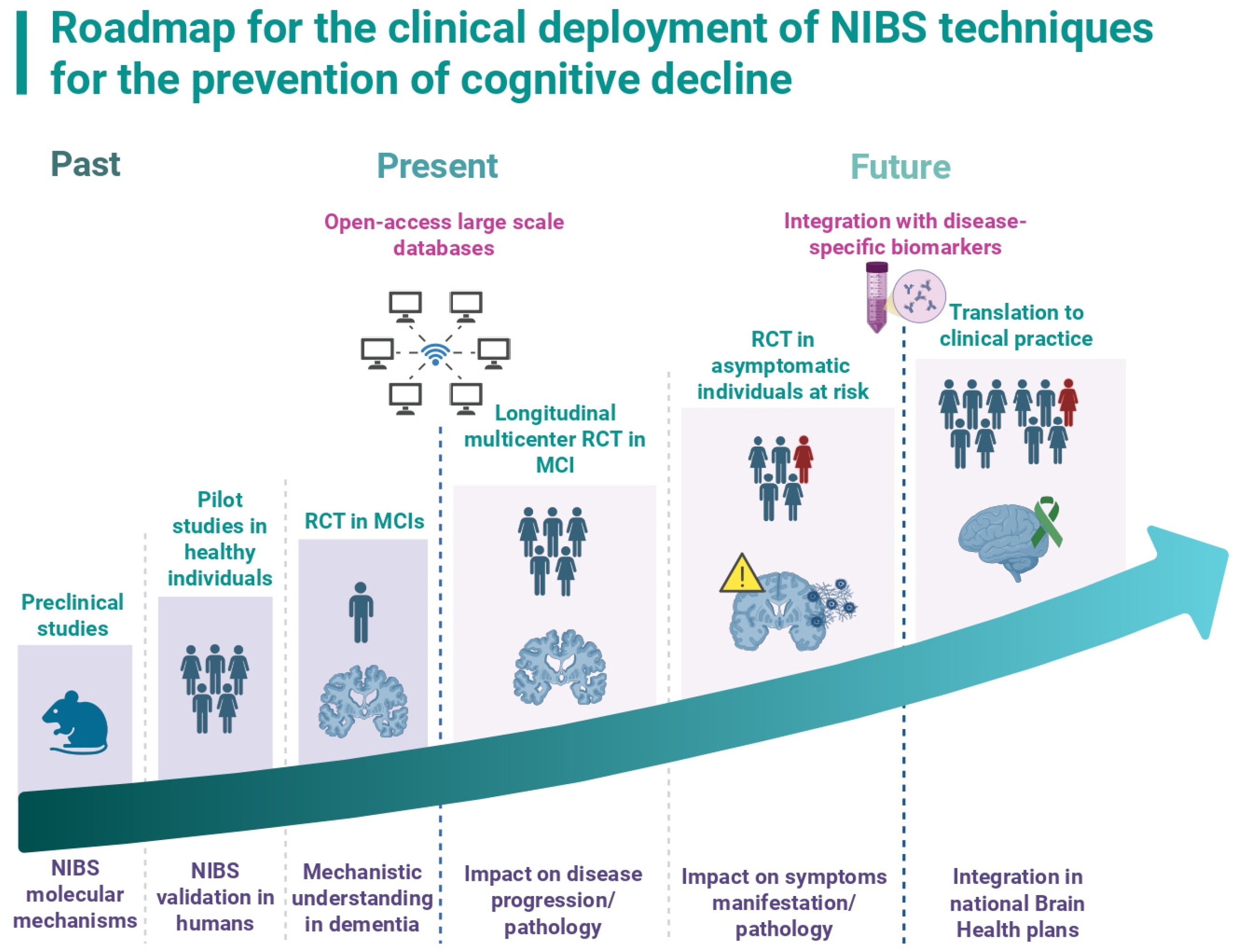
However, due to the intrinsic limitations of human studies that we described, many research questions remain to be answered to ensure the implementation of these techniques in clinical settings. Here, we propose a structured research roadmap to further develop these techniques and to achieve their future translation in clinical practice for neurodegenerative conditions (see Figure 2).

3.1. Building Large, Multicenter, Population-Based Longitudinal Neurophysiological Databases

To better understand and probe the impact of NIBS techniques on neurodegenerative diseases, there is an urgent need to build large, online, open-access international databases, collecting cross-sectional and longitudinal data, ideally from individuals across the entire spectrum of cognitive decline, including the preclinical stage of the disease. One very recent example of this type of platform is Big NIBS data, a free, open-access platform storing data from non-invasive brain stimulation interventions, which also acts as a public repository [103]. This is very similar to other well-established initiatives like the Alzheimer’s Disease Neuroimaging Initiative (ADNI) [104] or the more recent AMPAD-PNHS [105], which have mainly focused on longitudinal collection of imaging data along the course of the AD continuum. Critically, in the field of NIBS, there is a need for more sophisticated, multimodal tools, able to capture a wide variety of interventions, biological and neurophysiological parameters. In the future, this data should be collected following standardized international guidelines across multiple centers. Once available, this data could be used to inform and train machine-learning models or artificial intelligence tools to better predict the individual response to stimulation and allow for further personalization of future interventions.

3.2. Understanding the Differential Impact of NIBS on Specific Types of Dementias and Probing Their Values as Possible Disease-Modifying Therapies

Until present, most NIBS studies for cognitive rehabilitation in AD and related dementias have included only symptomatic patients and have relied mostly on clinical diagnostic criteria for their inclusion. Importantly, the vast majority of the studies only focused on AD, whereas much less data is available concerning related neurodegenerative disorders [106]. Moreover, these trials most often used a single stimulation modality and targeted a single or multiple superficial brain regions for a limited amount of time.
To accelerate the translational process of these techniques, future studies will need to systematically address their impact in different neurodegenerative diseases and probe their effects based on the network dysfunction underlying their specific pathophysiology. In this context, a promising avenue in the conceptualization of future stimulation studies by modulating network dysfunctions will consist in targeting different areas at the same time and/or through different stimulation modalities.
For instance, combining different techniques (e.g., TMS and tACS) to plan complementary interventions could theoretically already be implemented in research and clinical settings. TMS-based protocols allow for efficient modulation of the activity of cortical areas in a limited amount of time (usually in the tens of minutes range). The possibility to induce neural activity at a suprathreshold level or produce longer-lasting neural plasticity with specific protocols (e.g., intermittent theta burst, iTBS) makes this technique ideal to start a treatment, similarly to an “induction” phase for some pharmacological treatments. Later, the treatment could continue in a home-based setting, using a tES device for longer stimulation protocols during multiple days, either targeting the same areas or different areas of the same network. This would represent a “maintenance” phase, that the patient could couple to some cognitive training or a complementary intervention.
A second approach would be to design more complex circuit-based stimulation paradigms focusing on multiple hubs of the same network. In the future, also due to the development of novel techniques such as tTIS, another possibility would be to implement multi-channel stimulation protocols, using supplementary electrode pairs, in order to enhance the focality of the electric field and/or to modulate distant subcortical regions in different brain networks at the same time.
Combining different NIBS modalities and targeting different cortical and subcortical areas at the same time will allow, in a first phase, for studying the differential impact of stimulation on different pathologies that share overlapping clinical phenotypes but distinct pathophysiological mechanisms. Subsequently, these studies would inform about the mechanistic underpinnings of the stimulation across different disorders to help shape and personalize future interventions.
To further advance the clinical translation of NIBS techniques, another important aspect that needs consideration is their integration within the framework of pathology-specific biomarkers. As neurodegenerative diseases might present with highly overlapping clinical phenotypes, a great effort has been made to isolate reliable biomarkers (e.g., based on CSF, plasma or molecular imaging) of their underlying molecular pathology. A future step of stimulation studies will be to better identify patients who will benefit from this type of intervention through the implementation of these biomarkers. This is a crucial step that is already being integrated into current clinical studies and that will pave the way to better understand the impact of different NIBS interventions for specific diseases. Moreover, stratifying patients by using this integrated clinico-biological framework will be necessary to assess the value of different NIBS techniques as disease-modifying therapies in symptomatic individuals, with the ultimate aim of slowing cognitive decline and impacting the underlying molecular pathology.
Once a clear link between NIBS interventions and slowing cognitive decline or a positive impact on pathology-specific biomarkers is established in symptomatic patients, it will be crucial to explore whether there is an interest in implementing these techniques as a preventive strategy during the preclinical phase in asymptomatic individuals at risk of cognitive decline.

3.3. Validate the Use of NIBS for the Prevention of Cognitive Decline in At-Risk Populations

The integration of appropriate NIBS interventions with diagnostic or prognostic biomarkers has the potential to provide an invaluable tool to treat individuals based on their risk of cognitive decline, ultimately lowering the global burden of neurodegenerative diseases. For example, plasma p-tau217 has been actively investigated for its potential to predict amyloid status and cognitive decline, even in asymptomatic individuals, with relatively high performance, either in isolation or associated with other plasma biomarkers or tau-PET [107,108]. Moreover, the implementation of a blood-based biomarker diagnostic workflow has been proposed to identify individuals at-risk of cognitive decline and refer them for further confirmatory testing with tau-PET [109]. Once the diagnosis confirmed, future preventive strategies might benefit from a further stratification, to assess more precisely the individual’s response from a disease-modifying intervention. For instance, in the TRAILBLAZER trial that investigated the effect of Donanemab on AD progression, participants were stratified using tau-PET, to stage disease severity based on tau brain load [19]. Hence, a similar framework using disease-specific diagnostic and prognostic biomarkers might be implemented to assess: (i) risk of progression in asymptomatic individuals, (ii) probe their benefit from specific NIBS interventions, (iii) optimize the time for starting a treatment and (iv) follow-up the impact of these protocols on the underlying pathology. While in early symptomatic patients, interventions will need to focus on slowing the clinical progression, for individuals at risk of developing cognitive decline, preventive measures will aim to enhance cognitive performance and/or directly impact the underlying pathology. In this view, there is a need for longitudinal RCTs to probe the impact of NIBS techniques on the biological mechanisms of different dementias and to assess their impact on clinical progression. In asymptomatic individuals specifically, longitudinal RCTs with biomarker-informed preventive interventions will need to assess the benefits of neuroenhancement on the risk of progression towards objective cognitive decline. To account for the possible lack of adherence of asymptomatic individuals to a study protocol lasting many weeks to months, home-based interventions could be proposed, with the possibility to track their effect even from remote settings. Due to the digital nature of these techniques, adherence and side effects could also be remotely monitored and stimulation parameters corrected, when necessary, without the need for recurrent hospital visits.
Moreover, as future preventive strategies in asymptomatic individuals will likely integrate multidomain lifestyle interventions (e.g., physical exercise, cognitive training, dietary changes, etc.), NIBS techniques have the potential to nicely fit into this therapeutic framework even in home-based settings. In a first phase, these interventions could be implemented in a small number of participants, in the format of a pilot study, to evaluate their feasibility and further confirm the safety of these techniques in longer-lasting protocols. Subsequently, the standardization of stimulation parameters and the duration of the intervention will need to be replicated across different cohorts in different sites, within longitudinal randomized clinical trials. These studies will also systematically explore the disease-modifying potential of NIBS techniques and validate their use in the prevention of cognitive decline. Finally, when validated, these protocols could enter clinical practice and be integrated into national Brain Health programs with specific indications and guidelines.

4. Conclusions

With the forecast of the increasing socioeconomic burden of dementia in future years, NIBS techniques offer the promise of becoming a safe and non-invasive tool for slowing the progression of cognitive decline and potentially delaying symptom manifestation in people at risk by enhancing their cognitive performance. We are experiencing a rapid acceleration of neurotechnological advances that allow for unprecedented precision and flexibility in the field of non-invasive brain stimulation, with a clear possibility for clinical translation. However, many challenges remain before clinical deployment of these techniques. Among those, we acknowledge the establishment of a clear mechanistic understanding of these techniques in different dementias, the prediction of individual treatment response and the assessment of long-term efficacy in both patients and at-risk individuals, which have not yet been systematically explored. Ongoing studies are focusing on circuit-based interventions, longer interventions and starting to explore the impact of NIBS on the underlying molecular pathology. This will allow for evaluation of the potential of NIBS as disease-modifying therapies across different neurodegenerative diseases, once integrated in the framework of existing disease-specific biomarkers. Once a clear mechanistic understanding of the effect of stimulation on the disease course is established, longitudinal interventions will need to validate the clinical use of these techniques and their indications, firstly in symptomatic patients and subsequently in individuals at risk of cognitive decline. To complete this roadmap, an international and interdisciplinary effort between physicians, researchers, but also political stakeholders and patients’ societies is needed. There is an urgent need to standardize interventions, firstly in the academic settings, to translate them into safe and cost-effective clinical practices. The final step will be the systematic implementation of these protocols into national Brain Health programs via specifically trained healthcare professionals, to prevent or delay symptoms onset in at-risk individuals and slow cognitive decline in patients during the early phase of the disease. In the long term, this will help lower the burden of neurodegenerative diseases and offer a safe, non-invasive therapeutic option to support Brain Health and prevent cognitive decline in the context of an aging global population.

Author Contributions

U.N. and F.C.H. conceptualized and wrote the original draft of the manuscript, F.C.H. supervised the writing of the draft. All authors have read and agreed to the published version of the manuscript.

Funding

Swiss National Science Foundation (SNSF, 320030L_197899, NiBS-iCog) to F.C.H.; the Defitech Foundation (Morges, CH) to F.C.H. and the Social and hUman ceNtered XR (SUN) project that has received funding from the Horizon Europe Research & Innovation Programme under grant agreement N. 101092612 to F.C.H.

Institutional Review Board Statement

Not applicable.

Informed Consent Statement

Not applicable.

Data Availability Statement

Data is contained within the article.

Conflicts of Interest

The authors declare no conflicts of interest.

References

  1. Gustavsson, A.; Norton, N.; Fast, T.; Frölich, L.; Georges, J.; Holzapfel, D.; Kirabali, T.; Krolak-Salmon, P.; Rossini, P.M.; Ferretti, M.T.; et al. Global estimates on the number of persons across the Alzheimer’s disease continuum. Alzheimer’s Dement. 2023, 19, 658–670. [Google Scholar] [CrossRef]
  2. Frisoni, G.B.; Altomare, D.; Ribaldi, F.; Villain, N.; Brayne, C.; Mukadam, N.; Abramowicz, M.; Barkhof, F.; Berthier, M.; Bieler-Aeschlimann, M.; et al. Dementia prevention in memory clinics: Recommendations from the European task force for brain health services. Lancet Reg. Health—Eur. 2023, 26, 100576. [Google Scholar] [CrossRef]
  3. Ngandu, T.; Lehtisalo, J.; Solomon, A.; Levälahti, E.; Ahtiluoto, S.; Antikainen, R.; Bäckman, L.; Hänninen, T.; Jula, A.; Laatikainen, T.; et al. A 2 year multidomain intervention of diet, exercise, cognitive training, and vascular risk monitoring versus control to prevent cognitive decline in at-risk elderly people (FINGER): A randomised controlled trial. Lancet 2015, 385, 2255–2263. [Google Scholar] [CrossRef]
  4. Delrieu, J.; Payoux, P.; Carrié, I.; Cantet, C.; Weiner, M.; Vellas, B.; Andrieu, S. Multidomain intervention and/or omega-3 in nondemented elderly subjects according to amyloid status. Alzheimer’s Dement. 2019, 15, 1392–1401. [Google Scholar] [CrossRef]
  5. Chhetri, J.K.; De Souto Barreto, P.; Cantet, C.; Pothier, K.; Cesari, M.; Andrieu, S.; Coley, N.; Vellas, B. Effects of a 3-Year Multi-Domain Intervention with or without Omega-3 Supplementation on Cognitive Functions in Older Subjects with Increased CAIDE Dementia Scores. J. Alzheimer’s Dis. 2018, 64, 71–78. [Google Scholar] [CrossRef] [PubMed]
  6. Liu, K.Y.; Villain, N.; Ayton, S.; Ackley, S.F.; Planche, V.; Howard, R.; Thambisetty, M. Key questions for the evaluation of anti-amyloid immunotherapies for Alzheimer’s disease. Brain Commun. 2023, 5, fcad175. [Google Scholar] [CrossRef] [PubMed]
  7. Sperling, R.A.; Donohue, M.C.; Raman, R.; Rafii, M.S.; Johnson, K.; Masters, C.L.; Van Dyck, C.; Iwatsubo, T.; Marshall, G.A.; Yaari, R.; et al. Trial of Solanezumab in Preclinical Alzheimer’s Disease. N. Engl. J. Med. 2023, 389, 1096–1107. [Google Scholar] [CrossRef] [PubMed]
  8. AHEAD 3-45 Study: A Phase 3 Study Evaluating the Efficacy and Safety of Aducanumab in Preclinical Alzheimer’s Disease. ClinicalTrials.gov, Registration No. NCT04465898. 2023. Available online: https://clinicaltrials.gov/ct2/show/NCT04465898 (accessed on 22 August 2025).
  9. Wei, N.; Liu, H.; Ye, W.; Xu, S.; Lu, C.; Dai, A.; Hou, T.; Zeng, X.; Wu, J.; Chen, J. Repetitive transcranial magnetic stimulation may be superior to drug therapy in the treatment of Alzheimer’s disease: A systematic review and Bayesian network meta-analysis. CNS Neurosci. Ther. 2023, 29, 2912–2924. [Google Scholar] [CrossRef]
  10. Draaisma, L.R.; Wessel, M.J.; Hummel, F.C. Non-invasive brain stimulation to enhance cognitive rehabilitation after stroke. Neurosci. Lett. 2020, 719, 133678. [Google Scholar] [CrossRef]
  11. Lanni, I.; Chiacchierini, G.; Papagno, C.; Santangelo, V.; Campolongo, P. Treating Alzheimer’s disease with brain stimulation: From preclinical models to non-invasive stimulation in humans. Neurosci. Biobehav. Rev. 2024, 165, 105831. [Google Scholar] [CrossRef]
  12. Antal, A.; Luber, B.; Brem, A.K.; Bikson, M.; Brunoni, A.R.; Cohen Kadosh, R.; Dubljević, V.; Fecteau, S.; Ferreri, F.; Flöel, A.; et al. Non-invasive brain stimulation and neuroenhancement. Clin. Neurophysiol. Pract. 2022, 7, 146–165. [Google Scholar] [CrossRef]
  13. Liu, H.; Wu, M.; Huang, H.; Chen, X.; Zeng, P.; Xu, Y. Comparative efficacy of non-invasive brain stimulation on cognition function in patients with mild cognitive impairment: A systematic review and network meta-analysis. Ageing Res. Rev. 2024, 101, 102508. [Google Scholar] [CrossRef]
  14. Cespón, J.; Miniussi, C.; Pellicciari, M.C. Interventional programmes to improve cognition during healthy and pathological ageing: Cortical modulations and evidence for brain plasticity. Ageing Res. Rev. 2018, 43, 81–98. [Google Scholar] [CrossRef]
  15. Nissim, N.R.; Pham, D.V.H.; Poddar, T.; Blutt, E.; Hamilton, R.H. The impact of gamma transcranial alternating current stimulation (tACS) on cognitive and memory processes in patients with mild cognitive impairment or Alzheimer’s disease: A literature review. Brain Stimul. 2023, 16, 748–755. [Google Scholar] [CrossRef] [PubMed]
  16. Zimerman, M.; Nitsch, M.; Giraux, P.; Gerloff, C.; Cohen, L.G.; Hummel, F.C. Neuroenhancement of the aging brain: Restoring skill acquisition in old subjects. Ann. Neurol. 2013, 73, 10–15. [Google Scholar] [CrossRef] [PubMed]
  17. Rossi, S.; Antal, A.; Bestmann, S.; Bikson, M.; Brewer, C.; Brockmöller, J.; Carpenter, L.L.; Cincotta, M.; Chen, R.; Daskalakis, J.D.; et al. Safety and recommendations for TMS use in healthy subjects and patient populations, with updates on training, ethical and regulatory issues: Expert Guidelines. Clin. Neurophysiol. 2021, 132, 269–306. [Google Scholar] [CrossRef]
  18. Antal, A.; Alekseichuk, I.; Bikson, M.; Brockmöller, J.; Brunoni, A.R.; Chen, R.; Cohen, L.G.; Dowthwaite, G.; Ellrich, J.; Flöel, A.; et al. Low intensity transcranial electric stimulation: Safety, ethical, legal regulatory and application guidelines. Clin. Neurophysiol. 2017, 128, 1774–1809. [Google Scholar] [CrossRef] [PubMed]
  19. Sims, J.R.; Zimmer, J.A.; Evans, C.D.; Lu, M.; Ardayfio, P.; Sparks, J.D.; Wessels, A.M.; Shcherbinin, S.; Wang, H.; Serap Mokul nery, E.; et al. Donanemab in Early Symptomatic Alzheimer Disease: The TRAILBLAZER-ALZ 2 Randomized Clinical Trial. J. Am. Med. Assoc. 2023, 330, 512–527. [Google Scholar] [CrossRef]
  20. Budd Haeberlein, S.; Aisen, P.S.; Barkhof, F.; Chalkias, S.; Chen, T.; Cohen, S.; Dent, G.; Hansson, O.; Harrison, K.; Von Hehn, C.; et al. Two Randomized Phase 3 Studies of Aducanumab in Early Alzheimer’s Disease. J. Prev. Alzheimer’s Dis. 2022, 9, 197–210. [Google Scholar] [CrossRef]
  21. Van Dyck, C.H.; Swanson, C.J.; Aisen, P.; Bateman, R.J.; Chen, C.; Gee, M.; Kanekiyo, M.; Li, D.; Reyderman, L.; Cohen, S.; et al. Lecanemab Early Alzheimer’s Disease. N. Engl. J. Med. 2023, 388, 9–21. [Google Scholar] [CrossRef]
  22. Ross, E.L.; Weinberg, M.S.; Arnold, S.E. Cost-effectiveness of Aducanumab and Donanemab for Early Alzheimer Disease in the US. JAMA Neurol. 2022, 79, 478–487. [Google Scholar] [CrossRef] [PubMed]
  23. Cummings, J.; Apostolova, L.; Rabinovici, G.D.; Atri, A.; Aisen, P.; Greenberg, S.; Hendrix, S.; Selkoe, D.; Weiner, M.; Petersen, R.C.; et al. Lecanemab: Appropriate Use Recommendations. J. Prev. Alzheimer’s Dis. 2023, 10, 362–377. [Google Scholar] [CrossRef]
  24. Tatti, E.; Rossi, S.; Innocenti, I.; Rossi, A.; Santarnecchi, E. Non-invasive brain stimulation of the aging brain: State of the art and future perspectives. Ageing Res. Rev. 2016, 29, 66–89. [Google Scholar] [CrossRef]
  25. Reinhart, R.M.G.; Nguyen, J.A. Working memory revived in older adults by synchronizing rhythmic brain circuits. Nat. Neurosci. 2019, 22, 820–827. [Google Scholar] [CrossRef]
  26. Menardi, A.; Dotti, L.; Ambrosini, E.; Vallesi, A. Transcranial magnetic stimulation treatment in Alzheimer’s disease: A meta-analysis of its efficacy as a function of protocol characteristics and degree of personalization. J. Neurol. 2022, 269, 5283–5301. [Google Scholar] [CrossRef] [PubMed]
  27. Šimko, P.; Kent, J.A.; Rektorova, I. Is non-invasive brain stimulation effective for cognitive enhancement in Alzheimer’s disease? An updated meta-analysis. Clin. Neurophysiol. 2022, 144, 23–40. [Google Scholar] [CrossRef]
  28. Gu, L.; Xu, H.; Qian, F. Effects of Non-Invasive Brain Stimulation on Alzheimer’s Disease. J. Prev. Alzheimer’s Dis. 2022, 9, 410–424. [Google Scholar] [CrossRef]
  29. Vacas, S.M.; Stella, F.; Loureiro, J.C.; Simões do Couto, F.; Oliveira-Maia, A.J.; Forlenza, O.V. Noninvasive brain stimulation for behavioural and psychological symptoms of dementia: A systematic review and meta-analysis. Int. J. Geriatr. Psychiatry 2019, 34, 1336–1345. [Google Scholar] [CrossRef]
  30. Wang, X.; Mao, Z.; Yu, X. The role of noninvasive brain stimulation for behavioral and psychological symptoms of dementia: A systematic review and meta-analysis. Neurol. Sci. 2020, 41, 1063–1074. [Google Scholar] [CrossRef]
  31. Chu, C.S.; Li, C.T.; Brunoni, A.R.; Yang, F.C.; Tseng, P.T.; Tu, Y.K.; Stubbs, B.; Carvalho, A.F.; Thompson, T.; Rajji, T.K.; et al. Cognitive effects and acceptability of non-invasive brain stimulation on Alzheimer’s disease and mild cognitive impairment: A component network meta-analysis. J. Neurol. Neurosurg. Psychiatry 2021, 92, 195–203. [Google Scholar] [CrossRef] [PubMed]
  32. Teselink, J.; Bawa, K.K.; Koo, G.K.; Sankhe, K.; Liu, C.S.; Rapoport, M.; Oh, P.; Marzolini, S.; Gallagher, D.; Swardfager, W.; et al. Efficacy of non-invasive brain stimulation on global cognition and neuropsychiatric symptoms in Alzheimer’s disease and mild cognitive impairment: A meta-analysis and systematic review. Ageing Res. Rev. 2021, 72, 101499. [Google Scholar] [CrossRef] [PubMed]
  33. Koch, G.; Bonnì, S.; Pellicciari, M.C.; Casula, E.P.; Mancini, M.; Esposito, R.; Ponzo, V.; Picazio, S.; Di Lorenzo, F.; Serra, L.; et al. Transcranial magnetic stimulation of the precuneus enhances memory and neural activity in prodromal Alzheimer’s disease. Neuroimage 2018, 169, 302–311. [Google Scholar] [CrossRef]
  34. Koch, G.; Casula, E.P.; Bonnì, S.; Borghi, I.; Assogna, M.; Di Lorenzo, F.; Esposito, R.; Maiella, M.; D’Acunto, A.; Ferraresi, M.; et al. Effects of 52 weeks of precuneus rTMS in Alzheimer’s disease patients: A randomized trial. Alzheimer’s Res. Ther. 2025, 17, 69. [Google Scholar] [CrossRef] [PubMed]
  35. Chen, J.; Ma, N.; Hu, G.; Nousayhah, A.; Xue, C.; Qi, W.; Xu, W.; Chen, S.; Rao, J.; Liu, W.; et al. rTMS modulates precuneus-hippocampal subregion circuit in patients with subjective cognitive decline. Aging 2020, 13, 1314–1331. [Google Scholar] [CrossRef]
  36. Liang, X.; Xue, C.; Zheng, D.; Yuan, Q.; Qi, W.; Ruan, Y.; Chen, S.; Song, Y.; Wu, H.; Lu, X.; et al. Repetitive transcranial magnetic stimulation regulates effective connectivity patterns of brain networks in the spectrum of preclinical Alzheimer’s disease. Front. Aging Neurosci. 2024, 16, 1343926. [Google Scholar] [CrossRef]
  37. Antonioni, A.; Martorana, A.; Santarnecchi, E.; Hampel, H.; Koch, G. The neurobiological foundation of effective repetitive transcranial magnetic brain stimulation in Alzheimer’s disease. Alzheimer’s Dement. 2025, 21, e70337. [Google Scholar] [CrossRef]
  38. Ma, J.; Wang, J.; Lv, C.; Pang, J.; Han, B.; Wang, M.; Gen, Y. The role of hippocampal structural synaptic plasticity in repetitive transcranial magnetic stimulation to improve cognitive function in male SAMP8 mice. Cell. Physiol. Biochem. 2017, 41, 137–144. [Google Scholar] [CrossRef]
  39. Cao, H.; Zuo, C.; Gu, Z.; Huang, Y.; Yang, Y.; Zhu, L.; Jiang, Y.; Wang, F. High frequency repetitive transcranial magnetic stimulation alleviates cognitive deficits in 3xTg-AD mice by modulating the PI3K/Akt/GLT-1 Axis. Redox Biol. 2022, 54, 102354. [Google Scholar] [CrossRef]
  40. Windy McNerney, M.; Heath, A.; Narayanan, S.K.; Yesavage, J. Repetitive Transcranial Magnetic Stimulation Improves Brain-Derived Neurotrophic Factor and Cholinergic Signaling in the 3xTgAD Mouse Model of Alzheimer’s Disease. J. Alzheimer’s Dis. 2022, 86, 499–507. [Google Scholar] [CrossRef]
  41. Choung, J.S.; Kim, J.M.; Ko, M.H.; Cho, D.S.; Kim, M.Y. Therapeutic efficacy of repetitive transcranial magnetic stimulation in an animal model of Alzheimer’s disease. Sci. Rep. 2021, 11, 437. [Google Scholar] [CrossRef] [PubMed]
  42. Wang, J.; Zhou, C.; Huang, Z.; Ji, X.; Cui, R.; Kang, Y.; Zhang, G.; Wang, Y.; Zhang, T. Repetitive Transcranial Magnetic Stimulation-Mediated Neuroprotection in the 5xFAD Mouse Model of Alzheimer’s Disease Through GABRG2 and SNAP25 Modulation. Mol. Neurobiol. 2025, 62, 1971–1997. [Google Scholar] [CrossRef]
  43. Chen, X.; Chen, S.; Liang, W.; Ba, F. Administration of repetitive transcranial magnetic stimulation attenuates A β 1-42 -induced Alzheimer’s disease in mice by activating β-catenin signaling. Biomed Res. Int. 2019, 2019, 1431760. [Google Scholar] [CrossRef] [PubMed]
  44. Tao, Y.; Lei, B.; Zhu, Y.; Fang, X.; Liao, L.; Chen, D.; Gao, C. Repetitive Transcranial Magnetic Stimulation Decreases Serum Amyloid-β and Increases Ectodomain of p75 Neurotrophin Receptor in Patients with Alzheimer’s Disease. J. Integr. Neurosci. 2022, 21, 140. [Google Scholar] [CrossRef]
  45. Wang, J.X.; Rogers, L.M.; Gross, E.Z.; Ryals, A.J.; Dokucu, M.E.; Brandstatt, K.L.; Hermiller, M.S.; Voss, J.L. Memory Enhancement: Targeted enhancement of cortical-hippocampal brain networks and associative memory. Science 2014, 345, 1054–1057. [Google Scholar] [CrossRef]
  46. Vidal-Piñeiro, D.; Martín-Trias, P.; Falcón, C.; Bargalló, N.; Clemente, I.C.; Valls-Solé, J.; Junqué, C.; Pascual-Leone, A.; Bartres-Faz, D. Neurochemical modulation in posteromedial default-mode network cortex induced by transcranial magnetic stimulation. Brain Stimul. 2015, 8, 937–944. [Google Scholar] [CrossRef]
  47. Li, X.; Qi, G.; Yu, C.; Lian, G.; Zheng, H.; Wu, S.; Yuan, T.F.; Zhou, D. Cortical plasticity is correlated with cognitive improvement in Alzheimer’s disease patients after rTMS treatment. Brain Stimul. 2021, 14, 503–510. [Google Scholar] [CrossRef] [PubMed]
  48. Dayan, E.; Censor, N.; Buch, E.R.; Sandrini, M.; Cohen, L.G. Noninvasive brain stimulation: From physiology to network dynamics and back. Nat. Neurosci. 2013, 16, 838–844. [Google Scholar] [CrossRef] [PubMed]
  49. Benussi, A.; Cantoni, V.; Cotelli, M.S.; Cotelli, M.; Brattini, C.; Datta, A.; Thomas, C.; Santarnecchi, E.; Pascual-Leone, A.; Borroni, B. Exposure to gamma tACS in Alzheimer’s disease: A randomized, double-blind, sham-controlled, crossover, pilot study. Brain Stimul. 2021, 14, 531–540. [Google Scholar] [CrossRef]
  50. Benussi, A.; Cantoni, V.; Grassi, M.; Brechet, L.; Michel, C.M.; Datta, A.; Thomas, C.; Gazzina, S.; Cotelli, M.S.; Bianchi, M.; et al. Increasing Brain Gamma Activity Improves Episodic Memory and Restores Cholinergic Dysfunction in Alzheimer’s Disease. Ann. Neurol. 2022, 92, 322–334. [Google Scholar] [CrossRef]
  51. Dhaynaut, M.; Sprugnoli, G.; Cappon, D.; Macone, J.; Sanchez, J.S.; Normandin, M.D.; Guehl, N.J.; Koch, G.; Paciorek, R.; Connor, A.; et al. Impact of 40 Hz Transcranial Alternating Current Stimulation on Cerebral Tau Burden in Patients with Alzheimer’s Disease: A Case Series. J. Alzheimer’s Dis. 2022, 85, 1667–1676. [Google Scholar] [CrossRef]
  52. Majdi, A.; van Boekholdt, L.; Sadigh-Eteghad, S.; Mc Laughlin, M. A systematic review and meta-analysis of transcranial direct-current stimulation effects on cognitive function in patients with Alzheimer’s disease. Mol. Psychiatry 2022, 27, 2000–2009. [Google Scholar] [CrossRef]
  53. Wang, H.; Qin, N.; Maimaitiaili, D.; Wu, J.; Wang, S.; Zhou, Y.; Lu, J.; Li, Y. Transcranial electrical stimulation as a therapeutic strategy for Alzheimer’s disease: Current uses and challenges. J. Alzheimer’s Dis. 2025, 104, 297–305. [Google Scholar] [CrossRef] [PubMed]
  54. Ardolino, G.; Bossi, B.; Barbieri, S.; Priori, A. Non-synaptic mechanisms underlie the after-effects of cathodal transcutaneous direct current stimulation of the human brain. J. Physiol. 2005, 568, 653–663. [Google Scholar] [CrossRef] [PubMed]
  55. Hansen, N. Action mechanisms of transcranial direct current stimulation in Alzheimer’s disease and memory loss. Front. Psychiatry 2012, 3, 48. [Google Scholar] [CrossRef]
  56. Paulus, W. Chapter 75 Outlasting excitability shifts induced by direct current stimulation of the human brain. Suppl. Clin. Neurophysiol. 2004, 57, 708–714. [Google Scholar] [CrossRef]
  57. Luo, Y.; Yang, H.; Yan, X.; Wu, Y.; Wei, G.; Wu, X.; Tian, X.; Xiong, Y.; Wu, G.; Wen, H. Transcranial Direct Current Stimulation Alleviates Neurovascular Unit Dysfunction in Mice With Preclinical Alzheimer’s Disease. Front. Aging Neurosci. 2022, 14, 857415. [Google Scholar] [CrossRef]
  58. Keeser, D.; Meindl, T.; Bor, J.; Palm, U.; Pogarell, O.; Mulert, C.; Brunelin, J.; Möller, H.J.; Reiser, M.; Padberg, F. Prefrontal transcranial direct current stimulation changes connectivity of resting-state networks during fMRI. J. Neurosci. 2011, 31, 15284–15293. [Google Scholar] [CrossRef] [PubMed]
  59. Pini, L.; Pizzini, F.B.; Boscolo-Galazzo, I.; Ferrari, C.; Galluzzi, S.; Cotelli, M.; Gobbi, E.; Cattaneo, A.; Cotelli, M.S.; Geroldi, C.; et al. Brain network modulation in Alzheimer’s and frontotemporal dementia with transcranial electrical stimulation. Neurobiol. Aging 2022, 111, 24–34. [Google Scholar] [CrossRef]
  60. Antonenko, D.; Fromm, A.E.; Thams, F.; Kuzmina, A.; Backhaus, M.; Knochenhauer, E.; Li, S.C.; Grittner, U.; Flöel, A. Cognitive training and brain stimulation in patients with cognitive impairment: A randomized controlled trial. Alzheimer’s Res. Ther. 2024, 16, 6. [Google Scholar] [CrossRef]
  61. Liu, A.; Vöröslakos, M.; Kronberg, G.; Henin, S.; Krause, M.R.; Huang, Y.; Opitz, A.; Mehta, A.; Pack, C.C.; Krekelberg, B.; et al. Immediate neurophysiological effects of transcranial electrical stimulation. Nat. Commun. 2018, 9, 5092. [Google Scholar] [CrossRef]
  62. Vossen, A.; Gross, J.; Thut, G. Alpha power increase after transcranial alternating current stimulation at alpha frequency (a-tACS) reflects plastic changes rather than entrainment. Brain Stimul. 2015, 8, 499–508. [Google Scholar] [CrossRef] [PubMed]
  63. Adaikkan, C.; Middleton, S.J.; Marco, A.; Pao, P.C.; Mathys, H.; Kim, D.N.W.; Gao, F.; Young, J.Z.; Suk, H.J.; Boyden, E.S.; et al. Gamma Entrainment Binds Higher-Order Brain Regions and Offers Neuroprotection. Neuron 2019, 102, 929–943.e8. [Google Scholar] [CrossRef] [PubMed]
  64. Iaccarino, H.F.; Singer, A.C.; Martorell, A.J.; Rudenko, A.; Gao, F.; Gillingham, T.Z.; Mathys, H.; Seo, J.; Kritskiy, O.; Abdurrob, F.; et al. Gamma frequency entrainment attenuates amyloid load and modifies microglia. Nature 2016, 540, 230–235. [Google Scholar] [CrossRef]
  65. Martorell, A.J.; Paulson, A.L.; Suk, H.J.; Abdurrob, F.; Drummond, G.T.; Guan, W.; Young, J.Z.; Kim, D.N.W.; Kritskiy, O.; Barker, S.J.; et al. Multi-sensory Gamma Stimulation Ameliorates Alzheimer’s-Associated Pathology and Improves Cognition. Cell 2019, 177, 256–271.e22. [Google Scholar] [CrossRef]
  66. Duecker, F.; Sack, A.T. Rethinking the role of sham TMS. Front. Psychol. 2015, 6, 210. [Google Scholar] [CrossRef] [PubMed]
  67. Broadbent, H.J.; Van Den Eynde, F.; Guillaume, S.; Hanif, E.L.; Stahl, D.; David, A.S.; Campbell, I.C.; Schmidt, U. Blinding success of rTMS applied to the dorsolateral prefrontal cortex in randomised sham-controlled trials: A systematic review. World J. Biol. Psychiatry 2011, 12, 240–248. [Google Scholar] [CrossRef]
  68. Sheffield, J.G.; Ramerpresad, S.; Brem, A.K.; Mansfield, K.; Orhan, U.; Dillard, M.; McKanna, J.; Plessow, F.; Thomposon, T.; Santarnecchi, E.; et al. Blinding efficacy and adverse events following repeated transcranial alternating current, direct current, and random noise stimulation. Cortex 2022, 154, 77–88. [Google Scholar] [CrossRef] [PubMed]
  69. Janssen, A.M.; Oostendorp, T.F.; Stegeman, D.F. The coil orientation dependency of the electric field induced by TMS for M1 and other brain areas. J. Neuroeng. Rehabil. 2015, 12, 47. [Google Scholar] [CrossRef] [PubMed]
  70. Opitz, A.; Paulus, W.; Will, S.; Antunes, A.; Thielscher, A. Determinants of the electric field during transcranial direct current stimulation. Neuroimage 2015, 109, 140–150. [Google Scholar] [CrossRef]
  71. Kasten, F.H.; Duecker, K.; Maack, M.C.; Meiser, A.; Herrmann, C.S. Integrating electric field modeling and neuroimaging to explain inter-individual variability of tACS effects. Nat. Commun. 2019, 10, 5427. [Google Scholar] [CrossRef]
  72. Caulfield, K.A.; Fleischmann, H.H.; Cox, C.E.; Wolf, J.P.; George, M.S.; McTeague, L.M. Neuronavigation maximizes accuracy and precision in TMS positioning: Evidence from 11,230 distance, angle, and electric field modeling measurements. Brain Stimul. 2022, 15, 1192–1205. [Google Scholar] [CrossRef]
  73. Windel, F.; Gardier, R.M.M.; Fourchard, G.; Viñals, R.; Bavelier, D.; Padberg, F.J.; Rancans, E.; Bonne, O.; Nahum, M.; Thiran, J.P.; et al. Computer vision-based algorithm to sUppoRt coRrect electrode placemeNT (CURRENT) for home-based electric non-invasive brain stimulation. Clin. Neurophysiol. 2023, 153, 57–67. [Google Scholar] [CrossRef] [PubMed]
  74. Opitz, A.; Yeagle, E.; Thielscher, A.; Schroeder, C.; Mehta, A.D.; Milham, M.P. On the importance of precise electrode placement for targeted transcranial electric stimulation. Neuroimage 2018, 181, 560–567. [Google Scholar] [CrossRef] [PubMed]
  75. Koch, G.; Casula, E.P.; Bonnì, S.; Borghi, I.; Assogna, M.; Minei, M.; Pellicciari, M.C.; Motta, C.; D’Acunto, A.; Porrazzini, F.; et al. Precuneus magnetic stimulation for Alzheimer’s disease: A randomized, sham-controlled trial. Brain 2022, 145, 3776–3786. [Google Scholar] [CrossRef]
  76. Deng, Z.D.; Lisanby, S.H.; Peterchev, A.V. Electric field depth-focality tradeoff in transcranial magnetic stimulation: Simulation comparison of 50 coil designs. Brain Stimul. 2013, 6, 1–13. [Google Scholar] [CrossRef]
  77. Wagner, T.; Fregni, F.; Fecteau, S.; Grodzinsky, A.; Zahn, M.; Pascual-Leone, A. Transcranial direct current stimulation: A computer-based human model study. Neuroimage 2007, 35, 1113–1124. [Google Scholar] [CrossRef] [PubMed]
  78. Grossman, N.; Bono, D.; Dedic, N.; Kodandaramaiah, S.B.; Rudenko, A.; Suk, H.J.; Cassara, A.M.; Neufeld, E.; Kuster, N.; Tsai, L.H.; et al. Noninvasive Deep Brain Stimulation via Temporally Interfering Electric Fields. Cell 2017, 169, 1029–1041.e16. [Google Scholar] [CrossRef]
  79. Wessel, M.J.; Beanato, E.; Popa, T.; Windel, F.; Vassiliadis, P.; Menoud, P.; Beliaeva, V.; Violante, I.R.; Abderrahmane, H.; Dzialecka, P.; et al. Noninvasive theta-burst stimulation of the human striatum enhances striatal activity and motor skill learning. Nat. Neurosci. 2023, 26, 2005–2016. [Google Scholar] [CrossRef]
  80. Violante, I.R.; Alania, K.; Cassarà, A.M.; Neufeld, E.; Acerbo, E.; Carron, R.; Williamson, A.; Kurtin, D.L.; Rhodes, E.; Hampshire, A.; et al. Non-invasive temporal interference electrical stimulation of the human hippocampus. Nat. Neurosci. 2023, 26, 1994–2004. [Google Scholar] [CrossRef]
  81. Vassiliadis, P.; Beanato, E.; Popa, T.; Windel, F.; Morishita, T.; Neufeld, E.; Duque, J.; Derosiere, G.; Wessel, M.J.; Friedhelm, F.C. Non-invasive stimulation of the human striatum disrupts reinforcement learning of motor skills. Nat. Hum. Behav. 2024, 8, 1581–1598. [Google Scholar] [CrossRef]
  82. Beanato, E.; Moon, H.-J.; Windel, F.; Vassiliadis, P.; Wessel, M.J.; Popa, T.; Menoud, P.; Neufeld, E.; De Falco, E.; Gauthier, B.; et al. Noninvasive modulation of the hippocampal-entorhinal complex during spatial navigation in humans. Sci. Adv. 2024, 10, eado4103. [Google Scholar] [CrossRef]
  83. Mirzakhalili, E.; Barra, B.; Capogrosso, M.; Lempka, S.F. Biophysics of Temporal Interference Stimulation. Cell Syst. 2020, 11, 557–572.e5. [Google Scholar] [CrossRef]
  84. Caldas-Martinez, S.; Goswami, C.; Forssell, M.; Cao, J.; Barth, A.L.; Grover, P. Cell-specific effects of temporal interference stimulation on cortical function. Commun. Biol. 2024, 7, 1076. [Google Scholar] [CrossRef]
  85. Yang, C.; Xu, Y.; Feng, X.; Wang, B.; Du, Y.; Wang, K.; Lü, J.; Huang, L.; Qian, Z.; Wang, Z.; et al. Transcranial Temporal Interference Stimulation of the Right Globus Pallidus in Parkinson’s Disease. Mov. Disord. 2024, 40, 1061–1069. [Google Scholar] [CrossRef]
  86. Hummel, F.C.; Wessel, M.J. Non-invasive deep brain stimulation: Interventional targeting of deep brain areas in neurological disorders. Nat. Rev. Neurol. 2024, 20, 451–452. [Google Scholar] [CrossRef] [PubMed]
  87. Lamoš, M.; Bočková, M.; Missey, F.; Lubrano, C.; De Araújo e Silva, M.; Trajlínek, J.; Studnička, O.; Daniel, P.; Carron, R.; Jirsa, V.; et al. Noninvasive Temporal Interference Stimulation of the Subthalamic Nucleus in Parkinson’s Disease Reduces Beta Activity. Mov. Disord. 2025, 40, 1051–1060. [Google Scholar] [CrossRef] [PubMed]
  88. Ploumitsakou, M.; Beanato, E.; Windel, F.; Nencha, U.; Maceira-Elvira, P.; Di Natale, M.; Vassiliadis, P.; Leger, B.; Wessel, M.; Nouri, A.; et al. Effects of striatal transcranial temporal interference stimulation on motor learning after a traumatic brain injury. Brain Stimul. 2025, 18, 401–402. [Google Scholar] [CrossRef]
  89. Temporal Interference for Drug Resistant Epilepsy (TIE). Available online: https://clinicaltrials.gov/study/NCT06708143 (accessed on 22 August 2025).
  90. Temporal Interference for Essential Tremor. Available online: https://clinicaltrials.gov/study/NCT07016425 (accessed on 22 August 2025).
  91. The Efficacy of Temporal Interference Stimulation in the Treatment of Obsessive-Compulsive Disorder. Available online: https://clinicaltrials.gov/study/NCT07113652 (accessed on 22 August 2025).
  92. Amygdala MRI-TIS for Depression. Available online: https://clinicaltrials.gov/study/NCT06461260 (accessed on 22 August 2025).
  93. Temporal Interference Neurostimulation and Addiction. Available online: https://clinicaltrials.gov/study/NCT04432064 (accessed on 22 August 2025).
  94. Proulx, C.E.; Hummel, F.C. Beyond the surface: Advancing neurorehabilitation with transcranial temporal interference stimulation—Clinical applications and future prospects. Neural Regen. Res. 2025, 10, 4103. [Google Scholar] [CrossRef] [PubMed]
  95. Wessel, M.J.; Egger, P.; Hummel, F.C. Predictive models for response to non-invasive brain stimulation in stroke: A critical review of opportunities and pitfalls. Brain Stimul. 2021, 14, 1456–1466. [Google Scholar] [CrossRef]
  96. Zhang, Y.; Zhou, Z.; Zhou, J.; Qian, Z.; Lü, J.; Li, L.; Liu, Y. Temporal interference stimulation targeting right frontoparietal areas enhances working memory in healthy individuals. Front. Hum. Neurosci. 2022, 16, 918470. [Google Scholar] [CrossRef]
  97. Vassiliadis, P.; Stiennon, E.; Windel, F.; Wessel, M.J.; Beanato, E.; Hummel, F.C. Safety, tolerability and blinding efficiency of non-invasive deep transcranial temporal interference stimulation: First experience from more than 250 sessions. J. Neural Eng. 2024, 21, 24001. [Google Scholar] [CrossRef]
  98. Piao, Y.; Ma, R.; Weng, Y.; Fan, C.; Xia, X.; Zhang, W.; Qinghong Zeng, G.; Wang, Y.; Lu, Z.; Cui, J.; et al. Safety Evaluation of Employing Temporal Interference Transcranial Alternating Current Stimulation in Human Studies. Brain Sci. 2022, 12, 1194. [Google Scholar] [CrossRef]
  99. Esmaeilpour, Z.; Kronberg, G.; Reato, D.; Parra, L.C.; Bikson, M. Temporal interference stimulation targets deep brain regions by modulating neural oscillations. Brain Stimul. 2021, 14, 55–65. [Google Scholar] [CrossRef]
  100. Missey, F.; Acerbo, E.; Dickey, A.S.; Trajlinek, J.; Studnička, O.; Lubrano, C.; De Araújo e Silva, M.; Brady, E.; Vsiansky, V.; Szabo, J.; et al. Non-invasive Temporal Interference Stimulation of the Hippocampus Suppresses Epileptic Biomarkers in Patients with Epilepsy: Biophysical Differences between Kilohertz and Amplitude Modulated Stimulation. medRxiv 2024. [Google Scholar] [CrossRef]
  101. Wang, Y.; Zeng, G.Q.; Wang, M.; Zhang, M.; Chang, C.; Liu, Q.; Wang, K.; Ma, R.; Wang, Y.; Zhang, X. The safety and efficacy of applying a high-current temporal interference electrical stimulation in humans. Front. Hum. Neurosci. 2024, 18, 1484593. [Google Scholar] [CrossRef] [PubMed]
  102. Liu, R.; Zhu, G.; Wu, Z.; Gan, Y.; Zhang, J.; Liu, J.; Wang, L. Temporal interference stimulation targets deep primate brain. Neuroimage 2024, 291, 120581. [Google Scholar] [CrossRef] [PubMed]
  103. Corp, D.T.; Bereznicki, H.G.K.; Barham, M.P.; Clark, G.M.; Chadwick, B.J.; Jain, S.; Khalajzadeh, H.; Pascual-Leone, A.; Entincott, P.G. Big non-invasive brain stimulation data (Big NIBS data): An open-access platform and repository for NIBS data. Brain Stimul. 2025, 18, 278–279. [Google Scholar] [CrossRef] [PubMed]
  104. Mueller, S.G.; Weiner, M.W.; Thal, L.J.; Petersen, R.C.; Jack, C.R.; Jagust, W.; Trojanowski, J.Q.; Toga, A.W.; Beckett, L. Ways toward an early diagnosis in Alzheimer’s disease: The Alzheimer’s Disease Neuroimaging Initiative (ADNI). Alzheimer’s Dement. 2005, 1, 55–66. [Google Scholar] [CrossRef]
  105. Lopes Alves, I.; Collij, L.E.; Altomare, D.; Frisoni, G.B.; Saint-Aubert, L.; Payoux, P.; Kivipelto, M.; Jessen, F.; Drzezga, A.; Leeuwis, A.; et al. Quantitative amyloid PET in Alzheimer’s disease: The AMYPAD prognostic and natural history study. Alzheimer’s Dement. 2020, 16, 750–758. [Google Scholar] [CrossRef]
  106. Elder, G.J.; Taylor, J.P. Transcranial magnetic stimulation and transcranial direct current stimulation: Treatments for cognitive and neuropsychiatric symptoms in the neurodegenerative dementias? Alzheimers. Res. Ther. 2014, 6, 74. [Google Scholar] [CrossRef]
  107. Ossenkoppele, R.; Salvadó, G.; Janelidze, S.; Pichet Binette, A.; Bali, D.; Karlsson, L.; Palmqvist, S.; Mattson-Calgren, N.; Stomrud, E.; Therriault, J.; et al. Plasma p-tau217 and tau-PET predict future cognitive decline among cognitively unimpaired individuals: Implications for clinical trials. Nat. Aging 2025, 5, 883–896. [Google Scholar] [CrossRef] [PubMed]
  108. Mattsson-Carlgren, N.; Salvadó, G.; Ashton, N.J.; Tideman, P.; Stomrud, E.; Zetterberg, H.; Ossenkoppele, R.; Betthauser, T.J.; Cody, K.A.; Jonaitis, E.M.; et al. Prediction of Longitudinal Cognitive Decline in Preclinical Alzheimer Disease Using Plasma Biomarkers. JAMA Neurol. 2023, 80, 360–369. [Google Scholar] [CrossRef] [PubMed]
  109. Brum, W.S.; Cullen, N.C.; Therriault, J.; Janelidze, S.; Rahmouni, N.; Stevenson, J.; Servaes, S.; Benedet, A.L.; Zimmer, E.R.; Stomrud, E.; et al. A blood-based biomarker workflow for optimal tau-PET referral in memory clinic settings. Nat. Commun. 2024, 15, 2311. [Google Scholar] [CrossRef] [PubMed]
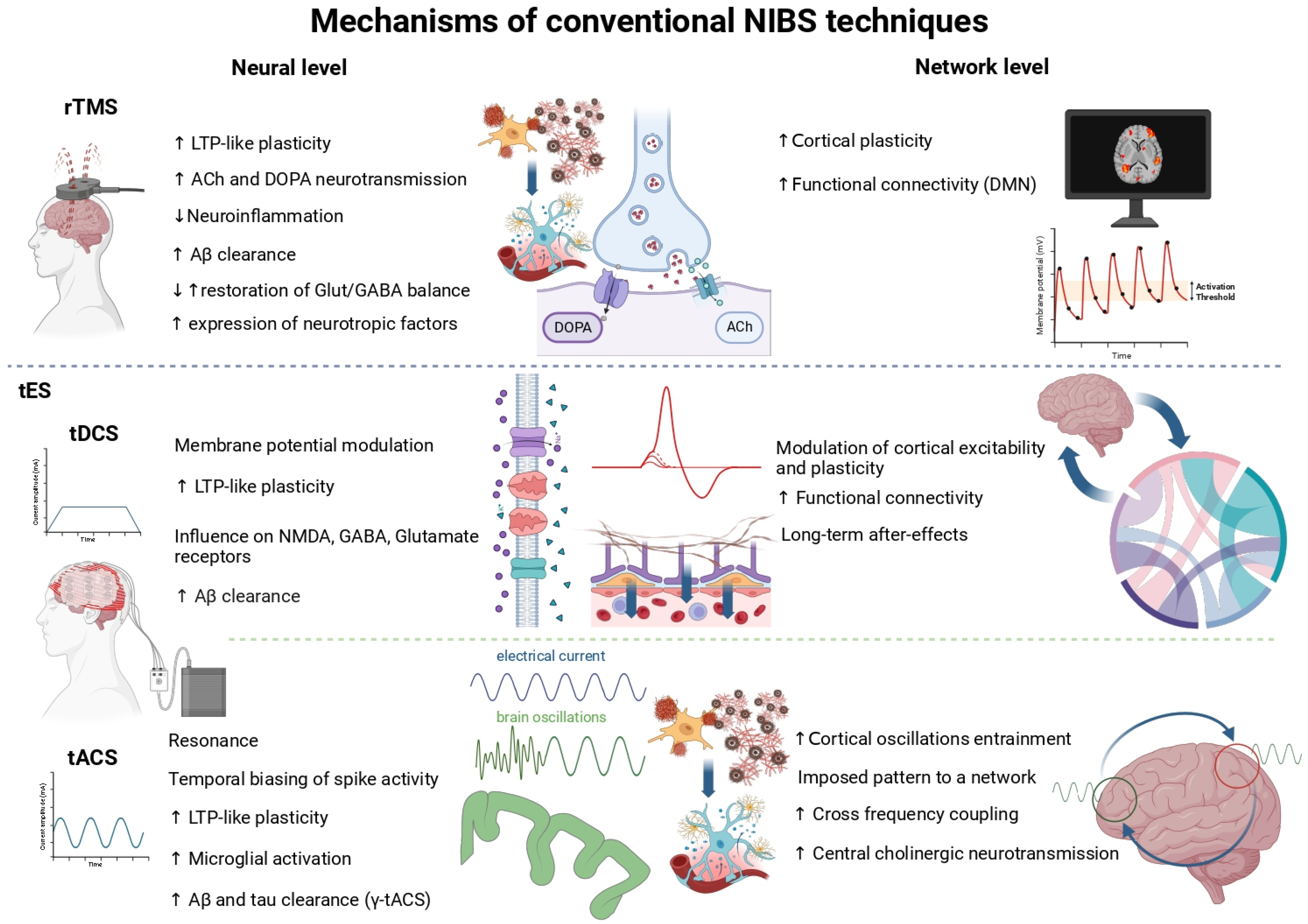
Figure 1. The known mechanisms of TMS and tES techniques and their effects in healthy humans and neurodegenerative diseases.
Ctn 09 00043 g001
Figure 2. A visual representation of the proposed scientific roadmap for implementing NIBS techniques in the prevention of neurodegenerative diseases.
Ctn 09 00043 g002
Disclaimer/Publisher’s Note: The statements, opinions and data contained in all publications are solely those of the individual author(s) and contributor(s) and not of MDPI and/or the editor(s). MDPI and/or the editor(s) disclaim responsibility for any injury to people or property resulting from any ideas, methods, instructions or products referred to in the content.