Abstract
Purpose: Accurate and precise navigation in space and postural stability rely on the central integration of multisensory input (vestibular, proprioceptive, visual), weighted according to its reliability, to continuously update the internal estimate of the direction of gravity. In this study, we examined both peripheral and central vestibular functions in a world-renowned 53-year-old male tightrope walker and investigated the extent to which his exceptional performance was reflected in our findings. Methods: Comprehensive assessments were conducted, including semicircular canal function tests (caloric irrigation, rotatory-chair testing, video head impulse testing of all six canals, dynamic visual acuity) and otolith function evaluations (subjective visual vertical, fundus photography, ocular/cervical vestibular-evoked myogenic potentials [oVEMPs/cVEMPs]). Additionally, static and dynamic posturography, as well as video-oculography (smooth-pursuit eye movements, saccades, nystagmus testing), were performed. The participant’s results were compared to established normative values. High-resolution diffusion tensor magnetic resonance imaging (DT-MRI) was utilized to assess motor tract integrity. Results: Semicircular canal testing revealed normal results except for a slightly reduced response to right-sided caloric irrigation (26% asymmetry ratio; cut-off = 25%). Otolith testing, however, showed marked asymmetry in oVEMP amplitudes, confirmed with two devices (37% and 53% weaker on the left side; cut-off = 30%). Bone-conducted cVEMP amplitudes were mildly reduced bilaterally. Posturography, video-oculography, and subjective visual vertical testing were all within normal ranges. Diffusion tensor MRI revealed no structural abnormalities correlating with the observed functional asymmetry. Conclusions: This professional tightrope walker’s exceptional balance skills contrast starkly with significant peripheral vestibular (otolithic) deficits, while MR imaging, including diffusion tensor imaging, remained normal. These findings highlight the critical role of central computational mechanisms in optimizing multisensory input signals and fully compensating for vestibular asymmetries in this unique case.
1. Introduction
Maintaining a stable, upright posture requires constant adjustment of body positioning to prevent falls and fall-related injuries in daily life. Among the various sensory systems contributing to this task, the vestibular organs play a crucial role by detecting angular and linear accelerations. Additional important input sources include skin pressure sensors, joint receptors, and vision. Within the central nervous system, these sensory signals are integrated and weighted according to their reliability []. When one or more sensory systems are impaired, the brain exhibits remarkable compensatory capabilities. For instance, approximately 80% of patients with persistent unilateral peripheral vestibular impairments recover to the extent that they experience no significant limitations in daily life [].
However, whether such compensation is sufficient for more demanding balance tasks, such as those faced by professional athletes like acrobats or ice skaters, remains an open question. In such cases, even minor sensory imbalances may pose significant challenges, potentially conflicting with the demands of their profession.
We had the rare opportunity to examine Freddy Nock, an extraordinary artist renowned for his mastery of balance, using advanced neuro-otologic diagnostic techniques. Our investigation focused on the integrity of his peripheral vestibular organs and his ability to compensate for external perturbations, such as unstable surfaces or visually disorienting surroundings.
Freddy Nock was a world-class performer with an unparalleled record of achievements. He famously walked a 347 m tightrope—just 18 mm in diameter—between two Swiss mountaintops without any form of safety equipment. In 2009, he became the fastest wire runner, earning the title of world champion in Seoul, South Korea. Among his many feats, he conquered Germany’s highest mountain, the Zugspitze (3000 m), navigating wet cable car wires. He also crossed Lake Zurich on a wire and, in 2011, set an astonishing seven records in seven days.
Given his extraordinary balance skills, we sought to determine whether these abilities were supported by exceptional peripheral or central vestibular functions. Our interest in studying Freddy arose after his awe-inspiring feat of crossing the Tamina Gorge before the construction of the legendary bridge in March 2013, a daring achievement that remains emblematic of his unparalleled skill and courage []. Tragically, Freddy Nock passed away on 7 February 2024 at the age of 59. Nevertheless, his remarkable legacy as both a world-renowned acrobat and a unique subject of scientific inquiry endures, leaving an indelible mark on the study of balance and human capability.
2. Methods
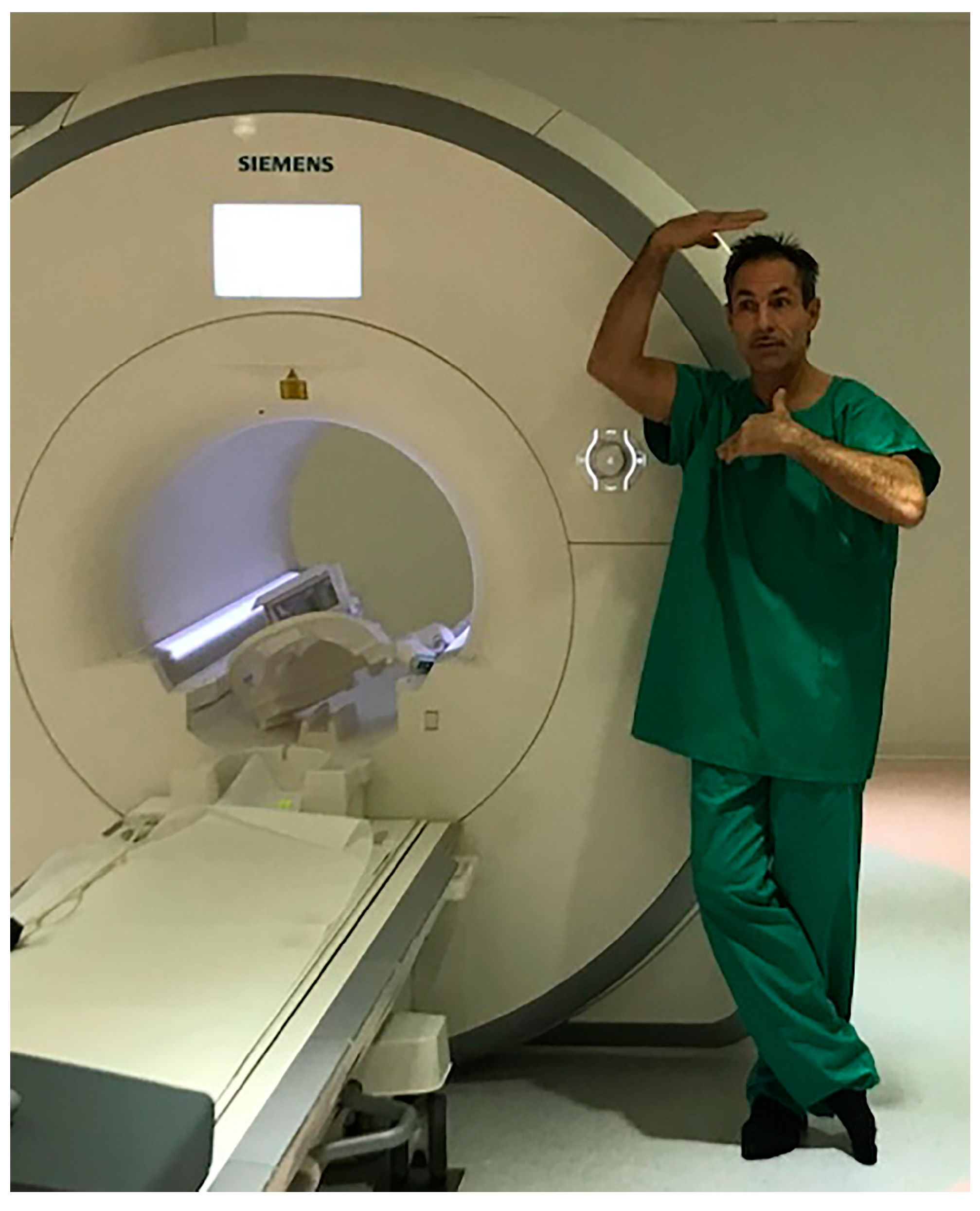
2.1. Experimental Setup
Vestibular testing was performed at the Interdisciplinary Center for Vertigo & Neurologic Visual Disorders of the University Hospital Zurich and an outpatient Concussion Center on a single day. By combining various tests, semicircular canal function and otolith function were assessed both at the level of brainstem reflexes (angular vestibulo-ocular reflex [aVOR], as assessed by the video head impulse test [vHIT] [] and caloric irrigation, and otolith-dependent reflexes, such as vestibular-evoked myogenic potentials [] and gravity-dependent ocular torsion []), and by use of functional tests evaluating postural stability (as assessed by static and dynamic posturography []), verticality perception (as assessed by the subjective visual vertical [SVV] []), and dynamic visual acuity [DVA] []).
2.2. Otolith Testing
Otolith function was quantified by means of vestibular-evoked myogenic-potentials (VEMPs) []. Cervical VEMPs (cVEMPs) mainly assess saccular function, and ocular VEMPs (oVEMPs) mainly assess utricular function. Air-conducted sound stimuli for cVEMPs (500 Hz, 6 ms tone bursts at 90–100 dB normal hearing level, total of 200 bursts) were applied to one ear and EMG activity was recorded from the ipsilateral sternocleidomastoid muscle. Afterwards, bone-conducted cVEMPs were obtained for comparison. Vibrations (unshaped 500 Hz bursts, duration 4 ms, 200 stimuli in total) were applied using a Minishaker (Model 4810, Brüel & Kjaer, P/L, Naerum, Denmark) placed over the hairline near Fz, as previously described by Weber and colleagues []. To improve reproducibility of measurements and to reduce noise from asymmetric muscle tension in individuals, response amplitudes were normalized (see [] for details). Reported values for air- and bone-conducted cVEMPS are therefore unitless. Bone-conducted oVEMPs (unshaped 500 Hz bursts, duration 4 ms, 200 stimuli in total) were applied by the same Minishaker, positioned again over the hairline near Fz. Stimuli were recorded with surface electrodes placed beneath the eyes during up-gaze. For further details see also [,].
Differences in response amplitude (left side vs. right side; asymmetry ratio (AR)) of more than 30% on cVEMPs or oVEMPs were considered abnormal. For comparison of VEMP latencies, we used data from a group of 26 healthy controls (aged 38.4 ± 15.8 years; 11 females) which were recorded with the same setup. For latency, values above the 95th percentile of values in the controls were considered abnormal, while for amplitudes, values below the 5th percentile of normal values were considered abnormal. Specifically, cut-off values (95th percentile) for latencies were 17.3 ms (p13) and 30.3 ms (n23) for cVEMPs and 13.8 ms (n10) and 18.7 ms (p15) for oVEMPs, respectively. For amplitudes, values below 0.8 (cVEMPs; normalized, unitless values) and 5.8 μV (oVEMPs), respectively, were considered abnormal.
2.3. Semicircular Canal Testing
We used quantitative video head impulse testing (vHIT) to assess the function of all six semicircular canals []. For video-oculography, an infrared camera recorded the right eye with a 250 Hz frame rate (ICS Impulse, Otometrics, Taastrup, Denmark). Vestibular hypofunction was defined as reduction in aVOR-gain (horizontal canals: <0.8, vertical canals < 0.7) [] or the occurrence of catch-up saccades during or after each head impulse.
Cold (30 °C) and warm (44 °C) water caloric irrigation was performed according to the Fitzgerald–Hallpike testing protocol [] and eye-movement responses were recorded by video-oculography. The asymmetry of peripheral-vestibular function was determined by the Jongkees formula []. Unilateral hypofunction was defined as a canal-paresis factor of >25% with a preserved response on the healthy side []. We also determined directional preponderance (DP). DP-values larger than 30% were considered abnormal, as suggested by [].
2.4. Quantitative Video-Oculography
By using video-oculography, ocular stability in the primary position, at eccentric gaze, after head shaking, and during mastoid vibration was recorded while the subject was in supine position with the head elevated by 30°. Then, ocular stability was measured while in supine position with the head roll-tilted 60° to either side in order to search for any positional nystagmus. Furthermore, smooth pursuit eye movements (center-to-peak amplitude: ±20°; peak-velocity range: 10–60°/s for the horizontal plane and 10–30°/s for the vertical plane) and saccadic eye movements (amplitudes: ±10°, ±15° and ±30° for horizontal saccades and ±10°, ±15° and ±20° for vertical saccades) were obtained in the horizontal and vertical plane. For assessing the angular vestibulo-ocular reflex (aVOR), constant-velocity chair rotations (60°/s) in either direction for at least 60 s duration was applied. For quantifying the optokinetic nystagmus (OKN) and the optokinetic after-nystagmus (OKAN), gains were determined during constant-velocity chair-rotation (50°/s) in either direction and immediately afterwards (chair stopped after 30 s). For assessing the VOR-suppression, chair oscillations at a frequency of 0.35 Hz and with peak-velocities of ±44°/s were applied while a chair-fixed visual target was shown.
2.5. Postural Stability
Postural stability was evaluated using two distinct methods. First, trunk movements (sway and surge) were quantified during the Romberg test using the gyroscopes of an Apple iPod (Apple, Cupertino, CA, USA). Four test conditions were applied: standing on solid ground with eyes open or closed and standing on a foam surface with eyes open or closed. Velocity and acceleration data were analyzed to calculate sway/surge areas, focusing on the region containing the central 95% of the data.
Second, postural stability was assessed using a computerized dynamic posturography system (EquiTest, Natus Medical Incorporated, Seattle, WA, USA). This system evaluated several parameters: the sensory organization test (SOT), the SOT with head shaking (HS SOT), the motor control test (MCT), the limits of stability (LOS) test, and the adaptation test (ADT). These measurements provided a comprehensive analysis of the subject’s postural control under various sensory and motor conditions.
2.6. Assessment of the Subjective Visual Vertical and Fundus Photography
When obtained under static conditions and in complete darkness, the subjective visual vertical strongly relies on otolith (utricular and saccular) input []. The perceived direction of gravity was assessed in a sitting position with the head upright or roll-tilted 45° to either side. Therefore, in otherwise complete darkness, a luminous line was shown on a computer screen, and the subject was asked to align the line in such a way that it was along the perceived direction of earth-vertical. In each head roll orientation, six adjustments were collected (no time limit). When upright, SVV adjustments within ±2.5 relative to the gravitational vertical are considered normal [,]. Using a non-mydriatic fundus camera (TRC-NW400, Topcon Europe Medical BV, Capelle aan den IJssel, The Netherlands), photographs of the right and left retina were obtained while the subject was sitting with the head upright. Static eye torsion was determined by measuring the angle of a line inter-connecting the fovea centralis and the disk.
2.7. MR-Imaging
In order to assess the structural integrity of those tracts that are eminent in motor control, i.e., the corticospinal tract and the rubrospinal tract [], fiber tracking was performed. Therefore, diffusion tensor imaging (DTI) was obtained on a 3 Tesla magnetic resonance imager (MRI) (Siemens Healthcare, Erlangen, Germany) with a 32-channel head coil. High angular resolution (64 encoded diffusion directions) as well as high spatial resolution (1.3 × 1.3 × 1.3 mm3) was acquired with a readout-segmented acquisition with a navigator-based reacquisition DTI MRI sequence [], planned on the motor cortico-spinal tract with a b-value of 1000 s/mm2. Other scan parameters are 7 readout-segments, 24 coronal slices, TR 3870 ms, TE 75 ms, TE Navigator 113 ms, acquisition time 36 min, GRAPPA acceleration factor 2, echo planar imaging factor 84, echo spacing 0.36 ms, receive bandwidth 709 Hz/pixel. Using SyngoVia (Siemens Healthcare, Erlangen, Germany), the cortico- and rubro-spinal tracts were reconstructed.
3. Results
The participant was Freddy Nock, a 53-year-old male professional tightrope walker. He had no history of vertigo, dizziness, or balance disorders, and he did not take any medication on a regular basis.
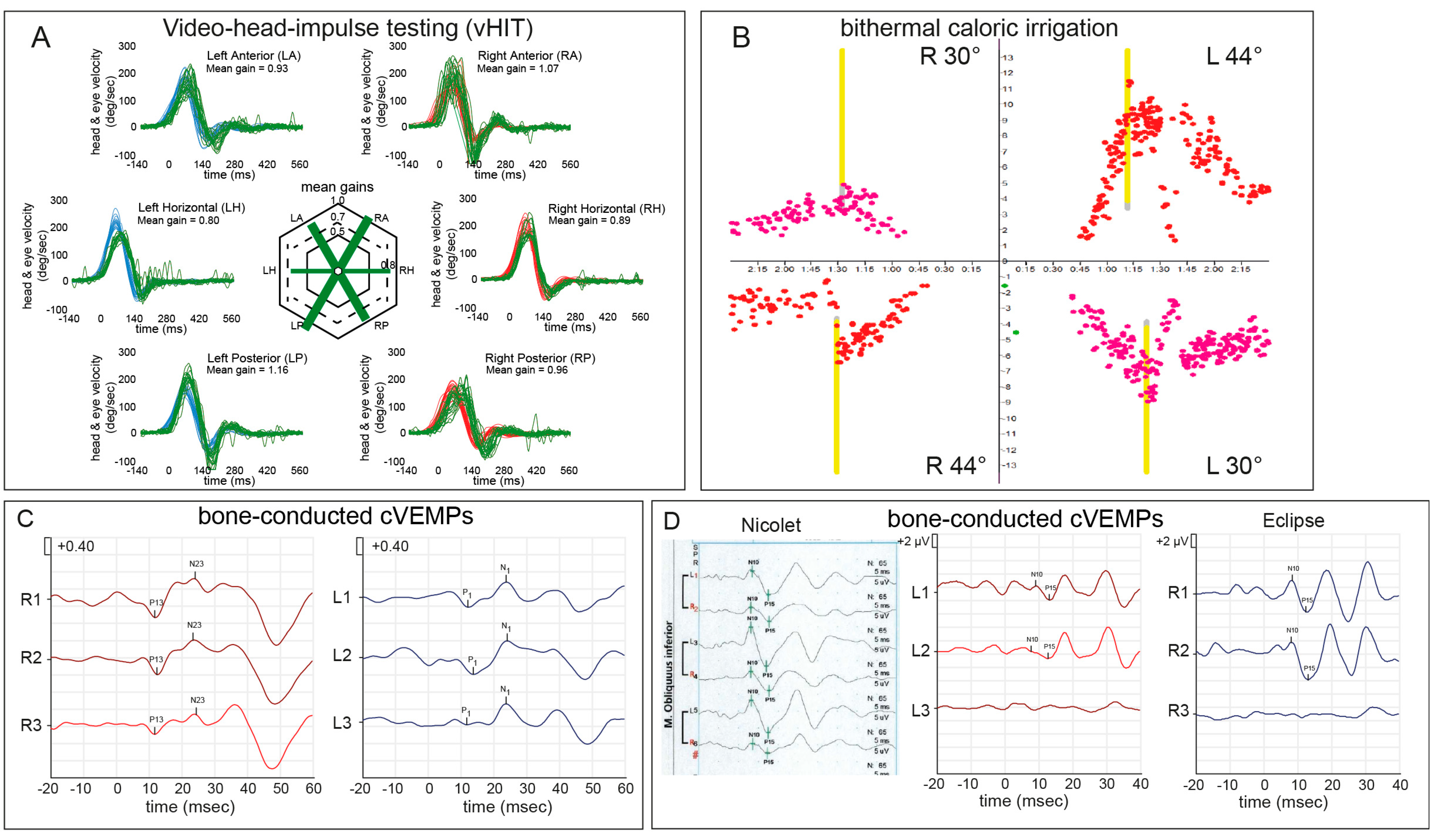
3.1. Assessment of Semicircular Canal Function
Semicircular canal function was assessed by video head impulse testing (Figure 1, panel A) demonstrated normal gain values for all six semicircular canals and no evidence for catch-up saccades (Table 1). Likewise, DVA testing showed reductions in visual acuity under dynamic conditions within the range of normal for both horizontal SCCs. On bithermal caloric irrigation (Figure 1, panel B) a borderline asymmetric response was noted with a canal paresis (CP) of 26% (weaker response on the left side), whereas directional preponderance was within normal limits (2%). On rotatory chair testing, per- and post-rotary VOR-gains and decay time constants for velocity steps were within normal range with the exception of a slightly reduced gain during rotations to the right. During oscillations marked per-rotatory VOR-gain reductions were noted and during optokinetic stimulation a poor response was observed. Smooth pursuit eye movements and VOR-suppression responses were within normal range.
Figure 1.
Quantitative vestibular testing including video head impulse testing (vHIT) (panel A), bithermal caloric irrigation (panel B), cVEMPs (panel C), and oVEMPs (panel D). For the vHIT (panel A) eye velocity traces (in green) and head velocity traces (in red for assessing the right vestibular organ and in blue for assessing the left vestibular organ) are plotted against time for each SCC (20 trials per canal recorded). Note that eye velocity traces were inverted for better visualization and comparison with the head velocity traces. In the center of both panels average gains are provided for all six semicircular canals. In the subject presented here, the normal function of all six semicircular canals was seen, thus all canals were plotted in green. For caloric irrigation (panel B), applying warm (44 °C, red dots) and cold (30 °C, pink dots) water to one ear, the nystagmus slow phase velocity was plotted against time. Noteworthy, a canal paresis factor of 26% was seen (panel B), pointing to a mildly reduced function of the right horizontal semicircular canal. Response asymmetries on bone-conducted cVEMPs (plotted against time, three sessions shown) were within normal range (panel C), whereas on oVEMP-testing (panel D) a significant asymmetry ratio was noted with left-sided impairment of utricular function both when using the Nicolet (37%) and the Eclipse (53%) testing devices. Abbreviations: R = right; L = left.
Table 1.
Detailed vestibular testing results.
3.2. Assessment of Otolith Function
Air-conducted cervical vestibular-evoked myogenic-potentials (cVEMPs) demonstrated an absent response on the right side, while the response on the left side showed a slightly reduced amplitude. Using bone-conducted cVEMPs instead (Figure 1, panel C), responses were present on both sides with a normal asymmetry ratio (10%, weaker on the left side). While p13/n23 latencies were normal, the amplitudes were slightly reduced on both sides.
Bone-conducted oVEMPs (Nicolet) showed a reduced peak-to-peak amplitude on the left side and an increased amplitude asymmetry ratio (37% weaker on the left side). n10 and p15 latencies were within normal. Testing was repeated on a different device (Eclipse), again showing an increased amplitude asymmetry ratio (53% weaker on the left side). Fundus photography demonstrated normal cyclotorsion of both eyes.
3.3. Assessment of Spatial Orientation and Postural Stability
Verticality perception as assessed by the SVV demonstrated accurate and precise internal estimates of direction of gravity while upright and roll over-compensation by 7° when roll-tilted 45° to either side. Static postural stability showed acceleration and velocity values within the normal range. Dynamic posturography (EquiTest) demonstrated normal testing results throughout all domains assessed, including adaptive capabilities to perturbations of the visual surroundings or the surface, probing visual and vestibular properties (SOT, HS SOT) and somatosensory systems (MCT).
3.4. Assessment for Central Eye Movement Disorders
Video-oculography demonstrated no signs of pathologic spontaneous, positional, gaze-evoked, vibration-induced or head-shaking-induced nystagmus. Horizontal and vertical smooth pursuit eye movements had a normal gain, and saccades were of normal latency, velocity and amplitude.
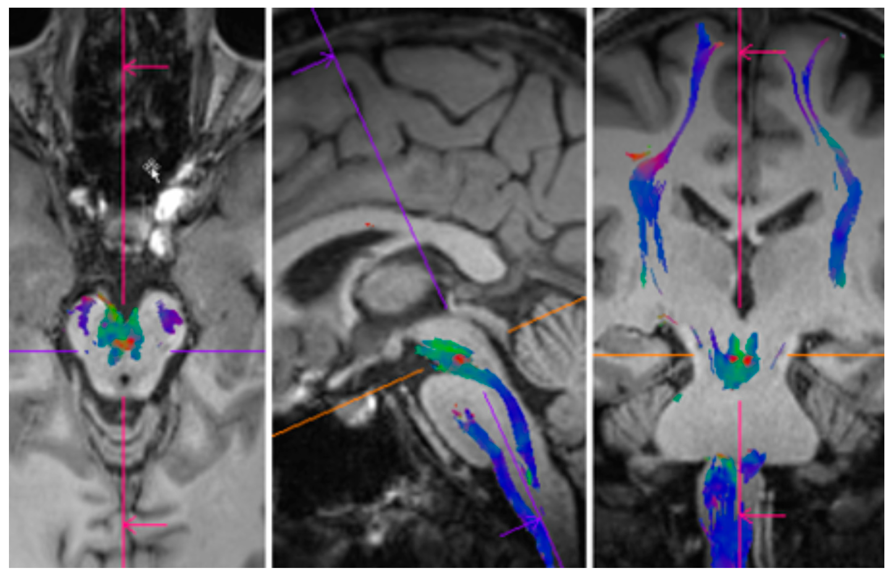
3.5. Analysis of MR-Imaging
The quality of the DTI data were very good, and no motion artifacts could be observed. The cortico-spinal tract and the rubro-spinal crossing at the red globule level could be depicted using tract reconstruction (see Figure 2 and Figure 3). Other tiny fiber structures were also visible. The left and right corticospinal tracts had similar diffusion properties, as well as the tracts crossing (hemisphere) between the pons and the motor cortex; all values were in these ranges: mean fractional anisotropy 0.51 ± 0.03, and mean diffusivity 1.0 ± 0.1, radial diffusivity 0.7 ± 0.1, axial diffusivity 1.7 ± 0.1, all in 10−3 mm2/s. The stability of these measurements underlines the quality of the DTI data and the symmetry of the tracts’ structural architecture.
Figure 2.
All tracts reconstructed from the high-resolution diffusion tensor imaging dataset. Data were sampled with a 1.3 mm isotropic spatial resolution and 64 encoded diffusion directions. Note the geometrical accuracy of the tracts due to the segmented image acquisition used. The quality of the data were also due to the absolute motion-less patient position during the acquisition. Overlay on a T1 weighted anatomical scan. Color encoding: blue: cranio-caudal, red: left-right, green: anterior–posterior.
Figure 3.
Rubro-spinal crossing of the motor tract, in red shown for the axial (left), sagittal (middle), and coronal (right) plane. The 1.3 mm isotropic resolution of the DTI data allowed the representation of such a small crossing with the depicted quality. For color coding see Legend of Figure 2.
4. Discussion
The exceptional balance performance observed in the professional tightrope walker studied (Figure 4) here was achieved despite confirmed peripheral vestibular deficits. Specifically, oVEMPs were unilaterally absent, indicating impaired utricular function. Additionally, bone-conducted cVEMP responses showed borderline amplitudes, and caloric irrigation revealed slight unilateral hypo-responsiveness. These findings of peripheral vestibular impairments are unexpected given the subject’s outstanding balance skills. However, no evidence of central vestibular dysfunction was found, as eye movement examinations and postural stability tests were all within normal ranges.
Figure 4.
Freddy Nock (1964–2024), renowned tightrope walker, captured in a moment of levity before undergoing MR imaging. Eager to participate in the study, Nock humorously remarked about his brain soon being visualized by the machine.
The detailed testing of semicircular canal and otolith organ function requires careful interpretation. Notably, bone-conducted oVEMPs reflect the activity of only a specific subset of utricular cells, primarily irregular type I cells [,]. This means that residual utricular function, even on the side with absent oVEMP responses, cannot be ruled out. While one might consider the absent oVEMPs as an artifact, the reproducibility of these results across two separate measurement setups and the subject’s age below 60 years suggest the asymmetry is pathological. Despite these deficits, the subject reported no vestibular symptoms, such as vertigo, dizziness, or imbalance in stance or gait. Moreover, the subject’s perceived direction of gravity was accurate when upright, and roll-tilt tests at 45° showed symmetric offsets consistent with central compensation for utricular dysfunction.
Our test battery covered a wide range of postural stability and spatial orientation measures, and the subject excelled in all assessments of higher cortical function. These included tests of static and dynamic postural stability under conditions of discrepant vestibular and visual stimuli. In contrast, reflexive otolith-ocular responses, mediated at the brainstem level, showed clear impairments. This dichotomy suggests that the brain developed advanced compensatory strategies to overcome peripheral vestibular deficits, allowing for exceptional balance performance. Importantly, while dynamic (EquiTest) and static posturography results fell within the normal range, these tests feature broad normative values, and performance deficiencies are the primary criteria for identifying abnormalities.
The diffusion tensor imaging (DTI) dataset was of excellent quality due to the subject’s compliance and lack of motion during acquisition. This enabled precise reconstruction of small bifurcations and crossings in the motor tracts. The absence of structural abnormalities in the cortico-spinal and rubro-spinal tracts further supports the assumption of a peripheral-vestibular origin for the slight asymmetry observed in vestibular responses with excellent central compensation.
In summary, the findings in this world-renowned tightrope walker underscore the critical role of higher brain functions in compensating for peripheral vestibular deficits. Despite significant utricular asymmetry, he achieved remarkable balance skills, likely through central compensatory mechanisms that fully adjusted for asymmetric utricular input. These observations strongly support the notion that balance performance relies predominantly on central processing capabilities and that peripheral sensory impairments can be effectively compensated, enabling extraordinary achievements even in demanding tasks.
Author Contributions
A.A.T. analyzed and interpreted the data and drafted the manuscript. G.B. was involved in the data collection and interpretation and critically reviewed and edited the manuscript. N.F.-D. was involved in the data collection and interpretation of the data and critically reviewed and edited the manuscript. F.R. was involved in data collection and interpretation and critically reviewed and edited the manuscript. U.S. was involved in data collection and critically reviewed and edited the manuscript. M.P. performed the dedicated MR-imaging and oversaw the imaging post-processing and analysis. He also critically reviewed and edited the manuscript. J.K. conceived of the study and critically reviewed and edited the manuscript. D.S. was involved in the study’s design and critically reviewed and edited the manuscript. All authors have read and agreed to the published version of the manuscript.
Funding
A. Tarnutzer and D. Straumann were supported by the Betty and David Koetser Foundation for Brain Research and the Zurich Center for Integrative Human Physiology, Switzerland.
Institutional Review Board Statement
Ethical review and approval were waived for this study due to the fact that this was a single case report.
Informed Consent Statement
Written informed consent has been obtained from the patient to publish this paper.
Data Availability Statement
Dataset available on request from the authors.
Acknowledgments
The authors thank Marco Penner and Elena Buffone for technical assistance. Study funding: none. Financial disclosure statements: A. Tarnutzer reports no disclosures relevant to the manuscript; F. Romano reports no disclosures relevant to the manuscript; N. Feddermann-Demont reports no disclosures relevant to the manuscript; U. Scheifele reports no disclosures relevant to the manuscript; M. Piccirelli reports no disclosures relevant to the manuscript; G. Bertolini reports no disclosures relevant to the manuscript; J. Kesselring reports no disclosures relevant to the manuscript; D. Straumann reports no disclosures relevant to the manuscript.
Conflicts of Interest
The authors declare no conflicts of interest.
References
- Fetsch, C.R.; DeAngelis, G.C.; Angelaki, D.E. Bridging the gap between theories of sensory cue integration and the physiology of multisensory neurons. Nat. Rev. Neurosci. 2013, 14, 429–442. [Google Scholar] [CrossRef] [PubMed]
- Halmagyi, G.M.; Weber, K.P.; Curthoys, I.S. Vestibular function after acute vestibular neuritis. Restor. Neurol. Neurosci. 2010, 28, 37–46. [Google Scholar] [CrossRef] [PubMed]
- Nock, F. 2013. Available online: https://www.youtube.com/watch?v=_rYkeWMOnkw (accessed on 21 December 2024).
- Curthoys, I.S.; McGarvie, L.A.; MacDougall, H.G.; Burgess, A.M.; Halmagyi, G.M.; Rey-Martinez, J.; Dlugaiczyk, J. A review of the geometrical basis and the principles underlying the use and interpretation of the video head impulse test (vHIT) in clinical vestibular testing. Front. Neurol. 2023, 14, 1147253. [Google Scholar] [CrossRef]
- Rosengren, S.M.; Colebatch, J.G.; Young, A.S.; Govender, S.; Welgampola, M.S. Vestibular evoked myogenic potentials in practice: Methods, pitfalls and clinical applications. Clin. Neurophysiol. Pract. 2019, 4, 47–68. [Google Scholar] [CrossRef]
- Evangelista, V.R.P.; Mermelstein, S.A.; da Silva, M.M.; Kaski, D. Bedside video-ophthalmoscopy as an aid in the diagnosis of central vestibular syndromes. J. Neurol. 2022, 269, 217–220. [Google Scholar] [CrossRef]
- Visser, J.E.; Carpenter, M.G.; van der Kooij, H.; Bloem, B.R. The clinical utility of posturography. Clin. Neurophysiol. 2008, 119, 2424–2436. [Google Scholar] [CrossRef] [PubMed]
- Dieterich, M.; Brandt, T. Perception of Verticality and Vestibular Disorders of Balance and Falls. Front. Neurol. 2019, 10, 172. [Google Scholar] [CrossRef] [PubMed]
- Chen, G.; Zhang, J.; Qiao, Q.; Zhou, L.; Li, Y.; Yang, J.; Wu, J.; Huangfu, H. Advances in dynamic visual acuity test research. Front. Neurol. 2022, 13, 1047876. [Google Scholar] [CrossRef] [PubMed]
- Curthoys, I.S. A critical review of the neurophysiological evidence underlying clinical vestibular testing using sound, vibration and galvanic stimuli. Clin. Neurophysiol. 2010, 121, 132–144. [Google Scholar] [CrossRef]
- Weber, K.P.; Rosengren, S.M.; Michels, R.; Sturm, V.; Straumann, D.; Landau, K. Single motor unit activity in human extraocular muscles during the vestibulo-ocular reflex. J. Physiol. 2012, 590, 3091–3101. [Google Scholar] [CrossRef] [PubMed]
- Blanquet, M.; Petersen, J.A.; Palla, A.; Veraguth, D.; Weber, K.P.; Straumann, D.; Tarnutzer, A.A.; Jung, H.H. Vestibulo-cochlear function in inflammatory neuropathies. Clin. Neurophysiol. 2018, 129, 863–873. [Google Scholar] [CrossRef] [PubMed]
- Rosengren, S.M.; Welgampola, M.S.; Colebatch, J.G. Vestibular evoked myogenic potentials: Past, present and future. Clin. Neurophysiol. 2010, 121, 636–651. [Google Scholar] [CrossRef]
- Weber, K.P.; Rosengren, S.M. Clinical utility of ocular vestibular-evoked myogenic potentials (oVEMPs). Curr. Neurol. Neurosci. Rep. 2015, 15, 22. [Google Scholar] [CrossRef] [PubMed]
- McGarvie, L.A.; MacDougall, H.G.; Halmagyi, G.M.; Burgess, A.M.; Weber, K.P.; Curthoys, I.S. The Video Head Impulse Test (vHIT) of Semicircular Canal Function—Age-Dependent Normative Values of VOR Gain in Healthy Subjects. Front. Neurol. 2015, 6, 154. [Google Scholar] [CrossRef] [PubMed]
- Fitzgerald, G.; Hallpike, C.S. Studies in human vestibular function. I. Observations on the directional preponderance of caloric nystagmus resulting from cerebral lesions. Brain 1942, 65, 115–137. [Google Scholar] [CrossRef]
- Jongkees, L.B. The caloric test and its value in evaluation of the patient with vertigo. Otolaryngol. Clin. N. Am. 1973, 6, 73–93. [Google Scholar] [CrossRef]
- Halmagyi, G.M.; Yavor, R.A.; McGarvie, L.A. Testing the vestibulo-ocular reflex. In Electrophysiologic Evaluation in Otolaryngology; Alford, B.R., Jerger, J., Jenkins, H.A., Eds.; Karger: Basel, Switzerland, 1997; pp. 132–154. [Google Scholar]
- Shepard, N.T.; Jacobson, G.P. The caloric irrigation test. Handb. Clin. Neurol. 2016, 137, 119–131. [Google Scholar] [CrossRef] [PubMed]
- Schoene, H. On the role of gravity in human spatial orientation. Aerosp. Med. 1964, 35, 764–772. [Google Scholar] [PubMed]
- Brandt, T.; Dieterich, M.; Danek, A. Vestibular cortex lesions affect the perception of verticality. Ann. Neurol. 1994, 35, 403–412. [Google Scholar] [CrossRef] [PubMed]
- Pérennou, D.A.; Mazibrada, G.; Chauvineau, V.; Greenwood, R.; Rothwell, J.; Gresty, M.A.; Bronstein, A.M. Lateropulsion, pushing and verticality perception in hemisphere stroke: A causal relationship? Brain 2008, 131, 2401–2413. [Google Scholar] [CrossRef]
- Olivares-Moreno, R.; Rodriguez-Moreno, P.; Lopez-Virgen, V.; Macias, M.; Altamira-Camacho, M.; Rojas-Piloni, G. Corticospinal vs Rubrospinal Revisited: An Evolutionary Perspective for Sensorimotor Integration. Front. Neurosci. 2021, 15, 686481. [Google Scholar] [CrossRef]
- Porter, D.A.; Heidemann, R.M. High resolution diffusion-weighted imaging using readout-segmented echo-planar imaging, parallel imaging and a two-dimensional navigator-based reacquisition. Magn. Reson. Med. 2009, 62, 468–475. [Google Scholar] [CrossRef] [PubMed]
- Tarnutzer, A.A.; Bockisch, C.J.; Buffone, E.; Weiler, S.; Bachmann, L.M.; Weber, K.P. Disease-specific sparing of the anterior semicircular canals in bilateral vestibulopathy. Clin. Neurophysiol. 2016, 127, 2791–2801. [Google Scholar] [CrossRef] [PubMed]
- Curthoys, I.S. The new vestibular stimuli: Sound and vibration-anatomical, physiological and clinical evidence. Exp. Brain Res. 2017, 235, 957–972. [Google Scholar] [CrossRef]
- Curthoys, I.S.; Kim, J.; McPhedran, S.K.; Camp, A.J. Bone conducted vibration selectively activates irregular primary otolithic vestibular neurons in the guinea pig. Exp. Brain Res. 2006, 175, 256–267. [Google Scholar] [CrossRef] [PubMed]
Disclaimer/Publisher’s Note: The statements, opinions and data contained in all publications are solely those of the individual author(s) and contributor(s) and not of MDPI and/or the editor(s). MDPI and/or the editor(s) disclaim responsibility for any injury to people or property resulting from any ideas, methods, instructions or products referred to in the content. |
© 2025 by the authors. Published by MDPI on behalf of the Swiss Federation of Clinical Neuro-Societies. Licensee MDPI, Basel, Switzerland. This article is an open access article distributed under the terms and conditions of the Creative Commons Attribution (CC BY) license (https://creativecommons.org/licenses/by/4.0/).