Review

Narcolepsy Type 1: Should We Only Target Hypocretin Receptor 2?

by
Rolf Fronczek
1,2 and
Gert Jan Lammers
1,2,*
1
Department of Neurology, Leiden University Medical Centre, 2300 RC Leiden, The Netherlands
2
Stichting Epilepsie Instellingen Nederland (SEIN), Sleep-Wake Centre, 2103 SW Heemstede, The Netherlands
*
Author to whom correspondence should be addressed.
Clin. Transl. Neurosci. 2023, 7(3), 28; https://doi.org/10.3390/ctn7030028
Submission received: 2 August 2023 / Revised: 11 September 2023 / Accepted: 15 September 2023 / Published: 19 September 2023
(This article belongs to the Special Issue Sleep–Wake Medicine)

Abstract

:
Nearly 25 years have passed since the ground-breaking discovery that hypocretin deficiency underlies human narcolepsy with cataplexy. Over time, it has become increasingly evident that hypocretin deficiency goes beyond the conventional core symptoms, or pentad, traditionally associated with narcolepsy. The emergence of hypocretin receptor 2 agonists presents an exciting opportunity, prompting us to explore the role of receptor 2 in the complete spectrum of NT1 symptoms. In this review, several clinical manifestations beyond the core symptoms will be discussed. We will outline what is currently known about the involvement of hypocretin receptors to reflect on what we expect with current knowledge from treatment with specific receptor agonists.

1. Introduction

Since the finding in 1999 that narcolepsy with cataplexy (type 1, NT1) is caused by hypocretin deficiency in the brain, research and expanding evidence on narcolepsy have mainly focused on the precise function of the hypocretins, the cause of the deficiency and the place of hypocretin measurement in the diagnosis of NT1 [1,2,3,4,5].
Little attention has focused on increasing knowledge and understanding of the symptoms and, thus, the clinical picture of NT1. Over the past 20 years, however, it has become increasingly clear, partly due to what we learned from the hypocretin system, that NT1 is not just about the pentad of core symptoms: excessive daytime sleepiness (EDS), cataplexy, hypnagogic hallucinations, sleep paralysis and disturbed nocturnal sleep. The complaints and symptoms range is much broader than previously thought. It has also become clear that classic symptoms can have diverse clinical manifestations. This is particularly true of EDS, which involves involuntary naps (or sleep attacks), impaired sustained attention, automatic behaviour, ‘brain fog’ and memory complaints. Other complaints and symptoms beyond the pentad include impaired executive functioning, memory problems, depression and anhedonia, anxiety and possibly an altered stress response, fatigue, obesity and possibly an altered reward system. Some of these symptoms are more prevalent in people than a ‘core symptom’ such as sleep paralysis [6,7].
As the first hypocretin receptor 2 (HCRTR-2) agonists become available for human studies, we can ascertain which symptoms are related to hypocretin deficiency and, more specifically, to the HCRTR-2 receptor, and which are not [8]. Studies in NT2 and IH, where hypocretin deficiency is not the major or even not at all a causative factor, will also contribute to this knowledge. It will be necessary and exciting to understand which aspects beyond the core symptoms will improve and which will not. It might even be that some symptoms will worsen due to a newly emerging imbalance between the activation of type 1 and type 2 receptors. It might also be that long-lasting hypocretin deficiency induces other downstream functional or structural changes in the brain. This information will guide future treatment and future drug development. It will also expand our understanding of the physiological interrelation between sleep, metabolism, mood, fatigue, stress and motivation.
Neither in the clinic nor in scientific research is it currently standard practice to measure narcolepsy symptoms beyond the core symptoms in a standardised way. It will be essential to start this to allow patients to optimise treatment efficacy but also for an accurate understanding of the pharmacological properties of the different types of HCRT-R agonists. This knowledge will guide the development of agonists with profiles other than a selective affinity for the type 2 receptor.
Here, we focus on the increased knowledge of the clinical manifestations of NT1, which is caused by severely impaired hypocretin transmission. Notably, the discovery of the role of hypocretin as the cause of NT1 has allowed for better specification of the clinical picture and more reliable diagnoses. It has been shown that hypocretin deficiency is a more critical biomarker for NT1 than the outcome of the multiple sleep latency test (MSLT), providing the opportunity to evaluate homogeneous groups of patients.
For each clinical manifestation/domain to be discussed, we will outline what is currently known about the involvement of hypocretin receptors to reflect on what we expect with current knowledge from treatment with specific receptor agonists. It is important to remember that there are two hypocretin receptors and two hypocretin peptides. The two hypocretin peptides, hypocretin 1 and 2, result from the cleavage of the precursor preprohypocretin [9]. Hypocretin 1 has an equal affinity for receptors 1 and 2, and hypocretin 2 has a much greater affinity for receptor 2 than for receptor 1 [10].

2. Core Symptoms

Animal studies have shown that HCRTR-2 knockout mice exhibit complete and similar core symptoms of narcolepsy with cataplexy, considering this can be measured in animals. Conversely, HCRTR-1 knockout mice show no significant abnormalities in sleep–wake behaviour. However, HCRTR 1 and 2 double-knockout mice display a more severe phenotype than HCRTR-2 mice, suggesting an additional role for HCRTR-1 in the narcolepsy phenotype [4]. Thus, although the core symptoms of narcolepsy with cataplexy can be explained by impaired HCRTR-2 function, it needs to be taken into account that the findings in knockout mice and narcolepsy in humans, is caused by a deficiency of hypocretin, which has an affinity for both receptors. Restoring HCRTR-2 receptor function may not fully reverse the symptoms. Theoretically, colocalising peptides in the hypocretin cells may also affect the phenotype when the hypocretin cells indeed disappear in human narcolepsy with cataplexy. Since there is little knowledge about this theoretical additional impact, we will not further discuss this aspect.

2.1. Expressions of Excessive Daytime Sleepiness

EDS has many manifestations [11]. It is not just what is defined in the ICSD3: “Daily episodes of irrepressible need to sleep or lapses into sleep occurring for at least three months”. Generally, there is indeed the inability to stay awake during the day, but there is much more. It is almost always accompanied by a subjective feeling of sleepiness throughout the day, only in monotonous situations, or when relaxing. The daytime naps usually occur during monotonous situations and begin gradually, but there may also be a more unexpected and sudden onset that resembles an actual sleep attack. Unscheduled daytime naps are usually short and refreshing. The frequency may vary from once to more than ten times a day. In most cases, falling asleep is the tip of the iceberg. There are much larger periods during the day with impaired vigilance, expressed as impaired sustained attention. The impaired vigilance causes performance impairment that is often more debilitating than falling asleep. People do not have many expectations of performance when someone is asleep, but they do have expectations of someone who appears awake and alert but is not. In those situations, when people fail to process information adequately or perform as requested, others become irritated and view patients as disinterested and forgetful. Animal studies suggest that impaired sustained attention is the primary problem in NT1 [12]. Reduced alertness can also lead to automatic behaviours such as putting pea pods in a pan to cook, throwing the green peas in the waste bin and missing an exit on the highway. Sleepiness and impaired vigilance can also be experienced as brain fog, although this experience may also be related to fatigue (see also below). Lastly, difficulty meeting demands because of the sleepiness or impaired vigilance of others often leads to irritability.
It seems to be mainly the hypocretin 2 receptor involved in sleepiness and attention [8,13]. However, an additional role of receptor 1 is suggested in mice studies [4].

2.2. Cataplexy

Cataplectic attacks are rarely observed during consultation and seldom when trying to elicit them or performing tests. Therefore, we must rely on history-taking and or questionnaires. After the discovery that narcolepsy with cataplexy is caused by hypocretin deficiency, the concept of typical cataplexy emerged when studying the characteristics of cataplectic attacks in people with a known hypocretin deficiency by use of questionnaires. Typical cataplexy is strongly associated with hypocretin deficiency; around 95% show hypocretin-1 deficiency in the CSF. The concept and diagnostic value of typical cataplexy are acknowledged and introduced in the recently updated diagnostic criteria for NT1 [14].
Typical cataplexy usually manifests as partial cataplexy. Classically, this involves bilateral loss of muscle tone in the face, neck or legs (e.g., buckling knees), with or without the involvement of the arms. Episodes are triggered by sudden and particularly positive emotions (e.g., laughing out loud, telling a funny joke/story), making a witty remark, meeting an acquaintance unexpectedly, or (less often) anger. Episodes usually last up to 1 min but generally less than 30 s. Sometimes episodes are fleeting. Successive attacks may occur if the trigger persists, leading to an apparent prolongation. The frequency of cataplexy can vary widely. Usually, it happens at least once a month (if untreated), but it can also occur daily up to even dozens of times per day, depending on the presence of triggers. During typical cataplexy, consciousness is preserved, and muscle tension is abruptly returned after the attack. Although these events are typically stereotyped within an individual, the progression and extension of the muscles involved, and the duration of the episodes, can vary [14].
Some of the patients also have complete attacks in addition to partial attacks. These attacks begin as partial attacks in which almost all skeletal muscles eventually become involved (except the respiratory and eye movement muscles) and may become totally flaccid. This leads to an inability to stand and falls if no support can be found. These episodes often last up to 2 min, but, again, consecutive attacks result in a much longer duration of symptoms. As with partial typical cataplexy, consciousness is preserved, and muscle tension is abruptly returned after the attack. Sometimes, however, patients may fall asleep during a prolonged attack.
Features of atypical cataplexy include the following: (1) purely unilateral attacks, (2) prolonged attacks (e.g., >3 min) in the absence of the precipitant, (3) no clear precipitants for episodes or only negative emotions serving as triggers, (4) hyperacute generalised muscle weakness without build-up over seconds, leading to falls and injuries, (5) prolonged recovery (several minutes) after a single attack, (6) uncertainty regarding the preservation of consciousness or (7) exclusively generalised attacks without a history of partial episodes. When less than three of these features are present in addition to typical cataplexy features, there is still an association with hypocretin deficiency, but it is much less intense than those with only typical features [14].
It has been clearly shown that receptor 2 is involved. If there is a relevant additional role for receptor 1 in humans, it is not clear. The well-studied dog model, caused by a receptor 2 mutation (ref. [2]), shows that a selective receptor 2 defect can cause typical and sometimes severe cataplexy.

2.3. Nocturnal Sleep

Experienced disturbed nocturnal sleep is mainly due to fragmentation and hypnagogic hallucinations with or without sleep paralysis. There may be an additional impact of severe comorbid sleep-related breathing problems. The role of frequent periodic leg movements and REM sleep behaviour disorder on (experienced) nocturnal sleep is unclear. The current concept is that hypocretin activity is significantly reduced or absent during nocturnal sleep [15]. This leads to the question of what happens to nocturnal sleep when the effect of a dose of a selective agonist wears off too early or too late? Regardless of the duration of action, it is clear that HCRTR-2 is involved in sleep–wake regulation, but there is also evidence that HCRTR-1 may be involved. In a rodent study, restoring this receptor in noradrenergic neurons of the locus coeruleus ameliorated sleep–wake fragmentation [16]. It might be that this effect will only be relevant in the context of reduced receptor 2 activation.

2.4. Hypnagogic Hallucinations and Sleep Paralysis

The sleep-related symptom hypnagogic hallucinations and sleep paralysis are also likely to be receptor 2-associated. Unfortunately, these symptoms are difficult to investigate in experimental animals and, to date, for the hypocretin receptor 2 agonists TAK 994 and 861 studied in humans, no information has been published about their effect on hypnagogic hallucinations [17].

3. Beyond the Core Symptoms

3.1. Executive Functioning

Executive functioning is defined as the group of complex mental processes and cognitive abilities (such as working memory, impulse inhibition and reasoning) that control the skills (such as organising tasks, remembering details, managing time and solving problems) required for goal-directed behaviour [18]. People with executive dysfunction cannot commonly start and finish tasks, recall and follow multi-step directions. Patients, but especially partners and family members, report these problems.
Unfortunately, problems with executive function and impaired sustained attention are often interpreted as ADHD or ADD by people without acquaintance with narcolepsy. Particularly in children, this leads to misdiagnoses of clear-cut NT1.
It is unknown if hypocretin deficiency directly induces impaired executive function. In that case, it will probably be mainly receptor 2-mediated. It may also be a consequence of disturbed nocturnal sleep and impaired sustained attention.

3.2. Memory

People with NT1 often report memory problems in daily life. Studies testing memory function generally do not show evident memory deficits in NT1. Scores are sometimes lower on specific memory functions compared to control subjects. This is mainly hypothesised due to lowered vigilance/alertness, which impairs memory storage and possibly retrieval. If one considers memory complaints part of the excessive daytime sleepiness spectrum, HCRTR-2 is probably the key player. Animal studies, however, have shown that there are HCRT receptors in the hippocampus and manipulation of receptor 1, in particular, has an impact on learning [19].

3.3. Depression and Anhedonia

There are hardly any studies on anhedonia in NT1. Probably, if present, it is often considered a symptom of depression. Depression is regarded as a common comorbidity in NT1. Still, there is the problem that questionnaires used to assess depression or depressive symptoms, such as the Beck Depression Inventory (BDI), often include questions about sleep and other manifestations of NT1. This leads to increased scores that do not necessarily reflect the expression of depression. In a large case-control study in which the Schedules for Clinical Assessment in Neuropsychiatry (SCAN) was used as a validated formal diagnostic tool, no increased prevalence of major depression diagnoses was found in people who suffered from narcolepsy [20]. Therefore, it remains unclear whether depression is a frequent comorbid problem in NT1. In chronic disorders, there is always the question of whether a depression is “reactive”, or if a depression is a (direct) expression of the disease or, in the case of narcolepsy, of hypocretin deficiency.
There are indications that both receptors 1 and 2 are involved, and the balance between the activation of both may be necessary [21,22].

3.4. Anxiety

Generally, people with NT1 score high on anxiety questionnaires, such as the anxiety subscale of the Hospital Anxiety and Depression Scale (HADS). A large inventory in the United States of more than 9000 narcolepsy subjects found an anxiety disorder in 25% of cases versus 12% in controls [23]. In a smaller study from 2022, one-third of NT1 subjects were diagnosed as having an anxiety disorder [24]. An anxiety disorder was also diagnosed in 10% of post-H1N1-vaccination children and adolescents with narcolepsy [25]. In a study of 88 children with narcolepsy, anxiety symptoms were equally common in children with and without depressive symptoms [26].
Rodent studies have shown that hypocretin administration can lead to anxious behaviour. However, whether hypocretin deficiency directly contributes to anxiety symptoms remains an open question. Alternatively, anxiety in NT1 might be attributed to coping with a chronic disease accompanied by severe EDS. Furthermore, the relationship between anxiety and depressive symptoms in NT1 is yet to be fully understood. In a post-mortem study, there was a surprisingly substantial reduction in the number of CRH-positive neurons in the paraventricular nucleus in NT1 [27]. The CRH signal was preserved in other brain areas. The exact reason for this reduced paraventricular CRH expression is unknown. There is evidence that hypocretin modulates CRH neurons. One explanation may be that a lack of hypocretin thus results in a lack of CRH. If true, an HCRTR-2 agonist may restore the altered CRH signal.
Conversely, the reduction in CRH neurons may directly affect the prime NT1 disease process and is probably autoimmune-related. If true, HCRTR-2 agonists will not restore the missing CRH signal. The paraventricular CRH neurons play a vital role in the hypothalamus–pituitary–adrenal (HPA) stress axis. As such, the reduced paraventricular CRH expression in NT1 may play a causal and perhaps essential role in mood and anxiety symptoms. These symptoms should thus be carefully monitored and studied in people with NT1 after the start of HCRTR-2 agonists.

3.5. Stress Response

In light of the abovementioned reduction in the number of CRH-positive neurons in the paraventricular hypothalamus, one might expect an altered stress response in NT1. A 60% reduction in the basal secretion of ACTH was found in NT1 [28], as well as lower plasma levels of cortisol after dexamethasone suppression [29]. However, cortisol levels were generally not significantly different compared to people without NT1. Furthermore, the high occurrence of anxiety symptoms in NT1 would suggest an increased stress response. Of note, there is a difference between acute stress response and chronic stress levels. In a follow-up study, almost all of the 12% ’surviving’ paraventricular CRH neurons expressed vasopressin [27]. This peptide also plays an essential role in the HPA axis and strongly potentiates the action of CRH on cortisol release. This might explain the relatively normal blood cortisol levels in NT1. Yet, the more acute stress response in NT1 may still be affected.
The HPA-axis and stress responses are understudied in NT1, while the contribution to patient burden may be significant. It is not clear which receptor type is (mainly) involved in anxiety and stress responses. There are indications that receptor 1 may be more critical than receptor 2. The effects of HCRTR-2 agonists on these disease aspects remain to be evaluated.

3.6. Fatigue

In clinical practice, distinguishing between fatigue and excessive daytime sleepiness is vital when assessing a patient. Sleepiness can be measured and evaluated. Fatigue is more challenging to grasp. It is a subjective experience of mental or physical exhaustion, not fully restored by sleep and not always related to sleep. There are many definitions and questionnaires aiming to tackle and measure fatigue. Fatigue can have many causes, and its multifactorial nature makes it difficult to quantify. For people with a sleep disorder, it can be difficult to distinguish between sleepiness and fatigue. This is often a question of semantics, depending on the language spoken. Despite these difficulties, it is clear that fatigue is common and a challenging symptom to treat in most chronic conditions. Likewise, a large majority of people with NT1 suffer from both fatigue and EDS. While sleep pressure may be reduced after stimulants and/or planned naps, this is often not the case for both physical and mental fatigue. Patients often report ‘brain fog’ with fatigue.
A validated questionnaire assessed fatigue in a large group of people with NT1 [30]. The majority of people reported severe fatigue. Interestingly, fatigued and non-fatigued subjects had similar Epworth Sleepiness Scores, confirming the separation between fatigue and sleepiness in NT1. Fatigue and depression scores were associated. An essential question is whether experienced fatigue is partly due to mood problems, or vice versa. In clinical experience, antidepressants do not seem to relieve fatigue symptoms in NT1. Similarly, stimulants often do not have significant effects on fatigue symptoms. It is hoped that HCRTR-2 agonists will significantly reduce fatigue symptoms in NT1 and its borderland.
It is unknown to what extent both HCRTR subtypes are involved. It is very well possible that fatigue, at least partially, is caused by indirect effects of hypocretin deficiency and/or the mentioned impact on mood.

3.7. Obesity

Obesity is a well-known feature of the NT1 phenotype, which generally develops around disease onset and remains stable [31]. This is also a prominent and burdensome feature of paediatric NT1. Previous studies have shown that obesity is not convincingly due to a lack of physical activity or too much food intake. Although some conflicting studies exist, it is generally assumed that the basal metabolic rate is normal in NT1 [32]. Food intake and body weight regulation are functions of hypothalamic networks. Rodent studies have shown the effects of the hypocretin system on appetite [9]. It is hypothesised that the lack of hypocretin might affect the hypothalamic ‘set point’ for appetite and body weight. Another hypothesis entails the effects of hypocretin on fat tissue, possibly direct, possibly indirect, through the autonomic nervous system [33]. In human NT1, the exact effect of hypocretins on sympathovagal balance is unclear. In rodents, hypocretin seems essential in the development and functioning of brown adipose tissue [34].
Interestingly, treatment with sodium oxybate can counteract the weight increase in NT1 [35]. In the months after initiating therapy with oxybate, there may be a striking weight reduction, sometimes even back to the initial or even lower weight pre-NT1. Stimulants do not have this effect.
The obesity common in NT1 may thus develop because of hypothalamic network alterations or by direct or indirect effects of the disappearing hypocretinergic effects on fat tissue. It remains unclear to what extent hypocretin receptors play a role. Animal studies have shown HCRTR-1 to be mainly involved in feeding behaviour and satiety, while HCRTR-2 is more involved in energy expenditure [36]. How an HCRTR-2 agonist affects obesity in NT1 thus remains to be seen.

3.8. Addiction and Reward

There are numerous connections between the hypocretin and dopamine systems [37]. Hypocretin is implied to play a significant part in reward mechanisms and motivational behaviour. In clinical experience, there is hardly ever a problem with drug abuse in NT1, even though clinicians prescribe potentially addictive compounds to treat the disorder. In rodents with disturbed hypocretin signalling, addictive behaviour to heroin or cocaine does not develop. When hypocretin signalling is reinstated, this behaviour does occur [38]. In a human post-mortem study, the number of hypocretin neurons was increased in opiate users [39].
It is thus hypothesised that hypocretin deficiency in NT1 protects against addiction [40]. Conversely, hypocretin antagonists may be beneficial in the treatment of addiction. Rodent studies show that receptors 1 and 2 are present in the VTA. However, manipulation of receptor 1 seems more significant, if not crucial, in addiction-like behaviours [13,41]. This would imply that the HCRT-2 agonist may not relevantly affect this behaviour. Nevertheless, it will be interesting to monitor NT1 patients using these agonists for addictive behaviour in the future. It may also be interesting to investigate whether there is an association between hedonism and addiction-like behaviours.

3.9. Pain

Hypocretin neurons project to the thalamus, periaqueductal grey and lamina I of the spinal cord [42]. Also, in the dorsal root ganglion, hypocretin receptors are found [43]. In animal studies, hypocretin-1 has antinociceptive effects, while hypocretin-2, in contrast, may lower pain thresholds [44,45]. Of interest, people with NT1 more often report migraine and pain in general [46]. A dual hypocretin receptor antagonist was tested as a migraine preventative but was not efficacious [47]. Since hypocretin-1 has antinociceptive properties in rodents, and preferably binds to receptor 1, a target specifically aimed at receptor 2 may not affect pain processing [44].

4. Summary and Conclusions

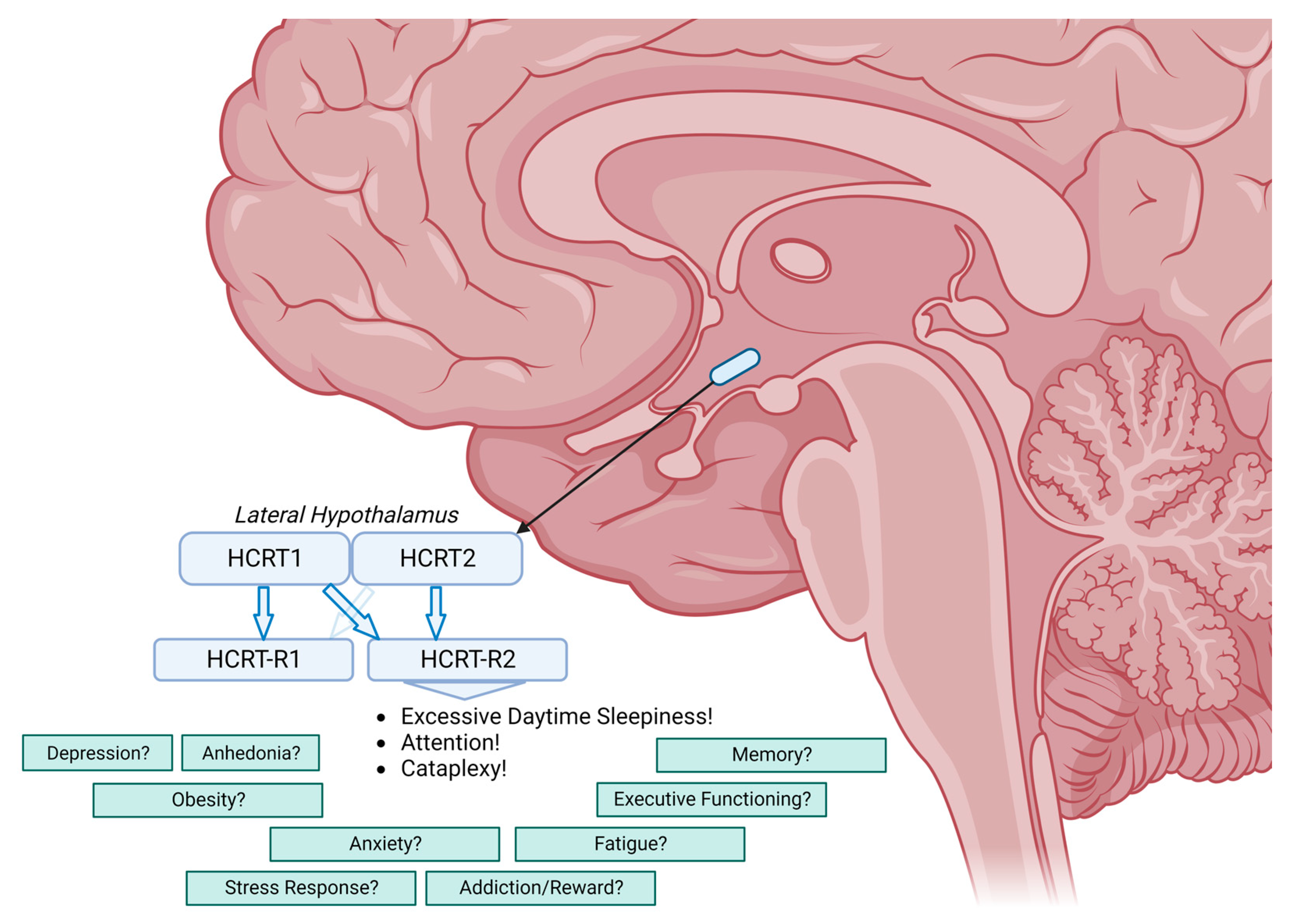
When the hypocretins are no longer produced, as in NT1, the entire range of sleep-related and non-sleep-related symptoms described above is seen. Both receptors must contribute to the phenotype (see Figure 1).
For the sleep-related symptoms (excessive daytime sleepiness, cataplexy, hypnagogic hallucinations and disturbed nocturnal sleep), receptor 2 is obviously the most important receptor. However, a contribution of receptor 1 failure to symptom severity is plausible, but only if receptor 2 is also down.
For the non-sleep-related symptoms, little is certain about the involvement of both receptors. In addiction, receptor 1 seems to be most important. In anxiety and depression, probably both receptors, and possibly the balance between the disturbance of both functions, plays a role. In obesity, receptor 1 seems to be important, but, interestingly, research with a dual 2 receptor shows that weight gain in the ataxin-3 mouse model for NT1 can be prevented by adding a receptor 2 agonist (Ishikawa T et al. Pharmacology Biochemistry and Behavior 2022). However, both appetite and metabolism play a role in the development of obesity, and it is likely that both receptors are involved in this complex interaction. For fatigue, it is even more unclear. Both receptors may be involved, but it may also occur as an indirect effect of the overall disruption of basal processes and/or be related to mood disturbance. For pain, receptor 1 may be the key player.
In a phase 2 randomised, placebo-controlled study with the hypocretin 2 agonist TAK-994—aborted for safety reasons—only the effect on the sleep-related symptoms of excessive daytime sleepiness and cataplexy is reported. The improvement of these symptoms, at least for the 8-week duration of the study, is spectacular [17].

Conclusions

Nearly 25 years have passed since the ground-breaking discovery that hypocretin deficiency underlies human narcolepsy with cataplexy (NT1). Over time, it has become increasingly evident that hypocretin deficiency goes beyond the conventional core symptoms, or pentad, traditionally associated with narcolepsy. The emergence of HCRTR-2 agonists presents an exciting opportunity, prompting us to explore the role of receptor 2 in the complete spectrum of NT1 symptoms.
It remains to be seen whether selective HCRTR-2 agonists will become the long-awaited holy grail for the effective treatment of NT1 in humans or serve only as a crucial stepping stone to even more advanced and causative therapies.
Figure 1. The two hypocretin peptides, hypocretin 1 and 2, result from the cleavage of the precursor preprohypocretin. Hypocretin 1 has an equal affinity for receptors 1 and 2, and hypocretin 2 has a much greater affinity for receptor 2 than for receptor 1. The role of both receptors in relation to the non-core symptoms of narcolepsy type 1 remains to be elucidated.
Ctn 07 00028 g001

Author Contributions

Writing—original draft preparation, review and editing, R.F. and G.J.L. All authors have read and agreed to the published version of the manuscript.

Funding

This research received no external funding.

Institutional Review Board Statement

Not applicable.

Informed Consent Statement

Not applicable.

Data Availability Statement

Not applicable.

Conflicts of Interest

The authors declare no conflict of interest.

References

  1. Lin, L.; Faraco, J.; Li, R.; Kadotani, H.; Rogers, W.; Lin, X.; Qiu, X.; de Jong, P.J.; Nishino, S.; Mignot, E. The Sleep Disorder Canine Narcolepsy Is Caused by a Mutation in the Hypocretin (Orexin) Receptor 2 Gene. Cell 1999, 98, 365–376. [Google Scholar] [CrossRef] [PubMed]
  2. Chemelli, R.M.; Willie, J.T.; Sinton, C.M.; Elmquist, J.K.; Scammell, T.; Lee, C.; Richardson, J.A.; Williams, S.C.; Xiong, Y.; Kisanuki, Y.; et al. Narcolepsy in Orexin Knockout Mice Molecular Genetics of Sleep Regulation. Cell 1999, 98, 437–451. [Google Scholar] [CrossRef] [PubMed]
  3. Liblau, R.S.; Latorre, D.; Kornum, B.R.; Dauvilliers, Y.; Mignot, E.J. The Immunopathogenesis of Narcolepsy Type 1. Nat. Rev. Immunol. 2023, 1–16. [Google Scholar] [CrossRef] [PubMed]
  4. Saitoh, T.; Sakurai, T. The Present and Future of Synthetic Orexin Receptor Agonists. Peptides 2023, 167, 171051. [Google Scholar] [CrossRef]
  5. Seifinejad, A.; Ramosaj, M.; Shan, L.; Li, S.; Possovre, M.-L.; Pfister, C.; Fronczek, R.; Garrett-Sinha, L.A.; Frieser, D.; Honda, M.; et al. Epigenetic Silencing of Selected Hypothalamic Neuropeptides in Narcolepsy with Cataplexy. Proc. Natl. Acad. Sci. USA 2023, 120, e2220911120. [Google Scholar] [CrossRef] [PubMed]
  6. Bassetti, C.L.A.; Adamantidis, A.; Burdakov, D.; Han, F.; Gay, S.; Kallweit, U.; Khatami, R.; Koning, F.; Kornum, B.R.; Lammers, G.J.; et al. Narcolepsy—Clinical Spectrum, Aetiopathophysiology, Diagnosis and Treatment. Nat. Rev. Neurol. 2019, 15, 519–539. [Google Scholar] [CrossRef]
  7. Maski, K.; Steinhart, E.; Williams, D.; Scammell, T.; Flygare, J.; McCleary, K.; Gow, M. Listening to the Patient Voice in Narcolepsy: Diagnostic Delay, Disease Burden, and Treatment Efficacy. J. Clin. Sleep Med. JCSM Off. Publ. Am. Acad. Sleep Med. 2016, 13, 419–425. [Google Scholar] [CrossRef]
  8. Evans, R.; Kimura, H.; Alexander, R.; Davies, C.H.; Faessel, H.; Hartman, D.S.; Ishikawa, T.; Ratti, E.; Shimizu, K.; Suzuki, M.; et al. Orexin 2 Receptor–Selective Agonist Danavorexton Improves Narcolepsy Phenotype in a Mouse Model and in Human Patients. Proc. Natl. Acad. Sci. USA 2022, 119, e2207531119. [Google Scholar] [CrossRef]
  9. Sakurai, T.; Amemiya, A.; Ishii, M.; Matsuzaki, I.; Chemelli, R.M.; Tanaka, H.; Williams, S.C.; Richardson, J.A.; Kozlowski, G.P.; Wilson, S.; et al. Orexins and Orexin Receptors: A Family of Hypothalamic Neuropeptides and G Protein-Coupled Receptors That Regulate Feeding Behavior. Cell 1998, 92, 573–585. [Google Scholar] [CrossRef] [PubMed]
  10. De Lecea, L.; Kilduff, T.S.; Peyron, C.; Gao, X.-B.; Foye, P.E.; Danielson, P.E.; Fukuhara, C.; Battenberg, E.L.F.; Gautvik, V.T.; Bartlett, F.S.; et al. The Hypocretins: Hypothalamus-Specific Peptides with Neuroexcitatory Activity. Proc. Natl. Acad. Sci. USA 1998, 95, 322–327. [Google Scholar] [CrossRef]
  11. Lammers, G.J.; Bassetti, C.L.A.; Dolenc-Groselj, L.; Jennum, P.J.; Kallweit, U.; Khatami, R.; Lecendreux, M.; Manconi, M.; Mayer, G.; Partinen, M.; et al. Diagnosis of Central Disorders of Hypersomnolence: A Reappraisal by European Experts. Sleep Med. Rev. 2020, 52, 101306. [Google Scholar] [CrossRef] [PubMed]
  12. Vassalli, A.; Franken, P. Hypocretin (Orexin) Is Critical in Sustaining Theta/Gamma-Rich Waking Behaviors That Drive Sleep Need. Proc. Natl. Acad. Sci. USA 2017, 114, E5464–E5473. [Google Scholar] [CrossRef] [PubMed]
  13. Xia, L.; Liu, H.Y.; Wang, B.Y.; Lin, H.N.; Wang, M.C.; Ren, J.-X. A Review of Physiological Functions of Orexin: From Instinctive Responses to Subjective Cognition. Medicine 2023, 102, e34206. [Google Scholar] [CrossRef] [PubMed]
  14. American Academy of Sleep Medicine. International Classification of Sleep Disorders, 3rd ed.; Text Revision; American Academy of Sleep Medicine: Darien, IL, USA, 2023. [Google Scholar]
  15. Lee, M.G.; Hassani, O.K.; Jones, B.E. Discharge of Identified Orexin/Hypocretin Neurons across the Sleep-Waking Cycle. J. Neurosci. 2005, 25, 6716–6720. [Google Scholar] [CrossRef]
  16. Hasegawa, E.; Yanagisawa, M.; Sakurai, T.; Mieda, M. Orexin Neurons Suppress Narcolepsy via 2 Distinct Efferent Pathways. J. Clin. Investig. 2014, 124, 604–616. [Google Scholar] [CrossRef]
  17. Dauvilliers, Y.; Mignot, E.; del Villegas, R.R.; Du, Y.; Hanson, E.; Inoue, Y.; Kadali, H.; Koundourakis, E.; Meyer, S.; Rogers, R.; et al. Oral Orexin Receptor 2 Agonist in Narcolepsy Type 1. N. Engl. J. Med. 2023, 389, 309–321. [Google Scholar] [CrossRef] [PubMed]
  18. Executive Functioning. Available online: Merriam-Webster.com (accessed on 1 August 2023).
  19. Gao, W.-R.; Hu, X.-H.; Yu, K.-Y.; Cai, H.-Y.; Wang, Z.-J.; Wang, L.; Wu, M.-N. Selective Orexin 1 Receptor Antagonist SB-334867 Aggravated Cognitive Dysfunction in 3xTg-AD Mice. Behav. Brain Res. 2023, 438, 114171. [Google Scholar] [CrossRef]
  20. Quaedackers, L.; Fortuyn, H.D.; Gilst, M.V.; Lappenschaar, M.; Overeem, S. Dissociative Symptoms Are Highly Prevalent in Adults with Narcolepsy Type 1. Behav. Sleep Med. 2022, 20, 63–73. [Google Scholar] [CrossRef]
  21. Fagan, H.; Jones, E.; Baldwin, D.S. Orexin Receptor Antagonists in the Treatment of Depression: A Leading Article Summarising Pre-Clinical and Clinical Studies. CNS Drugs 2023, 37, 1–12. [Google Scholar] [CrossRef]
  22. Scott, M.M.; Marcus, J.N.; Pettersen, A.; Birnbaum, S.G.; Mochizuki, T.; Scammell, T.E.; Nestler, E.J.; Elmquist, J.K.; Lutter, M. Hcrtr1 and 2 Signaling Differentially Regulates Depression-like Behaviors. Behav. Brain Res. 2011, 222, 289–294. [Google Scholar] [CrossRef]
  23. Ruoff, C.M.; Reaven, N.L.; Funk, S.E.; McGaughey, K.J.; Ohayon, M.M.; Guilleminault, C.; Black, J. High Rates of Psychiatric Comorbidity in Narcolepsy: Findings from the Burden of Narcolepsy Disease (BOND) Study of 9,312 Patients in the United States. J. Clin. Psychiatry 2016, 78, 171–176. [Google Scholar] [CrossRef] [PubMed]
  24. Abenza-Abildua, M.J.; Suárez-Gisbert, E.; Lores-Gutiérrez, V.; Algarra-Lucas, C.; Gómez-Aceña, Á.; Navacerrada-Barrero, F.J.; González-Martín, L.; Pérez-Villena, A.; Pérez-López, C. Anxiety and Depression in Patients with Narcolepsy. J. Sleep Res. 2023, 32, e13812. [Google Scholar] [CrossRef]
  25. Szakacs, Z.; Dauvilliers, Y.; Mikhaylov, V.; Poverennova, I.; Krylov, S.; Jankovic, S.; Sonka, K.; Lehert, P.; Lecomte, I.; Lecomte, J.-M.; et al. Safety and Efficacy of Pitolisant on Cataplexy in Patients with Narcolepsy: A Randomised, Double-Blind, Placebo-Controlled Trial. Lancet Neurol. 2017, 16, 200–207. [Google Scholar] [CrossRef]
  26. Inocente, C.O.; Gustin, M.-P.; Lavault, S.; Guignard-Perret, A.; Raoux, A.; Christol, N.; Gerard, D.; Dauvilliers, Y.; Reimão, R.; Bat-Pitault, F.; et al. Depressive Feelings in Children with Narcolepsy. Sleep Med. 2014, 15, 309–314. [Google Scholar] [CrossRef] [PubMed]
  27. Shan, L.; Linssen, S.; Harteman, Z.; Dekker, F.; Shuker, L.; Balesar, R.; Breesuwsma, N.; Anink, J.; Zhou, J.; Lammers, G.J.; et al. Activated Wake Systems in Narcolepsy Type 1. Ann. Neurol. 2023. [Google Scholar] [CrossRef] [PubMed]
  28. Kok, S.W.; Roelfsema, F.; Overeem, S.; Lammers, G.J.; Strijers, R.L.; Frölich, M.; Meinders, A.E.; Pijl, H. Dynamics of the Pituitary-Adrenal Ensemble in Hypocretin-Deficient Narcoleptic Humans: Blunted Basal Adrenocorticotropin Release and Evidence for Normal Time-Keeping by the Master Pacemaker. J. Clin. Endocrinol. Metab. 2002, 87, 5085–5091. [Google Scholar] [CrossRef] [PubMed]
  29. Maurovich-Horvat, E.; Keckeis, M.; Lattová, Z.; Kemlink, D.; Wetter, T.; Schuld, A.; Sonka, K.; Pollmächer, T. Hypothalamo–Pituitary–Adrenal Axis, Glucose Metabolism and TNF-α in Narcolepsy. J. Sleep Res. 2014, 23, 425–431. [Google Scholar] [CrossRef]
  30. Fortuyn, H.A.D.; Fronczek, R.; Smitshoek, M.; Overeem, S.; Lappenschaar, M.; Kalkman, J.; Renier, W.; Buitelaar, J.; Lammers, G.J.; Bleijenberg, G. Severe Fatigue in Narcolepsy with Cataplexy. J. Sleep Res. 2011, 21, 163–169. [Google Scholar] [CrossRef] [PubMed]
  31. Bassetti, C.L.; Baumann, C.R.; Scammell, T.E. Narcolepsy; Springer: Berlin/Heidelberg, Germany, 2011; ISBN 9781441983893. [Google Scholar]
  32. Fronczek, R.; Overeem, S.; Reijntjes, R.; Lammers, G.J.; van Dijk, J.G.; Pijl, H. Increased Heart Rate Variability but Normal Resting Metabolic Rate in Hypocretin/Orexin-Deficient Human Narcolepsy. J. Clin. Sleep Med. JCSM Off. Publ. Am. Acad. Sleep Med. 2008, 4, 248–254. [Google Scholar] [CrossRef]
  33. Straat, M.E.; Schinkelshoek, M.S.; Fronczek, R.; Lammers, G.J.; Rensen, P.C.N.; Boon, M.R. Role of Brown Adipose Tissue in Adiposity Associated with Narcolepsy Type 1. Front. Endocrinol. 2020, 11, 145. [Google Scholar] [CrossRef]
  34. Sellayah, D.; Bharaj, P.; Sikder, D. Orexin Is Required for Brown Adipose Tissue Development, Differentiation, and Function. Cell Metab. 2011, 14, 478–490. [Google Scholar] [CrossRef]
  35. Schinkelshoek, M.S.; Smolders, I.M.; Donjacour, C.E.; Meijden, W.P.; Zwet, E.W.; Fronczek, R.; Lammers, G.J. Decreased Body Mass Index during Treatment with Sodium Oxybate in Narcolepsy Type 1. J. Sleep Res. 2019, 28, e12684. [Google Scholar] [CrossRef] [PubMed]
  36. Kakizaki, M.; Tsuneoka, Y.; Takase, K.; Kim, S.J.; Choi, J.; Ikkyu, A.; Abe, M.; Sakimura, K.; Yanagisawa, M.; Funato, H. Differential Roles of Each Orexin Receptor Signaling in Obesity. iScience 2019, 20, 1–13. [Google Scholar] [CrossRef]
  37. Peyron, C.; Tighe, D.K.; van den Pol, A.N.; de Lecea, L.; Heller, H.C.; Sutcliffe, J.G.; Kilduff, T.S. Neurons Containing Hypocretin (Orexin) Project to Multiple Neuronal Systems. J. Neurosci. 1998, 18, 9996–10015. [Google Scholar] [CrossRef]
  38. Harris, G.C.; Wimmer, M.; Aston-Jones, G. A Role for Lateral Hypothalamic Orexin Neurons in Reward Seeking. Nature 2005, 437, 556–559. [Google Scholar] [CrossRef] [PubMed]
  39. Thannickal, T.C.; John, J.; Shan, L.; Swaab, D.F.; Wu, M.-F.; Ramanathan, L.; McGregor, R.; Chew, K.-T.; Cornford, M.; Yamanaka, A.; et al. Opiates Increase the Number of Hypocretin-Producing Cells in Human and Mouse Brain and Reverse Cataplexy in a Mouse Model of Narcolepsy. Sci. Transl. Med. 2018, 10, eaao4953. [Google Scholar] [CrossRef] [PubMed]
  40. Scammell, T.E.; Saper, C.B. Orexins: Looking Forward to Sleep, Back at Addiction. Nat. Med. 2007, 13, 126–128. [Google Scholar] [CrossRef]
  41. Yamamoto, H.; Nagumo, Y.; Ishikawa, Y.; Irukayama-Tomobe, Y.; Namekawa, Y.; Nemoto, T.; Tanaka, H.; Takahashi, G.; Tokuda, A.; Saitoh, T.; et al. OX2R-Selective Orexin Agonism Is Sufficient to Ameliorate Cataplexy and Sleep/Wake Fragmentation without Inducing Drug-Seeking Behavior in Mouse Model of Narcolepsy. PLoS ONE 2022, 17, e0271901. [Google Scholar] [CrossRef] [PubMed]
  42. Bingham, S.; Davey, P.T.; Babbs, A.J.; Irving, E.A.; Sammons, M.J.; Wyles, M.; Jeffrey, P.; Cutler, L.; Riba, I.; Johns, A.; et al. Orexin-A, an Hypothalamic Peptide with Analgesic Properties. Pain 2001, 92, 81–90. [Google Scholar] [CrossRef]
  43. Asano, H.; Arima, Y.; Yokota, S.; Fujitani, M. New Nociceptive Circuits to the Hypothalamic Perifornical Area from the Spinal Cord and Spinal Trigeminal Nucleus via the Parabrachial Nucleus. Biochem. Biophys. Res. Commun. 2019, 512, 705–711. [Google Scholar] [CrossRef]
  44. Holland, P.R.; Akerman, S.; Goadsby, P.J. Orexin 1 Receptor Activation Attenuates Neurogenic Dural Vasodilation in an Animal Model of Trigeminovascular Nociception. J. Pharmacol. Exp. Ther. 2005, 315, 1380–1385. [Google Scholar] [CrossRef] [PubMed]
  45. Yamamoto, T.; Saito, O.; Shono, K.; Aoe, T.; Chiba, T. Anti-Mechanical Allodynic Effect of Intrathecal and Intracerebroventricular Injection of Orexin-A in the Rat Neuropathic Pain Model. Neurosci. Lett. 2003, 347, 183–186. [Google Scholar] [CrossRef]
  46. Dahmen, N.; Kasten, M.; Wieczorek, S.; Gencik, M.; Epplen, J.; Ullrich, B. Increased Frequency of Migraine in Narcoleptic Patients: A Confirmatory Study. Cephalalgia 2003, 23, 14–19. [Google Scholar] [CrossRef]
  47. Chabi, A.; Zhang, Y.; Jackson, S.; Cady, R.; Lines, C.; Herring, W.J.; Connor, K.M.; Michelson, D. Randomized Controlled Trial of the Orexin Receptor Antagonist Filorexant for Migraine Prophylaxis. Cephalalgia 2014, 35, 379–388. [Google Scholar] [CrossRef] [PubMed]
Disclaimer/Publisher’s Note: The statements, opinions and data contained in all publications are solely those of the individual author(s) and contributor(s) and not of MDPI and/or the editor(s). MDPI and/or the editor(s) disclaim responsibility for any injury to people or property resulting from any ideas, methods, instructions or products referred to in the content.