Article

Physicochemical Morphological Evaluation and Stability Assessment of Nanoemulsions Containing Nutrients for Parenteral Nutrition

by
Panos Papandreou
1,*,
Efstathia Triantafyllopoulou
2,†,
Ioannis Pispas
3,4,†,
Sophia Havaki
5,
Aristeidis Papagiannopoulos
3,
Vassilis G. Gorgoulis
5,6,7,8,9 and
Natassa Pippa
2,*
1
Parenteral Nutrition and Intravenous Solutions Compounding Unit, IASO General Hospital, Marousi, 15123 Athens, Greece
2
Section of Pharmaceutical Technology, Department of Pharmacy, School of Health Sciences, National and Kapodistrian University of Athens (NKUA), Panepistimioupolis Zografou, 15771 Athens, Greece
3
Theoretical and Physical Chemistry Institute, National Hellenic Research Foundation, 48 Vassileos Constantinou Avenue, 11635 Athens, Greece
4
Department of Physics, School of Applied Mathematical and Physical Sciences, National Technical University of Athens, Heroon Polytechniou 9, Zografou, 15780 Athens, Greece
5
Molecular Carcinogenesis Group, Department of Histology and Embryology, Medical School, National and Kapodistrian University of Athens, 75 Mikras Asias Str., 11527 Athens, Greece
6
Ninewells Hospital and Medical School, University of Dundee, Dundee DD2 1UB, UK
7
Biomedical Research Foundation, Academy of Athens, 10679 Athens, Greece
8
Faculty Institute for Cancer Sciences, Manchester Academic Health Sciences Centre, University of Manchester, Manchester M20 4GJ, UK
9
Faculty of Health and Medical Sciences, University of Surrey, Surrey GU2 7XH, UK
*
Authors to whom correspondence should be addressed.
These authors contributed equally to this work.
Colloids Interfaces 2025, 9(5), 64; https://doi.org/10.3390/colloids9050064
Submission received: 8 August 2025 / Revised: 16 September 2025 / Accepted: 19 September 2025 / Published: 25 September 2025

Abstract

Parenteral nutrition is an integral part of the nutritional support of critically ill neonates, infants, and children in the intensive care units (ICUs) and at home. Therefore, the adequacy and the effectiveness of parenteral nutrition, PN, support are among the major concerns of doctors and pharmacists. The aim of this study is the physicochemical and stability evaluation of nanoemulsions, which are used for parenteral nutrition. These nanoemulsions are for intravenous (IV) administration of lipids, amino acids, glucose, electrolytes, trace elements as well as vitamins. Light scattering techniques are used for the identification of the hydrodynamic diameter (Dh), size polydispersity index (PDI), and the ζ-potential of the prepared nanoemulsions. Stability assessment is performed in different conditions, mimicking those of the hospital. The stability studies involve shelf-life measurement of these NEs over 10 days in two storage conditions (25 °C and 4 °C) using dynamic light scattering. According to the US Pharmacopeia, the droplet size should be under the upper limit of 500 nm (0.5 μm). Transmission electron microscopy (TEM) is used for the shape of the droplets of the nanoemulsion emulsion for parenteral nutrition for the first time. The results showed that the droplet size was around 300 nm, with a homogeneous population and negative ζ-potential. The morphology was vesicular and spherical, typical for NE droplet shape. The results from all the characterization techniques show that the formulations meet the high-quality standards of nanoemulsions for neonates, infants and children.

1. Introduction

Nanoemulsions (NE) are considered nanosystems due to the size of their droplets in the nanoscale [1]. They are composed of two immiscible liquids, water and oil. Surfactants play a key role in the structure and viscoelastic properties, as well as in the stability of NE. NEs are widely used in pharmaceutical technology for the encapsulation, protection, and controlled release of active substances [1]. NE is a formulation strategy for the increase in the bioavailability of poorly water-soluble active substances [2]. They can be administered by different routes, including the oral, transdermal, and parental. The rapid onset pharmacological action of NE is responsible for the fast therapeutic effect because the active substance reaches the systematic circulation quickly. NEs are prepared both in laboratory and industrial scale via different production protocols, including high- and low-energy input. Last but not least, NEs can be sterilized using filtration due to their nanosized droplets, making them identical for IV administration and for parenteral nutrition [1,2,3,4]. NEs are thermodynamically unstable isotropic nanosystems, and for this reason, the stability assessment is required after their preparation. The size which is synonymous to the droplet size, size distribution, ζ-potential, and stability over time are considered the Critical Quality Attributes (CQAs) of NEs. Additionally, the transparent appearance is also crucial for the usage of NEs. The surfactants mentioned above are one of the Critical Material Attributes (CMAs) of the NE, accompanied by the nature of the oil and the aqueous phase. The improved physical stability is strongly associated with the nature and the percentage of the surfactants [3,4]. Younger preterm newborns have lower nutritional resources, which puts them at a higher risk of postnatal malnutrition, particularly at very low gestational ages. Because of this, early growth and nutrition at this life period are crucial in determining the long-term health of premature infants [5]. It is well-known that in parenteral nutrition (parental nutrition ), intravenous fat emulsions are a dense source of energy that supply necessary fatty acids [6]. According to a recent prospective observational study, Wang and colleagues (2021) declared that the majority of preterm infants with birth weight less than 1500 g remain lower than the nutritional requirements [7]. A newly published randomized controlled trial showed the significant role of parental nutrition in the cases of parenteral nutrition-associated cholestasis, which is strongly associated with the low birth weight and preterm newborns [8]. From the Skouroliakou group, nanoemulsion formulations have already been developed. The all-in-one parenteral admixture with SMOFlipid (soybean oil; medium-chain triglycerides; olive oil; fish oil) as an alternative ingredient to soybean used for neonates was also found stable physicochemically for the time-period of clinical use. The SMOFlipid was a nanoemulsion with α-tocopherol-enriched lipid [9]. Its usage favors oxidation resistance due to its components: vitamin E, which is an antioxidant compound and a mixture of lipids including ones that are less prone to lipid peroxidation, such as medium-chain triglycerides, eicosapentaenoic and docosahexaenoic acids [10]. According to a double-blind clinical trial, the administration of these nanoemulsions exhibited an added value for the preterm newborns [11].
The aim of this study was the physicochemical and stability evaluation of nanoemulsions, which are used for parental nutrition. Light scattering techniques were used for the determination of the hydrodynamic diameter (Dh), polydispersity index (PDI), and the zeta potential (ζ-potential) of the prepared nanoemulsions. Stability assessment was performed under different conditions, mimicking those of the hospital. The stability studies involve shelf-life measurement of these NEs over 10 days in two storage conditions (25 °C and 4 °C) using dynamic light scattering. In general, the nanoemulsions intended for parenteral use are consumed within the hospital in 24 h or at most 48 h. The reason the physicochemical characterization took 9 days is to ensure that the physicochemical characteristics remained unaffected during a longer period for safety reasons [12,13]. These extended periods of stability tests are strong evidence of risk-based assessment for the self-life of the nanoemulsions. According to the US Pharmacopeia, the droplet size should be under the upper limit of 500 nm (0.5 μm). To the best of the authors’ knowledge, this is the first time that an analysis of the relationship between the formulation’s constituent parts and a clinically useful NE’s stability behavior has been carried out. These NEs are widely used all-in-one parenteral admixtures in hospitals [14].

2. Materials and Methods

2.1. Materials

The formulations of sample 1 (Table 1) and sample 2 (Table 2) are described in Table 1 and Table 2, respectively. The ingredients of the formulations, as well as the macronutrients and the energy, are included in Table 3 and Table 4 for the formulation of sample 1 and in Table 5 and Table 6 for sample 2. In some cases in the tables, the units are expressed per kilogram of the neonates, infants, and children. Peditrace, SMOFlipid, Soluvit, Magnesium sulfate heptahydrate, Glucose solutions, Calcium gluconate 10% solution, Vitalipid Infant, and Vamin Infant were obtained from Fresenius Kabi (Bad Homburg, Germany), while sodium chloride and potassium chloride were purchased from SDS (Peypin, France). The selected containers were ethyl vinyl acetate plastic bags that were automatically filled (MicroMacro 12, Baxa, Englewood, CO, USA).

2.2. Methods

2.2.1. Preparation of NEs

According to previously published data [12], when formulating an NE for parental nutrient daily formula for neonates, numerous factors should be taken into consideration such as age, weight, clinical state of the neonate/child, and environmental conditions—type of incubator, phototherapy, etc. In this investigation, the all-in-one parenteral admixture NEs that were formulating were placed into ethyl vinyl acetate plastic bags using an automated compounder (MicroMacro 12, Baxa, Englewood, CO, USA).

2.2.2. Dynamic Light Scattering

A standard ALV system (ALV GmbH, Hessen, Germany) was utilized to perform dynamic light scattering (DLS) measurements. The analysis was conducted by implementing an ALV-CG-3 goniometer and an ALV-5000/EPP multi-tau digital correlator with a He-Ne laser at λ = 632.8 nm. In DLS, the time-autocorrelation function of scattered light intensity g 2 ( q , τ ) is linked to the field autocorrelation function g 1 ( q , τ ) via the Siegert equation (Equation (1)), where β is a normalization constant [15,16]. The field autocorrelation function then provides a relaxation rate Γ distribution, which is transformed into a diffusion coefficient D distribution ( Γ = D · q 2 = 1 / τ ) . Then, the Stokes–Einstein equation (Equation (2)) is used to calculate the hydrodynamic radii Rh, with kB as the Boltzmann constant, T as the sample temperature, and η as the solvent viscosity. The CONTIN algorithm was used to examine the dispersed intensity-weighted distributions of hydrodynamic radii at θ = 90°. Each experiment was performed in triplicate with 30 s duration [17].
g 2 q , τ 1 = β | g 1 ( q , τ ) | 2
R h = k B T 6 π η D

2.2.3. Electrophoretic Light Scattering

A Zetasizer Nano-ZS (Malvern Instruments Ltd., Malvern, UK) was used to perform electrophoretic light scattering (ELS) measurements. The device uses a combination of laser Doppler velocimetry and phase analysis light scattering (PALS). Equation (3) shows the relation between electrophoretic mobility μe and frequency shift ∆f, with no as the solvent refractive index, λ as the wavelength of the laser, θ as the scattering angle, and E as the electric field. Equation (4), namely Henry’s equation, connects the electrophoretic mobility μe to the ζ-potential, with εo as the solvent dielectric constant in vacuum, εr as the relative dielectric constant, η as the solvent viscosity, f(ka) as the Henry function, k as the reciprocal Debye length λD, and a as the particle radius. The product ka refers to the ratio between the thickness of the electrical double layer and the particle radius.
μ e = λ n o sin θ · 1 E · Δ f
μ e = 2 3 · ε o ε r ζ f ( ka ) η
Under the Smoluchowski approximation, Henry’s function is constant and equal to 3/2, and ζ-potential can be given by Equation (5) [18,19]. The apparatus converts the electrophoretic mobilities into a ζ-potential distribution by performing several measurements of the mobilities at a fixed scattering angle of θ = 173° using PALS. To find the mean value and standard deviation of the ζ-potential, ten measurements were collected at T = 25 °C [20].
ζ = η μ e ε ο ε r

2.2.4. Transmission Electron Microscopy (TEM)

Two samples of nanoemulsions were examined with Transmission Electron Microscopy applying the negative staining technique. Specifically, 5 μL of each sample was allowed to be absorbed for 5 min to the surface of Formvar/Carbon coated copper, 200 mesh-square grids. The used grids were placed previously in a glow discharge unit to render them hydrophilic. After absorption, each grid was blotted with filter paper, washed thrice on drops of ultrapure water, and placed on a drop of 2% aqueous uranyl acetate (UA) solution for 2 min. The excess UA was removed, and the grids were left to air dry. The specimens were then observed in a JEM 2100Plus Transmission Electron Microscope (JEOL Ltd., Tokyo, Japan) operating at 120 kV and equipped with LaB6 filament. Images were photographed at original magnification 50,000× using a Gatan OneView digital camera (Gatan, Pleasanton, CA, USA). The digital camera was calibrated at each magnification setting using a reference grid with grating replica and latex spheres, according to the instructions of Gatan.

3. Results and Discussion

3.1. Nanoemulsion for Parental Nutrition

The prescribed regimens were calculated by the clinical decision support program NutriNet-parental nutrition, which analyzes the input data and calculates a daily regimen prescribed at IASO Hospital.
The prescribed regiment provides total energy, protein, glucose, and fatty substances. Also, the parental nutrition regimens included micronutrients (Table 1 and Table 2). The weight ratios of amino acids and fat (in mg) were less than the one referred to in the literature. The program calculates the ratio mEq Ca/mmol P, the osmolarity of parenteral solution, and the ratio non protein kcal/g. NutriNet-parental nutrition enables the user to print an order form for the patient record as well as a label that gets placed on the patient’s bag. It is also linked with an automated compounding device (ACD) for the preparation of the parental nutrition bag, and any data can be exported for statistical or data collection purposes. The prescribed solutions were to be examined by the compounding device.
The use of a specialized software program results in an adequate provision of energy and nutrients. Due to a variety of available current practice guidelines that exist (allowing individualized patient parental nutrition prescribing), patients’ requirements should be balanced with a standardized process regarding the stability to enhance their safety and reduce errors. Nowadays, healthcare automation is a useful tool to overcome the burden of calculations, including stability parameters, thus enabling the successful provision of individualized parental nutrition to become an efficient and safe standard routine procedure. NutriNet-parental nutrition and similar software will lead to the consistency of parental nutrition prescriptions through their screening of algorithm input information on stability substance and clinical application.
Four batches of five AIO admixtures containing macro- and micronutrients were compounded with commercially available lipid emulsions and amino acid solutions. Their stability was tested under conditions simulating clinical use. All the admixtures were assessed for criteria set by the USP: (a) under the upper limit (0.5 μm) set and (b) percentage of volume weighted particles with diameter more than 5 μm (PFAT5). In this study, the characteristics of the samples are analyzed in Table 3 and Table 4 (sample 1) and Table 5 and Table 6 (sample 2) (see Section 2.1). Details about the ingredients, the micronutrients, and the energy are also given.

3.2. Physicochemical Evaluation and Stability Assessment of the Nanoemulsions

Physicochemical properties of the two nanoemulsions were assessed in HPLC grade water on the day of preparation, with results summarized in Table 7 and Figure 1. The dispersed droplets exhibit a mean hydrodynamic diameter of 328 nm (sample 1) or 299 nm (sample 2) and comprise homogenous populations (PDI = 0.1). Based on our measurements, the SMOFlipid (four-oil lipid emulsion) with a concentration of 200 mg/mL forms lipid vesicles with a uniform size distribution. Their size is not significantly different. This is expected because the oil phase is not greatly different in the two samples. The lipids are surfactants that can self-assemble into aqueous media in different (vesicular and/or compact) structures. Moreover, they preserved their dimensions in PBS and at 37 °C, indicating that the samples could be stable in such environmental conditions (Table 7 and Figure 1). The subtle broadening of the size distribution of sample 2 in PBS and at 37 °C was probably related to mild and potential aggregation of the nanoemulsions. Such behavior is possible given the increased salinity present in PBS solutions in relation to HPLC grade water.
Moreover, they preserved their dimensions in PBS and at 37 °C, indicating that the samples could be stable in such environmental conditions (Table 7 and Figure 1). Zeta potential absolute values of dispersions larger than 30 mV suggest that the electrostatic repulsion is greater than the attractive forces according to Derjaguin–Landau–Verwey–Overbeek (DLVO) theory and are deemed as stable [21,22]. Therefore, both formulations seem to maintain adequate stability according to ζ-potential measurements with values approximately equal to −35 mV (Table 7). The presence of the trace elements is responsible for the high zeta potential values (Table 1 and Table 2). On the other hand, the formulation includes both chaotropic (i.e., Ca2+, Mg2+) and kosmotropic (i.e., K+, Na+ and PHO4) ions, according to the Hofmeister series. The presence of chaotropic ions is responsible for breaking the structure of water molecules onto the surface of the colloidal systems [23].
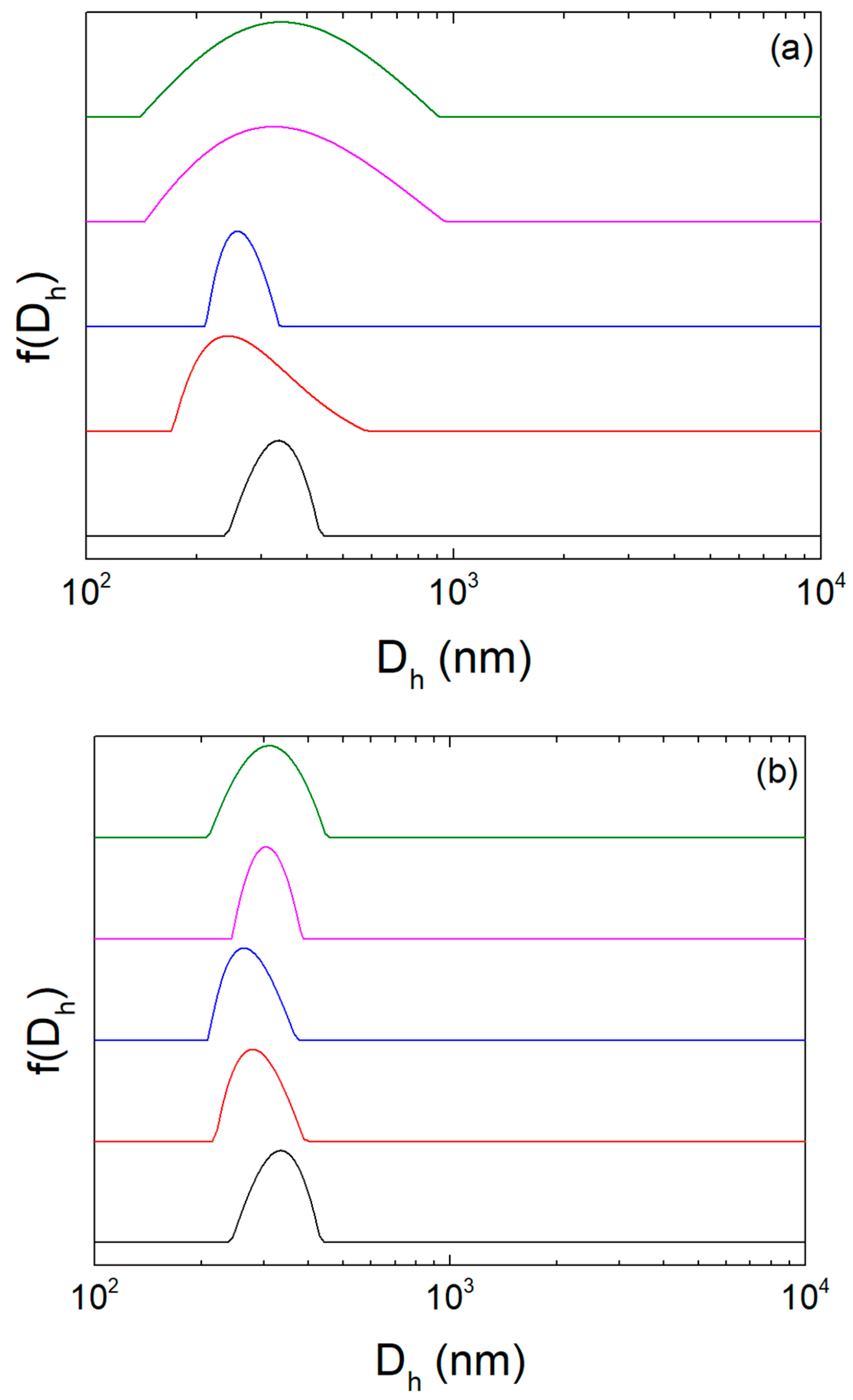
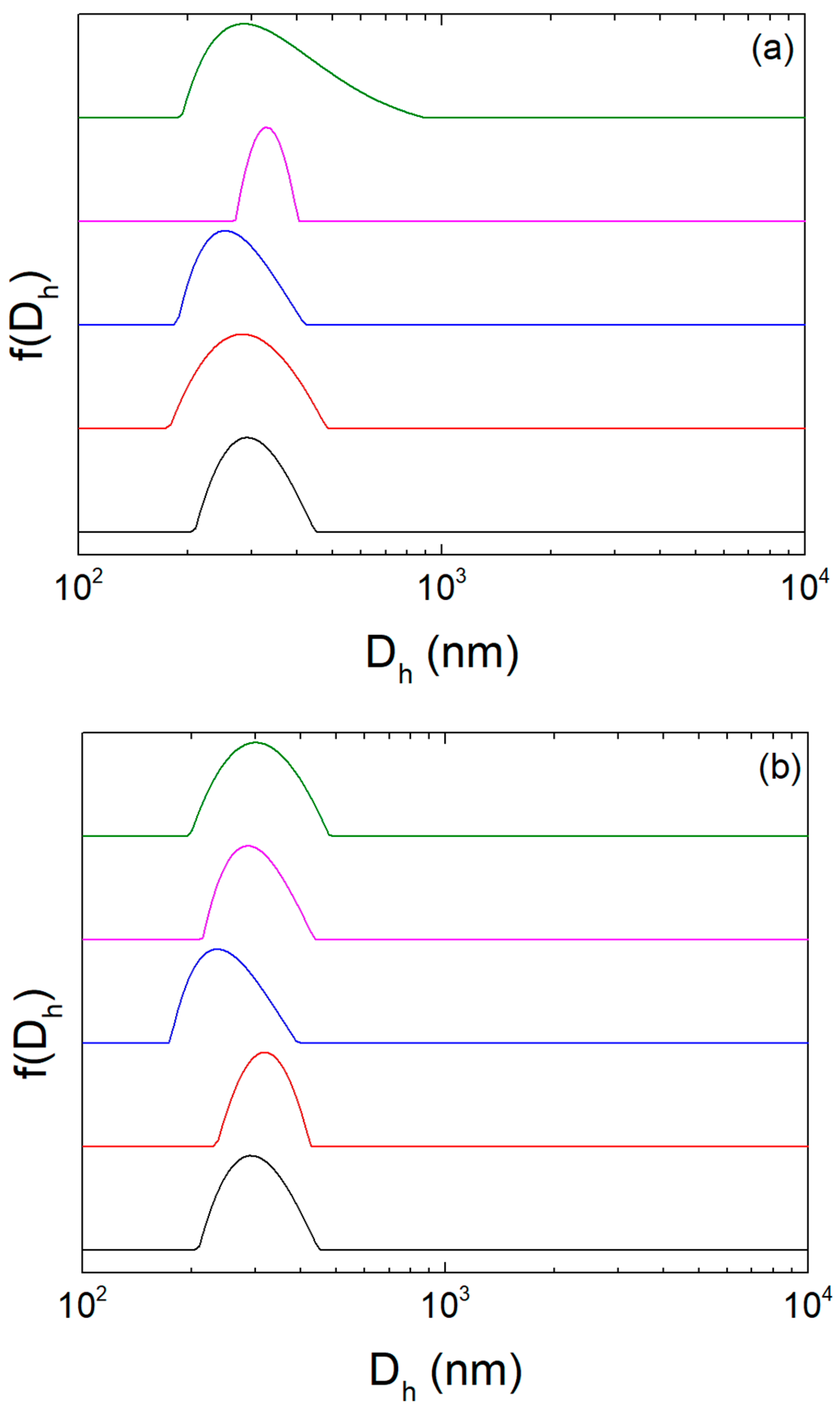
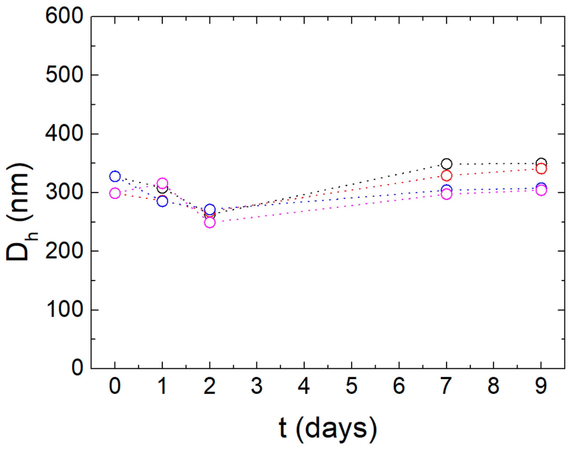
This is further supported by their stability assessment (Table 8 and Figure 2, Figure 3 and Figure 4). Considering lipid oxidation possibility, all formulations were protected from light and air during storage. It should be noted that these nanoemulsions are prepared in the hospital, and for this reason the preparation protocol is easy, and the duration of the stability assessment is 9 days. Namely, their dimensions did not change significantly for at least 48 h at ambient temperature, although there is an observed decrease in particle size from Day 0 to Day 2. Nanoemulsions may undergo interfacial rearrangements after preparation. The size decrease pattern could be attributed to interfacial stabilization [24]. After 9 days their size distributions corresponded to wider peaks implying that the formation of aggregates was initiated though. However, storage at 4 °C improved the dispersions stability for at least 9 days (Table 8, Figure 3). Even though the sensitivity of the dynamic light scattering (DLS) technique to larger particles may have contributed to an observed size variation (Table 8) [25], in fact the stability is demonstrated due to narrow size distributions of similar mean size for at least 9 days (Figure 3).
Namely, their dimensions did not change significantly for at least 48 h at ambient temperature, whereas after 9 days their size distributions corresponded to wider peaks, implying that the formation of aggregates was initiated. The high ζ-potential values, as well as the presence of glucose, gave the systems the required physicochemical stability over time. The electrostatic interactions caused by high ζ-potential values and the hydration forces due to the presence of glucose are responsible for keeping the lipidic droplets well separated from each other and lead to the absence of aggregation or sedimentation phenomena. According to the extended or modified DLVO theory, the hydration forces can also contribute to the stability of dispersions [26,27,28]. However, storage at 4 °C improved the dispersions stability, exhibiting narrow peaks of similar mean size for at least 9 days (Figure 3). Even though physical stability is not necessarily accompanied by chemical stability, lower storage temperature could enhance emulsion chemical stability as well due to lipid peroxidation dependance on storage conditions [9,10,29]. This can be attributed to lower coalescence and flocculation rates and more stable emulsifier organization at the oil–water interface that are expected at the lower temperature. Based on the physicochemical data, both dispersions fulfill the USP criteria for injectable lipid emulsions, since they have an average diameter that does not exceed 500 nm and could remain stable for at least 48 h at ambient temperature and 9 days at 4 °C [30].

3.3. Morphological Characterization of the Nanoemulsions

Shape and morphological characteristics of droplets are critical quality attributes of nanoemulsions, which affects their stability over time as well as interactions with living cells. TEM is widely used for the characterization of the microstructure of nanoemulsions, giving important information about the droplet size and morphology, as well as stability assessment [31,32].
Figure 5 and Figure 6 show the TEM micrographs of samples 1 and 2, respectively. In both cases, nanoparticulate structures and sample 1 aggregates especially are observed (Figure 5a,b). The sizes of the nanoparticles are in good agreement with those observed by DLS, keeping in mind that DLS is biased towards larger sizes as size distributions are weighted by the scattered intensity. It should be noted that the TEM images are obtained from the dispersed samples, avoiding the visualization of the particles/flocculates from the cake at the bottom of the vial. These samples are representative of the dispersed/solution state of the samples that were used for the characterization from DLS. Generally, dark droplets are observed via TEM images in the cases of formation of nanoemulsions [31,32]. Spherical droplets with a size around 300 nm in diameter and multicompartment structures with darker walls are present in sample 1 (Figure 5b). For sample 2, vesicular structures are visualized in Figure 6b. In our case, the darker walls appear like that because of the higher amount of uranyl stain absorption at the outer surface of the structures, in the context of negative staining, resulting in the “3D-depiction” of the structures (Figure 5 and Figure 6).
The different amounts of fats are probably responsible for the self-assembly into deformed bicompartmental structures. According to the literature, these structures are observed as intermediate and metastable phases during the formation of microemulsions, which are prepared by unicameral nanovesicles/nanoemulsions [33].

4. Conclusions

The designed nanoemulsions were found to have the desired physicochemical characteristics as characterization by light scattering methods indicated. This was proved by the mean sizes (~300 nm) and their relatively narrow size distributions (PDI~0.1) that were not perturbed in physiological conditions (PBS at 37 °C). To the best of the author’s knowledge, this is the first time TEM is used for the morphological characterization of emulsions for parenteral nutrition. The formulations showed sufficient colloidal stability for the intended application, which was attributed to their strongly negative ζ-potential. We believe that the extended stability assessment and morphological characterization could offer new insights into the regulatory framework of nanoemulsions, supporting a more risk-based approach to defining their shelf life.

Author Contributions

Conceptualization, P.P. and N.P.; methodology, E.T., I.P., S.H., A.P. and N.P.; validation, E.T., I.P., S.H., A.P. and N.P.; formal analysis, E.T., I.P., S.H., A.P. and N.P.; investigation, E.T., I.P. and S.H.; data curation, E.T., I.P., S.H., A.P. and N.P.; writing—original draft preparation, N.P.; writing—review and editing, E.T., I.P., S.H., A.P. and N.P.; supervision, A.P., V.G.G. and N.P. All authors have read and agreed to the published version of the manuscript.

Funding

This research received no external funding.

Data Availability Statement

The original contributions presented in this study are included in the article. Further inquiries can be directed to the corresponding authors.

Conflicts of Interest

The authors declare no conflicts of interest.

Abbreviations

The following abbreviations are used in this manuscript:
ACDAutomated Compounding Device
CΜACritical Material Attribute
CQACritical Quality Attribute
DLSDynamic Light Scattering
DLVODerjaguin–Landau–Verwey–Overbeek Theory
ICUIntensive Care Units
NENanoemulsions
PBSPhosphate-Buffer Saline
PDIPolydispersity Index
TEMTransmission Electron Microscopy
UAUranyl Acetate

References

  1. Singh, Y.; Meher, J.G.; Raval, K.; Khan, F.A.; Chaurasia, M.; Jain, N.K.; Chourasia, M.K. Nanoemulsion: Concepts, development and applications in drug delivery. J. Control. Release 2017, 252, 28–49. [Google Scholar] [CrossRef] [PubMed]
  2. Pandey, P.; Gulati, N.; Makhija, M.; Purohit, D.; Dureja, H. Nanoemulsion: A Novel Drug Delivery Approach for Enhancement of Bioavailability. Recent Patents Nanotechnol. 2020, 14, 276–293. [Google Scholar] [CrossRef]
  3. Banasaz, S.; Morozova, K.; Ferrentino, G.; Scampicchio, M. Encapsulation of Lipid-Soluble Bioactives by Nanoemulsions. Molecules 2020, 25, 3966. [Google Scholar] [CrossRef]
  4. Elzayat, A.; Adam-Cervera, I.; Álvarez-Bermúdez, O.; Muñoz-Espí, R. Nanoemulsions for synthesis of biomedical nanocarriers. Colloids Surfaces B Biointerfaces 2021, 203, 111764. [Google Scholar] [CrossRef] [PubMed]
  5. Rizzo, V.; Capozza, M.; Panza, R.; Laforgia, N.; Baldassarre, M.E. Macronutrients and Micronutrients in Parenteral Nutrition for Preterm Newborns: A Narrative Review. Nutrients 2022, 14, 1530. [Google Scholar] [CrossRef] [PubMed]
  6. Anez-Bustillos, L.; Dao, D.T.; Baker, M.A.; Fell, G.L.; Puder, M.; Gura, K.M. Intravenous Fat Emulsion Formulations for the Adult and Pediatric Patient. Nutr. Clin. Pract. 2016, 31, 596–609. [Google Scholar] [CrossRef]
  7. Wang, N.; Cui, L.; Liu, Z.; Wang, Y.; Zhang, Y.; Shi, C.; Cheng, Y. Optimizing parenteral nutrition to achieve an adequate weight gain according to the current guidelines in preterm infants with birth weight less than 1500 g: A prospective observational study. BMC Pediatr. 2021, 21, 303. [Google Scholar] [CrossRef]
  8. Wang, Y.-S.; Shen, W.; Yang, Q.; Lin, R.; Tang, L.-X.; Bai, R.-M.; Yang, D.; Zhang, J.; Zhang, Y.-J.; Yu, W.-T.; et al. Analysis of risk factors for parenteral nutrition-associated cholestasis in preterm infants: A multicenter observational study. BMC Pediatr. 2023, 23, 250. [Google Scholar] [CrossRef]
  9. Skouroliakou, M.; Kountouri, A.M.; Hatziantoniou, S.; Koutri, K.; Chiou, A. Physicochemical stability assessment of all-in-one parenteral emulsion for neonates containing SMOFlipid. Eur. J. Hosp. Pharm. 2012, 19, 514–518. [Google Scholar] [CrossRef]
  10. Boullata, J.I.; Mirtallo, J.M.; Sacks, G.S.; Salman, G.; Gura, K.; Canada, T.; Maguire, A. the ASPEN Parenteral Nutrition Safety Committee Parenteral nutrition compatibility and stability: A comprehensive review. J. Parenter. Enter. Nutr. 2021, 46, 273–299. [Google Scholar] [CrossRef]
  11. Papandreou, P.; Gioxari, A.; Ntountaniotis, D.; Korda, O.-N.; Skouroliakou, M.; Siahanidou, T. Administration of an Intravenous Fat Emulsion Enriched with Medium-Chain Triglyceride/ω-3 Fatty Acids is Beneficial Towards Anti-Inflammatory Related Fatty Acid Profile in Preterm Neonates: A Randomized, Double-Blind Clinical Trial. Nutrients 2020, 12, 3526. [Google Scholar] [CrossRef] [PubMed]
  12. Suliman, A.S.; Tom, R.; Palmer, K.; Tolaymat, I.; Younes, H.M.; Arafat, B.; Elhissi, A.M.A.; Najlah, M. Development, characterization and stability evaluation of ciprofloxacin-loaded parenteral nutrition nanoemulsions. Pharm. Dev. Technol. 2020, 25, 579–587. [Google Scholar] [CrossRef] [PubMed]
  13. Bouchoud, L.; Sadeghipour, F.; Klingmüller, M.; Fonzo-Christe, C.; Bonnabry, P. Long-term physico-chemical stability of standard parenteral nutritions for neonates. Clin. Nutr. 2010, 29, 808–812. [Google Scholar] [CrossRef]
  14. Skouroliakou, M.; Matthaiou, C.; Chiou, A.; Panagiotakos, D.; Gounaris, A.; Nunn, T.; Andrikopoulos, N. Physicochemical Stability of Parenteral Nutrition Supplied as All-in-One for Neonates. J. Parenter. Enter. Nutr. 2008, 32, 201–209. [Google Scholar] [CrossRef]
  15. Berne, B.J.; Pecora, R. Dynamic Light Scattering: With Applications to Chemistry, Biology, and Physics; Dover Publications Inc: Mineola, NY, USA, 2000. [Google Scholar]
  16. Alexander, M.; Dalgleish, D.G. Dynamic Light Scattering Techniques and Their Applications in Food Science. Food Biophys. 2006, 1, 2–13. [Google Scholar] [CrossRef]
  17. Triantafyllopoulou, E.; Selianitis, D.; Pippa, N.; Gazouli, M.; Valsami, G.; Pispas, S. Development of Hybrid DSPC:DOPC:P(OEGMA950-DIPAEMA) Nanostructures: The Random Architecture of Polymeric Guest as a Key Design Parameter. Polymers 2023, 15, 1989. [Google Scholar] [CrossRef]
  18. Hunter, R.J. Zeta Potential in Colloid Science: Principles and Applications; Colloid science; New paperback edition; Academic Press: London, UK; San Diego, CA, USA, 1988. [Google Scholar]
  19. Ohshima, H. Electrophoretic Mobility of a Spherical Colloidal Particle in a Salt-Free Medium. J. Colloid Interface Sci. 2002, 248, 499–503. [Google Scholar] [CrossRef]
  20. Pispas, I.; Spiliopoulos, N.; Papagiannopoulos, A. Biocompatible Preparation of Beta-Lactoglobulin/Chondroitin Sulfate Carrier Nanoparticles and Modification of Their Colloidal and Hydropathic Properties by Tween 80. Polymers 2024, 16, 1995. [Google Scholar] [CrossRef]
  21. Matthews, B.; Rhodes, C. Use of the Derjaguin, Landau, Verwey, and Overbeek Theory to Interpret Pharmaceutical Suspension Stability. J. Pharm. Sci. 1970, 59, 521–525. [Google Scholar] [CrossRef]
  22. Krumrey, M. Small angle x-ray scattering (SAXS). In Characterization of Nanoparticles; Elsevier: Amsterdam, The Netherlands, 2020; pp. 173–183. ISBN 978-0-12-814182-3. [Google Scholar]
  23. Gregory, K.P.; Elliott, G.R.; Robertson, H.; Kumar, A.; Wanless, E.J.; Webber, G.B.; Craig, V.S.J.; Andersson, G.G.; Page, A.J. Understanding specific ion effects and the Hofmeister series. Phys. Chem. Chem. Phys. 2022, 24, 12682–12718. [Google Scholar] [CrossRef] [PubMed]
  24. Gupta, A.; Eral, H.B.; Hatton, T.A.; Doyle, P.S. Nanoemulsions: Formation, properties and applications. Soft Matter 2016, 12, 2826–2841. [Google Scholar] [CrossRef]
  25. Wishard, A.; Gibb, B.C. Dynamic light scattering – an all-purpose guide for the supramolecular chemist. Supramol. Chem. 2019, 31, 608–615. [Google Scholar] [CrossRef] [PubMed]
  26. Sabín, J.; Prieto, G.; Messina, P.V.; Ruso, J.M.; Hidalgo-Alvarez, R.; Sarmiento, F. On the Effect of Ca2+ and La3+ on the Colloidal Stability of Liposomes. Langmuir 2005, 21, 10968–10975. [Google Scholar] [CrossRef] [PubMed]
  27. Sabın, J.; Prieto, G.; Ruso, J.M.; Hidalgo-Álvarez, R.; Sarmiento, F. Size and stability of liposomes: A possible role of hydration and osmotic forces. Eur. Phys. J. E 2006, 20, 401–408. [Google Scholar] [CrossRef] [PubMed]
  28. Sabın, J.; Prieto, G.; Ruso, J.M.; Sarmiento, F. Fractal aggregates induced by liposome-liposome interaction in the presence of Ca2+. Eur. Phys. J. E 2007, 24, 201–210. [Google Scholar] [CrossRef]
  29. Zorzetto, G.; Barzan, D.; Marzaro, G.; Pigozzo, S.; Valenti, A. One-chamber and two-chamber parenteral nutrition admixtures for pediatric and adult patients: An evaluation of physico-chemical stability at room and cold temperature. Nutrition 2022, 106, 111891. [Google Scholar] [CrossRef]
  30. Boullata, J.I.; Gilbert, K.; Sacks, G.; Labossiere, R.J.; Crill, C.; Goday, P.; Kumpf, V.J.; Mattox, T.W.; Plogsted, S.; Holcombe, B.; et al. A.S.P.E.N. Clinical Guidelines. J. Parenter. Enter. Nutr. 2014, 38, 334–377. [Google Scholar] [CrossRef]
  31. Klang, V.; Matsko, N.B.; Valenta, C.; Hofer, F. Electron microscopy of nanoemulsions: An essential tool for characterisation and stability assessment. Micron 2012, 43, 85–103. [Google Scholar] [CrossRef]
  32. Grapentin, C.; Barnert, S.; Schubert, R.; Bansal, V. Monitoring the Stability of Perfluorocarbon Nanoemulsions by Cryo-TEM Image Analysis and Dynamic Light Scattering. PLOS ONE 2015, 10, e0130674. [Google Scholar] [CrossRef]
  33. Lee, H.S.; Morrison, E.D.; Frethem, C.D.; Zasadzinski, J.A.; McCormick, A.V. Cryogenic Electron Microscopy Study of Nanoemulsion Formation from Microemulsions. Langmuir 2014, 30, 10826–10833. [Google Scholar] [CrossRef]
Figure 1. Size distributions of the dispersions from DLS on the day of preparation in HPLC grade water at 25 °C and in PBS at 37 °C. The line colors represent the different samples/conditions: sample 1 in HPLC grade water at 25 °C (black), sample 2 in HPLC grade water at 25 °C (red), sample 1 in PBS at 37 °C (blue), and sample 2 in PBS at 37 °C (magenta).
Colloids 09 00064 g001
Figure 2. Size distributions of sample 1 from DLS under different storage conditions: (a) 25 °C and (b) 4 °C. The colors represent different time points T (days): T = 0 (black line), T = 1 (red line), T = 2 (blue line), T = 7 (magenta line), Τ = 9 (green line).
Colloids 09 00064 g002
Figure 3. Size distributions of sample 2 from DLS under different storage conditions: (a) 25 °C and (b) 4 °C. The colors represent different time points from preparation T (days): T = 0 (black line), T = 1 (red line), T = 2 (blue line), T = 7 (magenta line), Τ = 9 (green line).
Colloids 09 00064 g003
Figure 4. Stability assessment for 9 days under different storage temperatures (25 °C and 4 °C). The colors represent the different samples and storage conditions: sample 1 at 25 °C (black points) and 4 °C (blue points) and sample 2 at 25 °C (red points) and 4 °C (magenta points). In all cases SD is lower than 1%.
Colloids 09 00064 g004
Figure 5. (ac) Transmission electron micrographs of sample 1 nanoemulsions; UA negative staining; original magnification: 50,000×. Scale bar: 200 nm.
Colloids 09 00064 g005
Figure 6. (ac) Transmission electron micrographs of sample 2 nanoemulsions; UA negative staining; original magnification: 50,000×. Scale bar: 200 nm.
Colloids 09 00064 g006
Table 1. The formulation of sample 1.
IngredientAmount (mL)
Calcium gluconate 10%3.85
Glucose 10%135.74
Glucose 35%24.06
Glycophos2.03
MgSO4∙7H2O 25%0.15
Peritrace1.35
Potassium chloride 10%6.44
SMOFlipid 200 mg/mL16.92
Sodium chloride15%5.35
Soluvit0.18
Vamin Infant57.00
Vitalipid Infant0.54
Table 2. The formulation of sample 2.
IngredientAmount (mL)
Calcium gluconate 10%6.32
Glucose 10%111.24
Glucose 35%77.92
Glycophos2.78
MgSO4∙7H2O 25%1.39
Peritrace5.25
Potassium chloride 10%4.15
SMOFlipid 200 mg/mL27.83
Sodium chloride 15%2.17
Soluvit5.25
Vamin Infant170.44
Vitalipid Infant5.25
Table 3. The macronutrients of the formulations of sample 1.
Amino AcidsGlucoseFatEnergyNon-Protein Energy
gg/kgkcalGg/kgkcalgg/kgkcalkcalkcal/kgkcal
Parental nutrition3.720.1214.8922.000.7374.793.380.1130.46120.134.00105.24
Table 4. The micronutrients and additives of the formulation of sample 1.
Micronutrients and AdditivesValue per BagValue per kg
Ca2+1.69mEq0.06mEq/kg
Trace elements1.35mL0.05mL/kg
K+8.63mEq0.29mEq/kg
Lipophilic vitamins0.54mL0.02mL/kg
Mg2+0.30mEq0.01mEq/kg
Na+17.77mEq0.59mEq/kg
Water-soluble vitamins0.18mL0.01mL/kg
PHO42.03mmol0.07mmol/kg
Table 5. The macronutrients of the formulations of sample 2.
Amino AcidsGlucoseFatEnergyNon-Protein Energy
gg/kgkcalgg/kgkcalgg/kgkcalkcalkcal/kgkcal
Parental nutrition10.603.5342.4047.7015.90162.185.301.7747.70252.2884.09209.88
Table 6. The micronutrients and additives of the formulation of sample 2.
Micronutrients and AdditivesValue per BagValue per kg
Ca2+2.65mEq0.88mEq/kg
Trace elements5.00mL1.67mL/kg
K+5.30mEq1.77mEq/kg
Lipophilic vitamins5.00mL1.67mL/kg
Mg2+2.65mEq0.88mEq/kg
Na+10.60mEq3.53mEq/kg
Water-soluble vitamins5.00mL1.67mL/kg
PHO42.65mmol0.88mmol/kg
Sodium chloride 15%2.17 mL (In the bag)
Soluvit5.25 mL (In the bag)
Vamin Infant170.44 mL (In the bag)
Vitalipid Infant5.25 mEq (In the bag)
Table 7. Physicochemical characteristics of the dispersions on the day of their preparation. In all cases SD is lower than 1%.
SampleDiluentDh (nm)PDIζ-Potential (mV)
1HPLC grade water3280.1−36
2HPLC grade water2990.1−33
1PBS2990.1-
2PBS3090.132-
Table 8. Physicochemical results at different storage conditions during time. In all cases SD is lower than 1%.
SampleStorage ConditionsTime from Preparation (Days)
01279
DhPDIDhPDIDhPDIDhPDIDhPDI
125 °C3280.13080.22630.23490.23500.2
4 °C--2850.12710.13040.13080.1
225 °C2990.12860.12680.13290.13410.2
4 °C--3160.12490.22980.13040.1
Disclaimer/Publisher’s Note: The statements, opinions and data contained in all publications are solely those of the individual author(s) and contributor(s) and not of MDPI and/or the editor(s). MDPI and/or the editor(s) disclaim responsibility for any injury to people or property resulting from any ideas, methods, instructions or products referred to in the content.