Article

Sulfonate Thiacalixarene-Modified Polydiacetylene Vesicles as Colorimetric Sensors for Lead Ion Detection

by
Angelina A. Fedoseeva
1,
Indira Yespanova
2,*,
Elza D. Sultanova
1,*,
Bulat Kh. Gafiatullin
1,
Regina R. Ibragimova
1,
Klara Kh. Darmagambet
2,
Marina A. Il’ina
1,
Egor O. Chibirev
1,
Vladimir G. Evtugyn
1,
Nurbol O. Appazov
2,3,
Vladimir A. Burilov
1,
Svetlana E. Solovieva
4 and
Igor S. Antipin
1
1
A. M. Butlerov Institute of Chemistry, Kazan Federal University, Kremlevskaya Str. 18, Kazan 420018, Russia
2
Institute of Engineering and Technology, Korkyt Ata Kyzylorda University, Ayteke bi Str. 29A, Kyzylorda 120014, Kazakhstan
3
“CNEC” LLP, Dariger Ali Lane 2, Kyzylorda 120001, Kazakhstan
4
A.E. Arbuzov Institute of Organic & Physical Chemistry, 8 Arbuzov Str., Kazan 420088, Russia
*
Authors to whom correspondence should be addressed.
Colloids Interfaces 2025, 9(2), 20; https://doi.org/10.3390/colloids9020020
Submission received: 11 February 2025 / Revised: 11 March 2025 / Accepted: 25 March 2025 / Published: 28 March 2025

Abstract

:
We report the first synthesis of zwitterionic thiacalixarenes featuring imidazolium and sulfonate groups on the upper rim and alkyl (butyl or octyl) fragments on the lower rim of the platform. Despite their amphiphilic structure, these macrocycles exhibit limited water solubility. However, dynamic light scattering detected the formation of associates for derivatives with octyl moieties at a concentration of 0.1 mM. To develop stable materials for aqueous environments and to investigate the functionality of zwitterionic sulfonate-imidazolium groups along with the thiacalixarene platform, mixed organo-organic systems based on polydiacetylene polymer were created. Characterization of the modified polydiacetylene systems through various analytical methods revealed a significant colorimetric response to lead ions in aqueous media, surpassing that of the unmodified polydiacetylene polymer. Additionally, the modified polymers demonstrated efficacy in purifying aqueous media from lead ions, as evidenced by anodic stripping voltammetry (ASV) and microwave plasma atomic emission spectroscopy (MP AES).

Graphical Abstract

1. Introduction

Supramolecular chemistry has seen significant advancements in recent decades, finding applications in diverse fields such as the construction of molecular machines, nanocontainers for catalysis, substrate delivery, medical diagnostic sensors, and ion extraction [1,2,3,4]. Supramolecular systems are formed through various non-covalent interactions, including hydrophobic and electrostatic interactions, hydrogen bonding, metal coordination, and more. Often, these interactions work cooperatively within a single supramolecular complex [5]. The properties of these complexes often surpass the sum of their individual components. Host–guest systems based on non-covalent interactions have been particularly useful in studying drug delivery, enzyme-substrate interactions, and developing colorimetric sensors [6,7]. Macrocyclic compounds such as cucurbituriles [8], calixarenes [9], and crown ethers [10] have further advanced the development of supramolecular systems by providing additional points of interaction with substrates.
A promising area of research involves the development of new modified systems based on amphiphilic diacetylene derivatives [11]. Amphiphilic diacetylenes not only self-organize but also polymerize to form conjugated polydiacetylene chains [12,13,14]. Polydiacetylenes (PDA) are a class of conjugated polymers based on 10,12-pentacosadiynoic acid (PCDA) and its analogs, known for their distinct colorimetric and fluorescent properties. UV treatment of PCDA derivatives leads to the formation of a conjugated ene-yne framework [15] within self-assembled polydiacetylene monomers, resulting in blue, non-fluorescent polymers [16]. These polymers exhibit a color change to red in response to various external stimuli (pH, temperature) [14,17,18], as well as in response to substrates [19,20,21]. This unique property makes PDAs suitable for use as active materials in applications such as chemosensors, biosensors, ion sensors, temperature sensors, and molecular switches [22,23,24,25,26,27]. Surface modification of polydiacetylene allows the creation of various sensors tailored to specified characteristics. For example, functionalization with amino acids [28], pentaethylene glycol headgroups [29], crown ethers [30], and 4-aminophenol fragments [31] enables these systems to detect heavy metal ions, including lead [32], mercury [31], and cadmium [33], as well as anions and surfactants [13]. Our research group has previously developed modified polydiacetylene particles based on the N-(2-aminoethyl)amide of 10,12-pentacosadiynoic acid and thiacalixarene with amine groups, which have been successfully applied in the detection of double-stranded DNA of calf thymus [34]. Additionally, PDAs incorporating carboxyl thiacalixarenes have shown colorimetric responses to lanthanide ions [35], and calixarene for the selective detection of adenosine triphosphate [36].
Among the various heavy metal ions, the detection of lead (Pb2+) ions remains a critical task due to its toxic effects on human health and the environment [37]. Even small amounts of Pb2+ can cause memory loss, muscle paralysis, anemia, and intellectual disability [13,38]. Several examples of PDA-based colorimetric sensors for the detection of Pb2+ have also been reported [39,40]. Detection by the naked eye is the simplest method for environmental monitoring. However, there are no studies in the literature where PDA systems have been used not only to recognize but also to recover toxic lead. The literature reports have demonstrated the efficient removal of Pb2+ ions from water using an anion exchange resin containing p-sulfonatothiacalix[4]arenes [41]. Therefore, modification of the polydiacetylene matrix with sulfonate calixarenes will allow not only the recognition but also the extraction of lead.
In this study, we synthesized new thiacalixarenes with amphiphilic structures, featuring zwitterionic sulfonate-imidazolium groups on the upper rim and alkyl fragments on the lower rim of the macrocyclic platform. These thiacalixarenes were non-covalently inserted into polydiacetylene vesicles based on 10,12-pentacosadiynoic acid. The modified polydiacetylenes were characterized using dynamic light scattering (DLS), UV-visible spectroscopy, and transmission electron microscopy (TEM). The resulting polydiacetylene systems demonstrated successful application as the colorimetric sensor for lead ions (Pb2+). This paper explores the use of these polymer-based systems not only as sensitive sensors but also as effective means of purifying aqueous media from the heavy metal lead.

2. Materials and Methods

Synthesis was performed using commercially available reagents obtained from Sigma-Aldrich (St. Louis, MO, USA), Alfa-Aesar (Ward Hill, MA, USA), and Macklin catalogs (Shanghai, China). TLC was performed using Merck UV 254 plates with Vilber Lourmat VL-6.LC UV lamp (6 W–254 nm tube) (Vilber, Marne-la-Vallée, France). NMR spectra were recorded using a Bruker Avance 400 Nanobay instrument (400 MHz for 1H and 100.6 MHz for 13C) (Ettlingen, Germany, Bruker). High-resolution ESI experiments were performed with an Agilent 6550 iFunnel Q-TOF LC/MS (Santa Clara, CA, USA, Agilent Technologies) in the positive mode using internal calibration for better accuracy of masses. FTIR spectra were conducted using Bruker Vector-22 using KBr pellets or a thin film in the frequency range of 4000–400 cm−1. The IR spectra of the synthesized compounds were recorded on a Bruker Vector-22 FTIR spectrometer (400–4000 cm−1) (Billerica, MA, USA, Bruker). The compounds were applied to KBr plates as solutions in chloroform, which evaporated to form a film. Melting points were measured using the OIptimelt MPA100 melting point apparatus (Stanford Research Systems, Sunnyvale, CA, USA). Column chromatography was performed on Merck silica gel (60A).
The mean micelle size and polydispersity index were determined by dynamic light scattering (DLS) measurement using Malvern ZetaSizer Nano (Malvern, UK, Malvern Instruments). The source of laser radiation was a He-Ne gas laser with a power of 10 mW and a wavelength of 633 nm. The light scattering angle was 173 °C. The pulse accumulation time was 5–8 min. The signals were analyzed using a single-plate multichannel correlator coupled with an IBM PC compatible computer equipped with the software package for the evaluation of the effective hydrodynamic radius of dispersed particles. All samples were analyzed in triplicate, and the average error of measurements was approximately 4%.
Electrophoretic light scattering (ELS) (Zetasizer Nano, Malvern Instruments Ltd., Worcestershire, UK) was used to measure the zeta potential of PDA and 0.05TCA-C8&PDA. Measurements were made in triplicate at 25 °C using a disposable folded capillary cell. The zeta potential was calculated using the Helmholtz–Smoluchowski equations
The UV-visible spectra were recorded on a Shimadzu UV-2600 spectrophotometer (Shimadzu Corporation, Kyoto, Japan) in an optical cell with a 10 mm light path at 298 K. All solutions were diluted 2.5 times after polymerization to register UV-visible spectra. Thermostating for 8 min in thermochromism experiments was carried out.
TEM was performed on Hitachi HT7700 ExaLens (Hitachi High-Tech Corporation, Tokyo, Japan) in the Interdisciplinary Center for Analytical Microscopy of Kazan Federal University. The images were acquired at an accelerating voltage of 100 kV. Samples were ultrasonicated for 10 min, dispersed on 200 mesh copper grids with continuous formvar support films, and then dried for 3 h. Energy dispersive X-ray spectroscopy was performed using an Oxford Instruments XMaxN 80T detector (Abingdon, UK, Oxford Instruments).
All reactions were carried out in an inert gas atmosphere (nitrogen or argon). Reactions in flasks were carried out using a reflux condenser with a chlorocalcium tube during heating.
The lead ions content in solution was determined by anodic stripping voltammetry (ASV) using a DropSens μSTAT200 bipotentiostat (Llanera (Asturias), Spain, DropSens) and microwave plasma atomic emission spectroscopy (MP AES) Agilent 4100M (Santa Clara, CA, USA, Agilent Technologies). In the ASV method, a three-electrode cell was used. A mercury film electrode was used as the working electrode, a silver chloride electrode as the reference electrode, and a platinum wire as the auxiliary electrode. Accumulation was carried out at E = −1.0 V for 60 s. Inversion voltammetry was recorded at a potential sweep rate of 50 mV/s. The concentration of Pb2+ was determined according to the calibration dependence (Figure S25A). Signal recording parameters for MP AES were as follows: Nitrogen flow rate: cooling 20 L/min, intermediate 1.5 L/min. Number of replicates: 3. Overflow pump speed: 15 rpm. Manual sample input. Sampling time: 15 s. Stabilization time: 15 s. Background correction is automatic. Signal read time: 5 s. Microwave frequency 2.45 GHz, power 1 kW. Single element, sequential mode. Wavelength at which the signal of the element under investigation was measured: Pb(II)—405.78 nm. Peak height was recorded. The concentration of Pb2+ was determined according to the calibration dependence (Figure S26). The study solutions were obtained by mixing PDA or 0.05TCA-C8&PDA (C(PCDA) = 0.4 mM, C(TCA-C8) = 0.02 mM), and Pb(NO3)2 (4 mM) and then filtering through a PTFE Hydrophilic Syringe Filter 0.45 μm and diluting by 50 time for MP AES and 10 times for ASV after 2 days of preparation.
Solvents were purified by standard methods [42]. P-tert-butylthaicalix[4]arenes [40], 5,11,17,23-tetra-tret-butyl-25,27-dibutyloxy-26,28-di-4-bromobutoxy-2,8,14,20-tetra-thiacalix[4]arenes, 5,11,17,23-tetra-tret-butyl-25,27-dioctyloxy-26,28-di-4-bromobutoxy-2,8,14,20-tetra-thiacalix[4]arene [43] were prepared following the literature procedures. A 10 mM Tris(hydroxymethyl)aminomethane (TRIS) of pH 7.4 was prepared in MilliQ water (~0.055 µS), and all solutions were prepared using this buffer. A 10 mM acetate buffer (pH 3.6, mix sodium acetate and acetic acid) was prepared in MilliQ water (~0.055 µS).

2.1. General Procedure for the Synthesis of Imidazole-Thiacalixarene Derivatives

NaH (0.11 g, 4.7 mmol) was washed with light petroleum on a Schott funnel, and imidazole (2.5 g, 3.6 mmol) was dissolved in 6 mL THF in a ‘GlassChem’ glass autoclave (USA, NC, Matthews, CEM® corporation) under cooling on an ice bath in an inert atmosphere. The reaction mixture was stirred for 1 h. Then, a solution of a dialkyl-dibromobutoxy derivative of p-tret-butylthiacalix[4]arene (0.9 mmol) in 3 mL THF was added by syringe, and the reaction mixture was heated under vigorous stirring (120 °C, 30 h). The progress of the reaction was monitored by TLC (eluent petroleum ether:ethyl acetate 4:1). To isolate the product, the reaction mixture was evaporated under vacuum, 50 mL of methylene chloride was added and extracted with water (3 × 50 mL). The organic phase was dried over anhydrous MgSO4 and it was evaporated under vacuum. The product was further purified by column chromatography (eluent petroleum ether:ethyl acetate 4:1).
  • 5,11,17,23-tetra-tret-butyl-25,27-dibutyloxy-26,28-bis[1-(imidazole) butyloxy]-2,8,14,20-tetra-thiacalix[4]arene (3)
Beige crystals, m.p. 262 °C. IR (KBr) νmaxcm−1: 2960 (Csp3–H), 2870 (Csp2–H), 1445 (C sp2–Csp2), 1265 (Csp2-O). NMR 1H (CDCl3, 400 MHz, 25 °C, δ, ppm, J/Hz): 0.82 (t, 6H, CH3, 3JHH 7.2 Hz), 0.99–1.10 (m, 4H, CH2), 1.10–1.22 (m, 8H, CH2), 1.26 (s, 18H, t-Bu), 1.28 (s, 18H, t-Bu), 1.58–1.70 (m, 8H, CH2), 3.76–3.91 (m, 12H, OCH2, N-CH2), 6.87 c (2H, NCHC), 7.06 c (2H, NCHC), 7.28 c (4H, CArH), 7.35 (s, 4H, HAr), 7.43 (s, 2H, NCHN). NMR 13C-{1H} (100.9 MHz, CDCl3, 25 °C, δ, ppm): 157.3 (C), 157.0 (C), 145.9 (C), 145.4 (C), 137.0 (CH), 129.8 (CH), 128.7 (C), 128.6 (CH), 128.0 (C), 127.6 (CH), 118.8 (CH), 69.0 (CH2), 68.0 (CH2), 46.7 (CH2), 34.4 (CH2), 31.6 (CH3), 31.4 (CH3), 30.9 (C), 27.9 (CH2), 26.1 (CH2), 19.1 (CH2), 14.1 (CH3). HRMS-ESI m/z: found m/z 1077.5451 [M + H]+ calcd. for C62H85N4O4S4+ 1077.5449.
  • 5,11,17,23-tetra-tret-butyl-25,27-dioctyloxy-26,28-bis[1-(imidazole) butyloxy]-2,8,14,20-tetra-thiacalix[4]arene (4)
Beige crystals, m.p. 253 °C. IR (KBr) νmaxcm−1: 2955 (Csp3–H), 2852 (Csp2–H), 1444 (C sp2–Csp2), 1268 (Csp2-O). NMR 1H (CDCl3, 400 MHz, 25 °C, δ, ppm, J/Hz): 0.89 (t, 6H, CH3, 3JHH 7.0 Hz), 0.99–1.10 (m, 4H, CH2), 1.00–1.39 (m, 64H, t-Bu, CH2), 1.65 (p 4H, CH2, 3JHH 7.0 Hz), 3.70–4.03 (m, 12H, OCH2, NCH2), 6.87 (br.d, 2H, NCHC), 7.06 (br.d, 2H, NCHC), 7.28 (s, 4H, CArH), 7.36 (s, 4H, CArH), 7.45 (s, 2H, NCHN). NMR 13C-{1H} (100.9 MHz, CDCl3, 25 °C, δ, ppm): 157.35 (C), 157.15 (C), 145.91 (C), 145.39 (C), 137.00 (CH), 129.66 (CH), 128.89 (CH), 128.72 (C), 128.02 (C), 127.74 (CH), 118.80 (CH), 69.26 (CH2), 68.26 (CH2), 46.77 (CH2), 34.38 (CH2), 32.02 (CH2), 31.81 (CH2), 31.58 (CH3), 31.44 (CH3), 29.82 (CH2), 29.77 (CH2), 29.32 (CH2), 29.07 (CH2), 27.91 (CH2), 26.24 (CH2), 25.94 (CH2), 22.76 (CH2), 14.25 (CH3). HRMS-ESI m/z: found m/z 1189.6694 [M + H]+ calcd. for C70H101N4O4S4+ 1189.6701.

2.2. General Procedure for the Synthesis of N-Sulfopropylimidazole Derivatives

1,3-Propanesultone (0.06 mL, 0.6 mmol) was dissolved in 3 mL of toluene under vigorous stirring, then solution 1 or 2 (0.31 g, 0.3 mmol) in 3 mL of toluene was added dropwise in an inert atmosphere under cooling. The reaction mixture was heated to 130 °C for 35 h. The progress of the reaction was monitored by TLC (methanol). To isolate the product, the reaction mixture was evaporated under vacuum, the precipitate was redissolved in a mixture of 30 mL of methylene chloride and 3 mL of methanol, then the organic phase was washed with water (3 × 30 mL), dried over anhydrous MgSO4, and evaporated under vacuum.
  • 5,11,17,23-tetra-tret-butyl-25,27-dibutyloxy-26,28-bis[4-(3-N-sulfopropylimidazolium) butyloxy-]-2,8,14,20-tetra-thiacalix[4]arene
Beige crystals, m.p. 260 °C. IR (KBr) νmaxcm−1: 2958 (Csp3–H), 2869 (Csp2–H), 1446 (Csp2–Csp2), 1265 (Csp2r-O), 1192 (S-O), 1033 (S=O). NMR 1H (400 MHz, DMSO-d6, 25 °C, δ, ppm, J/Hz): 0.46–0.90 (m, 6H, CH3), 0.90–1.52 (m, 48H, CH2), 1.60–1.92 (m, 4H, CH2), 1.92–2.28 (m, 4H, CH2), 3.59–3.97 (m, 12H, OCH2, NCH2), 4.05 (br.t, 4H, NCH2), 4.33 (br.t, 4H, SCH2), 7.08–7.49 (m, 8H, CArH), 7.59–7.99 (m, 4H, NCHC), 9.23 (s, 2H, NCH). NMR 13C-{1H} (100.9 MHz, (CDCl3: DMSO-d6 1:1, 25 °C, δ, ppm.): 156.9 (C), 156.6 (C), 146.1 (C), 145.6(C), 136.9 (CH), 128.6 (CH), 128.2 (C), 127.7 (CH), 126.7 (C), 123.1 (CH), 121.7 (CH), 68.7 (CH2), 67.3 (CH2), 47.8 (CH2), 44.1(CH2), 34.3 (CH2), 31.7 (C), 31.4 (CH3), 31.2 (CH3), 30.5 (CH2), 26.5 (CH2), 25.6 (CH2), 23.6 (CH2), 18.9 (CH2), 13.9 (CH3). HRMS-ESI m/z: found m/z 1321.5524 [M + H]+ calcd. for C68H97N4O10S6+ 1321.5524; found m/z 1355.5147 [M + Cl]; calcd. for C68H96ClN4O10S6 1355.5144.
  • 5,11,17,23-tetra-tret-butyl-25,27-dioctyloxy-26,28-bis[4-(3-N-sulfopropylimidazolium) butyloxy-]-2,8,14,20-tetra-thiacalix[4]arene
Brown crystals, m.p. 283 °C. IR (KBr) νmaxcm−1: 2967 (Csp3–H), 2867 (Csp2–H), 1442 (Csp2–Csp2), 1266 (Csp2-O), 1190 (S-O), 1043 (S=O). NMR 1H (400 MHz, D2O:DMSO-d6 = 1:1, 25 °C, δ, ppm, J/Hz): 0.79 (t, 6H, CH3, 3JHH 6.9 Hz), 0.96–1.17 (m, 24H, CH2), 1.20 (s, 18H, t-Bu), 1.22–1.28 (m, 22H, t-Bu, CH2) 1.68–1.84 (m, 4H, CH2), 2.05–2.17 (m, 4H, CH2), 3.73–3.90 (m, 16H, OCH2, OCH2, NCH2), 4.05 (br.t, 4H, NCH2), 4.32 (t, 4H, SCH2,3JHH 7.0 Hz), 7.32 (s, 4H, CArH), 7.35 (s, 4H, CArH), 7.79 (br.d, 2H, NCHC), 7.86 (br.d, 2H, NCHC), 9.22 (s, 2H, NCHN). NMR 13C-{1H} (100.9 MHz, (CDCl3, 25 °C, δ, ppm.): 169.1 (C), 156.7 (C), 146.1 (C), 146.0 (C), 137.8 (CH), 128.6 (CH), 128.0 (C), 127.8 (CH), 126.5 (C), 124.2 (CH), 120.5 (CH), 68.7 (CH2), 67.5 (CH2), 52.8 (CH2), 49.4 (CH2), 34.4 (CH2), 32.1 (CH2), 31.7 (CH2), 31.4 (CH3), 30.2 (CH2), 29.9 (CH3), 29.8 (C), 29.5 (CH2), 28.8 (CH2), 26.5 (CH2), 25.9 (CH2), 22.8 (CH2), 14.3 (CH3). HRMS-ESI m/z: found m/z 1433.6776 [M + H]+ calcd. for C76H113N4O10S6+ 1433.6776, found m/z 717.3423 [M + 2H]2+ calcd. for C76H114N4O10S62+ 717.3424, found m/z [M + Cl] 1467.6398 calcd. for C76H112Cl N4O10S6 1467.6391.

2.3. General Procedure of PDA and Modified PDA Preparation

Concentrated chloroform solutions of PDA (50 mM, 0.04 mL) and thiacalixarenes (2 mM, 0, 0.05, 0.1, 0.2 mL) were mixed together. The organic solvent was removed by purging with N2 at room temperature and dried at reduced pressure (0.01 Torr) for 2 h to remove all traces of organic solvent. Then, 2 mL of buffer solution (TRIS, 20 mM, pH 7.4) was added to the film. The samples were ultrasonicated at 60 °C for 2 h. The resulting solution was incubated at 4 °C for 12 h. Polymerisation was carried out by irradiating the solutions with 254 nm UV light for 10 min with vigorous stirring in 1 cm quartz cuvettes placed in a thermostatted holder at 25 °C.

3. Results and Discussion

3.1. Synthesis of Zwitterionic Sulfonate Derivatives of Thiacalix[4]arenes

For the synthesis of sulfonate derivatives of thiacalix[4]arenes, the first step was the synthesis of N-imidazole derivatives 34. The reaction of bromobutyl-containing thiacalix[4]arenes 12 with imidazole was carried out in the presence of NaH in a GlassChem autoclave (CEM® corporation) at 120 °C (Scheme 1, Table 1).
All the compounds obtained were characterized by a wide range of physicochemical methods (Supplementary Materials Figures S1–S23). The 1H NMR spectrum of compound 3 (Figure S1) shows characteristic signals of the protons of the imidazole groups appearing at δH 7.43 ppm and as two broadened δH at 7.06 and 6.87 ppm. In the high-resolution electrospray ionization mass spectra (HRESI MS) (Figure S7) of product 3, there is a peak of the quasi-molecular ion [M + H] + m/z = 1077.5451 (calculated for C62H85N4O4S4+ m/z = 1077.5449) detected, confirming the composition of this compound.
Furthermore, propyl sulfonate groups were introduced into the obtained imidazole derivatives 34. The reaction of compounds 34 with propan-1-sulphonate was carried out in a glass autoclave ‘GlassChem’ (CEM® corporation) at 120 °C. In the 1H NMR spectrum of compound 5 (Figure S13), proton signals characteristic of imidazolium salts appear as a singlet at 9.23 ppm and two overlapping broadened δH at 7.73 and 7.80 ppm. The HRESI MS spectrum (Figure S17) shows a peak of the quasi-molecular ion [M + H] + m/z = 1321.5524 (calculated for C68H97N4O10S6+ m/z = 1321.5524), which is consistent with the structure of this compound. The IR spectra of sulfonate derivatives 56 (Figures S15 and S20) show absorption bands at 1042 and 1186 cm−1, corresponding to the valence vibrations of the sulfonate group.

3.2. Preparation of Modified Thiacalixarene-PDA Systems

The resulting macrocycles (TCA-Cn; n = 4, 8) have an amphiphilic structure with charged functional groups and alkyl moieties separated by a thiacalixarene platform. However, the thiacalixarenes have limited solubility in aqueous media. Using dynamic light scattering (DLS), it was demonstrated that in an aqueous solution of TRIS, the formation of associates occurs only in the case of TCA-C8 at a concentration of 0.1 mM. In other instances, sedimentation and/or high polydispersity were observed, as indicated by elevated polydispersity index (PDI) values (Table 2).
It is known that thiacalix[4]arenes can be successfully non-covalently incorporated between molecules of the polydiacetylene monomer [7,44]. It was decided to use the diacetylene vesicle as an organic substrate to create an organic-organic colorimetric sensor for metals. The ratios of PCDA and thiacalixarene derivatives were carefully selected, and table-modified polydiacetylenes were selected.
A film hydrophilization method [45] was used to obtain mixed-type vesicular systems based on polydiacetylene (PDA) and thiacalixarenes (TCA-Cn; n = 4, 8). For this purpose, a highly volatile solvent (CHCl3) was removed with nitrogen currents from the mixtures of xTCA-Cn (n = 4, 8; x = C of macrocycle in the polymerization = 0.05; 0.1; 0.2 mM) and PCDA (1 mM), then the films were kept under vacuum at room temperature for 2 h. The mixed systems were then heated in TRIS (2 mL, pH 7.3, 20 mM) in an ultrasonic bath at 65–70 °C for 2 h. Then, the solutions were placed in a refrigerator for 15 h. Polymerization was carried out by ultraviolet irradiation (254 nm) at room temperature for 10 min. The color of the solution changed from milky white to dark blue (Figure 1A).
All systems were analyzed by UV-visible spectroscopy; the systems 0.05TCA-Cn&PDA (n = 4, 8) exhibited a higher optical density in comparison with other mixed systems (Figure 1B). The absorption capacity of vesicular systems decreases due to the effective incorporation of thiacalixarene molecules into the polymer chain and the partial destruction of hydrogen bonds between the head carboxyl groups [46]. An increase in the amount of the macrocycle leads to a decrease in the absorption intensity of the polymer, indicating the embedding of thiacalixarene and a reduction in the conjugated ene-yne chain [47,48]. A hypsochromic effect was also observed for relatively free PDA, which confirmed the efficient incorporation of the macrocycle into the polymer chain due to the partial disruption of hydrogen bonds between polydiacetylene carboxyl head groups [49,50,51].
The addition of thiacalixarene to diacetylene leads to the formation of supramolecular associates, which is confirmed by the DLS data (Table 3). It is clearly seen that mixed systems of xTCA-Cn + PCDA (where n = 4, 8; x = 0.05; 0.1; 0.2 mM), before polymerization, form associates with a PDI ranging from 0.2 to 0.5, whereas the macrocycles themselves practically do not self-organize as previously noted (Table 1). Polymerization of mixed vesicles leads to minor changes in hydrodynamic diameter and PDI; for example, for 0.1TCA-C4&PDA, there is a decrease in these characteristics compared to 0.1TCA-C4 + PCDA, and the polymerization of the 0.1TCA-C8+PCDA mix, on the contrary, leads to a slight increase (Figure S24).

3.3. The Effect of pH on Polymerization Thiacalixarene-Modified Polydiacetylene Vesicles

It is well known that some types of PDA can change their color in response to a change in the pH of the medium [52]. The addition of the hydroxide ion (OH) deprotonates carboxyl groups, thus breaking the hydrogen bonds on the surface of the vesicles [53]. Moreover, this acid-base reaction also converts the carboxyl group into a negatively charged carboxylate ion. The strong ionic repulsion between the carboxylate groups and the breaking of hydrogen bonds causes a segmental rearrangement of the polymer backbone, resulting in a color transition. A decrease in pH does not cause any color transition, but instead causes agglomeration at relatively low pH values [54].
We studied the effect of the pH of the medium on the polymerization by the preparation of 0.05TCA-C4&PDA and 0.05TCA-C8&PDA in different buffers (acetate buffer (pH 3.6) and TRIS (pH 7.4 and pH 9.0), 20 mM). The addition of thiacalixarene to the polymer chain results in a better stabilization of the system in comparison with unmodified PDA. As can be seen on the registered UV-visible spectra in alkaline medium (pH 9.0) (Figure 2A), unmodified polydiacetylene particles have the highest optical density, but after a day, the absorption band disappears at 650 nm and appears at 550 nm, which means a complete shift to the red shortwave region. In the case of PDA&TCA-Cn (n = 4, 8) systems, only a slight appearance of the red shoulder at 550 nm after 24 h is observed. In a neutral medium (pH 7.4), the modified systems and free PDA remain stable for 24 h, with a small shoulder recorded at 550 nm. In the acidic medium, no obvious blue-red shift is observed, but there is an agglomeration of particles visible to the naked eye, which precipitate, as confirmed by UV spectroscopy (Figure 2B). The presence of TCA-C8 results in a more intense absorption band of the polydiacetylene chain in PDA at 640 nm is observed under these conditions.

3.4. Thermochromism of Thiacalixarene-Modified Polydiacetylene Vesicles

The ability to control the thermochromic behavior of PDA-based materials is crucial for their applications. The modification of the polymer with various additives is an effective method to control the color transition behavior of PDA [55,56]. To use vesicles as thermal or chemical sensors, it is essential for them to have color stability. Perturbation of the PDA vesicles with increasing temperature contributes to the dynamics of all segments, including the alkyl tail, the polar group, and the conjugated backbone. The color transition occurs when the thermal motion of all segments overcomes the inter- and intrachain interactions within the vesicles of the PDA, which leads to significant segmental restructuring and stress relief in the conjugated backbone (torsion isomers of the main polymer chain): the “blue” chain is almost flat with all of the side groups in the same plane and the “red” one has alternating twisted lateral groups [57]. These processes cause a dramatic increase in the HOMO-LUMO energy gap [58,59]. The transition temperature is expected to correlate with the strength of these interactions.
The change in absorption spectra of pure polydiacetylene vesicles with an increase in temperature is shown in Figure 3A. The transition from blue to pinkish-red color of pure, unmodified PDA is observed with increasing temperature. The color transition occurs at a temperature of ~50 °C, as well as a noticeable increase in the absorption intensity of the peak at 550 nm. Figure 3B shows the temperature dependence of the colorimetric response for the systems obtained with different concentrations of thiacalixerene derivatives. It is noteworthy that an increase in the macrocycle concentration leads to a change in the thermochromism, and a decrease in the phase transition temperature is observed at higher concentrations. At a macrocycle concentration of 0.05 mM, the decrease in phase transition temperature is insignificant. The results confirm the incorporation of thiacalixarene into the polymer vesicle and indicate the potential of using mixed systems as sensors for metals at 50 °C. To evaluate the color transition of PDA vesicles and to determine the degree of color transition, the colorimetric response (CR, %) was calculated [32]. The formula is defined as follows: C R = [ ( P B 0 P B 1 ) / P B 0 ] × 100 % , where PB = Ablue/(Ablue + Ared). Ablue and Ared represent absorbance at 650 nm and at 550 nm, respectively, where PB0 is the ratio of absorbance at 650 nm to absorbance at 550 nm, while PB1 is the ratio of absorbance at 640 nm to absorbance at 550 nm after a temperature change.

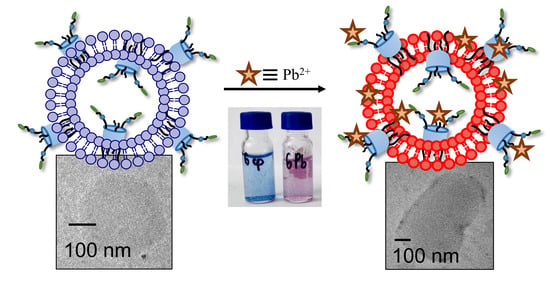
3.5. The Application of PDA Systems as Sensors for Metal Ions

PDA have demonstrated wide application as sensors based on the typical color change from blue to red. It has been previously shown in the literature that sulfonate groups can act as sensors for metal ions [60]. Considering the anionic nature of the calixarene head groups and the unique properties of the polymer base, the systems we obtained were used as a colorimetric sensor for lead (Scheme 2).
The stable systems PDA&0.05TCA-Cn (n = 4, 8) was selected as a sensor for metal ions due to the highest optical density, as well as different linker lengths along the lower rim of the platform. Unmodified polydiacetylene was used as a comparison. The photo (Figure 4A) shows that the addition of lead causes a more pronounced color transition. To investigate the sensitivity of the obtained systems, a correlation between the colorimetric response (CR) and different metals was plotted (Figure 4B) using the UV-visible spectroscopy data. Among the various metals tested, modified polydiacetylene polymers showed the highest response to lead ions, with an efficiency of 35% (Figure 4B). However, there is no significant increase in the intensity of the colorimetric response and color in the case of other metals. It can be concluded that modification of the polymer with thiacalix[4]arenes leads to an increase in the response to lead ions. The longer alkyl tail leads to greater perturbation of the polydiacetylene chain and, consequently, greater sensitivity to metal addition.
Due to its high toxicity to humans and other living organisms, the development of a sensitive method for detecting Pb2+ in an aqueous solution has been attracting increasing attention [61,62]. After developing the optimal detection system, we examined the changes in the optical spectrum of 0.05TCA-Cn&PDA (n = 4, 8) vesicles, as well as PDA, under varying concentrations of Pb2+ (ranging from 0 to 4 mM). As shown in Figure 5A, an increase in Pb2+ concentration resulted in a pronounced decrease in absorption intensity at 650 nm, accompanied by the emergence of a new absorption band at 550 nm. This observation signifies a characteristic blue-to-red shift for PDA sensors. Furthermore, the colorimetric response was remarkably rapid, with a colorimetric coefficient of approximately 35%, and the color change was readily discernible to the naked eye (Figure 5B). The colorimetric response was quantified using the previously described formula.
The modified polymer interacts with lead ions through electrostatic forces, a similar interaction that is observed between lead ions and unmodified vesicles (Figure 5B). In the presence of the macrocycle, the interaction between lead ions and the system is enhanced, suggesting the formation of a thiacalixarene-lead complex, which also induces alterations in the backbone of the polydiacetylene chain. Thiacalixarenes possess multiple coordination centers, facilitating the potential formation of chelates with sulfur and oxygen atoms [63,64,65,66,67]. Donor groups play an important role in the formation of complexes with metals: the presence of donor oxygen leads to higher stability of metal-ligand complexes with metals with large ionic radii, such as Sr2+, Ba2+, and Pb2+. The presence of large donor groups, such as sulfur, leads to difficulties in coordination with small metal ions with densely packed solvation spheres. However, the paper presents the rule ’an increase in the size of the chelate ring leads to a greater degree of destabilization of complexes for larger metal ions than for smaller metal ions’, because in the course of the experiments, it is shown that an increase in the size of the chelate ring leads to a significant decrease in the stability of complexes with metal ions of large radius [68]. This means that there must be a balance between the size of the ligand and the size of the metal ion.
The 0.05TCA-C8&PDA-based systems were examined using transmission electron microscopy (TEM) both before and after the addition of lead ions. As depicted in Figure 6B, the presence of Pb2+ results in the vesicular becoming more contrasted, and the addition of metal ions induces coagulation of modified PDA. The polymers exhibit an ellipsoidal shape (Figure 6A), which is consistent with the DLS data, where two hydrodynamic diameters were observed (Table 2).
An energy dispersive X-ray (EDX) analysis shows the presence of sulfur (thiacalixarene macrocycle platform) and lead in the sample (Figure 6C).
Previous work [69] has demonstrated that the mechanism behind the colorimetric response of calixarene-decorated polydiacetylene vesicles involves deformation of the calixarene cavity, causing perturbation of the polymer backbone. This process is accompanied by metal-induced aggregation of the vesicles, leading to particle sedimentation. As mentioned earlier, the high selectivity towards lead ions is attributed to the strong electrostatic interaction and complementarity between the negatively charged thiacalixarene platform and the lead ion. Consequently, lead ions lead to polymer aggregation by reducing the electrostatic repulsion of the vesicles. Indeed, PDA and TCA-C8&PDA have a surface charge of about −53 and −35 mV, respectively, and the addition of lead ions leads to a decrease in charge followed by a recharging of the systems (Figure 6D). The zeta potential of the polymers in the presence of Pb2+ is about +13 and +5 PDA and 0.05TCA-C8&PDA, respectively, indicating their low colloidal stability.
In this study, anodic stripping voltammetry (ASV) and microwave plasma atomic emission spectroscopy (MP AES) were employed to quantify the amount of unbound lead two days after mixing and subsequent filtration. Table 4 indicates that the 0.05TCA-C8&PDA system removed approximately 44% of the added excess lead as measured by ASV, and 42% as measured by MP AES. In contrast, the PDA system was found to be less effective at removing lead, as evidenced by the difference in peak current for Pb2+ in the presence of PDA and 0.05TCA-C8 (Figure S24B).

4. Conclusions

New zwitterionic receptors based on sulfonate-imidazolium thiacalixarenes in the 1,3-alternate configuration were synthesized and used for decoration of polydiacetylene vesicles. Macrocycles with butyl or octyl alkyl moieties can be incorporated into polydiacetylene vesicles, allowing them to form water-soluble, stable systems. Using dynamic light scattering, UV-visible spectroscopy, TEM, and EDX it was shown that irradiation of the mixture of macrocycles with PCDA gives polydiacetylene particles. An increase in thiacalixarene loading decreases the degree of PDA polymerization. However, the addition of thiacalixarene to PCDA increases its colloidal stability under a change in pH. Addition of zwitterionic thiacalixarenes into PDA vesicles dramatically changes their colorimetric response toward lead: modified PDA has a colorimetric response toward lead detectable with the naked eye. The mechanism of the colorimetric response of thiacalixarene-decorated PDAs to lead involves the formation of a lead—thiacalixarene cavity complex that provokes perturbation of the PDA backbone. Modified polymers were also shown to be effective systems for the sorption of lead ions from aqueous media. Thus, modified PDAs are promising colorimetric sensors for qualitative analysis of heavy metals with the naked eye, as well as effective systems for water purification.

Supplementary Materials

The following supporting information can be downloaded at https://www.mdpi.com/article/10.3390/colloids9020020/s1, Figure S1: 1H NMR spectrum (CDCl3, 400 MHz, 25 °C) of compound 3; Figure S2. 13C NMR spectrum {1H} CDCl3, 100.9 MHz, 25 °C) of compound 3; Figure S3. DEPT spectrum (CDCl3, 100.9 MHz, 25 °C) of compound 3; Figure S4. IR spectrum of compound 3. Figure S5. 2D-NOESY 1H-1H spectrum (CDCl3, 100.9 MHz, 25 °C) of compound 3. Figure S6. 2D-HSQC 1H-13C spectrum (CDCl3, 100.9 MHz, 25 °C) of compound 3. Figure S7. HRMS-ESI mass spectrum of compound 3. Figure S8. HRMS-ESI mass spectrum of compound 3. Figure S9. 1H NMR spectrum (CDCl3, 400 MHz, 25 °C) of compound 4. Figure S10. 13C{1H} NMR spectrum (CDCl3, 100.9 MHz, 25 °C) of compound 4. Figure S11. IR spectrum of compound 4. Figure S12. HRMS-ESI mass spectrum of compound 4. Figure S13. 1H NMR spectrum (D2O:DMSO-d6 1:1, 400 MHz, 25 °C) of the compound TCA-C4. Figure S14. 13C{1H} NMR spectrum (CDCl3:DMSO-d6 1:1, 100.9 MHz, 25 °C) of compound TCA-C4. Figure S15. IR spectrum of compound TCA-C4. Figure S16. HRMS-ESI mass spectrum of compound TCA-C4. Figure S17. HRMS-ESI mass spectrum of compound TCA-C4. Figure S18. 1H NMR spectrum (D2O:DMSO-d6 1:1, 400 MHz, 25 °C) of compound TCA-C8. Figure S19. 13C{1H} NMR spectrum (CDCl3, 100.9 MHz, 25 °C) of the compound TCA-C8. Figure S20. IR spectrum of compound TCA-C8. Figure S21. HRMS-ESI mass spectrum of compound TCA-C8. Figure S22. HRMS-ESI mass spectrum of compound TCA-C8. Figure S23. HRMS-ESI mass spectrum of compound TCA-C8. Figure S24. Diagram of PDI before and after polymerization of the system. Figure S25. A) Calibration plot of lead (II) versus potential, B) anodic stripping voltammograms of lead(II) in the filtrate after two days from mixture PDA+Pb2+ (black) and 0.05TCA-C8&PDA+Pb2+ (red), (C(PCDA) = 0.4 mM, C(TCA-C8) = 0.02 mM). Figure S26. Calibration plot of lead (II) for MP AES.

Author Contributions

Synthesis: R.R.I., B.K.G., I.Y. and K.K.D.; supervision (chemistry): V.A.B., N.O.A. and S.E.S.; physico-chemical studies: A.A.F., M.A.I. and E.O.C.; supervision (physico-chemical studies): E.D.S.; microscopy studies: V.G.E.; data curation: E.D.S.; conceptualization: E.D.S. and V.A.B.; writing—original draft preparation: A.A.F. and E.D.S.; writing—review and editing: V.A.B. and I.S.A.; project administration: E.D.S., V.A.B. and I.S.A. All authors have read and agreed to the published version of the manuscript.

Funding

Synthesis of zwitterionic sulfonate derivatives of thiacalix[4]arenes and study of PDA systems as sensors for metal ions were funded by the Russian Science Foundation (grant No. 23-73-01140). The synthesis of starting reagents was performed and funded by the non-profit joint-stock company “Korkyt Ata Kyzylorda University”.

Data Availability Statement

Data are contained within the article or Supplementary Material.

Conflicts of Interest

Author Nurbol O. Appazov was employed by the company “CNEC” LLP. The remaining authors declare that the research was conducted in the absence of any commercial or financial relationships that could be construed as a potential conflict of interest.

References

  1. Huang, F.; Anslyn, E.V. Introduction: Supramolecular Chemistry. Chem. Rev. 2015, 115, 6999–7000. [Google Scholar] [CrossRef] [PubMed]
  2. Lackinger, M.; Heckl, W.M. Carboxylic Acids: Versatile Building Blocks and Mediators for Two-Dimensional Supramolecular Self-Assembly. Langmuir 2009, 25, 11307–11321. [Google Scholar] [CrossRef] [PubMed]
  3. Khan, S.B.; Lee, S.-L. Supramolecular Chemistry: Host–Guest Molecular Complexes. Molecules 2021, 26, 3995. [Google Scholar] [CrossRef]
  4. Truong, P.L.; Yin, Y.; Lee, D.; Ko, S.H. Advancement in COVID-19 detection using nanomaterial-based biosensors. Exploration 2023, 3, 20210232. [Google Scholar] [CrossRef] [PubMed]
  5. Cebula, J.; Fink, K.; Boratyński, J.; Goszczyński, T.M. Supramolecular chemistry of anionic boron clusters and its applications in biology. Coord. Chem. Rev. 2023, 477, 214940. [Google Scholar] [CrossRef]
  6. Friedman, S.; Kolusheva, S.; Volinsky, R.; Zeiri, L.; Schrader, T.; Jelinek, R. Lipid/Polydiacetylene Films for Colorimetric Protein Surface-Charge Analysis. Anal. Chem. 2008, 80, 7804–7811. [Google Scholar] [CrossRef]
  7. Akhmadeev, B.S.; Podyachev, S.N.; Katsyuba, S.A.; Spicher, S.; Sudakova, S.N.; Gimazetdinova, G.S.; Syakaev, V.V.; Sinyashin, O.G.; Mustafina, A.R. The incorporation of upper vs. lower rim substituted thia- and calix[4]arene ligands into polydiacethylene polymeric bilayers for rational design of sensors to heavy metal ions. Polymer 2022, 245, 124728. [Google Scholar] [CrossRef]
  8. Chernikova, E.Y.; Berdnikova, D.V. Cucurbiturils in nucleic acids research. Chem. Commun. 2020, 56, 15360–15376. [Google Scholar] [CrossRef]
  9. Sharma, V.S.; Vishwakarma, V.K.; Shrivastav, P.S.; Ammathnadu Sudhakar, A.; Sharma, A.S.; Shah, P.A. Calixarene Functionalized Supramolecular Liquid Crystals and Their Diverse Applications. ACS Omega 2022, 7, 45752–45796. [Google Scholar] [CrossRef]
  10. Gokel, G.W.; Leevy, W.M.; Weber, M.E. Crown Ethers: Sensors for Ions and Molecular Scaffolds for Materials and Biological Models. Chem. Rev. 2004, 104, 2723–2750. [Google Scholar] [CrossRef]
  11. Huang, Q.; Wu, W.; Ai, K.; Liu, J. Highly Sensitive Polydiacetylene Ensembles for Biosensing and Bioimaging. Front. Chem. 2020, 8, 565782. [Google Scholar] [CrossRef]
  12. Pankaew, A.; Traiphol, N.; Traiphol, R. Tuning the sensitivity of polydiacetylene-based colorimetric sensors to UV light and cationic surfactant by co-assembling with various polymers. Colloids Surf. A Physicochem. Eng. Asp. 2021, 608, 125626. [Google Scholar] [CrossRef]
  13. Jannah, F.; Kim, J.-H.; Lee, J.-W.; Kim, J.-M.; Kim, J.-M.; Lee, H. Immobilized Polydiacetylene Lipid Vesicles on Polydimethylsiloxane Micropillars as a Surfactin-Based Label-Free Bacterial Sensor Platform. Front. Mater. 2018, 5, 57. [Google Scholar] [CrossRef]
  14. Lee, S.; Kim, J.-Y.; Chen, X.; Yoon, J. Recent progress in stimuli-induced polydiacetylenes for sensing temperature, chemical and biological targets. Chem. Commun. 2016, 52, 9178–9196. [Google Scholar] [CrossRef]
  15. Okada, S.; Peng, S.; Spevak, W.; Charych, D. Color and Chromism of Polydiacetylene Vesicles. Acc. Chem. Res. 1998, 31, 229–239. [Google Scholar] [CrossRef]
  16. Chakarborty, S.; Suklabaidya, S.; Bhattacharjee, D.; Arshad Hussain, S. Polydiacetylene (PDA) Film: A unique sensing element. Mater. Today Proc. 2018, 5, 2367–2372. [Google Scholar] [CrossRef]
  17. Jannah, F.; Kim, J.-M. pH-sensitive colorimetric polydiacetylene vesicles for urease sensing. Dye. Pigment. 2019, 169, 15–21. [Google Scholar] [CrossRef]
  18. He, L.; Lu, Y.; Wang, F.; Gao, X.; Chen, Y.; Liu, Y. Bare eye detection of Hg(II) ions based on enzyme inhibition and using mercaptoethanol as a reagent to improve selectivity. Microchim. Acta 2018, 185, 174. [Google Scholar] [CrossRef]
  19. Akhmadeev, B.S.; Retyunskaya, O.O.; Podyachev, S.N.; Katsyuba, S.A.; Gubaidullin, A.T.; Sudakova, S.N.; Syakaev, V.V.; Babaev, V.M.; Sinyashin, O.G.; Mustafina, A.R. Supramolecular Optimization of Sensory Function of a Hemicurcuminoid through Its Incorporation into Phospholipid and Polymeric Polydiacetylenic Vesicles: Experimental and Computational Insight. Polymers 2023, 15, 714. [Google Scholar] [CrossRef]
  20. Chen, X.; Kang, S.; Kim, M.J.; Kim, J.; Kim, Y.S.; Kim, H.; Chi, B.; Kim, S.; Lee, J.Y.; Yoon, J. Thin-Film Formation of Imidazolium-Based Conjugated Polydiacetylenes and Their Application for Sensing Anionic Surfactants. Angew. Chem. Int. Ed. 2010, 49, 1422–1425. [Google Scholar] [CrossRef]
  21. Meng, Y.; Jiang, J.; Liu, M. Self-assembled nanohelix from a bolaamphiphilic diacetylene via hydrogelation and selective responsiveness towards amino acids and nucleobases. Nanoscale 2017, 9, 7199–7206. [Google Scholar] [CrossRef]
  22. dos Santos Pires, A.C.; de Fátima Ferreira Soares, N.; da Silva, L.H.M.; de Andrade, N.J.; Silveira, M.F.A.; de Carvalho, A.F. Polydiacetylene as a Biosensor: Fundamentals and Applications in the Food Industry. Food Bioprocess Technol. 2010, 3, 172–181. [Google Scholar] [CrossRef]
  23. Demikhovsky, Y.; Kolusheva, S.; Geyzer, M.; Jelinek, R. Polydiacetylene-supported silica films formed at the air/water interface. J. Colloid Interface Sci. 2011, 364, 428–434. [Google Scholar] [CrossRef]
  24. Chanakul, A.; Traiphol, N.; Traiphol, R. Controlling the reversible thermochromism of polydiacetylene/zinc oxide nanocomposites by varying alkyl chain length. J. Colloid Interface Sci. 2013, 389, 106–114. [Google Scholar] [CrossRef] [PubMed]
  25. Kim, C.; Lee, K. Polydiacetylene (PDA) Liposome-Based Immunosensor for the Detection of Exosomes. Biomacromolecules 2019, 20, 3392–3398. [Google Scholar] [CrossRef]
  26. Thakuri, A.; Acharya, R.; Banerjee, M.; Chatterjee, A. A polydiacetylene (PDA) based dual-mode optical sensor for the ppb level selective detection of biogenic polyamines. Sens. Actuators B Chem. 2024, 409, 135573. [Google Scholar] [CrossRef]
  27. Jang, H.; Jeon, J.; Shin, M.; Kang, G.; Ryu, H.; Kim, S.M.; Jeon, T.-J. Polydiacetylene (PDA) Embedded Polymer-Based Network Structure for Biosensor Applications. Gels 2025, 11, 66. [Google Scholar] [CrossRef]
  28. Yang, G.; Nie, Z.; Zhang, S.; Ge, Z.; Zhao, J.; Zhang, J.; Li, B. Sensitive Colorimetric Sensor for Lead Ions and VOCs Based on Histidine-Functionalized Polydiacetylene. Macromol. Res. 2020, 28, 1192–1197. [Google Scholar] [CrossRef]
  29. Li, Y.; Wang, L.; Yin, X.; Ding, B.; Sun, G.; Ke, T.; Chen, J.; Yu, J. Colorimetric strips for visual lead ion recognition utilizing polydiacetylene embedded nanofibers. J. Mater. Chem. A 2014, 2, 18304–18312. [Google Scholar] [CrossRef]
  30. Wang, M.; Wang, F.; Wang, Y.; Zhang, W.; Chen, X. Polydiacetylene-based sensor for highly sensitive and selective Pb2+ detection. Dye. Pigment. 2015, 120, 307–313. [Google Scholar] [CrossRef]
  31. Lee, C.G.; Kang, S.; Oh, J.; Eom, M.S.; Oh, J.; Kim, M.-G.; Lee, W.S.; Hong, S.; Han, M.S. A colorimetric and fluorescent chemosensor for detection of Hg2+ using counterion exchange of cationic polydiacetylene. Tetrahedron Lett. 2017, 58, 4340–4343. [Google Scholar] [CrossRef]
  32. Chen, S.-W.; Chen, X.; Li, Y.; Yang, Y.; Dong, Y.; Guo, J.; Wang, J. Polydiacetylene-based colorimetric and fluorometric sensors for lead ion recognition. RSC Adv. 2022, 12, 22210–22218. [Google Scholar] [CrossRef] [PubMed]
  33. Pham, T.C.; Kim, Y.K.; Park, J.B.; Jeon, S.; Ahn, J.; Yim, Y.; Yoon, J.; Lee, S. A Selective Colorimetric and Fluorometric Chemosensor Based on Conjugated Polydiacetylenes for Cadmium Ion Detection. ChemPhotoChem 2019, 3, 1133–1137. [Google Scholar] [CrossRef]
  34. Valiyakhmetova, A.M.; Sultanova, E.D.; Burilov, V.A.; Solovieva, S.E.; Antipin, I.S. New DNA-sensor based on thiacalix [4]arene-modified polydiacetylene particles. Russ. Chem. Bull. 2019, 68, 1067–1074. [Google Scholar] [CrossRef]
  35. Burilov, V.; Valiyakhmetova, A.; Mironova, D.; Safiullin, R.; Kadirov, M.; Ivshin, K.; Kataeva, O.; Solovieva, S.; Antipin, I. “Clickable” thiacalix[4]arene derivatives bearing polymerizable 1,3-butadiyne fragments: Synthesis and incorporation into polydiacetylene vesicles. RSC Adv. 2016, 6, 44873–44877. [Google Scholar] [CrossRef]
  36. Sultanova, E.; Gazalieva, A.; Makarov, E.; Belov, R.; Evtugyn, V.; Burilov, V.; Solovieva, S.; Antipin, I. Novel aminocalixarene-modified polydiacetylene vesicles: Synthesis and naked-eye detection of ATP. Colloids Surf. A Physicochem. Eng. Asp. 2021, 630, 127642. [Google Scholar] [CrossRef]
  37. Needleman, H. Lead Poisoning. Annu. Rev. Med. 2004, 55, 209–222. [Google Scholar] [CrossRef]
  38. Finkelstein, Y. Low-level lead-induced neurotoxicity in children: An update on central nervous system effects. Brain Res. Rev. 1998, 27, 168–176. [Google Scholar] [CrossRef]
  39. Xing, H.; Song, Y.; Ma, L.; Wang, F.; Zhang, M.; Li, Z.; Wan, Z.; Cui, X.; Yang, G. Preparation of Bis-Polydiacetylene with Reversible Thermochromism and High Sensitivity toward Pb2+ Detection. ACS Appl. Polym. Mater. 2024, 6, 15035–15042. [Google Scholar] [CrossRef]
  40. Sagong, H.Y.; Son, M.H.; Park, S.W.; Kim, J.S.; Li, T.; Jung, Y.K. Dual-signal optical detection of Lead(II) ions (Pb2+) using galloyl group-functionalized polydiacetylene. Analytica Chimica Acta 2022, 1230, 340403. [Google Scholar] [CrossRef]
  41. Hu, X.J.; Zhang, P.; Li, Y.S. The Preparation of TCAS-Loaded Resin and its Adsorption Property to Pb2+ Ion. Adv. Mater. Res. 2011, 396–398, 421–424. [Google Scholar] [CrossRef]
  42. Armarego, W.L. Purification of Laboratory Chemicals; Elsevier: Amsterdam, The Netherlands, 2013; ISBN 9780123821614. [Google Scholar]
  43. Sultanova, E.D.; Gafiatullin, B.K.; Ocherednyuk, E.A.; Garipova, R.I.; Volodina, A.A.; Daminova, A.G.; Evtugyn, V.G.; Burilov, V.A.; Solovieva, S.E.; Antipin, I.S. N-Oxyethylimidazolium Calix[4]arenes and Thiacalix[4]arenes: Difference in Solubilization Property and Detection of Adenine-Containing Nucleotides. Macroheterocycles 2023, 16, 168–176. [Google Scholar] [CrossRef]
  44. Kolusheva, S.; Zadmard, R.; Schrader, T.; Jelinek, R. Color Fingerprinting of Proteins by Calixarenes Embedded in Lipid/Polydiacetylene Vesicles. J. Am. Chem. Soc. 2006, 128, 13592–13598. [Google Scholar] [CrossRef]
  45. Weston, M.; Pham, A.-H.; Tubman, J.; Gao, Y.; Tjandra, A.D.; Chandrawati, R. Polydiacetylene-based sensors for food applications. Mater. Adv. 2022, 3, 4088–4102. [Google Scholar] [CrossRef]
  46. Wilk-Kozubek, M.; Potaniec, B.; Gazińska, P.; Cybińska, J. Exploring the Origins of Low-Temperature Thermochromism in Polydiacetylenes. Polymers 2024, 16, 2856. [Google Scholar] [CrossRef] [PubMed]
  47. Yoon, B.; Lee, S.; Kim, J.-M. Recent conceptual and technological advances in polydiacetylene-based supramolecular chemosensors. Chem. Soc. Rev. 2009, 38, 1958. [Google Scholar] [CrossRef]
  48. Lee, D.-C.; Sahoo, S.K.; Cholli, A.L.; Sandman, D.J. Structural Aspects of the Thermochromic Transition in Urethane-Substituted Polydiacetylenes. Macromolecules 2002, 35, 4347–4355. [Google Scholar] [CrossRef]
  49. Charoenthai, N.; Pattanatornchai, T.; Wacharasindhu, S.; Sukwattanasinitt, M.; Traiphol, R. Roles of head group architecture and side chain length on colorimetric response of polydiacetylene vesicles to temperature, ethanol and pH. J. Colloid Interface Sci. 2011, 360, 565–573. [Google Scholar] [CrossRef]
  50. Kamphan, A.; Charoenthai, N.; Traiphol, R. Fine tuning the colorimetric response to thermal and chemical stimuli of polydiacetylene vesicles by using various alcohols as additives. Colloids Surf. A Physicochem. Eng. Asp. 2016, 489, 103–112. [Google Scholar] [CrossRef]
  51. Tjandra, A.D.; Weston, M.; Tang, J.; Kuchel, R.P.; Chandrawati, R. Solvent injection for polydiacetylene particle synthesis—Effects of varying solvent, injection rate, monomers and needle size on polydiacetylene properties. Colloids Surf. A Physicochem. Eng. Asp. 2021, 619, 126497. [Google Scholar] [CrossRef]
  52. Chanakul, A.; Traiphol, N.; Faisadcha, K.; Traiphol, R. Dual colorimetric response of polydiacetylene/Zinc oxide nanocomposites to low and high pH. J. Colloid Interface Sci. 2014, 418, 43–51. [Google Scholar] [CrossRef]
  53. Kew, S.J.; Hall, E.A.H. pH Response of Carboxy-Terminated Colorimetric Polydiacetylene Vesicles. Anal. Chem. 2006, 78, 2231–2238. [Google Scholar] [CrossRef]
  54. de Oliveira, C.P.; de Fátima Ferreira Soares, N.; Fontes, E.A.F.; de Oliveira, T.V.; Filho, A.M.M. Behaviour of polydiacetylene vesicles under different conditions of temperature, pH and chemical components of milk. Food Chem. 2012, 135, 1052–1056. [Google Scholar] [CrossRef]
  55. Kamphan, A.; Traiphol, N.; Traiphol, R. Versatile route to prepare reversible thermochromic polydiacetylene nanocomposite using low molecular weight poly(vinylpyrrolidone). Colloids Surf. A Physicochem. Eng. Asp. 2016, 497, 370–377. [Google Scholar] [CrossRef]
  56. Nakayama, S.; Suga, K.; Kamata, T.; Watanabe, K.; Namigata, H.; Welling, T.A.J.; Nagao, D. Bio-Inspired Polydiacetylene Vesicles for Controlling Stimulus Sensitivity. Macromol. React. Eng. 2025, 19, 2400016. [Google Scholar] [CrossRef]
  57. Filhol, J.-S.; Deschamps, J.; Dutremez, S.G.; Boury, B.; Barisien, T.; Legrand, L.; Schott, M. Polymorphs and Colors of Polydiacetylenes: A First Principles Study. J. Am. Chem. Soc. 2009, 131, 6976–6988. [Google Scholar] [CrossRef] [PubMed]
  58. Jung, Y.K.; Park, H.G. Colorimetric polydiacetylene (PDA) liposome-based assay for rapid and simple detection of GST-fusion protein. Sens. Actuators B Chem. 2019, 278, 190–195. [Google Scholar] [CrossRef]
  59. Traiphol, N.; Faisadcha, K.; Potai, R.; Traiphol, R. Fine tuning the color-transition temperature of thermoreversible polydiacetylene/zinc oxide nanocomposites: The effect of photopolymerization time. J. Colloid Interface Sci. 2015, 439, 105–111. [Google Scholar] [CrossRef]
  60. Huang, M.-R.; Ding, Y.-B.; Li, X.-G. Lead-ion potentiometric sensor based on electrically conducting microparticles of sulfonic phenylenediamine copolymer. Analyst 2013, 138, 3820. [Google Scholar] [CrossRef]
  61. Narkwiboonwong, P.; Tumcharern, G.; Potisatityuenyong, A.; Wacharasindhu, S.; Sukwattanasinitt, M. Aqueous sols of oligo(ethylene glycol) surface decorated polydiacetylene vesicles for colorimetric detection of Pb2+. Talanta 2011, 83, 872–878. [Google Scholar] [CrossRef]
  62. Zhang, S.; Shi, B.; Yang, G. A Selective Colorimetric Sensor for Pb2+ Detection by Using Phenylboronic Acid Functionalized Polydiacetylene Liposomes. Macromol. Res. 2020, 28, 51–56. [Google Scholar] [CrossRef]
  63. Kuriki, R.; Ishii, Y.; Kuwabara, T. Exo- and endo -Isomers of Plumbylenes Supported by 1,3-Diethers of Calix[4]arene. Eur. J. Inorg. Chem. 2023, 26, e202200655. [Google Scholar] [CrossRef]
  64. Eddaif, L.; Shaban, A.; Telegdi, J. Sensitive detection of heavy metals ions based on the calixarene derivatives-modified piezoelectric resonators: A review. Int. J. Environ. Anal. Chem. 2019, 99, 824–853. [Google Scholar] [CrossRef]
  65. Ohto, K. Review of adsorbents incorporating calixarene derivatives used for metals recovery and hazardous ions removal: The concept of adsorbent design and classification of adsorbents. J. Incl. Phenom. Macrocycl. Chem. 2021, 101, 175–194. [Google Scholar] [CrossRef]
  66. Rathod, N.V.; Rao, A.; Kumar, P.; Ramakumar, K.L.; Malkhede, D.D. Complexation with calixarenes and efficient supercritical CO2 extraction of Pb(ii) from acidic medium. New J. Chem. 2014, 38, 5331–5340. [Google Scholar] [CrossRef]
  67. Ferreira, A.S.D.; Ascenso, J.R.; Marcos, P.M.; Schurhammer, R.; Hickey, N.; Geremia, S. Calixarene-Based lead receptors: An NMR, DFT and X-Ray synergetic approach. Supramol. Chem. 2021, 33, 231–244. [Google Scholar] [CrossRef]
  68. Hancock, R.D.; Martell, A.E. Ligand design for selective complexation of metal ions in aqueous solution. Chem. Rev. 1989, 89, 1875–1914. [Google Scholar] [CrossRef]
  69. Burilov, V.; Valiyakhmetova, A.; Mironova, D.; Sultanova, E.; Evtugyn, V.; Osin, Y.; Katsyuba, S.; Burganov, T.; Solovieva, S.; Antipin, I. Novel amphiphilic conjugates of p-tert -butylthiacalix[4]arene with 10,12-pentacosadiynoic acid in 1,3-alternate stereoisomeric form. Synthesis and chromatic properties in the presence of metal ions. New J. Chem. 2018, 42, 2942–2951. [Google Scholar] [CrossRef]
Scheme 1. Synthesis of imidazolium sulphonate salts based on thiacalix[4]arene.
Colloids 09 00020 sch001
Figure 1. (A) The change of color of the solution before and after polymerization and (B) UV-visible spectra of xTCA-Cn&PDA (x = 0.05, 0.1, 0.2, n = 4, 8) systems; C(PCDA) = 0.4 mM, C(TCA-Cn) = 0.02, 0.4, 0.08 mM, TRIS, 1 cm−1.
Colloids 09 00020 g001
Figure 2. Effect of pH on polymerization of the PDA&0.05TCA-Cn system (n = 4, 8) (UV-visible spectrum): (A) pH 9.4, (B) pH 7.4, (C) pH 3.2, and (D) photo of obtained PDA solutions at different pH: (a) pH 3.2; (b) pH 7.4; and (c) pH 9.0.
Colloids 09 00020 g002
Figure 3. (A) UV-visible spectrum of unmodified PDA, measured at different temperatures; (B) colorimetric response of xTCA-Cn&PDA (n = 4, 8, x = 0.05, 0.1); C(PCDA) = 0.4 mM, C(TCA-Cn) = 0.02, 0.4 mM, TRIS, 1 cm−1.
Colloids 09 00020 g003
Scheme 2. Schematic representation of the sensory properties of the modified PDA on lead.
Colloids 09 00020 sch002
Figure 4. (A) Photo of changes in the color of the PDA&0.05mM TCA-C8 upon addition of various metal ions in TRIS buffer (pH 7.4) at room temperature, (B) colorimetric response (%) of the systems to various metal ions.
Colloids 09 00020 g004
Figure 5. (A) UV-visible spectrum of 0.05TCA-C4& in the absence and presence of different lead concentrations and (B) CR (%) of vesicular systems in TRIS buffer after adding Pb(NO3)2 (0–4 mM) at room temperature, C(PCDA) = 0.4 mM, C(TCA-Cn) = 0.02 and mM, TRIS, 1 cm−1.
Colloids 09 00020 g005
Figure 6. TEM images of (A) 0.05TCA-C8&PDA, (B) 0.05TCA-C8&PDA + Pb2+, (C) EDX spectrum of 0.05TCA-C8&PDA + Pb2+, and (D) Zeta-potential of PDA (blue) and 0.05TCA-C8&PDA (orange) versus Pb(NO3)2 concentration, C(PCDA) = 0.4 mM, C(TCA-C8) = 0.02 mM, TRIS, 1 cm−1.
Colloids 09 00020 g006
Table 1. Substituents, yields, and times for compounds 34.
MacrocyclenYield (%)Time (h)
346540
487030
TCA-C447020
TCA-C887527
Table 2. DLS data for TCA-Cn; n = 4, 8 solutions.
Concentration, mMPDIHydrodynamic Diameter, nm
TCA-C40.001-
0.01 0.89   ± 0.06 570   ±   204 ,   124   ±   39 ,   19   ± 32
0.05 0.76   ± 0.08 713   ± 73
0.1sedimentation
0.5
TCA-C80.001
0.01 0.58   ± 0.05 501   ±   151 ,   187   ± 41
0.05 0.67   ± 0.07 718   ±   22 ,   148   ± 36
0.1 0.51   ± 0.01 580   ± 86 ,   246   ± 43
0.5 0.65   ± 0.03 330   ±   79 ,   374   ±   471 ,   357   ± 713
Table 3. DLS data for systems xTCA-Cn (n = 4, 8; x = C of macrocycle = 0.05; 0.1; 0.2 mM) and PCDA (1 mM) before and after polymerization.
Before PolymerizationAfter Polymerization
SystemPDIHydrodynamic Diameter, nmSystemPDIHydrodynamic Diameter, nm
PCDA0.164 ± 0.018228 ± 31 (86%), 53 ± 63 (14%)PDA0.226 ± 0.013270 ± 45 (77%), 95 ± 15 (23%)
0.05TCA-C4+PCDA0.380 ± 0.019483 ± 178 (68%), 545 ± 705 (34%)0.05TCA-C4&PDA0.413 ± 0.041391 ± 68 (78%), 92 ± 82 (22%)
0.1TCA-C4+PCDA0.448 ± 0.015976 ± 120 (66%), 222 ± 57 (34%)0.1TCA-C4&PDA0.434 ± 0.012888 ± 140 (64%), 188 ± 27 (36%)
0.2TCA-C4+PCDA0.263 ± 0.02243 ± 64 (79%), 1523 ± 2598 (21%)0.2TCA-C4&PDA0.266 ± 0.01254 ± 57 (89%), 41 ± 72 (11%)
0.05TCA-C8+PCDA0.229 ± 0.008195 ± 21 (94%), 28 ± 48 (6%)0.05TCA-C8&PDA0.337 ± 0.003251 ± 96 (65%), 177 ± 150 (35%)
0.1TCA-C8+PCDA0.203 ± 0.024252 ± 37 (82%), 101 ± 16 (18%)0.1TCA-C8&PDA0.257 ± 0.011318 ± 46 (71%), 115 ± 19 (29%)
0.2TCA-C8+PCDA0.373 ± 0.019422 ± 42 (74%), 143 ± 19 (26%)0.2TCA-C8&PDA0.322 ± 0.024356 ± 96 (74%), 273 ± 263 (26%)
Table 4. The concentration of lead in the filtrate of the mixture PDA + Pb2+ and 0.05TCA-C8&PDA + Pb2+ (C(PCDA) = 0.4 mM, C(TCA-C8) = 0.02 mM) after two days.
MP AES After Diluted *ASV After Diluted *
Entered, mg/LFound, mg/LAdsorbed Lead, %RSD %Entered, mg/LFound, mg/LAdsorbed Lead, %RSD %
PDA10.377.54270.5951.8637.29281.36
0.05TCA-C8&PDA10.375.96420.5751.8629.01442.06
* C0 of Pb2+ 518.65 mg/L.
Disclaimer/Publisher’s Note: The statements, opinions and data contained in all publications are solely those of the individual author(s) and contributor(s) and not of MDPI and/or the editor(s). MDPI and/or the editor(s) disclaim responsibility for any injury to people or property resulting from any ideas, methods, instructions or products referred to in the content.

