Article

Determination of Particle Mixture Composition by Visible Spectroscopy

by
Mauricio Escudey
1,2,
Lizethly Cáceres-Jensen
3 and
Manuel Gacitúa
2,4,*
1
Facultad de Química y Biología, Universidad de Santiago de Chile, Avenida Alameda Libertador Bernardo O’Higgins 3363, Santiago 9170124, Chile
2
Center for the Development of Nanoscience and Nanotechnology (CEDENNA), Santiago 9170124, Chile
3
Laboratorio de Fisicoquímica & Analítica (PachemLab), Núcleo Pensamiento Computacional y Educación para el Desarrollo Sostenible (NuCES), Centro de Investigación en Educación (CIE-UMCE), Departamento de Química, Universidad Metropolitana de Ciencias de la Educación, Santiago 7760197, Chile
4
Facultad de Ingeniería & Ciencias, Universidad Diego Portales, Ejercito 441, Santiago 8370191, Chile
*
Author to whom correspondence should be addressed.
Colloids Interfaces 2025, 9(2), 16; https://doi.org/10.3390/colloids9020016
Submission received: 9 January 2025 / Revised: 27 February 2025 / Accepted: 10 March 2025 / Published: 12 March 2025

Abstract

:
Limited methods exist to determine the composition of particle mixtures. This research presents a simple UV-vis-spectroscopy-based method for the separate quantification of particles mixtures considering the following: synthesized ferrihydrite, commercial Fe2O3, and natural allophane. Calibration curves and adsorption/scattering coefficients are determined for each particle at different wavelengths. This is the main input to solve equation systems and, ultimately, quantify particle concentration in binary mixtures. The limit of detection varies with wavelength and particle type, yielding values as low as 1.5, 0.2, and 1.6 mg L−1 for ferrihydrite (500 nm), Fe2O3 (450 nm), and natural allophane (450 nm), respectively. This study provides a simple, low-cost and straightforward method, compared to atomic-spectroscopy- or chromatography-based techniques, for resolving the composition of binary particle mixtures in suspension.

Graphical Abstract

1. Introduction

The quantification of particles in liquid suspension is a relevant aspect in the implementation of industrial solutions and crucial environmental impact studies. Despite its importance, the methods for particle quantification are often hard to implement and expensive. Depending on the particle composition, the most adequate method for quantification in liquid suspension could vary. For instance, atomic spectroscopies such as Flame Atomic Adsorption Spectroscopy (FAAS) [1] and Inductively Coupled Plasma Optical Emission Spectroscopy (ICP-OES) [2,3,4,5] are extensively used for metallic and metallic-oxide particles, requiring previous dissolution steps before quantification. Also, vibrational spectroscopies such as Fourier Transform Infrared Spectroscopy [6,7,8] and Raman Spectroscopy [9,10,11,12] are suitable to characterize carbon based nanomaterials such as carbon nanotubes, graphene, and micro- and nano-plastics. Although these methods do not require pre-steps, sometimes matrix interferences hinder proper quantification. Finally, molecular spectroscopies such as Fluorescence Spectroscopy and UV–visible light spectroscopy are suitable for all kinds of particulates, including quantum dots [11,12,13]. In particular, UV–visible light spectroscopy-based methods are extensively used to monitor the syntheses, stability, aggregation, and other features of particle suspensions [14] due to their low cost, feasible implementation, and decent sensitivity. Several researchers use UV-vis spectroscopy to calculate particle concentration during synthesis [15], liberation to natural media [16,17,18,19], deposition [20], transport [21,22], and functionalization [23].
Particles (including micro- and nanoparticles) are used in a range of applications and consumer products, including electronics, optics, medical devices, food packaging, sunscreens, cosmetics, paints, water treatment technologies, fuel cells, catalysts, biosensors, environmental remediation agents, and in agricultural production [24,25,26]. Engineered particles are man-made particles that are often produced in large amounts [26]. For example, iron oxide-based nanoparticles (IONPs) are used in many environmental applications involving remediation treatments and the recovery of inorganic, organic, and heavy metal contaminants from wastewater due to their higher sorption affinity and faster sorption rate compared to other sorbents [27,28,29,30,31]. While these remarkable properties of IONPs make them excellent candidates for the remediation of contaminated soil and water, the augmentation of any engineered particles requires regulation and monitoring to control unintended consequences on the environment and ultimately human health [29,32,33,34]. In contrast, natural particles are compatible with the local environment and its organisms [26]. One example is ferrihydrite (Fh, Fe5HO8·4H2O), an iron oxide mineral present in diverse environments such as water springs, drainage lines, lake oxide precipitates, groundwater, river sediments, and oceans, where it plays a significant role in many biogeochemical processes [28,35,36]. Another interesting natural particle is the main component of the clay fraction of volcanic ash-derived soils, natural allophane (NA, Al2O3·2SiO2·nH2O), a low-range ordered aluminosilicate mineral that occurs widely in Andisols [37]. Due to the presence of Al and Si on its surface, NA generates a variable surface charge. Together along with the high specific surface area of NA, this property confers a high capacity for the specific and nonspecific adsorption of ions, cation and anion exchange, and water sorption [37,38,39]. Considering these characteristics, particles suspensions of natural and engineered particles are thought to form complex mixtures when IONPs such as commercial Fe2O3 are applied to remediate contaminated soil and water. Therefore, understanding the dynamics of these particle mixtures is critical to both the success of the remediation program and the mitigation of unintended consequences to environmental or human health. Thus, it is important to have handy low-cost methods to characterize the impact (environmental fate, consequences) of the intended particle augmentations and their disposal in the environment. Methods to characterize particle mixtures must consider the complexity of the matrix as well as the concentrations and characteristics of the particles [2,40].
In a UV-vis spectroscopy cell, incident radiation is scattered and absorbed by the suspended particles, decreasing the intensity of the light that passes through the optical path, b, which is used by the device to calculate the reading, R λ , a variation from Beer’s Law [41,42,43].
R λ = A λ + S λ = log I I 0 = K λ bC
The reading, R λ , corresponds to the sum between the absorbed, A λ , and the scattered light, S λ ; both magnitudes depend on the radiation wavelength, λ . Thus, following the same relationship with Beer’s law, the R λ depends on the optical path, b, the particle concentration, C, and a constant, K λ . This constant is related to both the absorption, a λ , and the turbidity coefficient, k λ , that depend on the selected wavelength:
R λ = A λ + S λ = a λ bC + k λ bC = ( a λ b + k λ b ) C = K λ bC
Now, if in a colloidal suspension there is a mixture of different “n” particles, the reading could be determined considering the additivity properties of the different Rλn [44,45]. In n-component mixtures, one can determine the concentration, C n , of each component if the total Rλ is measured at n different wavelengths and the Kλ constant is determined for each particle at n wavelengths. In this sense, all the relationships to determine the individual concentrations of n particle types in the mixture can be expressed by
R λ 1 = K 1 λ 1 b C 1 + K 2 λ 1 b C 2   + K n λ 1 b C n
R λ 2 = K 1 λ 2 b C 1 + K 2 λ 2 b C 2   + K n λ 2 b C n
R λ n = K 1 λ n b C 1 + K 2 λ n b C 2   + K n λ n b C n
where the Kλ values can be determined from calibration curves constructed for each particle at each n wavelengths considered.
Here, we report the use of an additivity property work protocol to solve particle mixtures in colloid suspensions. To do so, the stabilization of a particle suspension is a key factor to take care of. The quantification of particles in suspension is usually conducted for single-particle systems. In this research, we prove that visible light spectroscopy can be used for the design of a quantification method for particle mixtures in suspension. Particles were first characterized as a function of their composition and size distribution. Calibration curves were constructed at different wavelengths and the particle mixtures were resolved by means of equation systems analogous to Equations (2)–(5) from above. The stability through time and sensitivity of the method also were evaluated.

2. Materials and Methods

Three particles were considered for this research: synthetic ferrihydrite (Fh), a commercial dispersion of Fe2O3, and natural allophane (NA), extracted from a volcanic ash-derived soil. The Fe2O3 dispersion and all the reagents used for this research were purchased from Sigma-Aldrich (Burlington, MA, USA).

2.1. Synthesis of Ferrihydrite

Fh was prepared using the method described by Schwertmann and Cornell (1991) [46]. First, 40 g of Fe(NO3)3·9H2O was added to 0.5 L of water, and the pH was adjusted to 7–8 using 0.1 M KOH. The resulting solid was washed with double-distilled water and separated by centrifugation for 30 min at 10,000 rpm. The washing of the particles was repeated four times. A stock suspension of 17.76 mg mL−1 was brought to pH 3 with HCl to optimize the quality of dispersion. The particles were kept in suspension for a week, then the first 20 cm of suspension was separated and kept refrigerated. According to calculations made using Stoke’s law, after one week of settling there should have been no Fh particles with dimensions higher than 14 µm. This part of the suspension was used as the concentrated ferrihydrite suspension.

2.2. Extraction of Natural Allophane

The extraction of NA from soil involved the destruction of the organic matter by treatment with 30% H2O2 until no dark residues remained [47]. After this peroxide attack, the sample was suspended in 1.0 L of double-distilled water and dispersed with an ultrasonic probe. Material < 2 µm was removed with a siphon from a depth of 20 cm after a period of 30 h. This < 2 µm sample was then treated for iron oxide removal by the dithionite–citrate–bicarbonate (DCB) procedure [48]. After iron oxide removal, the sample was treated again with 30% H2O2 to eliminate the adsorbed organic compounds involved in iron removal treatment [49]. Finally, the resulting suspension was centrifuged for 30 min at 10,000 rpm and re-suspended in double-distilled water to wash the sample; this procedure was repeated four times.

2.3. Morphological and Elemental Composition of Particles

Particle morphology was observed using a Zeiss Scanning Electron Microscope EVO MA 10 model (Jena, Germany). High vacuum was maintained during characterization. Accelerating voltage varied between 8 and 20 kV depending on the sample. Also, the elemental composition of the particles was determined by energy dispersive spectrometry using a Penta FET Precision EDS detector (Oxford Instrument, Oxford, UK) coupled to the SEM. One drop of each particle suspension was placed on a silicon film and allowed to dry after characterization.

2.4. Particle Size Distribution

The particle size distribution measurements were carried out on a Malvern Zetasizer Nano ZS Dynamic Light Scattering (DLS) system (Almeno, The Netherlands) [50,51]. The device uses classical single-scattering methods to calculate particle sizes in a range from 0.3 nm to 10 μm with a 633 nm laser and detects the backscattered light at 173°. Measurements were made at 25 °C and represent the average of 10 runs of 10 s duration for a total of 100 s. The sample in the cuvette was diluted to avoid multiple scattering.

2.5. Suspended Particle Quantification by UV–Visible Spectroscopy

All Rλ measurements were carried out in a Thermo Electron Spectronic Helios Alpha Beta UV–Visible (Waltham, MA, USA) double-beam spectrophotometer with a wavelength step of 1 nm. After measurements, suspensions were stirred for 30 s in vortex to ensure repeatable outcomes. Measurements were made using a 1 cm optical path quartz cell, with bi-distilled water as blank. Room temperature was maintained during the measurements. Concentrated and stable particle suspensions were diluted to prepare different reference materials and, ultimately, calibration curve construction ionic strength was fixed using a 100 mM concentration of NaNO3 as background electrolyte to prepare standards. Particle mass concentration for the reference materials was controlled by the gravimetric method. A set of three 5 mL beakers of known mass were used to deposit an exact volume of particle suspension and left to dry in lab oven at 85 °C until they reached constant mass. Mass gain from beakers was accounted as suspended particle max and related to a certain Rλ measurement for calibration curve point-by-point construction. Experimental pH was monitored during experiments. For the determination of K, calibration curves were constructed at 450, 500, and 550 nm wavelengths, using pure particle standards of different concentrations within the range of 0 to 1.5 mg mL−1.
A set of 12 binary particle mixtures were prepared using different concentrations. The reading Rλ was measured for each prepared mix at 450, 500, and 550 nm. These mixes were prepared and measured in triplicate. To ensure reproducibility, standard deviations were evaluated. Analysis of variance (ANOVA) performed on the results showed that there were no effective statistical differences (95% confidence) between the repetitions.
Regardless of the outcomes, we proved stability through 1-week-old standards; to avoid source of error, it was important to vigorously stir the standards before any measurement.

2.6. Stability of Calibration Curves over Time

To assess the stability of the calibration suspensions over time, calibration curves were constructed from freshly made suspensions and after one week of storage under refrigeration. The concentrations of the particle mixtures were calculated and compared using calibration curve data from both fresh and stored calibration suspensions.

2.7. Analytical Quality Parameters

For the proposed method, analytical quality parameters were evaluated as the limit of detection (LOD) and limit of quantification (LOQ) [52], based on Equations (6) and (7):
L O D = 3 × σ b l k m
L O Q = 10 × σ b l k m
where σ b l k and m correspond to blank standard deviation and calibration curve slope, respectively. These parameters were calculated for each particle suspension at 450, 500, and 550 nm.

3. Results

3.1. Particle Characterization

The particle suspensions presented characteristic pH values of 1.9 ± 0.2, 3.5 ± 0.1, and 2.5 ± 0.5 for Fh, Fe2O3, and NA, respectively. The size distribution of the suspensions after DLS characterization is presented in Figure 1.
The size distribution reveals that more than the 90% of the synthesized Fh corresponded to nanoparticles with a mean diameter of 81 nm. In contrast, the other two materials exhibited a monodisperse signal, with 100% of particles measuring 83 nm for Fe2O3 and 206 nm for NA. This agrees with the Fe2O3 supplier’s description of a nanoparticle dispersion with less than 100 nm particle size. Also, the size distribution of NA agrees well with past reports on this natural particle that naturally forms sub-micro- and micro-sized aggregates [53].
The SEM micrographs (Figure S1) obtained revealed similar size distributions for the particles as were described after DLS characterization. While Fh presented a broader size-distribution, Fe2O3 and NA displayed more homogeneously sized particles. In addition, EDS characterization established that the Fh particles contained comparable amounts of Fe and O as their major constituents (Figure S1). Some K and Cl were also found, probably from their synthesis and acid–water rinses for particle-dispersion enhancement. For Fe2O3, oxygen and iron were found at the expected ratio of 3:2, while some amounts of C and N were detected due to the dispersing agent, as described by the provider. Finally, the major constituents of NA were Al, Si, and O in a 3:2:2 ratio. Additionally, some amounts of Cl, Na, and C were found in the NA samples, likely because of the extraction procedure. Other elements found in smaller proportion may belong to the natural mineral particles present in the volcanic soil samples used to extract the NA.

3.2. Determination of Particle Mixtures Using UV–Visible Spectroscopy

Wavelength selection is a critical but flexible aspect of the quantification of particle mixtures in suspension. While it is important to choose different wavelengths for the determination of each particle, it is not essential to choose a specific wavelength or to follow a particular protocol for wavelength selection. Unlike molecular absorption spectra, the dispersion of radiation by suspended particles does not necessarily have a maximum whose wavelength can preferably be selected. This is corroborated by Figure 2, where the single-particle spectra did not display maximum absorption bands at any wavelength. As described above (Equation (1)), the total R corresponds to the contribution by each particle present in the mixture due to the absorption and dispersion of radiation, as illustrated in Figure 2 by the spectra of Fh, Fe2O3, and combinations.
As presented in Figure 2, each particle under study displays a different spectrum. Since these particles do not react with each other or with other suspension components, the sum of the independent spectra agrees well with the spectrum obtained from a suspension made with a binary mixture, maintaining a pH near 2.2 ± 0.2. For example, the sum (Colloids 09 00016 i001
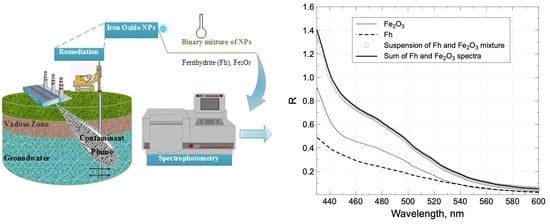
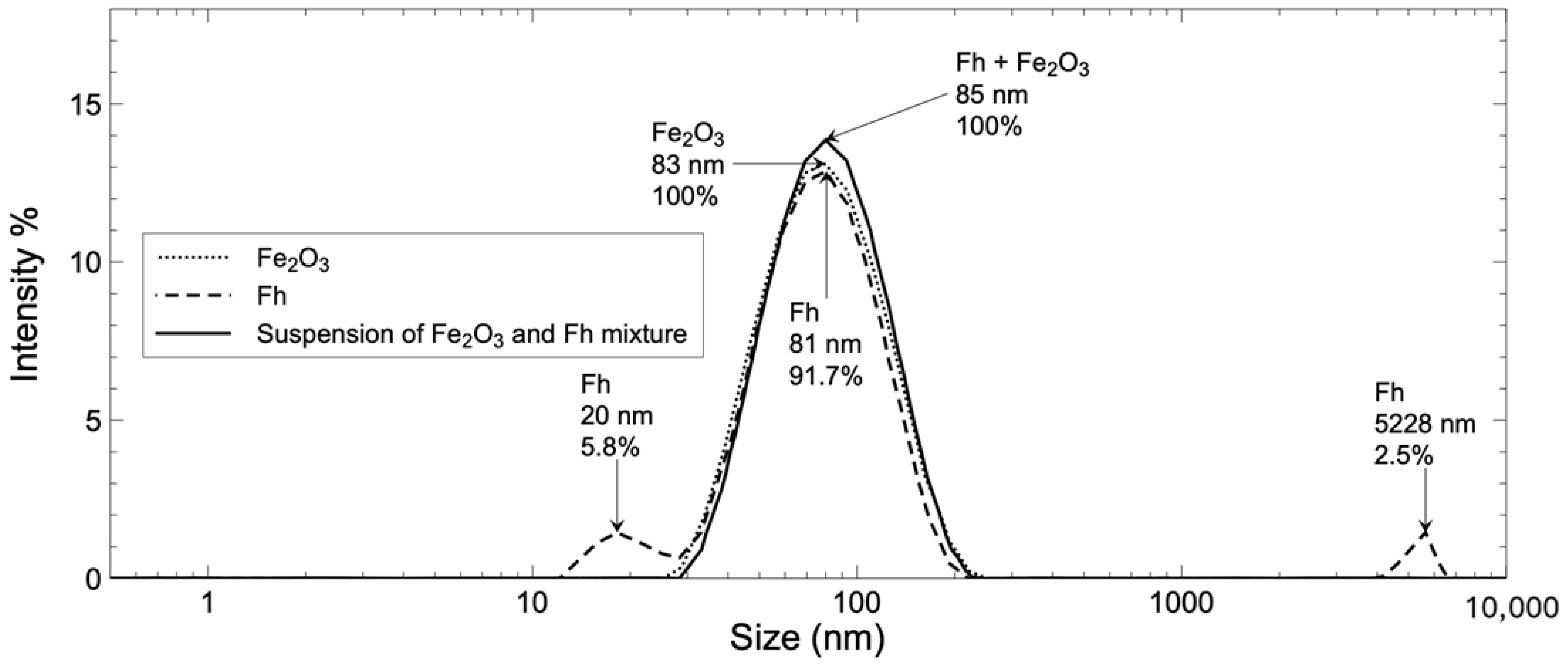
) of the spectra made with Fe2O3 and Fh agrees well with the spectrum obtained from a suspension made with both particles (◯) at same concentration. This behavior is consistent at each wavelength and is independent of the particle or medium composition if there is no reaction between either of the components. Another way to demonstrate the absence of interaction between the particles is to compare the DLS size distribution of the single particles to the distribution obtained for the mix (Figure S2). In the case of an interaction between the Fh and Fe2O3 particles, one should expect a strong size distribution shift from the single-particle to the mixed-particle system. This is not the case, since after combination the intense distribution located at 81 and 83 nm for the single-particle Fh and Fe2O3 DLS size distributions, respectively, (Figure 1) grew into a more intense distribution present for the mix at 85 nm (Figure S2). The secondary, less important distribution for Fh was not detected for the mix with Fe2O3, probably due to a dilution effect.
For any binary system, solving the concentration of mixed-particle suspensions requires that particles should not interact with each other or with other suspension components, a low level of background interference from the matrix (total R, absorption + dispersion measurement should not surpass 2.0 reading values), and maintaining stability in the suspension during the measurements, avoiding coagulation and sedimentation processes as much as possible.
All in all, any binary systems need to have their R measured at two different wavelengths. For instance, the binary mixtures of Fh, Fe2O3, and NA were quantified by measuring R at 450, 500, and 550 nm, as displayed in Figure 3.
The experimental points for calibration curve construction are presented for each particle. Concentration was determined after gravimetry measurements using reference material prepared from stable suspensions of each particle, displaying high precision with low standard deviation (horizontal error bars) at each point. Then, each reference material was measured, obtaining Rλ at three different wavelengths (450, 500, and 550 nm). Linear regression was selected as the statistical model [54] to explain the relationship between concentration (independent variable) and the reading, R (dependent variable). The model’s limitations required us to consider different concentrations and reading values for each binary system, as would be proven going forward. As can be observed, there is a linear relationship between the magnitude of R, and concentration, CP, for each particle at the different wavelengths. Linear regression analysis was performed after this, and slope values were calculated. Thus, these systems can be used to construct a data matrix with the calculated slopes at different wavelengths, Kλ, for each particle, as presented in Table 1.
Theoretically, with this information it is possible to formulate an equation system considering at least two different particles measured at two different wavelengths. Solving this system of equations yields the concentration for each particle in the mixed solution. For example, to solve a binary mixture between Fh and Fe2O3, an equation system can be established using the measurements at 500 and 550 nm wavelengths values.
R 500 = 1.497   ×   C Fe 2 O 3 + 1.062   ×   C Fh
R 550 = 0.345   ×   C Fe 2 O 3 + 0.411   ×   C Fh
Then, the measurements at 500 and 550 nm replace the values for R500 and R550 in the equation system. For instance, a mixture of Fe2O3 and Fh with the respective theoretical concentration of 0.2 and 1.0 mg mL−1 was prepared. R500 and R550 were measured, resulting in 1.388 and 0.491, respectively. Solving the equation system with these data provides experimental concentrations of 0.20 and 1.03 mg mL−1 of Fe2O3 and Fh, respectively, corresponding to a high accuracy with a percentual error less than +3%. Similar equation systems may be developed for other particle pairs if Rλ measurements are made considering at least two different wavelengths for each.
Table 2 provides the complete set of paired-particle mixtures prepared at different theoretical concentrations and their effective experimental concentrations after light scattering measurement, using Kλ from Table 1 for calculations.
As illustrated in Table 2, the determined experimental concentrations were in close agreement for most theoretical values, except for M9. Notably, the M9 mixture surpasses the method’s limit in terms of the maximum concentration of Fe2O3 that maintains a linear response with R450. As can be observed in Figure 3, at 450 nm Fe2O3 displays a linear response until reaching 0.8 mg mL−1. This observation indicates that the procedure requires that the particle mixture measurements are made under single-particle concentrations less than or equal to those used for the construction calibration curves. The R450 values for M9 were as high as 2.5, meaning that the sample practically transmitted less than 1% of the incident light. This clearly surpasses the method’s limits, since concentrated samples do not obey Beer’s law. This is also observed for M10, M11, and M12, presented in the Supplementary Information (Table S1), where the high R450 values generate low accuracy. Theoretically speaking, measurements can be made at any two different wavelengths, but in the case of Fe2O3, measurements at 450 nm have the highest standard deviation (Table 1). This example demonstrates how high-absorption/scattering samples are not adequate and will provoke erroneous concentration calculus, lowering the accuracy of the method. Taken together, these data demonstrate that the present method allows for the resolution of particle mixtures stably suspended using a simple and straightforward method, irrespective of particle origin, composition, or size. Nevertheless, method design needs to consider wavelength selection, avoiding high absorption/scattering datapoints that will surely lower the method’s accuracy.
The limit of detection (LOD) and limit of quantification (LOQ) are key parameters to evaluate the sensitivity of a method. Table 3 provides these analytical parameters for the particles in this study at different wavelengths.
Both LOD and LOQ were highest at 550 nm, coinciding with the lowest values of K, indicating decreased sensitivity at higher wavelengths. The LOD and LOQ of Fe2O3 at 450 nm were 0.2 and 0.8 mg L−1, respectively, corresponding to the highest sensitivity observed during the study. These values are acceptable considering the simplicity of the methodology. Zhou et al. (2016) [30] developed a method employing a Size Exclusion Chromatography–Inductively Coupled Plasma Mass Spectrometry (SEC-ICP-MS) technique for the determination of mixtures between metal oxide particles (MOs) and free-metal (Mn+), and obtained an LOD of 0.390 μg L−1 for MOs. This represents an LOD 500 times lower than present UV-vis spectroscopy techniques. SEC-ICP-MS offers superior sensitivity, selectivity, and size characterization, making it ideal for precise quantification and differentiation of nanomaterials, especially in complex matrices. However, it is costly and requires sophisticated instrumentation. In contrast, UV-Vis spectroscopy provides a simple and cost-effective alternative for rapid screening but suffers from lower specificity and sensitivity, particularly for polydisperse or complex samples.
The method developed herein to resolve particle mixtures in liquid suspensions represents an accessible, cost-effective approach with adequate sensitivity. Binary particle mixtures were resolved; however, ternary or more complex combinations (particles and dissolved solutes) are possible systems that would require us to escalate present methods. For instance, this straightforward method could be adapted to the real-time monitoring of nano-/microparticles discharge from industrial effluents into water bodies or to follow up with particle-related parameters (concentration, size-shifts, etc.) in controlled industrial processes.
Finally, the stability of the standards through time is analyzed.

3.3. Aging of Reference Material

The stability of the reference materials proves the usefulness of the proposed methods to characterize samples using standards stored for a long time. Calibration curves were re-measured and constructed after one week of reference material preparation, as displayed in Figure 4.
Figure 4 shows three different calibration curves for Fe2O3 suspensions measured at 500 nm. The freshly prepared and stored standards yielded almost the same calibration curve. For instance, the evaluation of the Fe2O3 suspension with K500 equal to 0.888 with curves (1), (2), and (3) yields concentrations of 0.582, 0.576, and 0.580 mg mL−1, respectively. There is a maximum of 1% difference in the resulting Fe2O3 concentration if fresh or stored standards are employed for the construction of the calibration curve, indicating that the method is stable across at least this period.
From the reference material measured in Figure 4, Kλ determination was compared between freshly prepared standards and the standards that had been stored for one week, as shown in Table 4.
As presented in Table 4, there is a good stability of the reference material within one week of preparation, with less than +2% variation observed between the fresh and stored standards. The stability of a calibration curve over time depends on the stability of the suspension and the size of the nanoparticle over time. In this case, the commercial Fe2O3 particle suspension possesses a stabilizer agent to guarantee suspension stability over time. In general terms, the effect of calibration curve aging must be treated on a case-by-case basis, as similarly sized nanoparticles can vary in their stability.

4. Conclusions

The results demonstrate that this method, based on the measurement of the reduction in light intensity (transmitted radiation), effectively resolves mixtures of commercial, natural, and synthesized nano- and sub-micrometric particles with adequate sensitivity. Our outcomes showed that, for stable particle suspensions, the calibration curves maintain their quality through time for at least one week after being prepared. The sensitivity of the method varied with wavelength and particle type, yielding LOD values at tested wavelengths as low as 1.5, 0.2, and 1.6 mg L−1 for ferrihydrite (500 nm), Fe2O3 (450 nm), and natural allophane (450 nm), respectively. The limitations of the method are that particles should not interact with each other or with other suspension components, no other species which absorb or disperse radiation at the same wavelength should be present, and combined light absorption/scattering values should not surpass reading values of 2.0. This method represents an economic, straightforward approach to characterizing the composition of particle mixtures, something that is important when monitoring particle synthesis protocols and the environmental impact of particle-based remediation strategies, among others. Further application to real-life applications or consideration of ternary mixture quantifications are possible, but require a case-by-case evaluation of the system, as presented in this research.

Supplementary Materials

The following supporting information can be downloaded at: https://www.mdpi.com/article/10.3390/colloids9020016/s1. Figure S1. Morphological and elemental characterization of particles using scanning electron microscopy (SEM) and energy dispersive spectrometry (EDS). Fh and Fe2O3 micrograph scale bar correspond to 10 µm while NA is of 1 µm; Figure S2. DLS Size distribution of single and combined Fe2O3 and Fh suspension. Particle concentration 0.16 mg mL−1; Table S1. Theoretical and experimental concentrations of particle in binary Fe2O3 and NA.

Author Contributions

Conceptualization, M.E.; methodology, M.E.; software, M.G.; validation, L.C.-J.; formal analysis, M.G.; investigation, M.E.; resources, M.E. and M.G.; data curation, M.G.; writing—original draft preparation, M.E., L.C.-J. and M.G.; writing—review and editing, M.G.; visualization, L.C.-J.; supervision, M.E.; project administration, M.E.; funding acquisition, L.C.-J. and M.G. All authors have read and agreed to the published version of the manuscript.

Funding

This work was funded by: Basal Funding for Scientific and Technological Centers of Excellence, CEDENNA [AFB220001]; FONDECYT [grant #1221634]; FONDECYT [grant #1220272].

Data Availability Statement

The raw data supporting the conclusions of this article will be made available by the authors on request.

Acknowledgments

We thank Alyssa Grube for assistance with language support.

Conflicts of Interest

The authors declare no conflict of interest.

Abbreviations

The following abbreviations are used in this manuscript:
IONPsIron oxide-based nanoparticles
FhFerrihydrite
NANatural allophane
FAASFlame Atomic Adsorption Spectroscopy
ICP-OESInductively Coupled Plasma Optical Emission Spectroscopy
DCBDithionite–citrate–bicarbonate
SEMScanning Electron Microscope
EDSEnergy dispersive spectroscopy
DLSDynamic Light Scattering
LODLimit of detection
LOQLimit of quantification
SEC-ICP-MSSize Exclusion Chromatography Inductively Coupled Plasma Mass Spectrometry

References

  1. Zaman, B.T.; Akbiyik, H.; Girgin, A.; Bozyigit, G.D.; Bakirdere, E.G.; Bakirdere, S. Removal of cadmium ions from synthetic wastewater samples by copper ferrite magnetic nanoparticle-assisted batch-type adsorption-based removal strategy. Environ. Monit. Assess. 2024, 196, 1210. [Google Scholar] [CrossRef] [PubMed]
  2. Klaine, S.J.; Alvarez, P.J.J.; Batley, G.E.; Fernandes, T.F.; Handy, R.D.; Lyon, D.Y.; Mahendra, S.; McLaughlin, M.J.; Lead, J.R. Nanomaterials in the environment: Behavior, fate, bioavailability, and effects. Environ. Toxicol. Chem. 2008, 27, 1825–1851. [Google Scholar] [CrossRef]
  3. Liang, Y.; Bradford, S.A.; Simunek, J.; Heggen, M.; Vereecken, H.; Klumpp, E. Retention and Remobilization of Stabilized Silver Nanoparticles in an Undisturbed Loamy Sand Soil. Environ. Sci. Technol. 2013, 47, 12229–12237. [Google Scholar] [CrossRef] [PubMed]
  4. Arruda, S.C.C.; Silva, A.L.D.; Galazzi, R.M.; Azevedo, R.A.; Arruda, M.A.Z. Nanoparticles applied to plant science: A review. Talanta 2015, 131, 693–705. [Google Scholar] [CrossRef]
  5. Otsuki, A.; Dodbiba, G.; Fujita, T. Two-Liquid Flotation for Separating Mixtures of Ultra-Fine Rare Earth Fluorescent Powders for Material Recycling—A Review. Colloids Interfaces 2018, 2, 7. [Google Scholar] [CrossRef]
  6. Cai, Z.Q.; Feng, W.W.; Wang, H.Q.; Lang, X.H.; Yang, J.L.; Wu, X.; Wang, Q. Identification method of microplastics based on Raman—Infrared spectroscopy fusion. In Proceedings of the Conference on Optical Design and Testing XII, Electr Network, Online, 5–11 December 2022. [Google Scholar]
  7. Liu, J.B.; Du, Y.C.; Zhao, Y.H. Soil microplastics spectrum based on visible near-infrared spectroscopy. Bangladesh J. Bot. 2022, 51, 971–977. [Google Scholar] [CrossRef]
  8. Shang, S.M.; Guo, Y.W.; Song, J.; Liu, L.P. Study on marine microplastics monitoring based on infrared spectroscopy technology. Mater. Express 2023, 13, 1582–1589. [Google Scholar] [CrossRef]
  9. Dresselhaus, M.S.; Jorio, A.; Souza, A.G.; Dresselhaus, G.; Saito, R. Raman spectroscopy on one isolated carbon nanotube. Phys. B-Condens. Matter 2002, 323, 15–20. [Google Scholar] [CrossRef]
  10. Graupner, R. Raman spectroscopy of covalently functionalized single-wall carbon nanotubes. J. Raman Spectrosc. 2007, 38, 673–683. [Google Scholar] [CrossRef]
  11. Babenco, M.G.; Tao, L.; Akinwande, D. Graphene Raman imaging and spectroscopy processing: Characterization of graphene growth. In Proceedings of the Conference on Instrumentation, Metrology, and Standards for Nanomanufacturing, Optics, and Semiconductors VI, San Diego, CA, USA, 13–14 August 2012. [Google Scholar]
  12. Yang, S.N.; Chen, Q.; Shi, M.Y.; Zhang, Q.Q.; Lan, S.K.; Maimaiti, T.; Li, Q.; Ouyang, P.; Tang, K.X.; Yang, S.T. Fast Identification and Quantification of Graphene Oxide in Aqueous Environment by Raman Spectroscopy. Nanomaterials 2020, 10, 770. [Google Scholar] [CrossRef]
  13. Patel, D.; Patel, B.; Patani, A.; Yadav, V.K.; Alharbi, S.A.; Alarfaj, A.A.; Choudhary, N.; Patel, A. Biogenic silver nanoparticles derived from the marine brown algae Iyengaria stellata for plant growth promotion under saline conditions. Physiol. Plant. 2024, 176, 14638. [Google Scholar] [CrossRef]
  14. Begum, R.; Farooqi, Z.H.; Naseem, K.; Ali, F.; Batool, M.; Xiao, J.; Irfan, A. Applications of UV/Vis Spectroscopy in Characterization and Catalytic Activity of Noble Metal Nanoparticles Fabricated in Responsive Polymer Microgels: A Review. Crit. Rev. Anal. Chem. 2018, 48, 503–516. [Google Scholar] [CrossRef]
  15. Attar, A.; Yapaoz, M.A. Biosynthesis of palladium nanoparticles using Diospyros kaki leaf extract and determination of antibacterial efficacy. Prep. Biochem. Biotechnol. 2018, 48, 629–634. [Google Scholar] [CrossRef] [PubMed]
  16. Masood, N.; Ahmed, R.; Tariq, M.; Ahmed, Z.; Masoud, M.S.; Ali, I.; Asghar, R.; Andleeb, A.; Hasan, A. Silver nanoparticle impregnated chitosan-PEG hydrogel enhances wound healing in diabetes induced rabbits. Int. J. Pharm. 2019, 559, 23–36. [Google Scholar] [CrossRef] [PubMed]
  17. Saleh, N.; Kim, H.-J.; Phenrat, T.; Matyjaszewski, K.; Tilton, R.D.; Lowry, G.V. Ionic Strength and Composition Affect the Mobility of Surface-Modified Fe0 Nanoparticles in Water-Saturated Sand Columns. Environ. Sci. Technol. 2008, 42, 3349–3355. [Google Scholar] [CrossRef] [PubMed]
  18. Saleh, N.; Sirk, K.; Liu, Y.; Phenrat, T.; Dufour, B.; Matyjaszewski, K.; Tilton, R.D.; Lowry, G.V. Surface Modifications Enhance Nanoiron Transport and NAPL Targeting in Saturated Porous Media. Environ. Eng. Sci. 2007, 24, 45–57. [Google Scholar] [CrossRef]
  19. Spek, S.; Haeuser, M.; Schaefer, M.M.; Langer, K. Characterisation of PEGylated PLGA nanoparticles comparing the nanoparticle bulk to the particle surface using UV/vis spectroscopy, SEC, H-1 NMR spectroscopy, and X-ray photoelectron spectroscopy. Appl. Surf. Sci. 2015, 347, 378–385. [Google Scholar] [CrossRef]
  20. Sanchez-Valdes, S.; Munoz-Jimenez, L.; Ramos-deValle, L.F.; Sanchez-Martinez, Z.V.; Flores-Gallardo, S.; Ramirez-Vargas, R.R.; Ramirez-Vargas, E.; Castaneda-Flores, M.; Betancourt-Galindo, R.; Martinez-Colunga, J.G.; et al. Antibacterial silver nanoparticle coating on oxo-biodegradable polyethylene film surface using modified polyethylene and corona discharge. Polym. Bull. 2018, 75, 3987–4002. [Google Scholar] [CrossRef]
  21. Yu, C.; Guo, X.; Gao, X.; Yu, Z.; Jiang, J. Transport of graphene quantum dots (GQDs) in saturated porous media. Colloids Surf. A Physicochem. Eng. Asp. 2020, 589, 124418. [Google Scholar] [CrossRef]
  22. Zhou, D.; Cai, Y.; Yang, Z. Transport of polystyrene microplastics in bare and iron oxide-coated quartz sand: Effects of ionic strength, humic acid, and co-existing graphene oxide. Sci. Total Environ. 2024, 946, 174270. [Google Scholar] [CrossRef]
  23. Sebby, K.B.; Mansfield, E. Determination of the surface density of polyethylene glycol on gold nanoparticles by use of microscale thermogravimetric analysis. Anal. Bioanal. Chem. 2015, 407, 2913–2922. [Google Scholar] [CrossRef] [PubMed]
  24. Teo, W.Z.; Pumera, M. Simultaneous Direct Voltammetric Determination of Metal-Oxide Nanoparticles from Their Mixture (CuO/NiO). ChemElectroChem 2014, 1, 249–253. [Google Scholar] [CrossRef]
  25. Christian, P.; Von der Kammer, F.; Baalousha, M.; Hofmann, T. Nanoparticles: Structure, properties, preparation and behaviour in environmental media. Ecotoxicology 2008, 17, 326–343. [Google Scholar] [CrossRef]
  26. Pan, B.; Xing, B. Applications and implications of manufactured nanoparticles in soils: A review. Eur. J. Soil Sci. 2012, 63, 437–456. [Google Scholar] [CrossRef]
  27. Nashaat, N.N. The application of nanoparticles for wastewater remediation. In Applications of Nanomaterials for Water Quality; Future Science Ltd.: London, UK, 2013; pp. 52–65. [Google Scholar] [CrossRef]
  28. Dickson, D. The Effect of Iron Oxide Nanoparticles on the Fate and Transformation of Arsenic in Aquatic Environments. Ph.D. Thesis, Florida International University, Miami, FL, USA, 2013. [Google Scholar]
  29. Brar, S.K.; Verma, M.; Tyagi, R.D.; Surampalli, R.Y. Engineered nanoparticles in wastewater and wastewater sludge—Evidence and impacts. Waste Manag. 2010, 30, 504–520. [Google Scholar] [CrossRef]
  30. Zhou, X.-X.; Liu, J.-F.; Geng, F.-L. Determination of metal oxide nanoparticles and their ionic counterparts in environmental waters by size exclusion chromatography coupled to ICP-MS. NanoImpact 2016, 1, 13–20. [Google Scholar] [CrossRef]
  31. Ali, A.M.; Hill, H.J.; Elkhouly, G.E.; Raya, N.R.; Tawfik, N.F.; Bakkar, M.R.; El-Basaty, A.B.; Stamataki, Z.; Abo-zeid, Y. Green and chemical synthesis of iron oxide nanoparticles: Comparative study for antimicrobial activity and toxicity concerns. J. Drug Deliv. Sci. Technol. 2025, 103, 106434. [Google Scholar] [CrossRef]
  32. Gaffet, E. Nanomaterials: A review of the definitions, applications, health effects. How to implement secure development. arXiv 2011, arXiv:1106.2206. [Google Scholar]
  33. Auffan, M.; Bottero, J.-Y.; Chaneac, C.; Rose, J. Inorganic manufactured nanoparticles: How their physicochemical properties influence their biological effects in aqueous environments. Nanomedicine 2010, 5, 999–1007. [Google Scholar] [CrossRef]
  34. Faria, J.M.D.; Morozesk, M.; Souza, I.D.; da Silva, V.C.; Bataus, L.A.M.; de Sabóia-Morais, S.M.T.; Fernandes, M.N. Glyphosate and glyphosate-based herbicides induce Poecilia reticulata to maintain redox equilibrium during and after coexposure to iron oxide nanoparticles (y-Fe2O3). Aquat. Toxicol. 2025, 279, 107175. [Google Scholar] [CrossRef]
  35. Ding, Y.F.; Sheng, A.X.; Li, X.X.; Liu, Y.Y.; Yan, M.Q.; Takahashi, Y.; Liu, J. Triplet-Excited Riboflavin Promotes Labile Fe(III) Accumulation and Changes Mineralization Pathways in Fe(II)-Catalyzed Ferrihydrite Transformation. Environ. Sci. Technol. 2024, 58, 22148–22158. [Google Scholar] [CrossRef] [PubMed]
  36. Tang, M.; Wu, D.; Nie, Y.L.; Yang, C.; Li, Y. Efficiently catalytic ozonation of 2,4-dichlorophenoxyacetic acid by natural ferrihydrite: A pH dependent and surface-OH group involved reaction mechanism. Environ. Res. 2025, 264, 120410. [Google Scholar] [CrossRef] [PubMed]
  37. Kawano, M.; Nanamura, K. Allophanes in the weathered volcanic ash deposits distributed in southern Kyushu, Japan and their ion adsorption characteristics. J. Mineral. Petrol. Sci. 2024, 119, 240729. [Google Scholar] [CrossRef]
  38. Cea, M.; Seaman, J.C.; Jara, A.A.; Fuentes, B.; Mora, M.L.; Diez, M.C. Adsorption behavior of 2,4-dichlorophenol and pentachlorophenol in an allophanic soil. Chemosphere 2007, 67, 1354–1360. [Google Scholar] [CrossRef]
  39. Jara, A.A.; Goldberg, S.; Mora, M.L. Studies of the surface charge of amorphous aluminosilicates using surface complexation models. J. Colloid Interface Sci. 2005, 292, 160–170. [Google Scholar] [CrossRef]
  40. Ben Moshe, S.; Gelman, F.; Furman, A. Noninvasive Investigation of Gold Nanoparticle Transport and Fate in Soil. ACS EST Water 2024, 4, 5787–5794. [Google Scholar] [CrossRef]
  41. Bouguer, M. Essai D’optique, sur la Gradation de la Lumière; Claude Jombert: Paris, France, 1729. [Google Scholar]
  42. Lambert, J.H. Photometria sive de Mensura et Gradibus Luminis, Colorum et Umbrae; Eberhardt Klett: Augsburg, Germany, 1760. [Google Scholar]
  43. Beer, A. Bestimmung der Absorption des rothen Lichts in farbigen Flüssigkeiten. Ann. Phys. 1852, 162, 78–88. [Google Scholar] [CrossRef]
  44. Morris, R. Spectrophotometry. Curr. Protoc. Essent. Lab. Tech. 2015, 11, 2.1.1–2.1.30. [Google Scholar] [CrossRef]
  45. Morais, I.P.; Tóth, I.V.; Rangel, A.O. Turbidimetric and nephelometric flow analysis: Concepts and applications. Spectrosc. Lett. 2006, 39, 547–579. [Google Scholar] [CrossRef]
  46. Schwertmann, U.; Cornell, R.M. General Preparative Techniques. In Iron Oxides in the Laboratory: Preparation and Characterization; Wiley-VCH: Weinheim, Germany, 2000; pp. 19–25. [Google Scholar] [CrossRef]
  47. Kunze, G.W. Pretreatment for mineralogical analysis. In Methods of Soil Analysis; Black, C.A., Ed.; American Society of Agronomy: Madison, WI, USA, 1965; Volume 9, pp. 568–577. [Google Scholar]
  48. Mehra, O.P.; Jackson, M.L. Iron oxide removal from soils and clays by a dithionite–citrate system buffered with sodium bicarbonate A2—Ingerson, earl. In Clays and Clay Minerals, Proceedings of the Seventh National Conference on Clays and Clay Minerals, Washington, DC, USA, 20–23 October 1958; Pergamon: Washington, DC, USA, 1960; pp. 317–327. [Google Scholar] [CrossRef]
  49. Escudey, M.; Galindo, G.; Ervin, J. Effect of Iron Oxide Dissolution Treatment on the Isoelectric Point of Allophanic Soils. Clays Clay Miner. 1986, 34, 108–110. [Google Scholar] [CrossRef]
  50. de Kanter, M.; Meyer-Kirschner, J.; Viell, J.; Mitsos, A.; Kather, M.; Pich, A.; Janzen, C. Enabling the measurement of particle sizes in stirred colloidal suspensions by embedding dynamic light scattering into an automated probe head. Measurement 2016, 80, 92–98. [Google Scholar] [CrossRef]
  51. Freud, P.J. Nanoparticle Sizing—Dynamic Light Scattering Analysis in the Frequency Spectrum Mode. Available online: https://analysis.rs/wp-content/uploads/2022/01/0193_M_Nanoparticles-with-DLS_Nanotrac_EN_RevF-min.pdf (accessed on 9 March 2025).
  52. Christian, G.D. Química Analítica; Mcgraw-Hill/Interamericana: Ciudad de México, Mexico, 2009; pp. 111–113. [Google Scholar]
  53. Kaufhold, S.; Kaufhold, A.; Dohrmann, R. Comparison of the Critical Coagulation Concentrations of Allophane and Smectites. Colloids Interfaces 2018, 2, 12. [Google Scholar] [CrossRef]
  54. Belenkiy, A.; Vila Echagüe, E. History of one defeat: Reform of the Julian calendar as envisaged by Isaac Newton. Notes Rec. R. Soc. 2005, 59, 223–254. [Google Scholar] [CrossRef]
Figure 1. DLS size distribution of single-particle suspensions.
Colloids 09 00016 g001
Figure 2. UV-vis absorption/dispersion spectra of particles. Particle concentration 0.16 mg mL−1.
Colloids 09 00016 g002
Figure 3. Calibration curve construction. Experimental points and linear regression of data presented at different wavelengths for (a) ferrihydrite, (b) iron oxide, and (c) natural allophane suspensions.
Colloids 09 00016 g003
Figure 4. Calibration curves for Fe2O3 suspensions at 500 nm, prepared fresh (duplicate preparations) and following storage for one week.
Colloids 09 00016 g004
Table 1. Values of Kλ calculated from calibration curves in Figure 3.
K450K500K550
(mL mg−1)
Fh-1.062 ± 0.0010.411 ± 0.001
Fe2O32.681 ± 0.1091.497 ± 0.0160.345 ± 0.016
NA0.380 ± 0.0020.277 ± 0.0020.204 ± 0.000
Table 2. Theoretical and experimental concentrations of particles in binary mixtures after measurements at different wavelengths, and corresponding Rλ values. All binary particle mixtures maintained pH values around 2.0–2.9.
MixTheoretical Concentration
(mg mL−1)
RλExperimental Concentration ± Standard Deviation (%error) (mg mL−1)
Fe2O3Fh500 nm550 nmFe2O3Fh
M10.21.01.3880.4910.20 ± 0.00 (0.0)1.03 ± 0.06 (+3.0)
M20.60.61.5750.4650.61 ± 0.03 (+1.7)0.61 ± 0.02 (+1.7)
M31.00.21.7480.4341.03 ± 0.08 (+3.0)0.19 ± 0.02 (−5.0)
Fe2O3NA500 nm550 nmFe2O3NA
M40.21.00.6060.2810.21 ± 0.00 (+5.0)0.99 ± 0.07 (−1.0)
M50.60.61.1260.3530.60 ± 0.04 (0.0)0.61 ± 0.02 (+1.7)
M61.00.21.6760.4331.03 ± 0.06 (+3.0)0.23 ± 0.05 (+15.0)
Fe2O3NA450 nm550 nmFe2O3NA
M70.21.00.9740.2810.23 ± 0.00 (+15.0)0.96 ± 0.05 (−4.0)
M80.60.61.9050.3530.63 ± 0.01 (+5.0)0.58 ± 0.01 (−3.3)
M91.00.22.5010.4330.83 ± 0.03 (−17.0)0.72 ± 0.02 (+260.0)
Table 3. Quality analytical parameters for Fh, Fe2O3, and NA at three wavelengths.
LOD (mg L−1)LOQ (mg L−1)
Wavelengths (nm)450500550450500550
Fh-1.53.0-5.19.8
Fe2O30.21.13.50.83.611.7
NA1.65.96.05.319.519.8
Table 4. Slope variation through time for Fh and Fe2O3 calibration curves.
FhFe2O3
FreshOne-Week OldFreshOne-Week Old
K500(mL mg−1)1.0621.0691.4971.525
K5500.4110.4160.3450.346
Disclaimer/Publisher’s Note: The statements, opinions and data contained in all publications are solely those of the individual author(s) and contributor(s) and not of MDPI and/or the editor(s). MDPI and/or the editor(s) disclaim responsibility for any injury to people or property resulting from any ideas, methods, instructions or products referred to in the content.

