Article

Characterization of Chitosan/Hyaluronan Complex Coacervates Assembled by Varying Polymers Weight Ratio and Chitosan Physical-Chemical Composition

1
Department of Life Sciences, University of Trieste, Via L. Giorgieri 5, I-34127 Trieste, Italy
2
Department of Medicine, Surgery and Health Sciences, University of Trieste, Piazza dell’Ospitale 1, I-34129 Trieste, Italy
*
Author to whom correspondence should be addressed.
Colloids Interfaces 2020, 4(1), 12; https://doi.org/10.3390/colloids4010012
Submission received: 7 February 2020 / Revised: 21 February 2020 / Accepted: 25 February 2020 / Published: 2 March 2020
(This article belongs to the Special Issue Food Colloids)

Abstract

:
Herein, we synthetized and characterized polysaccharide-based complex coacervates starting from two water-soluble biopolymers, i.e., hydrochloride chitosans and sodium hyaluronan. We used chitosans encompassing a range of molecular weights from 30,000 to 400,000 and showing different fraction of acetylated units (i.e., FA = 0.16, 0.46, and 0.63). This set of chitosans was mixed with a low molecular weight hyaluronan to promote electrostatic interactions. Resulting colloids were analyzed in terms of size, polydispersity and surface charge by Dynamic Light Scattering. The weight ratio between the two polyelectrolytes was studied as additional parameter influencing the liquid-liquid phase separation. Main results include the following: the polymers weight ratio was fundamental in dictating the colloids surface charge, whereas chitosan physical-chemical features influenced the dimension and homogeneity of colloids. This contribution presents additional understanding of the complex coacervation between these two oppositely charged polysaccharides, with the potential translation of present system in food and biomedical sectors.

1. Introduction

The molecular constituents of food colloids are proteins, polysaccharides and lipids. Dynamic and structural features of the dissimilar phases related with food systems and the various interactions with guest constituents represent important parameters for food processing covering huge amounts of applications [1,2,3,4,5].
Among different polymers, the use of chitosans in food applications has gained increasing attention in recent years [6,7,8,9,10]. Chitosans are biopolymers obtained mainly from the deacetylation of chitin. They are composed of two building β-1→4 linked sugars, i.e., glucosamine, GlcNH2 (deacetylated, D unit) and N-acetyl-glucosamine, GlcNAc (acetylated, A unit) randomly or partly blockwise distributed along the polymer chain. The fraction of A-type unit, i.e., FA, defines the acetylation degree of chitosans, which is of pivotal importance in modulating polysaccharide properties such as linear charge density, solubility and susceptibility to enzymatic degradation [11]. Chitosans are low toxic, non-immunogenic and they could represent a vehicle for the delivery of drug molecules [12,13]. These biopolymers are extremely versatile, and can be assembled in different macro/nano-systems as hydrogels, scaffolds and hydrocolloids [14,15,16,17,18]. Furthermore, chitosans can be chemically modified to synthetize derivatives showing improved solubility at neutral pH [14,19,20,21,22,23,24,25].
Complex coacervation represents a liquid-liquid phase separation in which a polymer-rich phase is in equilibrium with a polymer-poor phase [26,27]. In this context, the polycation chitosan may electrostatically interact with negatively charged macromolecules such as hyaluronan for assembling colloids to be used as carriers of payloads in different fields of application, ranging from food to pharmaceutical sectors [28,29,30,31,32,33,34,35,36,37,38].
The goal of this work is to investigate on some key experimental variables affecting complex coacervation of chitosans when mixed with a low molecular weight hyaluronan. Hyaluronan was selected as polyanion due to its biological relevance as main component of extracellular matrix and the ability to form colloids when mixed with chitosans. In more detail, the role played by polymers weight ratio and chitosan physical-chemical features, such as FA and molecular weight, in forming colloids is here disclosed. In our previous contribution, we identified a structure–function relationship between chitosan acetylation degree and immune cells response, showing that instable (heterogeneous) but not stable (homogeneous) colloids manifested a pro-inflammatory activity towards human macrophages [39]. Hence, it results of paramount importance understanding factors that could influence complex coacervation of these two polyelectrolytes. By systematically varying chitosan molecular weight, acetylation degree, and polymer mixing, herein, we propose an in-depth characterization of the resulting colloids.

2. Materials and Methods

2.1. Materials

Hydrochloride chitosans (CHs) showing different viscosity average molecular weight, M v ¯ , (determined by viscometry) and different fraction of acetylated units, FA, (determined by 1H-NMR) were kindly provided by Novamatrix/FMC Biopolymer (Sandvika, Norway) and by the late Prof. Kjell Morten Vårum (NTNU, Trondheim, Norway). The physical-chemical features of CHs are reported in Table 1. Sodium hyaluronan (HA), ( M v ¯ = 90,000, Bioibérica S.A.) was kindly provided by Sigea Srl (Trieste, Italy). Sodium tripolyphosphate pentabasic (TPP ≥ 98%) was from Sigma-Aldrich Co. (St. Louis, MO). Deionized water was used in all experiments.

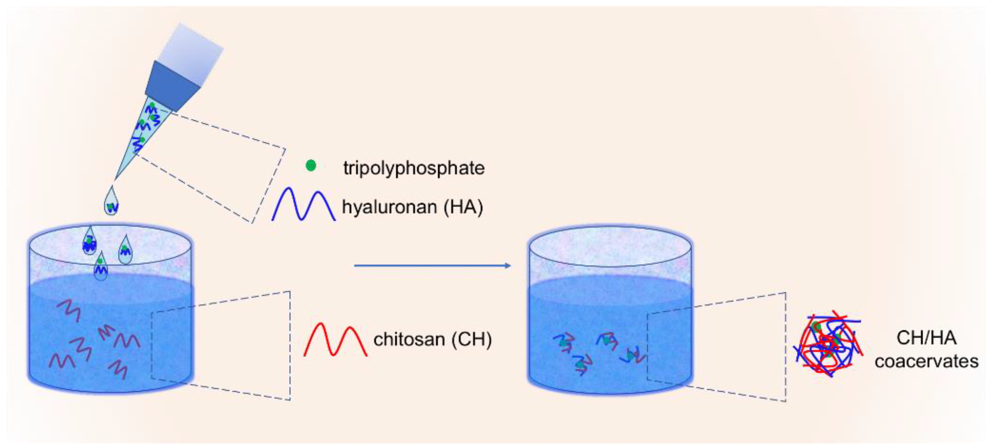
2.2. Preparation of Complex Coacervates

The synthesis of colloids was performed according to a previously reported procedure [36]. Briefly, polymers and TPP were solubilized in deionized water at a concentration equal to 0.6 mg/mL for chitosans, 1.25 mg/mL for sodium hyaluronan and 0.5 mg/mL for TPP. After complete solubilization, 150 μL of TPP solution were added dropwise to 3 mL of hyaluronate solution under stirring. Solutions were filtered through 0.22 μm filters (Biosigma, Italy) and stored at room temperature until use. In a 5 mL beaker 500 μL of the HA-TPP solution were added to 0.5 or 2.0 mL of chitosan solutions under stirring to allow the formation of colloids at different CH/HA weight ratio, i.e., equal to 1:2 and 2:1, respectively. Solutions were kept under stirring for 10 min and left at rest for 20 min before being analyzed. The final pH of solutions was in the range 4–6.5.

2.3. Physical-Chemical Characterization of Complex Coacervates

Dynamic Light Scattering (DLS) Analyses

Formulations were investigated by means of dynamic light scattering (DLS) on a Zetasizer Nano ZS with 173° detection optics (Malvern Instruments) to evaluate their size (hydrodynamic diameter), PolyDispersity Index (PDI) and surface charge ( ζ -potential). Each formulation was analyzed at least in triplicate at T = 25 °C after dilution 1:10 v/v in deionized filtered water using disposable cuvettes. The size was expressed as the Z-average hydrodynamic diameter, obtained by a cumulative analysis of the correlation function using the viscosity and refractive index of water in the calculations. ζ -potential was determined via the laser Doppler velocimetry (LDV) technique.

3. Results and Discussion

Characterization of complex coacervates at different chitosan physical-chemical composition and polymers weight ratio. We mixed chitosans showing different chemical composition and mass with a low molecular weight hyaluronan in order to build a large set of colloids (Figure 1). Upon injection of the polyanion-TPP solution, liquid-liquid phase separation (complex coacervation) occurred following to the formation of (i) low energy interactions, as well as hydrogen, electrostatic and hydrophobic between the two biopolymers [33,36], and (ii) the entropy gain due to the release of counterions and water molecules from polyelectrolytes. Preliminary (qualitative) considerations have been drawn on the basis of visual observations: aggregates were detected to the naked eye in the case of formulations composed of high molecular weight or high acetylated chitosans (Table 2). Therefore, we can conclude that low chitosan acetylation degree, as well as medium-to-low molecular weight, are essential in promoting an efficient complex coacervation event. Conversely, chitosan-to-hyaluronan weight ratio plays a marginal role.
Physical properties (i.e., dimensions, polydispersity and surface charge) of colloids at different chemical composition or polymers weight ratio were investigated by a DLS analysis. A different behavior of colloids composed of FA 0.16 chitosan and different CH/HA weight ratio was detected as a function of chitosan molecular weight (Figure 2a). In general terms, in the case of CH/HA 1:2 weight ratio colloids dimensions were almost independent on chitosan molecular weight. Overall, our results are in line with previous findings, wherein a 1:1 CH/HA weight ratio and a medium molecular weight chitosan were considered in the analysis [39]. Conversely, the dimension of colloids increased abruptly by increasing chitosan chain length for CH/HA 2:1 weight ratio up to the formation of micrometric colloids. A further increment of chitosan degree of polymerization was accompanied by a decrease of hydrodynamic diameter. Concerning colloids composed of medium acetylated chitosan, i.e., FA = 0.46, no apparent influence of CH/HA weight ratio was detected (Figure 2b) up to chitosan molecular weight of 210,000. Upon further increasing the degree of polymerization, CH/HA 1:2 colloids manifested an increment of dimensions at a variance with CH/HA 2:1 counterparts. For FA = 0.63 chitosans, dimensions of both types of colloids resulted to be independent on chitosan molecular weight up to M v ¯ = 170,000 (Figure 2c), whereas an increment of hydrodynamic diameter was spotted for M v ¯ = 310,000 chitosan.
Further considerations have been made with respect to the polydispersity index (PDI) (Figure 3). For FA 0.16 chitosan, a correlation between dimensional homogeneity and chitosan molecular weight emerged for the two types of polymers weight ratio analyzed (Figure 3a). In more detail, by using a weight ratio equal to 1:2, PDI values were independent on chitosan molecular weight and lower than 0.2. These results are in line with what previously reported for CH/HA of 1:1 and medium molecular weight chitosan-based colloids [39]. In the case of CH/HA 2:1, PDI values (linearly) increased as a function of chitosan molecular weight, suggesting a correlation between dimensional dispersity and polymer chain length. In the case of colloids fabricated with high acetylated chitosans, i.e., FA = 0.46 and 0.63 - none straightforward correlation between polymers weight ratio and PDI was detected (Figure 3b,c). All in all, PDI values 0.2 were recorded for all these formulations, suggesting the formation of more heterogeneous colloids with respect to lower acetylated and low molecular weight chitosan-based counterparts.
Finally, surface charge measurements were undertaken to give further insights onto colloids physical properties. ζ -potential analyses pointed at a different colloids surface charge depending on the weight ratio between chitosan and hyaluronan (Figure 4). As a general consideration, a positive surface charge was detected for chitosan-prevalent colloids (i.e., weight ratio CH/HA = 2:1), whereas a negative surface charge was found for their counterparts (i.e., weight ratio CH/HA = 1:2). This opposite behavior clearly reflects the different polycation/polyanion balance throughout the surface matrix. In the case of FA = 0.16 chitosan, the increment of molecular weight associated with an increase of colloids surface charge, indicating that a greater amount of D-type sugars, i.e., glucosamines, are available at the colloids surface. This typical behavior was detected both for CH/HA 1:2 and 2:1 coacervates (Figure 4a). For FA = 0.46 chitosan, ζ -potential was found to increase up to chitosan M v ¯ = 210,000, and subsequently decrease for higher chitosan degree of polymerization (Figure 4b). Lastly, an almost independency on chitosan molecular weight was noticed with FA = 0.63 chitosan for both CH/HA 1:2 and 2:1 colloids (Figure 4c).

4. Conclusions

In this work, the complex coacervation between chitosans of varying physical-chemical composition and a low molecular weight hyaluronan has been exploited as method to produce polysaccharide-based colloids. Experimental variables as well as polymers weight ratio, chitosan acetylation degree and molecular weight have been varied to verify the formation and quality of related colloids. The main conclusions arisen from experimental evidences are the following:
(i)
Low acetylation as well as low polycation molecular weight determine a true liquid-liquid phase separation without formation of aggregates; in these conditions, the dilute phase (the supernatant) is in equilibrium with the dense phase (the coacervate);
(ii)
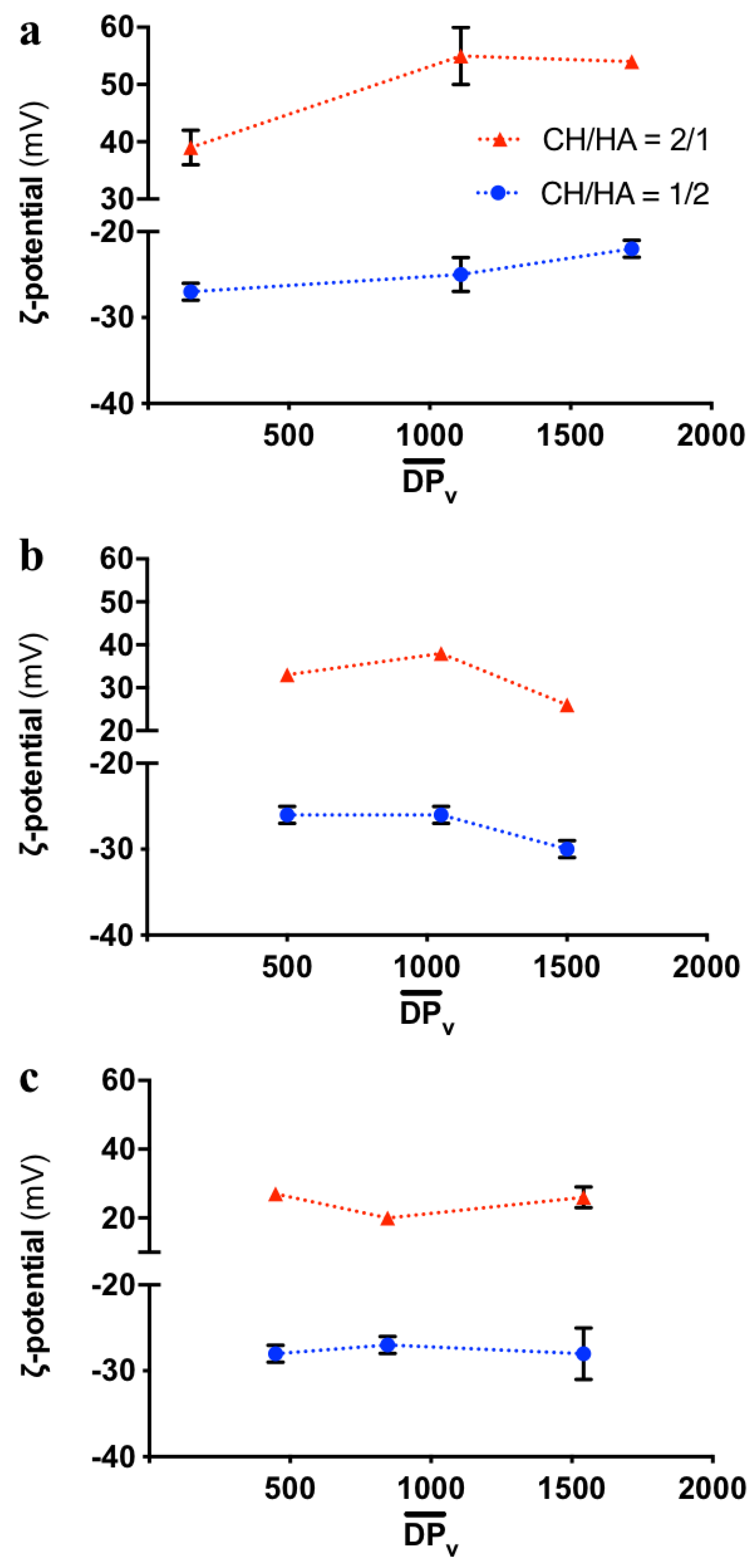
Chitosan-to-hyaluronan mass ratio directs colloids surface charge;
(iii)
Chitosan physical-chemical features influence colloids dimension and homogeneity.
Overall, this contribution provides additional knowledge about the complex coacervation between chitosan and hyaluronan, with the potential translation of related networks in food and biomedical sectors.

Author Contributions

Conceived and designed the experiments: I.D., P.S. Performed the experiments: F.F. Analyzed DLS data: F.F., I.D., E.M., P.S. Wrote the paper: F.F., P.S. All authors have read and agreed to the published version of the manuscript.

Conflicts of Interest

The authors declare no conflict of interest.

References

  1. Perugini, L.; Cinelli, G.; Cofelice, M.; Ceglie, A.; Lopez, F.; Cuomo, F. Effect of the coexistence of sodium caseinate and Tween 20 as stabilizers of food emulsions at acidic pH. Colloids Surf. B Biointerfaces 2018, 168, 163–168. [Google Scholar] [CrossRef]
  2. Kroll, J. Food Colloids Proteins, Lipids and Polysaccharides; Dickinson, E., Bergenståhl, B., Eds.; WILEY-VCH Verlag: Weinheim, Germany, 1997. [Google Scholar]
  3. Dickinson, E. On the road to understanding and control of creaminess perception in food colloids. Food Hydrocoll. 2018, 77, 372–385. [Google Scholar] [CrossRef]
  4. McClements, D.J. The future of food colloids: Next-generation nanoparticle delivery systems. Curr. Opin. Colloid Interface Sci. 2017, 28, 7–14. [Google Scholar] [CrossRef] [Green Version]
  5. Dickinson, E. Colloids in Food: Ingredients, Structure, and Stability. Annu. Rev. Food Sci. Technol. 2015, 6, 211–233. [Google Scholar] [CrossRef]
  6. Gutiérrez, T.J. Chitosan: Derivatives, Composites and Applications. In Chitosan: Derivatives, Composites and Applications; Ahmed, S., Ikram, S., Eds.; Wiley: Hoboken, NJ, USA, 2017; pp. 183–232. [Google Scholar]
  7. Dutta, J.; Tripathi, S.; Dutta, P.K. Progress in antimicrobial activities of chitin, chitosan and its oligosaccharides: A systematic study needs for food applications. Food Sci. Technol. Int. 2012, 18, 3–34. [Google Scholar] [CrossRef]
  8. Guo, M.; Yadav, M.P.; Jin, T.Z. Antimicrobial edible coatings and films from micro-emulsions and their food applications. Int. J. Food Microbiol. 2017, 263, 9–16. [Google Scholar] [CrossRef]
  9. Lekshmi, R.G.K.; Rahima, M.; Chatterjee, N.S.; Tejpal, C.S.; Anas, K.K.; Vishnu, K.V.; Sarika, K.; Asha, K.K.; Anandan, R.; Suseela, M. Chitosan—Whey protein as efficient delivery system for squalene: Characterization and functional food application. Int. J. Biol. Macromol. 2019, 135, 855–863. [Google Scholar] [CrossRef]
  10. de Farias, B.S.; Sant’Anna Cadaval Junior, T.R.; de Almeida Pinto, L.A. Chitosan-functionalized nanofibers: A comprehensive review on challenges and prospects for food applications. Int. J. Biol. Macromol. 2019, 123, 210–220. [Google Scholar] [CrossRef]
  11. Vårum, K.M.; Smidsrød, O. Structure-Property Relationship in Chitosans. In Polysaccharides: Structural Diversity and Functional Versatility, 2nd ed.; Dumitriu, S., Ed.; CRC Press: Boca Raton, FL, USA, 2004. [Google Scholar]
  12. Nilsen-Nygaard, J.; Strand, S.P.; Vårum, K.M.; Draget, K.I.; Nordgård, C.T. Chitosan: Gels and interfacial properties. Polymers (Basel) 2015, 7, 552–579. [Google Scholar] [CrossRef] [Green Version]
  13. Cuomo, F.; Cofelice, M.; Venditti, F.; Ceglie, A.; Miguel, M.; Lindman, B.; Lopez, F. In-vitro digestion of curcumin loaded chitosan-coated liposomes. Colloids Surfaces B Biointerfaces 2018, 168, 29–34. [Google Scholar] [CrossRef]
  14. Rinaudo, M. Chitin and chitosan: Properties and applications. Prog. Polym. Sci. 2006, 31, 603–632. [Google Scholar] [CrossRef]
  15. Racine, L.; Texier, I.; Auzély-Velty, R. Chitosan-based hydrogels: Recent design concepts to tailor properties and functions. Polym. Int. 2017, 66, 981–998. [Google Scholar] [CrossRef]
  16. Agnihotri, S.A.; Mallikarjuna, N.N.; Aminabhavi, T.M. Recent advances on chitosan-based micro- and nanoparticles in drug delivery. J. Control. Release 2004, 100, 5–28. [Google Scholar] [CrossRef]
  17. Aramwit, P.; Ekasit, S.; Yamdech, R. The development of non-toxic ionic-crosslinked chitosan-based microspheres as carriers for the controlled release of silk sericin. Biomed. Microdevices 2015, 17, 1–9. [Google Scholar] [CrossRef]
  18. Sacco, P.; Furlani, F.; de Marzo, G.; Marsich, E.; Paoletti, S.; Donati, I.; Sacco, P.; Furlani, F.; de Marzo, G.; Marsich, E.; et al. Concepts for Developing Physical Gels of Chitosan and of Chitosan Derivatives. Gels 2018, 4, 67. [Google Scholar] [CrossRef] [Green Version]
  19. Liu, Q.; Sacco, P.; Marsich, E.; Furlani, F.; Arib, C.; Djaker, N.; Lamy De La Chapelle, M.; Donati, I.; Spadavecchia, J. Lactose-Modified Chitosan Gold(III)-PEGylated Complex-Bioconjugates: From Synthesis to Interaction with Targeted Galectin-1 Protein. Bioconjug. Chem. 2018, 29, 3352–3361. [Google Scholar] [CrossRef]
  20. Cok, M.; Viola, M.; Vecchies, F.; Sacco, P.; Furlani, F.; Marsich, E.; Donati, I. N-isopropyl chitosan. A pH- and thermo-responsive polysaccharide for gel formation. Carbohydr. Polym. 2020, 230, 115641. [Google Scholar] [CrossRef]
  21. Cok, M.; Sacco, P.; Porrelli, D.; Travan, A.; Borgogna, M.; Marsich, E.; Paoletti, S.; Donati, I. Mimicking mechanical response of natural tissues. Strain hardening induced by transient reticulation in lactose-modified chitosan (chitlac). Int. J. Biol. Macromol. 2018, 106, 656–660. [Google Scholar] [CrossRef]
  22. Vecchies, F.; Sacco, P.; Decleva, E.; Menegazzi, R.; Porrelli, D.; Donati, I.; Turco, G.; Paoletti, S.; Marsich, E. Complex Coacervates between a Lactose-Modified Chitosan and Hyaluronic Acid as Radical-Scavenging Drug Carriers. Biomacromolecules 2018, 19, 3936–3944. [Google Scholar] [CrossRef]
  23. Alves, N.M.; Mano, J.F. Chitosan derivatives obtained by chemical modifications for biomedical and environmental applications. Int. J. Biol. Macromol. 2008, 43, 401–414. [Google Scholar] [CrossRef] [Green Version]
  24. Lim, S.H.; Hudson, S.M. Synthesis and antimicrobial activity of a water-soluble chitosan derivative with a fiber-reactive group. Carbohydr. Res. 2004, 339, 313–319. [Google Scholar] [CrossRef]
  25. Furlani, F.; Sacco, P.; Marsich, E.; Donati, I.; Paoletti, S. Highly monodisperse colloidal coacervates based on a bioactive lactose-modified chitosan: From synthesis to characterization. Carbohydr. Polym. 2017, 174, 360–368. [Google Scholar] [CrossRef]
  26. Kizilay, E.; Kayitmazer, A.B.; Dubin, P.L. Complexation and coacervation of polyelectrolytes with oppositely charged colloids. Adv. Colloid Interface Sci. 2011, 167, 24–37. [Google Scholar] [CrossRef]
  27. Antonov, M.; Mazzawi, M.; Dubin, P.L. Entering and exiting the protein-polyelectrolyte coacervate phase via nonmonotonic salt dependence of critical conditions. Biomacromolecules 2010, 11, 51–59. [Google Scholar] [CrossRef] [Green Version]
  28. Umerska, A.; Paluch, K.J.; Inkielewicz-Stepniak, I.; Santos-Martinez, M.J.; Corrigan, O.I.; Medina, C.; Tajber, L. Exploring the assembly process and properties of novel crosslinker-free hyaluronate-based polyelectrolyte complex nanocarriers. Int. J. Pharm. 2012, 436, 75–87. [Google Scholar] [CrossRef] [Green Version]
  29. Zambito, Y.; Felice, F.; Fabiano, A.; Di Stefano, R.; Di Colo, G. Mucoadhesive nanoparticles made of thiolated quaternary chitosan crosslinked with hyaluronan. Carbohydr. Polym. 2013, 92, 33–39. [Google Scholar] [CrossRef]
  30. Tan, C.; Xie, J.; Zhang, X.; Cai, J.; Xia, S. Polysaccharide-based nanoparticles by chitosan and gum arabic polyelectrolyte complexation as carriers for curcumin. Food Hydrocoll. 2016, 57, 236–245. [Google Scholar] [CrossRef]
  31. Almalik, A.; Donno, R.; Cadman, C.J.; Cellesi, F.; Day, P.J.; Tirelli, N. Hyaluronic acid-coated chitosan nanoparticles: Molecular weight-dependent effects on morphology and hyaluronic acid presentation. J. Control. Release 2013, 172, 1142–1150. [Google Scholar] [CrossRef]
  32. de la Fuente, M.; Seijo, B.; Alonso, M.J. Novel hyaluronan-based nanocarriers for transmucosal delivery of macromolecules. Macromol. Biosci. 2008, 8, 441–450. [Google Scholar] [CrossRef]
  33. de la Fuente, M.; Seijo, B.; Alonso, M.J. Novel hyaluronic acid-chitosan nanoparticles for ocular gene therapy. Investig. Ophthalmol. Vis. Sci. 2008, 49, 2016–2024. [Google Scholar] [CrossRef] [Green Version]
  34. de la Fuente, M.; Seijo, B.; Alonso, M.J. Design of novel polysaccharidic nanostructures for gene delivery. Nanotechnology 2008, 19, 075105. [Google Scholar] [CrossRef]
  35. Lallana, E.; Rios De La Rosa, J.M.; Tirella, A.; Pelliccia, M.; Gennari, A.; Stratford, I.J.; Puri, S.; Ashford, M.; Tirelli, N. Chitosan/Hyaluronic Acid Nanoparticles: Rational Design Revisited for RNA Delivery. Mol. Pharm. 2017, 14, 2422–2436. [Google Scholar] [CrossRef]
  36. Sacco, P.; Decleva, E.; Tentor, F.; Menegazzi, R.; Borgogna, M.; Paoletti, S.; Kristiansen, K.A.; Vårum, K.M.; Marsich, E. Butyrate-Loaded Chitosan/Hyaluronan Nanoparticles: A Suitable Tool for Sustained Inhibition of ROS Release by Activated Neutrophils. Macromol. Biosci. 2017, 17. [Google Scholar] [CrossRef]
  37. Luo, Y.; Wang, Q. Recent development of chitosan-based polyelectrolyte complexes with natural polysaccharides for drug delivery. Int. J. Biol. Macromol. 2014, 64, 353–367. [Google Scholar] [CrossRef]
  38. Oyarzun-Ampuero, F.A.; Brea, J.; Loza, M.I.; Torres, D.; Alonso, M.J. Chitosan-hyaluronic acid nanoparticles loaded with heparin for the treatment of asthma. Int. J. Pharm. 2009, 381, 122–129. [Google Scholar] [CrossRef]
  39. Furlani, F.; Sacco, P.; Decleva, E.; Menegazzi, R.; Donati, I.; Paoletti, S.; Marsich, E. Chitosan Acetylation Degree Influences the Physical Properties of Polysaccharide Nanoparticles: Implication for the Innate Immune Cells Response. ACS Appl. Mater. Interfaces 2019, 11, 9794−9803. [Google Scholar] [CrossRef]
  40. Sacco, P.; Cok, M.; Asaro, F.; Paoletti, S.; Donati, I. The role played by the molecular weight and acetylation degree in modulating the stiffness and elasticity of chitosan gels. Carbohydr. Polym. 2018, 196, 405–413. [Google Scholar] [CrossRef]
Figure 1. Sketchy representation of complex coacervation process between chitosan (the polycation) and hyaluronan (the polyanion). Tripolyphosphate serves as “adjuvant” cross-linker for chitosan. Electrostatic interactions—together with gain of entropy due to counterions and water release from polyelectrolytes—drive complex coacervates formation.
Colloids 04 00012 g001
Figure 2. Hydrodynamic diameter of CH/HA complex coacervates showing different polymers weight ratio (CH:HA = 1:2 and 2:1). Colloids were assembled using chitosans with fraction of acetylated units (FA) of 0.16 (a), 0.46 (b) and 0.63 (c), respectively. Data are reported as a function of chitosan average degree of polymerization, i.e., D P v ¯ . Colloids were analyzed by DLS after dilution 1:10 (v/v) in deionized water. Data are means (±SD) of at least three measurements.
Colloids 04 00012 g002
Figure 3. PolyDispersity Index (PDI) of CH/HA complex coacervates showing different polymers weight ratio (CH/HA = 1:2 and 2:1). Colloids were assembled using chitosans with fraction of acetylated units (FA) of 0.16 (a), 0.46 (b) and 0.63 (c), respectively. Data are reported as a function of the chitosan average degree of polymerization, i.e., D P v ¯ . Colloids were analyzed by DLS after dilution 1:10 (v/v) in deionized water. Data are means (±SD) of at least three measurements.
Colloids 04 00012 g003
Figure 4. Surface charge, i.e., ζ -potential, of CH/HA complex coacervates showing different polymers weight ratio (CH/HA = 1:2 and 2:1). Colloids were assembled using chitosans with fraction of acetylated units (FA) of 0.16 (a), 0.46 (b) and 0.63 (c), respectively. Data are reported as a function of chitosan average degree of polymerization, i.e., D P v ¯ . Colloids were analyzed by DLS after dilution 1:10 (v/v) in deionized water. Data are means (±SD) of at least three measurements.
Colloids 04 00012 g004
Table 1. Fraction of acetylated units (FA), intrinsic viscosity, [ η ], viscosity average molecular weight, M v ¯ , molecular weight of chitosan repetitive unit, M W r . u . , and viscosity average degree of polymerization, D P v ¯ , of hydrochloride chitosans used for the synthesis of complex coacervates. M v ¯ is calculated using the following Mark-Houwink-Sakurada parameters: K = 8.43 × 10⁻³ mL/g and a = 0.92. [40]
FA [ η ] (mL/g) M v ¯ M W r . u .   ( g / mol ) D P v ¯
0.1611030,000198152
681220,000 1111
1026340,000 1717
0.46340100,000200500
650210,000 1050
920300,000 1500
0.6330090,000>201448
550170,000 846
950310,000 1542
Table 2. Characterization of CH/HA complex coacervates after dilution 1:10 (v/v) in deionized water. Colloids were assembled using chitosans with different fraction of acetylated units (FA = 0.16, 0.46 and 0.63) and different viscosity average molecular weights ( M v ¯ ). Polymers weight ratio and aggregation at time zero of the resulting formulations are reported.
FA M v ¯   CH:HA
Weight Ratio
Notes
30,0001:2no aggregation
2:1no aggregation
0.16220,0001:2no aggregation
2:1no aggregation
340,0001:2no aggregation
2:1limited aggregation
100,0001:2no aggregation
2:1no aggregation
0.46210,0001:2no aggregation
2:1no aggregation
300,0001:2no aggregation
2:1limited aggregation
90,0001:2no aggregation
2:1no aggregation
0.63170,0001:2limited aggregation
2:1no aggregation
310,0001:2no aggregation
2:1limited aggregation

