Article

Bioactive Properties of Chitosan/Nanocellulose Films Loaded with Sage Essential Oil: From In Vitro Study to In Situ Application in Shelf-Life Extension of Fresh Poultry Meat

by
João R. A. Pires
1,
Raquel Pereira
1,
Sara Paz
1,
Leandro A. Gomes
1,
Victor G. L. Souza
1,
Maria H. Godinho
2,
Maria P. Duarte
1 and
Ana L. Fernando
1,*
1
MEtRICs, Departamento de Química, NOVA School of Science and Technology|FCT NOVA, Universidade Nova de Lisboa, Campus de Caparica, 2829-516 Caparica, Portugal
2
CENIMAT|I3N, Departamento de Ciências dos Materiais, NOVA School of Science and Technology|FCT NOVA, Universidade Nova de Lisboa, Campus de Caparica, 2829-516 Caparica, Portugal
*
Author to whom correspondence should be addressed.
J. Compos. Sci. 2025, 9(8), 428; https://doi.org/10.3390/jcs9080428
Submission received: 25 June 2025 / Revised: 28 July 2025 / Accepted: 2 August 2025 / Published: 8 August 2025
(This article belongs to the Section Biocomposites)

Abstract

The overuse of nonrenewable resources has motivated intensive research and the development of new types of green bio-based and degradable feedstocks derived from natural sources, such as cellulose derivates, also in nanoforms. The inclusion of such nanoparticles in bio-based polymers with the aim of providing reinforcement is a trend, which, when associated with the incorporation active compounds, creates active packaging suitable for the packaging of highly perishable food, thus contributing to the product’s shelf-life extension. Chitosan (Ch)/sage essential oil (SEO) bionanocomposite reinforced with nanocrystalline cellulose (CNC) was cast as active packaging for the preservation of fresh poultry meat. Meat samples were wrapped in different bioplastics (pristine chitosan, chitosan with commercial CNC, chitosan with CNC obtained from three different lignocellulosic crops, giant reed (G), kenaf (K), and miscanthus (M), chitosan with SEO, and chitosan with SEO and CNC), while unwrapped samples were tested as the control. Periodically, samples were evaluated in terms of their physicochemical properties and microbial growth. Additionally, bionanocomposites were also evaluated in terms of their in situ antimicrobial properties, as well as migration toward food simulants. Meat samples protected with bionanocomposites showed lower levels of microbiological growth (2–3 logs lower than control) and lipid oxidation (20–30% lower than in control), over time. This was attributed to the intrinsic antimicrobial capacity of chitosan and the high oxygen barrier properties of the films resulting from the CNC inclusion. The SEO incorporation did not significantly improve the material’s antimicrobial and antioxidant activity yet interfered directly with the meat’s color as it migrated to its surface. In the in vitro assays, all bionanocomposites demonstrated good antimicrobial activity against B. cereus (reduction of ~8.2 log) and Salmonella Choleraesuis (reduction of ~5–6 log). Through the in vitro migration assay, it was verified that the SEO release rate of phenolic compounds to ethanol 50% (dairy products simulate) was higher than to ethanol 95% (fatty food simulate). Furthermore, these migration tests proved that nanocellulose was capable of delaying SEO migration, thus reducing the negative effect on the meat’s color and the pro-oxidant activity recorded in TBARS. It was concluded that the tested chitosan/nanocellulose bionanocomposites increased the shelf life of fresh poultry meat.

Graphical Abstract

1. Introduction

The global food system and its value chains rely heavily on packaging to ensure proper functioning. With the increasing population in urban areas and demand for convenience foods, the packaging market is expected to grow, leading to the generation of millions of tons of plastic waste [1,2]. Moreover, the production of plastic is also linked to greenhouse gas emissions, contributing to global warming [3]. Therefore, the application of sustainable, renewable resources to create functional alternatives to non-biodegradable plastics is an opportunity to reduce the ecological impact of packaging [4].
Currently, these bioplastics are becoming more popular in the food packaging industry and are considered a viable alternative [1]. Chitin is a primary constituent of marine arthropods’ exoskeletons, and it can be extracted from seafood industry waste. This bioresource can be deacetylated to produce a cationic polysaccharide denominated as chitosan [5]. The biocompatible and non-toxic biopolymer becomes soluble in slightly acidic environments, allowing the development of films/coatings with intrinsic antimicrobial and antioxidant properties [6].
In terms of functionality, chitosan films/coatings effectively block gas transfer due to their well-ordered matrix arrangement [7]. However, like other polysaccharides, chitosan is hydrophilic and has poor water vapor barrier properties, which is a disadvantage in preserving perishable food products [8]. Aiming to improve such properties, nanofillers are incorporated into the polymeric matrix to create bionanocomposites [9].
Nanocrystalline cellulose is an organic filler commonly used to reinforce bio-based films [9,10,11]. Their surface is abundant in hydroxyl groups, allowing chemical crosslinking with other polymers, like chitosan. Among its distinctive properties, these reinforcement agents own a high aspect ratio and significant levels of crystallinity, therefore an increase in the functional properties of the film is expected, even at low concentrations [12]. Additionally, CNC films offer fewer public health risks and are more eco-friendly when compared with inorganic fillers, since they are biodegradable, non-toxic, and can be extracted from several low-cost renewable lignocellulosic sources [13].
Sage (Salvia officinalis L.) is an aromatic plant, rich in terpenes and phenolic compounds, used for culinary and medicinal purposes due to its antibacterial and antioxidant properties [14,15,16], which is also used as both an extract or an essential oil. To improve the stability of the active compounds and prevent excessive oil absorption by the food, sage essential oil (SEO) can be incorporated into bionanocomposites, allowing controlled release, which prevents excessive odor and taste transfer negatively affecting the food. While there is no documented information about the toxicological effects of SEO on humans, the no-observed-adverse-effect level (NOAEL) for orally administered SEO on rats is 250 mg/kg body weight/day, according to the European Medicines Agency (EMA) report [17]. However, further studies are necessary to clarify any potential carcinogenic effects.
Although the application of films based on Ch/CNC [18] or Ch/SEO [16] has been cited in the literature, very little is known about the effect of CNC as reinforcement and an encapsulation agent of natural antioxidant compounds, like SEO, and the associated effect as active packaging to perishable food matrices. Therefore, this research aimed to evaluate the bioactive properties of chitosan/CNC/SEO films either in vitro through migration assay and antimicrobial tests; and in situ as primary packaging of fresh poultry meat to elucidate their effect in the extension of such perishable food items. This novel approach followed the concept of circular economy, reusing biomass to develop a new high-added-value material with the aim of it being used in the future as a fully biodegradable and active element in food packaging. The nanocellulose used as nanofillers were extracted from three different lignocellulosic biomasses, Arundo donax L. (also identified as giant reed), Hibiscus cannabinus L. (also identified as kenaf), and Miscanthus x giganteus Greef et Deu. (or simply miscanthus), following a previous work [19], where these chitosan bionanocomposites demonstrated good mechanical, thermal, and barrier properties to oxygen for application as food packaging.

2. Materials and Methods

2.1. Materials and Reagents

High-molecular-weight chitosan (31–37 kDa and 75% deacetylation degree) and 1,1,3,3-tetraethoxypropane (TEP) were acquired from Sigma-Aldrich (Steinheim, Germany). Trichloroacetic acid (TCA) and 2-thiobarbituric acid (TBA) were obtained from PanReac (Barcelona, Spain). Glacial acetic acid (CH3COOH), glycerol (C3H8O3), sodium hydroxide (NaOH), hydrogen peroxide (H2O2), sulfuric acid 95–97% (H2SO4), calcium nitrate (Ca(NO3)2), and Tween® 80 (polyethylene glycol sorbitan monolaurate) were purchased from Alfa Aesar (Kandel, Germany). The microbiological reagents “Violet Red Bile Glucose” (VRBG) “Plate Count Agar” (PCA), “Trypto-Casein Agar” (TSA), and “Trypto-Casein Soy Broth” (TSB) were purchased from Biokar (Allonne, France). Commercial nanocellulose was kindly supplied by Nanocrystacell (Podcerkev, Slovenia). The food-grade essential oil of sage (Salvia officinalis L.) from Biover (Nazareth, Belgium) was purchased at a local market. All water used was purified using a Barnstead™ GenPure™ water purification system (Thermo Scientific™, Gothenburg, Sweden) and all chemicals were of analytical reagent grade and used as purchased. Giant reed, kenaf, and miscanthus biomass were harvested from pilot fields settled in Caparica, near Lisbon, in the Peninsula of Setúbal, Portugal (latitude 38°40′03′′N, longitude 9°12′8′′W, altitude of 50 m). The reaped material was dried at 40 °C in a vacuum oven, cut into small pieces, and stored in darkness, in a dry place at room temperature until further use.

2.2. Bionanocomposite Production

The bionanocomposites were prepared according to [19], and their respective formulations are depicted in Table 1.
To prepare the film-forming dispersion (FFD), 1.5% (w/v) of chitosan was dissolved in 1% (v/v) glacial acetic acid solution under mechanical stirring for 24 h at room temperature. Then, glycerol was added as a plasticizer at the percentage of 30% (w/w Ch) corresponding to only 0.45% (w/v FFD), along with 1% (v/v FFD) of sage essential oil (SEO). For the films incorporated with SEO, Tween® 80 at a level of 0.2% (w/v SEO) was added as emulsifier. Following that, the dispersion was submitted to 5 min agitation using a T18 Ultra-Turrax disperser (IKA® T18, Staufen, Germany) at 15.000 rpm, followed by 15 min degasification in an ultrasound bath (360 W) (Selecta, Barcelona, Spain). The nanofillers, 2.5% CNC (w/w Ch), produced from the three lignocellulosic biomasses (giant reed, miscanthus and kenaf [19]) or the commercial one, were then added. Thereafter, two more cycles of Ultra-Turrax and ultrasound under the same conditions were carried out to guarantee the correct dispersion of all components into the polymeric chain. The finished solution was cast in glass molds (18 × 25 cm), dried at 30 °C/24 h, and peeled for use.
Aiming to study the encapsulation effect of CNC on SEO, a Ch film with only the addition of essential oil was assembled. Moreover, a Ch film reinforced with commercial nanocellulose (at the same percentages) and a pristine Ch film were also prepared for comparison.

2.3. In Vitro Bionanocomposite Study

2.3.1. Specific Migration of Antioxidant Compounds

The in vitro quantification of antioxidant compound migration from the bionanocomposites over time was performed using the diffusion test [20]. Two simulants were chosen to mimic dairy products (50% ethanol solution) and fatty foods (95% ethanol solution), and the assay was carried out at 40 °C ± 2 °C/10 days. These food simulations were chosen since the packaging’s main objective is to delay the oxidative process in food matrices; in this way, these were considered the most susceptible to such spoilage processes.
Periodically, the total phenolic content (TPC) in the simulant medium was determined by the Folin–Ciocalteu method [21]. Briefly, 1 mL of the simulant medium was added to 3 mL of water and 0.25 mL of the Folin–Ciocalteu reagent. After 6 min, 0.75 mL of sodium carbonate (5% w/v) was added, and the mixture was incubated for 60 min in the dark and at room temperature (20 °C ± 5 °C). The TPC was performed using a UV/VIS spectrophotometer by reading the absorbance at 760 nm. A gallic acid calibration curve (0–200 mg/L) was used to calculate the results, which were expressed in mg gallic acid equivalent (mg GAE/L).
In addition, based on Fick’s second law, described in Equation (1) and using initial migration data, the diffusion coefficient (D) of phenolic compounds was calculated from the plot of MF,t/MP,0 versus t0.5 [22].
M F , t M P , 0 = 2 δ D t π 0.5
where MF,t (mg GAE) is the TPC in the food simulant at time t, MP,0 (mg GAE) is the initial TPC in the film, D (cm2/s) is the diffusion coefficient of TPC and δ(m) is the film thickness.

2.3.2. Antioxidant Activity

In vitro antioxidant activity was determined in the simulant media by the DPPH radical capture assay (2,2-diphenyl-1-picrylhydrazyl) according to the methodology described by [20]. Briefly, 3 mL of 60 μM ethanolic DPPH solution was mixed with 1 mL of the simulant media. The mixture was kept protected from light at room temperature (20 °C ± 5 °C) for 20 min. Quantification was performed using a UV/VIS spectrophotometer at 517 nm. The inhibition percentage (%) was calculated using Equation (2).
I n h i b i t i o n % = A b s c o n t r o l A b s s a m p l e A b s c o n t r o l × 100
where Abssample is the sample absorbance, while Abscontrol is the blank absorbance.

2.3.3. Antimicrobial Activity—Colony-Forming Unit Counting Method

In order to complement the study, assessment of the bionanocomposites’ antimicrobial activity, by the method of counting colony-forming units [23] for Gram-negative (Salmonella enterica subsp. enterica serovar Choleraesuis ATCC® 10708TM) and Gram-positive (Bacillus cereus ATCC® 11778TM) foodborne bacteria, was carried out. Initially, 0.2 g of each film was immersed in 4 mL of TSB nutrient medium containing approximately 106 CFU/mL of the bacteria under test. Then, the system was maintained at 37 °C (S. Choleraesuis) or 30 °C (B. cereus) for 24 h with continuous agitation (150 RPM) using an incubator (Innova model, New Brunswick Scientific, Edison, NJ, USA). Tubes without films were used as a control. After incubation, 100 μL of serial dilutions were inoculated by spreading on TSA plates, which were subsequently incubated at the respective optimal growth temperature of each evaluated bacterium for 16–24 h. The number of colony-forming units was counted, and the antimicrobial activity was expressed as the number of log reductions (CFU)/mL, calculated according to Equation (3).
L o g   r e d u c t i o n s = L o g B L o g A
where B and A correspond to the counts of viable bacteria (CFU/mL) in the control treatment and in the treatments evaluated after 24 h of incubation, respectively.

2.4. In Situ Application of Bionanocomposites as Primary Packaging of Poultry Meat

2.4.1. Fresh Poultry Meat Preparation

Fresh poultry meat was purchased from the local market with the longest possible shelf life. Meat was minced and divided into small “hamburgers” of 30 g, which were wrapped in the respective bionanocomposite (5 × 15 cm) and conditioned inside sterile plastic boxes, under refrigeration (5 °C ± 2 °C) (Figure 1). These preparation processes (grinding, fractioning and packaging) were carried out in a sterilized environment (vertical laminar flow air hood Steril Helios, Bionova, Italy) and with sterilized tools to avoid contamination. Each treatment was collected and evaluated at 3, 6, 9, and 12 days of storage. At the initial time (0 days of storage), only the unwrapped meat was characterized. Unpackaged meat samples were used as a control over the test times. The experiment was performed in triplicate, and results are the means ± standard deviation.

2.4.2. Microbiological Growth

Total mesophilic aerobic microorganism (TMAM), total psychrotrophic aerobic microorganism (TPAM), and Enterobacteriaceae quantification were determined to estimate the microbiological quality of the meat according to ISO 4833-1:2013 [24], ISO 17410:2019 [25], and ISO 21528-2:2017 [26], respectively. TMAM and TPAM counts were performed in PCA after incubation at 30 °C for 72 h or 7 °C for 168 h, respectively. For Enterobacteriaceae counts, VRBG agar was used with incubation at 30 °C/24 h. Results are expressed as log CFU (colony forming units)/g of meat.

2.4.3. Physicochemical Characterization

The physicochemical characterization consisted of pH, total titratable acidity, total volatile basic nitrogen (TVB-N) and moisture content according to the AOAC method (Association of Official Analytical Chemists) [27].
The color was registered on the meat surface through the measurement of CIE-L*a*b* (International Commission on Illumination) coordinates using a CR 410 colorimeter (Minolta Co., Tokyo, Japan) with D65 light source, and visual angle of 10°. The data were analyzed and converted into two evaluation parameters, lightness (L*), which describes the overall intensity of how light or dark a color is perceived (L* = 0 yields black, and L* = 100 indicates diffuse white), and hue angle, which corresponds to the color angular position around a central or neutral point or axis on the CIE-Lab space coordinate diagram. The color measurements were performed 3 times at 3 different points of the sample, for averaging purposes, on a standardized white background. Hue angle (hue) was calculated using Equations (4) and (5).
h u e = arctan b * a *   ( i f   a * > 0 )   or
h u e = arctan b * a * + 180 °   ( i f   a * < 0 )

2.4.4. Lipid Oxidation (TBARS Index)

The oxidative state of the samples were evaluated by the thiobarbituric acid reactive substances (TBARS) assay, according to [28]. For each treatment, 15 g of poultry meat was mixed with 30 mL of TCA 7.5% (w/v) and agitated briefly. The supernatants were filtered, and 5 mL of the filtrate was mixed with 5 mL of TBA 0.02 M. The tubes were heated at 95 °C/30 min in a water bath (Memmert, Schwabach, Germany), and then cooled to room temperature. Finally, the absorbance of the resulting system was measured using a UV/VIS spectrophotometer (Model Spekol 1500, Analytikjena, Jena, Germany) at 530 nm. The quantification of malondialdehyde (MDA), used to determine the TBARS index, was calculated from calibration curves made with known concentrations of MDA (from 1,1,3,3-Tetraethoxypropane (TEP) solution). Results are expressed as mg of MDA/kg of sample.

2.5. Statistical Analysis

All experiments were conducted in a completely randomized design with three replications. Statistical analysis of the obtained data was performed using a one-way analysis of variance using IBM SPSS Statistics version 23 (IBM, Armonk, NY, USA), and differences between mean values were processed using Tukey’s test. Significance was set at p < 0.05.

3. Results

3.1. In Vitro Bionanocomposite Study

3.1.1. Specific Migration of Antioxidant Compounds

The introduction of novel materials in food packaging requires careful monitoring of bioactive compound migration present in its formulation. Understanding the migration profile of these substances is crucial to predict the packaging behavior when in direct contact with food. Additionally, with this test it is possible to analyze the magnitude order of the phenolic compounds released, essential to understanding the substance’s antioxidant activity and the possible danger these may yield to the final consumer, as well as their potential to modify the food’s nutritional and organoleptic properties [29,30].
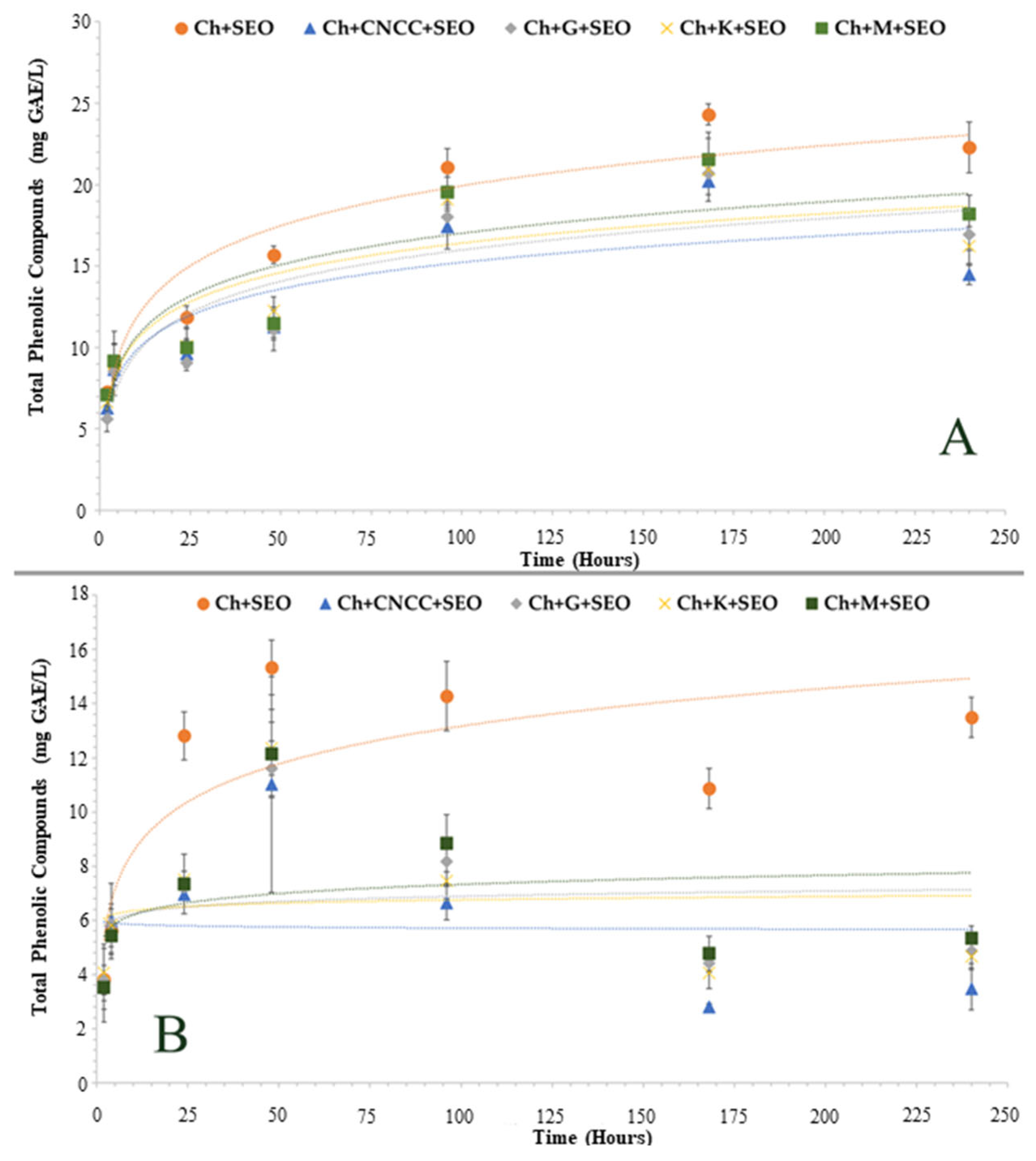
Food is made up of a complex diversity of substances, making it difficult to accurately evaluate their migration. As an alternative, to make this determination, it is usual to resort to food-simulating agents, duly regulated by competent authorities, such as the European Food Safety Authority (EFSA) or the Food and Drug Administration (FDA). Several factors influence the migration severity when the packaging comes into contact with the food, like time, temperature, compound structure and concentration, type of polymeric matrix, and the food’s physical–chemical characteristics. The main objective of applying bionanocomposites in fresh poultry meat was to hinder the progression of deterioration processes, like lipid oxidation, thus justifying the choice of the two simulating media [31]. Moreover, the migration profile was not contemplated for other hydrophilic simulants, like water, and ethanol 10% (for alcoholic products), since in previous works a partial degradation of the bionanocomposites was verified between 24 and 48 h. The total phenolic compound migration profile for the two simulants is presented in Figure 2. The diffusion coefficient, according to Fick’s second law, and the rate and amount of the phenolic compounds released were calculated for each sample and simulating medium (Table 2).
The migration profile of phenolic compounds was similar for both simulating media, showing a pattern of “exponential growth to a maximum”. As an aid to decoding the TPC migration pattern, logarithmic trend lines were drawn. In this way, it was verified that the TPC migration profile reached an equilibrium after the maximum point was reached. However, the amount released as well as the speed at which the compounds were diffused to the outside was not the same for both cases, as seen in Table 2. Lower diffusion coefficients were obtained for ethanol 50%, indicating a more gradual release. Moreover, the samples showed a higher release rate of phenolic compounds for ethanol 50% (between 71–85% of the total incorporated) than for ethanol 95% (between 54–75% of the total incorporated), with the maximum release peak occurring after 168 h and 48 h, respectively.
The exponential TPC release verified in the first hours can be explained by the non-adsorbed SEO present in the bionanocomposite surface, which rapidly migrated to the outside [32]. After reaching the maximum, a dispersion of points was observed, characterized by a slight decrease followed by a final growth. The decrease could be related to the lack of stability of the phenolic compounds. Despite the high applicability potential, the agri-food industry often struggles with the instability of these bioactive substances. Polyphenols have unsaturated bonds and a high antioxidant capacity, which makes them sensitive to a large number of factors, including temperature and long-term storage time, which are related to this work [33]. Determining the exact point where the degradation of these compounds began is difficult to set with accuracy, since at the point of maximum migration it may already be occurring. However, the decrease observed after 48 h for ethanol 95%, and 168 h for ethanol 50%, may indicate that from that time on, the amount of degraded TPC is greater than the amount released [30]. In the last evaluation time, an increase in TPC was observed in ethanol 95%, which is due to the beginning of the bionanocomposites decomposition, also verified in the last evaluation time (12 days) when the samples were in contact with the fresh poultry meat. The migration profile from the samples to ethanol 95% was similar to that exposed in the work of Souza et al. (2018) [34].
The higher total phenolic compounds observed for ethanol 50%, throughout all evaluation times, could be related to different factors. A similar result was recorded in the work of [35], although the migration profile of phenolic compounds from banana peels extract was only evaluated after 4 h. According to the authors, the TPC values were significantly higher for more hydrophilic media, like pure water and ethanol 50%, due to the hydrophilicity of the Ch film, thus, for superior solubility of the film, a higher release of phenolic compounds. As stated by Buonocore et al. (2003) [36], the release of an active compound from a polymeric network occurs in several steps. When diffusing from the simulating solution to the polymeric matrix, the water molecules weaken and change the structure of the network, allowing phenolic compound diffusion, until thermodynamic equilibrium is reached. However, other works stated the opposite results. In the work of [37], the highest limonene, a major component of bergamot oil, released from Ch was verified in all films for ethanol 95% aqueous solutions. For higher ethanol concentrations, the polarity of the solvent decreased, and therefore, the chemical affinity and solubility of limonene increased, justified by the authors. In another similar work, the migration profile of ginger (GEO) and rosemary (REO) essential oils from Ch films were compared to different simulant media. The results showed differences between the two samples, with REO having a greater affinity for ethanol 10%, while GEO had higher release amounts for ethanol 95%. The authors justified the results by stating that the components present in GEO were more lipophilic than those in REO, which on the other hand were more hydrophilic [30]. It can thus be seen that the chosen polymer matrix, as well as the composition of the essential oil and the food matrix where the bionanocomposite will be applied, are important factors in determining the migration profile of total phenolic compounds over time.
Continuing to review the work of Souza et al. (2019) [30], the authors verified that the main components of GEO (zingiberene and α-curcumene) have a more complex structure with a higher molecular weight than the components of REO (camphor and eucalyptol), causing a reduction in the intermolecular friction with the polymer (higher plasticizing effect) that contributed to active compound release, especially in the more hydrophobic simulant (ethanol 95%). In our work, the study of the chemical composition of the essential oil of sage (Salvia officinalis L.) was not carried out; however, resorting to the literature, it was found that there was a consensus that the most predominant compounds in this essential oil are the oxygenated monoterpenes, like camphor and α-thujone [14,38,39]. The fact that sage’s chemical composition is more like rosemary than ginger, since both belong to the same botanical family and genus (Lamiaceae, Salvia), may help to justify why there was a greater affinity for more hydrophilic media. However, both camphor and α-thujone are practically insoluble in water and easily soluble in alcohol and many organic solvents, contradicting our results [40,41]. Consequently, analyzing all the factors, the most plausible reason for understanding the migration profile of the phenolic compounds from SEO obtained from the two simulated media lies in the hydrophilic profile and the wettability properties of the chitosan bionanocomposites, in which the swelling and solubility of them govern the higher substance release for more hydrophilic systems. In future work, a complete characterization of the properties of bionanocomposites loaded with SEO, an analysis of the chemical composition of the essential oil, as well as a specific migration analysis for the largest compounds detected, are fundamental to better understanding this phenomenon.
Analyzing Figure 2, it was verified that the release of phenolic compounds was more limited in the bionanocomposites incorporated with nanocellulose compared to the “Ch + SEO” film. In comparison, the release rate from the bionanocomposites was shown to be inferior: between 15–20% (Table 2). Among the added nano-reinforcements, no significant differences were verified (p > 0.05); however, the commercial nanocellulose bionanocomposite showed a slightly higher encapsulation capacity. The lower release of essential oil into the simulating media from the reinforced bionanocomposites was in agreement with the results from other studies in which nanoparticles were added to reinforce chitosan films [42,43]. The compact network formed between the nanocellulose and chitosan trapped the essential oil, hindering its diffusion from the interior to the surface of the bionanocomposite. Moreover, it was verified in our previous work that the addition of nanocellulose decreases the swelling and solubility of the chitosan film, making it more hydrophobic [19]. This factor determines that the polymeric matrix structure is more resistant to modifications and therefore fewer gaps are created when in contact with water, impairing the diffusion of oil to the outside.

3.1.2. Antioxidant Activity

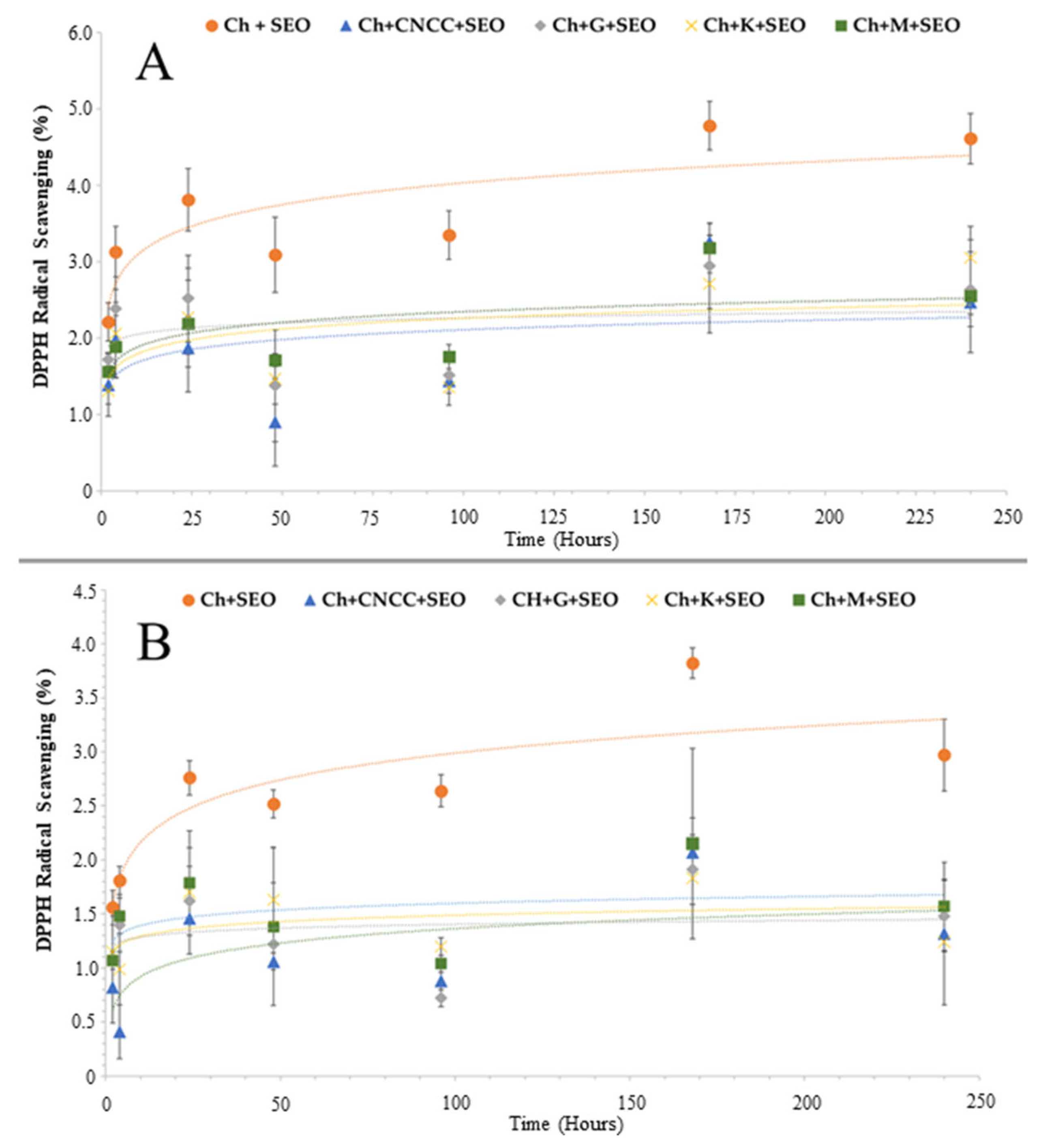
The antioxidant activity of the compounds that migrated from the bionanocomposite to the simulating media was evaluated using the radical scavenging method (DPPH). The study was conducted at the same time as the migration of phenolic compounds and the results are expressed as the percentage of inhibition of the DPPH radical, as can be seen in Figure 3.
Observing the results, it was noted that there was a higher percentage of DPPH radical inhibition in the simulating media ethanol 50% for all evaluation times. These values may be due to the greater phenolic compounds released into this simulant, as observed in the previous assay. For both media, it was observed that the antioxidant activity results adopted an increased tendency over the first 24 h, followed by a decrease until a new rise with a peak of activity at 168 h. The recorded antioxidant activity profile was similar to that of TPC for ethanol 95%; however, it did not follow the longer gradual release observed in ethanol 50%. This may indicate a relevant degradation of the phenolic compounds released after this time, thus decreasing their antioxidant activity. The peak of activity recorded at 168 h was mostly due to the beginning of the bionanocomposites’ degradation process, which granted a greater release of phenolic compounds. Additionally, the antioxidant activity for both media was lower for the nanocellulose-reinforced bionanocomposites than for the “Ch + SEO” film, justified by the lower release of phenolic compounds.
Inhibition values never exceeded 5% for either of the analyzed media, corresponding to a slight antioxidant activity. These values proved to be below those obtained for rosemary and ginger [30]. In the literature, it is possible to verify that sage essential oil demonstrated good DPPH scavenging activity, being commonly associated with a compound with good antioxidant activity. In fact, two different works, when comparing Rosmarinus officinalis L. and Salvia officinalis L. essential oils, attributed the greater neutralization of the DPPH radical to sage, since the percentage of inhibition of 50% was achieved for considerably lower oil concentrations [44,45]. Therefore, it was expected that, by using sage essential oil in chitosan bionanocomposites, it would be possible to enhance their antioxidant capacity. However, the results obtained showed that, although there was a release of total phenolic compounds to the simulating media, these did not demonstrate a marked ability to inhibit the DPPH radical. Moreover, these observations may help to explain why no significant improvement in lipid oxidation results was observed in meat wrapped in bionanocomposites incorporated with SEO, as discussed in the next chapters.

3.1.3. Antimicrobial Activity—Colony-Forming Unit Counting Method

The results presented in Table 3 show that the analyzed bionanocomposites had an elevated antimicrobial activity against both foodborne bacteria. Results for the remaining films reinforced with CNC did not statistically differ (p > 0.05) from CNCC, thus are not included in Table 3.
All samples tested were more efficient against Gram-positive bacteria (B. cereus) with an almost total reduction, superior to ~8.2 log, after 24 h incubation time. Comparatively, for the Gram-negative bacteria (S. Choleraesuis), the maximum reduction registered was for the pristine chitosan film, ~6.0 log. Nanocellulose incorporation did not significantly affect antibacterial activity (p > 0.05), consistent with findings in Section 3.1.1. However, adding SEO slightly but significantly reduced (p < 0.05) activity against S. Choleraesuis, suggesting chitosan’s intrinsic antimicrobial properties mainly drive the reduction in viable counts. These results align with the in situ evaluation of microbial growth in packaged meat and support findings by Souza et al. (2019) [30].
Chitosan has been shown to have antimicrobial properties against a wide range of microorganisms, including both Gram-positive and -negative bacteria [23]. There are various factors that can impact the efficiency of chitosan against distinct bacterial species. These may include the degree of deacetylation and the molecular weight of the chitosan, as well as the specific environmental conditions in which the bacteria are grown. Moreover, particular bacterial strains could have different susceptibilities to chitosan, and the emergence of chitosan resistance is a possibility that should be taken into account [46]. Gram-negative bacteria are typically more resistant to antimicrobial agents due to the presence of an outer membrane that serves as a permeability barrier, limiting the access of antimicrobial agents to the bacterial cell wall and membrane. However, chitosan has been shown to exhibit significant antimicrobial activity against Gram-negative bacteria due to the cationic nature of chitosan, which allows it to interact with the negatively charged bacterial outer membrane and penetrate the cell wall, leading to disruption of the cytoplasmic membrane and cell death. On the other hand, Gram-positive bacteria lack the outer membrane but possess a thicker layer of peptidoglycan that binds non-covalently with chitosan through teichoic acids. This interaction can also lead to the disruption of the bacterial cell wall and subsequent cell death [47,48].
In general, the wide-ranging ability of chitosan to prevent the growth of both Gram-negative and Gram-positive bacteria makes it a promising option for use as an antimicrobial agent in various areas, such as preserving food, healing wounds, and coating medical devices. Nonetheless, additional investigations are necessary to comprehend the underlying mechanisms of chitosan’s antimicrobial activity and to refine its effectiveness against diverse types and strains of bacteria.

3.2. “In Situ” Meat Characterization—Shelf-Life Study

3.2.1. Microbiological Growth

Antimicrobial activity represents an essential property to be evaluated during the development of novel active packaging applications, especially for perishable foods. Fresh meat constitutes a wealthy source of nutrients (like sugars, amino acids, and vitamins), having a pH close to neutrality and plenty of available water, essential characteristics for microbiological development, thus, distinguishing itself as an ideal substrate for the rapid proliferation of numerous types of microorganisms [49]. Individually, poultry meat is considered a highly perishable food product, known to have elevated initial rates of microbiological contamination, deteriorating above tolerable levels after four days post-slaughter under typical refrigerated storage conditions [50,51].
At the end of the experiment, spoilage was observed in all treatments, registered by the significant increase (p < 0.05) in all microbiological indicators evaluated (Table 4). As expected, the unwrapped meat presented the highest and fastest microbial contamination, recording the counting of 2.11 ± 0.21 log CFU/g for TMAM and 2.31 ± 0.49 log CFU/g for TPAM on the first evaluation date. These numbers increased steadily until the last day to 9.81 ± 0.37 log CFU/g and 9.58 ± 0.01 log CFU/g, respectively. Regarding Enterobacteriaceae, an initial value of 2.26 ± 0.12 log CFU/g and 9.41 ± 0.32 log CFU/g on the final day were found.
Compared to control, all packaged samples showed significantly lower contamination levels (p < 0.05), approximately 2 log CFU/g less for TMAM and TPAM, and 2–3 log CFU/g for Enterobacteriaceae, confirming that the produced bionanocomposites hold an antimicrobial activity capable of delaying microbial development. These positive results should be mainly attributed to the recognized Ch antimicrobial property. The most commonly accepted mechanism behind this intrinsic activity is ascribed to the electrostatic interactions between the positively charged amine groups of the polymer and the negatively charged microorganism cell surface, resulting in cell death by permeabilization and emptying of intracellular material [30]. High-molecular-weight chitosan, like the one used in this work, generally has difficulty penetrating the cell membrane, so its antimicrobial effect may also be related to selective metal chelation, inhibiting various metabolic enzymes of microbial cells by blocking their active centers, or forming an impermeable layer on the microbial surface, blocking the entry of nutrients into the cell [6].
Regarding the samples wrapped in reinforced chitosan films, it was observed that the introduction of commercial CNC resulted in less contamination in the first six days compared to the Ch pure film; even so, this difference was not found to be significant (p > 0.05). However, CNC-reinforced bionanocomposites extracted from lignocellulosic cultures proved to be more effective, as they significantly delayed (p < 0.05) the contamination of mesophilic and psychrotrophic microorganisms in the first three days. From then until day nine, the results continued to be better than those of the protected control sample, although significant differences were no longer detected (p > 0.05). The decrease in microbiological contamination rates is due, to a certain extent, to the increase in the film’s oxygen barrier enhanced by the incorporation of nanocellulose, as observed in our previous work [19]. In fact, compared to pristine chitosan films, the reductions in the oxygen permeability registered were of 57% for Ch + M; 54% for Ch + G; 46% for Ch + K, followed by 38% for Ch + CNCC [19]. The oxygen barrier is essential in food packaging, as oxygen favors the growth of aerobic microorganisms, contributing to spoiling fresh products and consequent loss of their nutritional qualities [12]. CNC forms hydrogen bonds with biopolymers, forming a compact network with smaller pore sizes, which makes the diffusion path of oxygen more difficult, thus less oxygen reaches the surface of the meat [8]. Nevertheless, cellulose by itself and without any functionalization does not hold intrinsic antimicrobial mechanisms that allow it to grant broader activity [52]. As demonstrated in [53], CMC films reinforced with different concentrations of nanocellulose (0.1, 0.5, and 1%) did not show any inhibition zone against P. aeruginosa, S. enteritidis, E. coli, B. cereus, and S. aureus.
The antimicrobial activity of sage reported in the literature, either as an essential oil or as an extract, could be associated with its content of phenolic acids, monoterpenes, and diterpenes [15,54,55]. Currently, only the work by Ehsani et al. (2020) [16] applied chitosan films incorporated with sage essential oil in a food matrix (fish burgers), and concluded that the addition of SEO significantly improved the film’s antimicrobial activity. In contradiction, this work found that the incorporation of SEO in Ch films (at a concentration of 1% v/v FFD) did not statistically improve (p > 0.05) its antimicrobial activity, which is in good agreement with the in situ results previously discussed. Moreover, the incorporation of essential oil into polymeric matrices is associated with an increase in oxygen permeability, as they act as a plasticizer [56], which at some level would deplete the protective characteristic of the film. Therefore, the delay of microbiological contamination in the packed samples in Ch + SEO + CNC was mainly attributed to the intrinsic antimicrobial property of chitosan combined with its increase in the oxygen-barrier property related to the CNC inclusion, which counterbalanced the negative effect of the essential oil. For the same percentage of oil added to the films, similar results were obtained for different essential oils, like rosemary, ginger [57], thyme, and basil [58]. As reported in [59], the growth of pathogens can be influenced by the type of polymer matrix where the essential oil is distributed. One hypothesis is that chitosan could have been bonded with phenolic compounds or other active substances, like terpenes present in the essential oil, thus limiting not only the release of these molecules to the meat but also decreasing the free chitosan amino groups that would react with the cell membrane [60,61], which also corroborates the in situ assay previously discussed. In a similar study, Sedlaříková et al. (2017) [62] combined two essential oils, oregano and marjoram, with chitosan-based solutions. Through SEM imaging, the authors found that the combination of chitosan/marjoram resulted in a much more compact structure than that of chitosan/oregano, which resulted in limited antimicrobial activity against the microorganisms assessed. The authors attributed the more compact structure to the better compatibility between chitosan and the hydrophilic compound terpinene-4-ol (present in marjoram) while the major component identified in oregano essential oil was a more hydrophobic compound, carvacrol, thus forming a less compact structure.
Following the guidelines of the European Commission Regulation (EC) Nº 2073/2005 [63], foods must not contain microorganisms or their toxins or metabolites in amounts that pose an unacceptable risk to human health, in which the count of aerobic colonies in uncooked minced meat must not exceed 6.70 log CFU/g of meat. Additionally, when this value of microbiological contamination is exceeded, unpleasant odors derived from biogenic amines, like putrescine and cadaverine, are noticeable and will irreversibly change the flavor of the meat [64]. Observing the results, it is possible to verify that this limit was exceeded by the unwrapped meat on day three. Comparatively, the same limit was surpassed for almost all wrapped treatments on day nine, making it possible to verify that packing the meat with the novel bio-based films allowed the poultry meat’s microbiological conservation period to be extended by at least six extra days. Moreover, of the protected specimens, those that were packed in Ch films reinforced with CNC demonstrated a slower microbiological growth, without significant differences (p > 0.05) observed regarding the other samples.

3.2.2. Physicochemical Characterization

Meat Color
The visual perception of the fresh meat product is the first evaluation parameter available to the consumer at the time of purchase. Therefore, color plays a significant role in the acceptance of meat to be purchased, with the bright red/pink tone being more attractive to the consumer as it is associated with a higher level of freshness. This property is dependent on factors like the chemical form and myoglobin concentration, the morphology of the muscle structure, and its ability to absorb or scatter incident light [65].
Fresh poultry meat presented an initial hue angle of 45 ± 1° (Table 5), which is located in the red range. For the unwrapped sample, the hue was maintained (p > 0.05) during the first six days. From that day until the end, the meat showed a significant change in its tonality (p < 0.05), increasing to 56 ± 2°, meaning that as time passes and deterioration increases, the unwrapped meat color tends to approach a yellowish hue (Figure 4). During meat storage, atmospheric oxygen diffuses into the meat, which is initially beneficial as the O2 favors the formation of oxymyoglobin (the reduced pigment state of myoglobin (Fe2+) in which O2 occupies the ligand position), responsible for the bright red color. However, after a few days of exposure to oxygen, oxidation processes occur, modifying this chemical form to metmyoglobin (the oxidized pigment state of myoglobin (Fe3+)), giving rise to discolored and undesirable meat, darker and more brownish [66,67].
Regarding the luminosity parameter, the unpackaged meat became darker (p < 0.05) right after day three, a value that remained equal until day nine. From that day until the end of the experiment, the L* value increased significantly (p < 0.05) due to the protein denaturation of the meat, leading to a reduction in the water holding capacity. The extent of light scattering, influenced by the muscle’s structural attributes, is directly proportional to the degree of protein denaturation. As storage time proceeds, structural changes occur in the muscle, like protein denaturation or aggregation, myofibrillar or fiber shrinkage, and osmolarity of the sarcoplasm extracellular space. These changes lead to an increase in the drip, which will influence the amount of light reflected from the meat surface. It is possible to observe in Figure 4 some of this drip accumulated on the meat’s surface. Accordingly, the refractive index of the medium (sarcoplasm and extracellular space) will modify and an increase in lightness is perceived [68,69].
The sample wrapped in the Ch film maintained the hue angle intact for nine days (p > 0.05), three more days than the unwrapped one. The consistent behavior was also observed for bionanocomposites incorporated with CNC; however, on the last assessment day, hue angle values were slightly lower (p > 0.05) than those presented by the control trials, even being significantly different (p < 0.05) for the sample wrapped in the Ch + CNCC, consequently suggesting that a more efficient color protection has occurred. The biomass source of the incorporated CNC did not affect the outcomes for this parameter (p > 0.05). The mechanism responsible for the preservation of meat color by chitosan has not yet been fully identified; nevertheless, it was anticipated that the chitosan would help to maintain the meat color for a long time as this biopolymer demonstrates the intrinsic chelating ability to scavenge the iron (Fe3+) present in the meat, contributing to retarding the oxidative process catalyzed by this metal. As exposed in the early work of Knorr, (1991), [70] chitosan absorbed Fe3+ at a rate of 17.6 mg per gram in 30 min. The fact that bionanocomposites displayed more satisfactory results in meat color retention than Ch films without reinforcement may be related to the increase in the oxygen barrier with the incorporation of nanocellulose, as demonstrated in our previous work, thus reducing the oxygen exchange between the outside and inside of the package, limiting the gas interactions with the iron present in myoglobin [19].
Regarding the L* parameter, all packaged samples showed a significant reduction (p > 0.05) in the first three days, as was observed for the unpacked specimen. In contrast to the control, for treatments without SEO, this variable remained unchanged until the end. This result may indicate that the meat did not undergo such severe degradation processes, maintaining its water retention capacity, or that the water released by the meat over time was absorbed by the bionanocomposite, not being able to interfere with the luminosity parameter.
The incorporation of sage essential oil directly influenced the meat’s hue angle as shown by the data and as displayed in Figure 4. In the beginning, the values increased significantly (p < 0.05) in contrast to the other samples, which may indicate that some oil (which presents a yellowish color) has migrated from the film surface to the meat, affecting the color perception. In the work of Unal et al. (2014) [71], minced beef samples were homogenized with different essential oils at 2% (w/w), including sage, and the hue angle was evaluated. The authors observed that, among the evaluated oils, sage and oregano were the ones that most altered the meat tonality. A similar effect was also monitored in our previous work with ginger essential oil [56]. Although the results were not significant (p > 0.05), the chitosan bionanocomposites in which reinforcement particles were added after SEO proved to be more effective in maintaining the hue angle in the first six days, which may indicate an encapsulating effect on the essential oil (corroborating with the results reported for the diffusion process in Section 3.1.1). From that day on, the bionanocomposites, due to their hydrophilic character, began degrading, going from a film to a gel, thus releasing the essential oil into the meat surface, which significantly increased the final values of the hue angle. The bionanocomposite deterioration phenomenon also affected the L* parameter, with a significant increase (p < 0.05) in meat lightness observed on the last evaluation day.
To conclude, the developed bionanocomposites demonstrated potential as preservative agents for poultry meat color, improving the visual qualities of this type of fresh food over a longer shelf life. However, the incorporation of essential oils in bionanocomposites can directly interfere with the color of the food if this substance is not properly encapsulated within the biopolymer matrix, thus migrating to the surface of the meat.
Moisture Content
Moisture represents a crucial feature of meat’s sensory aspects, influencing quality attributes like tenderness, juiciness, and processing. In addition, it is significant for the shelf life of meat since higher moisture values increase the probability of microbial growth. This parameter is equally relevant from an economic point of view since water contributes to the meat weight, therefore, influencing the product price. Moisture content depends on livestock species, typically ranging from 41% to 76%. In addition, the water-holding capacity is different for each species, with poultry meat possessing one of the lowest retention rates [72,73].
At the beginning of the experiment, the moisture content of the meat was settled at 74.7 ± 0.3% (Table 5). The unpacked meat showed a gentle increment (p > 0.05) in water content over the storage time, recording an increase of 4% on the twelfth day. In contrast, all the packed samples evidenced a prompt decrease in moisture on the third evaluation day without significant changes (p > 0.05) until the end of the experiment. The fact that there was no variability in the moisture during this period can be explained by the barrier created by the bionanocomposites, hindering the transport of water vapor between the meat and the environment, preventing moisture gain or loss [74]. On average, there was a considerable reduction (p < 0.05) in this variable, of between 4 and 10%, with any significant differences (p > 0.05) stated for the specimens. These results may be explained by the chitosan film’s hydrophilic character, absorbing water from the meat’s surface, as previously noticed in other works with chitosan bionanocomposites packaging fresh poultry [57,75]. The results recorded in this parameter agree with the data obtained in the microbiological evaluation, as the samples packaged with lower moisture levels over time were able to preserve the microbial contamination for a longer period of time.
pH and Total Titratable Acidity
Under regular conditions, the pH of poultry living muscle should be close to neutral values, decreasing to values between 5.6 and 6.4 after the animal’s death. Tissue acidification is an outcome of postmortem glycolysis. Once all oxygen is consumed in the muscle, glycogen along with phosphate compounds are mobilized to maintain adenosine triphosphate (ATP), accumulating lactate and ultimately H+ ions due to the lack of an effective elimination mechanism. Therefore, pH is determined by the quantity of glycogen present in the muscle preceding slaughter and the rate of glycogen conversion into lactic acid after slaughter. The decrease in pH has a direct influence on the fresh meat quality attributes like tenderness, water-holding capacity, color, juiciness, and shelf life [69,76,77].
On the initial storage day, the meat registered a pH value of 6.0 ± 0.0 (Table 6). The unwrapped meat pH underwent the first significant increase (p < 0.05) at six days of testing, following a rising trend until the last day, where it lodged the highest pH value among all samples, 7.3 ± 0.2. The significant increase in pH after six days is an indication that from this date onwards the unwrapped meat is in an advanced state of decomposition, being in concordance with the microbiological results (above 7 log CFU/g). Poultry meat, being highly perishable with a relatively high initial pH, readily supports microbial growth under refrigeration or at room temperature [65]. During storage, meat degradation produces alkaline compounds (e.g., ammonia, amines, sulfides), raising the pH and accelerating biochemical processes that promote microbial growth [78].
As reported in previous parameters, the developed bionanocomposites proved to be suitable for extending the meat’s shelf-life. The pH evaluation also supported that analysis, as it was shown that the packaged samples kept the meat’s pH equal for longer. The pH of the media is a crucial parameter to activate the antimicrobial capacity of Ch, demonstrating more efficiency at pH values below 6.5. The manifestation of this phenomenon is predicted to be due to the large amount of positively charged amine groups –NH3+ that bind to negatively charged membrane constituents from pathogenic bacteria [48]. In comparison with the unwrapped treatment, all protected specimens presented significantly lower pH values (p < 0.05) from the sixth day onwards. Although not being a significant result (p > 0.05), a slight decrease in pH was observed in the meat packaged with the pristine Ch film, probably because chitosan owns an acidic character since it was dissolved in an aqueous solution of acetic acid [57]. In contrast, this small acidification was not observed for samples wrapped in bionanocomposites, which may reflect the suitable interlinkage between the reinforcement nanoparticles and the polymer that can decrease the number of Ch amino groups able to react in the membrane of microorganisms [79]. The pH of samples packed in Ch film without SEO remained unchanged (p > 0.05) during the first nine days. However, it was noticed that the pH starts to increase significantly (p < 0.05) on the last evaluation day, demonstrating that the meat degradation becomes too substantial to be contradicted by the chitosan’s antimicrobial properties. Furthermore, the introduction of SEO in the bionanocomposites caused the meat samples to have the lowest pH (p < 0.05) from day six onwards. For these samples, after an initial slight decrease (p > 0.05), the pH value remained unchanged until the end of the trial. Although these samples showed a lower pH, it was not verified that it had a direct influence on microbiological contamination rates. These results are in agreement with those presented by our previous works, in which poultry meat was wrapped in Ch combined with other natural preservatives, like rosemary essential oil and ginger essential oil [30,57].
The total titratable acidity measures the total acid concentration in a food. In addition to pH, it is another interrelated concept in food analysis in determining the acidity of a sample. Nevertheless, the methodology of these two analytical parameters is different, providing particular insights into food quality [80]. As observed in Table 6, the acid concentration of the control meat decreased significantly (p < 0.05) over time, following the tendency observed for the pH, as already explained. For the meat samples wrapped in the bionanocomposites, this was only observed with an initial significant decrease (p < 0.05) for the first evaluation day. From day three to the end of the experiment, no significant changes (p > 0.05) were observed in this parameter.
Total Volatile Basic Nitrogen
Total volatile basic nitrogen (TVB-N) represents volatile substances, like amines and ammonia, resulting from the degradation of proteins and other nitrogen-containing compounds. In the course of the postmortem state of meat, there is a progressive denaturation of muscle proteins due to changes in muscle structure resulting from biochemical processes that occur during meat maturation, in which energy resources (glycogen, ATP, CK) are degraded [81]. As it is commonly associated with spoilage mechanisms, like proteolytic enzyme exudation and microorganism growth, the quantification of these compounds is considered an important physicochemical indicator of meat freshness. Above certain levels, some of the amines could have a negative influence on the meat product, changing its color and flavor; they may also exhibit toxicity, being poisonous if inhaled, ingested, or absorbed through the skin [78].
The TVB-N values were evaluated at three different times and can be viewed in Table 6. All treatments showed a significant increase (p < 0.05) between the initial and the last assessment day. The unwrapped meat had an initial value of 14.1 ± 1.0 mg/100 g of meat, and exhibited the highest result in the end, of 87.8 ± 15.4 mg/100 g of meat, reported to be the most deteriorated sample. The meat protected with the bionanocomposites presented significantly lower values (p < 0.05), less than 33–52% compared to the unpacked specimen. Therefore, these proved to hold back the evolution of the TVB-N, maintaining the meat in an acceptable freshness state through the refrigeration storage time. Other works have reported similar effects [82,83,84], in which the chitosan’s antimicrobial activity is the most plausible justification for delaying the progress of TVB-N. Indeed, the formation of compounds categorized as TVB-N follows the meat deterioration process, establishing a correlation with the results obtained in the pH and microbiological growth assays. After the animal dies, contamination by microorganisms on the meat’s surface increases, depleting the main energy sources, consequently causing a gradual shift from glycogen-dependent microbes to protein-degrading types of bacteria. The degradation of proteins into low-molecular-weight compounds, such as amino acids, increases the pH, creating a favorable environment to potentiate an even greater proliferation of microorganisms and endogenous enzymes able to decompose alkaline ammonia compounds [78].
The samples wrapped in Ch films reinforced with CNC showed significantly lower results (p < 0.05) when compared with the ones protected by the pristine Ch film, less than 13–19%. Costa et al. (2021) [18] also proved that chicken meat in contact with Ch/CNC membranes had lower TVB-N values when compared to meat in contact with a Ch or commercial membrane, thus being more effective in delaying meat deterioration at the end of fourteen storage days. Analogously, a significant decrease (p < 0.05) was observed in meat samples wrapped in films with SEO. It should be highlighted that the treatment Ch + SEO presented the lowest TVB-N value at the end of twelve days, 42.1 ± 5.4 mg/100 g of meat. Although not a significant result (p > 0.05), the treatments in which CNC was combined with SEO had slightly higher values than the sample in which the SEO acted alone, which might indicate a slight entrapment of the essential oil by the cellulosic nanoparticles. The beneficial effect obtained against the TVB-N after the introduction of SEO in Ch films is in agreement with the results of similar works in which sage, in the form of essential oil [85] or extract [86], was added directly to fish products.
Lipid Oxidation (TBARS Index)
Poultry meat possesses high concentrations of unsaturated fat that is highly susceptible to lipid oxidation. Lipid oxidation is one of the principal causes of food spoilage, being responsible for the alteration of significant sensory attributes (like odor development, rancidity, and meat discoloration), and is associated with most cases of rejection or loss of value of food products by consumers. Consequently, increasing attention has been allocated to developing biomaterials with film-forming capacity in conjunction with antioxidant properties to improve food safety and shelf life [87].
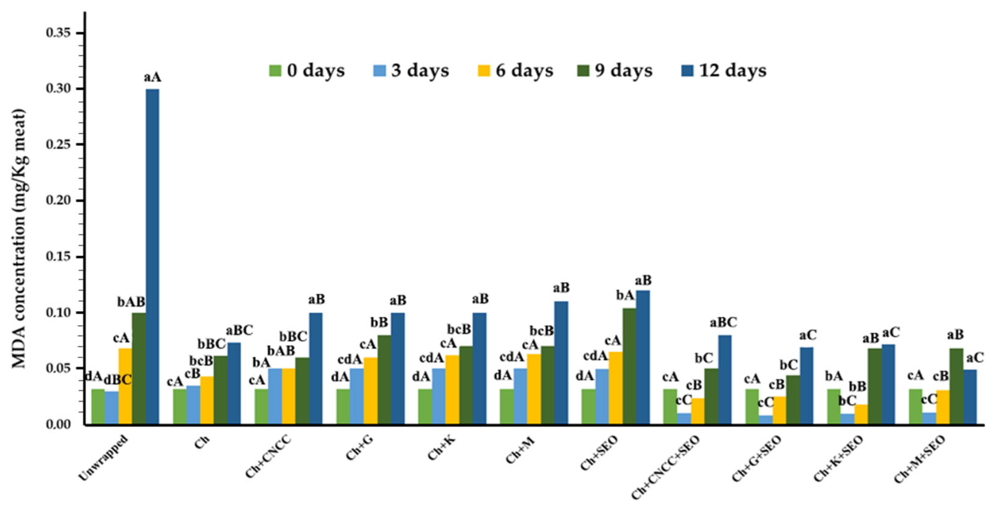
The increase in TBARS values is linked to lipid oxidation product (mainly aldehydes) accumulation, in which higher TBARS values are associated with unacceptable off-flavors/-odors in meat products. Therefore, in this study, malondialdehyde, one of the major hydroperoxide decomposition products of polyunsaturated fatty acids, formed from the oxidation of fatty acid was calculated as the value of thiobarbituric acid reactive substances [88]. It was noted that the initial TBARS values were approximately 0.03 mg MDA/kg of meat, followed by an increasing tendency in all samples over time (Figure 5). As expected, the unpackaged meat reached the highest average value of 0.32 mg MDA/kg of meat at the end of the 12 days of testing, while the samples protected by the chitosan film and respective Ch bionanocomposites maintained significantly lower (p < 0.05) MDA levels for all evaluation times, thus delaying the meat degradation by lipid oxidation mechanisms.
Over the 12 days, there were no significant differences (p > 0.05) between the sample packed in the Ch film compared to the CNC-reinforced bionanocomposites. Furthermore, the meat wrapped in the Ch films had slightly lower MDA concentration values at each evaluation time. The protection effect associated to chitosan may be attributed to the good oxygen barrier and the chelator ability of this biopolymer to donate electrons and stabilize free radicals, acting as antioxidants [89]. The OH radical has the most active reaction to oxygen between several species, thus, it can readily react with active hydrogen atoms available in chitosan biomolecules, constituting a more stable compound. The incorporation of CNC in the polymeric matrix and good binding with Ch decreased the reactive sites and consequently caused a decrease in the film’s antioxidant activity [90]. It was also stated that no observable differences (p > 0.05) occurred between bionanocomposites reinforced with the different nanocelluloses extracted from lignocellulosic crops. Moreover, these samples reported similar MDA concentrations as the specimen wrapped in the bionanocomposite with commercial CNC.
Several reports found in the literature stated that the introduction of small amounts of essential oils (EOs) into chitosan films has the ability to significantly delay or inhibit the oxidation of oxidizable substrates, and may act at different levels in an oxidative sequence [90,91]. The EO’s antioxidative properties have been commonly related to their phenolic content, which acts as hydrogen donators and radical scavengers [92]. The results presented in this work showed that SEO incorporation in the Ch film significantly (p < 0.05) increased the meat’s MDA values soon after day 3, compared to the control sample wrapped in Ch. This result could indicate a possible pro-oxidant effect associated with SEO. These results do not agree with those presented by [93], in which Salvia officinalis L. powder was added to salmon hamburgers. The results of this study showed that Salvia officinalis L. at three different concentrations (0.5, 1, and 1.5%) was able to stop the oxidation process of the hamburgers, presenting a good antioxidant and antimicrobial potential. In another study, sage essential oil and sage extract obtained from sage (Salvia officinalis L.) herbal dust (food industry by-product) were directly applied to fresh pork sausages [15]. Once again, it was verified that the addition of these two active compounds significantly (p < 0.05) reduced the TBARS values. The study developed by Kenar et al. (2010) [94] supports our results, which evaluated the effects of rosemary and sage tea extracts on the sensory, chemical, and microbiological changes in vacuum-packed and refrigerated sardine (Sardina pilchardus) fillets. In this work, the sample treated with sage tea showed significantly higher MDA values when compared to the control, indicating the pro-oxidant effect of sage tea on fish muscle.
As can be seen, there is an ambiguity in results, and it is difficult to predict whether SEO has an anti- or pro-oxidant profile. In the work presented by Akyuz et al. (2017) [95], a novel protein-based solid-biosensor for determining the pro-oxidant activity of phenolic compounds was developed and extracts (testing different dilutions) of sage, green tea, mint, and marjoram plants were studied as real samples to test total pro-oxidant activity (TPA). It was observed that sage presented the highest TPA values for the 1:5 dilution (v/v). The authors pointed out that it is quite difficult to make a general assessment of the anti- or pro-oxidant behavior of polyphenols since the ability of polyphenols to behave under in vitro and in situ systems depends on different factors like the polyphenol concentration and structure, the free radical source, and the molecules present in the substrate to be protected.
Additionally, contrary to the results presented by Ch + SEO bionanocomposites, samples packed in Ch + CNC + SEO bionanocomposites showed the lowest MDA values (p < 0.05) among all the samples studied, between days 3 and 6. These results may be due to the incorporation of components into the biopolymer matrix, increasing the barrier capacity to gases, like oxygen. It was verified once again that the CNC introduction had an essential oil retention effect and therefore the results for these samples were lower than those presented by the Ch + SEO.

4. Conclusions

This study demonstrated that although there was a logical degradation in the meat’s properties over the 12 days of evaluation, there was a significant delay in the food degradation processes when preserved in the diverse bionanocomposites. Through the different microbiological parameters studied, it was verified that the packaged samples presented lower contamination values by about 2–3 logs. The delay of microbiological contamination in these samples showed that the protected meat is acceptable for human consumption for a longer useful time. The good antimicrobial activity shown was supported by the in vitro assays in which was reflected the inhibition of the liquid media growth of both Gram-positive (total inhibition) and Gram-negative (reduction of 0.999998–0.999999%) bacteria tested. Furthermore, the positive results obtained in the evaluation of the physicochemical parameters of meat freshness, like pH, total titratable acidity, color, humidity, total volatile basic nitrogen, and lipid oxidation, help to justify the extension of fresh poultry meat shelf-life.
Nanocellulose extracted from lignocellulosic biomass had a similar performance to the commercial nanocellulose in improving food conservation. However, the excellent bond between the nanocellulose and the polymer matrix could cause a decrease in the active properties as there was a reduction in the chitosan free active groups.
The addition of sage essential oil to the bionanocomposites slightly lowered the pH of the meat sample, which gave lower results for TVB-N (in comparison to unwrapped meat, average reductions of 32% for samples protected with pristine chitosan films; ≅ 43% for the samples wrapped in chitosan films + nano-cellulose and ≅ 49% when SEO was included into the films). However, this phenomenon did not result in significant improvements in microbiological control. Negatively, the SEO directly interfered with meat color from the first day of evaluation and showed a slightly pro-oxidant activity. These aspects were mitigated with the introduction of nanocellulose, proving that this nanoreinforcement had an encapsulating effect, promoting a more controlled release of the oil into the food.
It was possible to extend the shelf life of a perishable food (from a microbiological point of view by 6 days, as protected meat only reached the TMAM limit on the ninth day of storage, while unwrapped meat surpassed it on the third day of storage), like fresh poultry meat, by packaging it in a bionanocomposite made from a renewable and biodegradable material, capable of replacing packaging layers made from fossil-based polymers in the future. The hydrophilicity associated with chitosan, albeit improved with the introduction of nanocellulose, hinders its applicability in foods with high water content. Therefore, for now, the use of these materials should focus on preserving dried foods. Consequently, optimizations will still have to be made for this type of material to be produced efficiently at the industry level, with greater applicability.

Author Contributions

J.R.A.P.: conceptualization, methodology, formal analysis, investigation, writing—original draft, visualization. V.G.L.S.: conceptualization, methodology, investigation, resources, writing—review and editing, supervision. R.P.: formal analysis, investigation, visualization. S.P.: formal analysis, investigation, visualization. L.A.G.: formal analysis, investigation, visualization. M.H.G.: conceptualization, methodology, investigation, resources, writing—review and editing, supervision, funding acquisition. M.P.D.: conceptualization, formal analysis, investigation, visualization. A.L.F.: conceptualization, methodology, resources, writing—review and editing, supervision, funding acquisition. All authors have read and agreed to the published version of the manuscript.

Funding

This research was funded by national funding by FCT, Foundation for Science and Technology (FCT/MECI), through the individual PhD research grant (SFRH/BD/144346/2019) of J.R.A.P, and individual contract (https://doi.org/10.54499/2023.09446.CEECIND/CP2836/CT0011) of V.G.L.S. This work was also supported by the MEtRICs research unit which is funded by national funds from FCT/MECI (UID/4077: Mechanical Engineering and Resources Sustainability Center), and by I3N-CENIMAT unit which is funded by FCT/MECI (UIDB/50025/2020-2023).

Data Availability Statement

All data are available within the manuscript.

Conflicts of Interest

The authors declare no conflicts of interest. The funders had no role in the design of the study; in the collection, analyses, or interpretation of data; in the writing of the manuscript; or in the decision to publish the results.

References

  1. Otoni, C.G.; Azeredo, H.M.C.; Mattos, B.D.; Beaumont, M.; Correa, D.S.; Rojas, O.J. The Food–Materials Nexus: Next Generation Bioplastics and Advanced Materials from Agri-Food Residues. Adv. Mater. 2021, 33, 2102520. [Google Scholar] [CrossRef] [PubMed]
  2. Pires, J.R.A.; Souza, V.G.L.; Fuciños, P.; Pastrana, L.; Fernando, A.L. Methodologies to Assess the Biodegradability of Bio-Based Polymers—Current Knowledge and Existing Gaps. Polymers 2022, 14, 1359. [Google Scholar] [CrossRef]
  3. Lau, W.; Shiran, Y.; Bailey, R.M.; Cook, E.; Stuchtey, M.R.; Koskella, J.; Velis, C.A.; Godfrey, L.; Boucher, J.; Murphy, M.B.; et al. Evaluating Scenarios toward Zero Plastic Pollution. Science 2020, 369, 1455–1461. [Google Scholar] [CrossRef]
  4. Zhu, Y.; Romain, C.; Williams, C.K. Sustainable Polymers from Renewable Resources. Nature 2016, 540, 354–362. [Google Scholar] [CrossRef]
  5. Flórez, M.; Guerra-Rodríguez, E.; Cazón, P.; Vásquez, M. Chitosan for Food Packaging: Recent Advances in Active and Intelligent Films. Food Hydrocoll. 2022, 124, 107328. [Google Scholar] [CrossRef]
  6. Kumar, S.; Mukherjee, A.; Dutta, J. Chitosan Based Nanocomposite Films and Coatings: Emerging Antimicrobial Food Packaging Alternatives. Trends Food Sci. Technol. 2020, 97, 196–209. [Google Scholar] [CrossRef]
  7. Rodrigues, C.; Polesca, C.; Bicalho, I.; Souza, V.G.L.; Coelhoso, I.; Fernando, A.L. Quality Preservation and Shelf-Life Extension of Prickly Pear (Opuntia ficus-indica L. Mill) Using Edible Coatings. Foods 2025, 14, 161. [Google Scholar] [CrossRef] [PubMed]
  8. Pires, J.; de Paula, C.D.; Souza, V.G.L.; Fernando, A.L.; Coelhoso, I. Understanding the Barrier and Mechanical Behavior of Different Nanofillers in Chitosan Films for Food Packaging. Polymers 2021, 13, 721. [Google Scholar] [CrossRef] [PubMed]
  9. de Sousa, M.S.; Schlogl, A.E.; Estanislau, F.R.; Souza, V.G.L.; dos Reis Coimbra, J.S.; Santos, I.J.B. Nanotechnology in Packaging for Food Industry: Past, Present, and Future. Coatings 2023, 13, 1411. [Google Scholar] [CrossRef]
  10. Bourbon, A.I.; Costa, M.J.; Maciel, L.C.; Pastrana, L.; Vicente, A.A.; Cerqueira, M.A. Active Carboxymethylcellulose-Based Edible Films: Influence of Free and Encapsulated Curcumin on Films’ Properties. Foods 2021, 10, 1512. [Google Scholar] [CrossRef] [PubMed]
  11. Yadav, M.; Behera, K.; Chang, Y.H.; Chiu, F.C. Cellulose Nanocrystal Reinforced Chitosan Based UV Barrier Composite Films for Sustainable Packaging. Polymers 2020, 12, 202. [Google Scholar] [CrossRef]
  12. Ahankari, S.S.; Subhedar, A.R.; Bhadauria, S.S.; Dufresne, A. Nanocellulose in Food Packaging: A Review. Carbohydr. Polym. 2021, 255, 117479. [Google Scholar] [CrossRef] [PubMed]
  13. Pires, J.R.A.; Souza, V.G.L.; Fernando, A.L. Production of Nanocellulose from Lignocellulosic Biomass Wastes: Prospects and Limitations. In Innovation, Engineering and Entrepreneurship; Machado, J., Soares, F., Veiga, G., Eds.; Lecture Notes in Electrical Engineering; Springer International Publishing: Cham, Switzerland, 2019; Volume 505, pp. 719–725. ISBN 978-3-319-91333-9. [Google Scholar]
  14. Vosoughi, N.; Gomarian, M.; Ghasemi Pirbalouti, A.; Khaghani, S.; Malekpoor, F. Essential Oil Composition and Total Phenolic, Flavonoid Contents, and Antioxidant Activity of Sage (Salvia officinalis L.) Extract under Chitosan Application and Irrigation Frequencies. Ind. Crops Prod. 2018, 117, 366–374. [Google Scholar] [CrossRef]
  15. Branislav, Š.; Pavlić, B.; Zeković, Z.; Tomović, V.; Ikonić, P.; Džinić, N. The Effect of Essential Oil and Extract from Sage (Salvia officinalis L.) Herbal Dust (Food Industry by-Product) on the Oxidative and Microbiological Stability of Fresh Pork Sausages. LWT-Food Sci. Technol. 2018, 89, 749–755. [Google Scholar] [CrossRef]
  16. Ehsani, A.; Hashemi, M.; Afshari, A.; Aminzare, M.; Raeisi, M.; Zeinali, T. Effect of Different Types of Active Biodegradable Films Containing Lactoperoxidase System or Sage Essential Oil on the Shelf Life of Fish Burger during Refrigerated Storage. LWT-Food Sci. Technol. 2020, 117, 108633. [Google Scholar] [CrossRef]
  17. EMA. Assessment Report on Salvia officinalis L., folium and Salvia officinalis L., aetheroleum; EMA: London, UK, 2016.
  18. Costa, S.M.; Ferreira, D.P.; Teixeira, P.; Ballesteros, L.F.; Teixeira, J.A.; Fangueiro, R. Active Natural-Based Films for Food Packaging Applications: The Combined Effect of Chitosan and Nanocellulose. Int. J. Biol. Macromol. 2021, 177, 241–251. [Google Scholar] [CrossRef]
  19. Pires, J.R.A.; Souza, V.G.L.; Gomes, L.A.; Coelhoso, I.M.; Godinho, M.H.; Fernando, A.L. Micro and Nanocellulose Extracted from Energy Crops as Reinforcement Agents in Chitosan Films. Ind. Crops Prod. 2022, 186, 115247. [Google Scholar] [CrossRef]
  20. Souza, V.G.L.; Rodrigues, C.; Ferreira, L.; Pires, J.R.A.; Duarte, M.P.; Coelhoso, I.; Fernando, A.L. In Vitro Bioactivity of Novel Chitosan Bionanocomposites Incorporated with Different Essential Oils. Ind. Crops Prod. 2019, 140, 111563. [Google Scholar] [CrossRef]
  21. Singleton, V.L.; Orthofer, R.; Lamuela-Raventós, R.M. Analysis of Total Phenols and Other Oxidation Substrates and Antioxidants by Means of Folin-Ciocalteu. Methods Enzymol. 1999, 299, 152–178. [Google Scholar]
  22. Chung, D.; Papadakis, S.E.; Yam, K.L. Simple Models for Assessing Migration from Food-Packaging Films. Food Addit. Contam. 2002, 19, 611–617. [Google Scholar] [CrossRef]
  23. Nouri, A.; Yaraki, M.T.; Ghorbanpour, M.; Agarwal, S.; Gupta, V.K. Enhanced Antibacterial Effect of Chitosan Film Using Montmorillonite/CuO Nanocomposite. Int. J. Biol. Macromol. 2017, 109, 1219–1231. [Google Scholar] [CrossRef] [PubMed]
  24. ISO 4833-1:2013; Microbiology of the Food Chain—Horizontal Method for the Enumeration of Microorganisms—Part 1: Colony Count at 30 Degrees C by the Pour Plate Technique. ISO: Geneva, Switzerland, 2013.
  25. ISO 17410:2019; Microbiology of the Food Chain—Horizontal Method for the Enumeration of Psychrotrophic Microorganisms. ISO: Geneva, Switzerland, 2019.
  26. ISO 21528-2:2017; Microbiology of the Food Chain—Horizontal Method for the Detection and Enumeration of Enterobacteriaceae—Part 2: Colony-Count Technique. ISO: Geneva, Switzerland, 2017.
  27. AOAC. Official Methods of Analysis of the Association of Official Analytical Chemists, 20th ed.; AOAC: San Diego, CA, USA, 2016. [Google Scholar]
  28. Rosmini, M.R.; Perlo, F.; Pérez-Alvarez, J.A.; Pagán-Moreno, M.J.; Gago-Gago, A.; López-Santoveña, F.; Aranda-Catalá, V. TBA Test by an Extractive Method Applied to “Paté”. Meat Sci. 1996, 42, 103–110. [Google Scholar] [CrossRef]
  29. Zhang, X.; Ismail, B.B.; Cheng, H.; Jin, T.Z.; Qian, M.; Ali, S.; Liu, D.; Guo, M. Emerging Chitosan-Essential Oil Films and Coatings for Food Preservation—A Review of Advances and Applications. Carbohydr. Polym. 2021, 273, 118616. [Google Scholar] [CrossRef]
  30. Souza, V.G.L.; Pires, J.R.A.; Vieira, É.T.; Coelhoso, I.M.; Duarte, M.P.; Fernando, A.L. Activity of Chitosan-Montmorillonite Bionanocomposites Incorporated with Rosemary Essential Oil: From in Vitro Assays to Application in Fresh Poultry Meat. Food Hydrocoll. 2019, 89, 241–252. [Google Scholar] [CrossRef]
  31. Ribeiro-Santos, R.; de Melo, N.R.; Andrade, M.; Sanches-Silva, A. Potential of Migration of Active Compounds from Protein-Based Films with Essential Oils to a Food and a Food Simulant. Packag. Technol. Sci. 2017, 30, 791–798. [Google Scholar] [CrossRef]
  32. Montero, Y.; Souza, A.G.; Oliveira, É.R.; Rosa, S. Nanocellulose Functionalized with Cinnamon Essential Oil: A Potential Application in Active Biodegradable Packaging for Strawberry. Sustain. Mater. Technol. 2021, 29, e00289. [Google Scholar] [CrossRef]
  33. Esparza, I.; Cimminelli, M.J.; Moler, J.A.; Jiménez-Moreno, N.; Ancín-Azpilicueta, C. Stability of Phenolic Compounds in Grape Stem Extracts. Antioxidants 2020, 9, 720. [Google Scholar] [CrossRef] [PubMed]
  34. Souza, V.G.L.; Rodrigues, P.F.; Duarte, M.P.; Fernando, A.L. Antioxidant Migration Studies in Chitosan Films Incorporated with Plant Extracts. J. Renew. Mater. 2018, 6, 548–558. [Google Scholar] [CrossRef]
  35. Zhang, W.; Li, X.; Jiang, W. Development of Antioxidant Chitosan Film with Banana Peels Extract and Its Application as Coating in Maintaining the Storage Quality of Apple. Int. J. Biol. Macromol. 2020, 154, 1205–1214. [Google Scholar] [CrossRef] [PubMed]
  36. Buonocore, G.G.; Del Nobile, M.A.; Panizza, A.; Corbo, M.R.; Nicolais, L. A General Approach to Describe the Antimicrobial Agent Release from Highly Swellable Films Intended for Food Packaging Applications. J. Control Release 2003, 90, 97–107. [Google Scholar] [CrossRef] [PubMed]
  37. Sánchez-González, L.; Cháfer, M.; González-Martínez, C.; Chiralt, A.; Desobry, S. Study of the Release of Limonene Present in Chitosan Films Enriched with Bergamot Oil in Food Simulants. J. Food Eng. 2011, 105, 138–143. [Google Scholar] [CrossRef]
  38. Abbasi, A.; Mohammad, K.; Ahmad, M.; Mohammad, A.; Sourestani, M. Nutritive Composition, Growth, Biochemical Traits, Essential Oil Content and Compositions of Salvia officinalis L. Grown in Different Nitrogen Levels in Soilless Culture. J. Soil Sci. Plant Nutr. 2021, 21, 3320–3332. [Google Scholar] [CrossRef]
  39. Cuceu, A.P.; Tofană, M.; Socaci, S.A.; Vârban, D.; Nagy, M.; Borş, M.-D.; Fărcaş, A. Essential Oil Composition, Phenolic Content and Antioxidant Activity in Romanian Salvia officinalis L. J. Agroaliment. Process. Technol. 2015, 21, 241–246. [Google Scholar]
  40. O’Neil, M.J. The Merck Index—An Encyclopedia of Chemicals, Drugs, and Biologicals, 15th ed.; Chemistry, R.S., Ed.; Royal Society of Chemistry: Cambridge, UK, 2013; ISBN 9781849736701. [Google Scholar]
  41. Yalkowsky, S.H.; He, Y.; Jain, P. Handbook of Aqueous Solubility Data, 2nd ed.; CRC Press: Boca Raton, FL, USA, 2010; ISBN 9780367384173. [Google Scholar]
  42. Lan, W.; Wang, S.; Zhang, Z.; Liang, X.; Liu, X.; Zhang, J. Development of Red Apple Pomace Extract/Chitosan-Based Films Reinforced by TiO2 Nanoparticles as a Multifunctional Packaging Material. Int. J. Biol. Macromol. 2021, 168, 105–115. [Google Scholar] [CrossRef]
  43. Surendhiran, D.; Chandra, V.; Park, J.; Chun, B. Fabrication of Chitosan-Based Food Packaging Film Impregnated with Turmeric Essential Oil (TEO)-Loaded Magnetic-Silica Nanocomposites for Surimi Preservation. Int. J. Biol. Macromol. 2022, 203, 650–660. [Google Scholar] [CrossRef]
  44. Bozin, B.; Mimica-Dukic, N.; Samojlik, I.; Jovin, E. Antimicrobial and Antioxidant Properties of Rosemary and Sage (Rosmarinus officinalis L. and Salvia officinalis L.; Lamiaceae) Essential Oils. J. Agric. Food Chem. 2007, 55, 7879–7885. [Google Scholar] [CrossRef]
  45. Viuda-Martos, M.; Navajas, Y.R.; Zapata, E.S.; Fernández-López, J.; Pérez-Álvarez, J.A. Antioxidant Activity of Essential Oils of Five Spice Plants Widely Used in a Mediterranean Diet. Flavour Fragr. J. 2009, 25, 13–19. [Google Scholar] [CrossRef]
  46. Kou, S.G.; Peters, L.; Mucalo, M. Chitosan: A Review of Molecular Structure, Bioactivities and Interactions with the Human Body and Micro-Organisms. Carbohydr. Polym. 2022, 282, 119132. [Google Scholar] [CrossRef]
  47. Ke, C.-L.; Deng, F.-S.; Chuang, C.-Y.; Lin, C.-H. Antimicrobial Actions and Applications of Chitosan. Polymers 2021, 13, 904. [Google Scholar] [CrossRef]
  48. Kong, M.; Chen, X.G.; Xing, K.; Park, H.J. Antimicrobial Properties of Chitosan and Mode of Action: A State of the Art Review. Int. J. Food Microbiol. 2010, 144, 51–63. [Google Scholar] [CrossRef]
  49. Pires, J.R.A.; Almeida, K.M.; Augusto, A.S.; Vieira, É.T.; Fernando, A.L.; Souza, V.G.L. Application of Biocomposite Films of Chitosan/Natural Active Compounds for Shelf Life Extension of Fresh Poultry Meat. J. Compos. Sci. 2022, 6, 342. [Google Scholar] [CrossRef]
  50. Maria Pelissari, F.; Andrade-Mahecha, M.M.; José, P.; Cecilia, F. Nanocomposites Based on Banana Starch Reinforced with Cellulose Nanofibers Isolated from Banana Peels. J. Colloid Interface Sci. 2017, 505, 154–167. [Google Scholar] [CrossRef]
  51. Samapundo, S.; De Baenst, I.; Aerts, M.; Cnockaert, M.; Devlieghere, F.; Van Damme, P. Tracking the Sources of Psychrotrophic Bacteria Contaminating Chicken Cuts during Processing. Food Microbiol. 2019, 81, 40–50. [Google Scholar] [CrossRef] [PubMed]
  52. Norrrahim, M.N.F.; Nurazzi, N.M.; Jenol, M.A.; Farid, M.A.A.; Janudin, N.; Ujang, F.A.; Yasim-Anuar, T.A.T.; Najmuddin, S.U.F.S.; Ilyas, R.A. Emerging Development of Nanocellulose as an Antimicrobial Material: An Overview. Mater. Adv. 2021, 2, 3538–3551. [Google Scholar] [CrossRef]
  53. Jannatyha, N.; Shojaee-aliabadi, S.; Moslehishad, M.; Moradi, E. Comparing Mechanical, Barrier and Antimicrobial Properties of Nanocellulose/CMC and Nanochitosan/CMC Composite Films. Int. J. Biol. Macromol. 2020, 164, 2323–2328. [Google Scholar] [CrossRef] [PubMed]
  54. Cegielka, A.; Hac-Szymanczuk, E.; Piwowarek, K.; Dasiewicz, K.; Slowinski, M.; Wronska, K. The Use of Bioactive Properties of Sage Preparations to Improve the Storage Stability of Low-Pressure Mechanically Separated Meat from Chickens. Poult. Sci. 2019, 98, 5045–5053. [Google Scholar] [CrossRef]
  55. Pateiro, M.; Munekata, P.E.S.; Sant’Ana, A.S.; Domínguez, R.; Rodríguez-Lázaro, D.; Lorenzo, J.M. Application of Essential Oils as Antimicrobial Agents against Spoilage and Pathogenic Microorganisms in Meat Products. Int. J. Food Microbiol. 2021, 337, 108966. [Google Scholar] [CrossRef] [PubMed]
  56. Souza, V.G.L.; Pires, J.R.A.; Rodrigues, P.F.; Lopes, A.A.S.; Fernandes, F.M.B.; Duarte, M.P.; Coelhoso, I.M.; Fernando, A.L. Bionanocomposites of Chitosan/Montmorillonite Incorporated with Rosmarinus officinalis Essential Oil: Development and Physical Characterization. Food Packag. Shelf Life 2018, 16, 148–156. [Google Scholar] [CrossRef]
  57. Pires, J.R.A.; de Souza, V.G.L.; Fernando, A.L. Chitosan/Montmorillonite Bionanocomposites Incorporated with Rosemary and Ginger Essential Oil as Packaging for Fresh Poultry Meat. Food Packag. Shelf Life 2018, 17, 142–149. [Google Scholar] [CrossRef]
  58. Bonilla, J.; Vargas, M.; Atarés, L.; Chiralt, A. Effect of Chitosan Essential Oil Films on the Storage-Keeping Quality of Pork Meat Products. Food Bioprocess Technol. 2014, 7, 2443–2450. [Google Scholar] [CrossRef]
  59. Sánchez-González, L.; Cháfer, M.; Hernández, M.; Chiralt, A. Antimicrobial Activity of Polysaccharide Films Containing Essential Oils. Food Control 2011, 22, 1302–1310. [Google Scholar] [CrossRef]
  60. Matei, P.M.; Buzón-Durán, L.; Pérez-Lebeña, E.; Martín-Gil, J.; Iacomi, B.M.; Ramos-Sánchez, M.C.; Martín-Ramos, P. In Vitro Antifungal Activity of Chitosan-Polyphenol Conjugates against Phytophthora cinnamomi. AgriEngineering 2020, 2, 72–77. [Google Scholar] [CrossRef]
  61. Qin, Y.; Li, P. Antimicrobial Chitosan Conjugates: Current Synthetic Strategies and Potential Applications. Int. J. Mol. Sci. 2020, 21, 499. [Google Scholar] [CrossRef]
  62. Sedlaříková, J.; Doležalová, M.; Egner, P.; Pavlačková, J.; Krejčí, J.; Rudolf, O.; Peer, P. Effect of Oregano and Marjoram Essential Oils on the Physical and Antimicrobial Properties of Chitosan Based Systems. Int. J. Polym. Sci. 2017, 2017, 2593863. [Google Scholar] [CrossRef]
  63. The Commision of the European Communities. COMMISSION REGULATION (EC) No 2073/2005 of 15 November 2005 on Microbiological Criteria for Foodstuffs; Elsevier: Amsterdam, The Netherlands, 2005; Volume L338. [Google Scholar]
  64. Marmion, M.; Ferone, M.T.; Whyte, P.; Scannell, A.G.M. The Changing Microbiome of Poultry Meat; from Farm to Fridge. Food Microbiol. 2021, 99, 103823. [Google Scholar] [CrossRef] [PubMed]
  65. Robertson, G.L. Packaging of Flesh Foods. In Food Packaging: Principles and Practice; CRC Press: Boca Raton, FL, USA, 2013; pp. 445–476. [Google Scholar]
  66. Cardoso, G.P.; Dutra, M.P.; Fontes, P.R.; Ramos, A.d.L.S.; Gomide, L.A.d.M.; Ramos, E.M. Selection of a Chitosan Gelatin-Based Edible Coating for Color Preservation of Beef in Retail Display. Meat Sci. 2016, 114, 85–94. [Google Scholar] [CrossRef]
  67. Singh, P.; Wani, A.A.; Saengerlaub, S.; Langowski, H.-C. Understanding Critical Factors for the Quality and Shelf-Life of MAP Fresh Meat: A Review. Crit. Rev. Food Sci. Nutr. 2011, 51, 146–177. [Google Scholar] [CrossRef] [PubMed]
  68. Hughes, J.M.; Oiseth, S.K.; Purslow, P.P.; Warner, R.D. A Structural Approach to Understanding the Interactions between Colour, Water-Holding Capacity and Tenderness. Meat Sci. 2014, 98, 520–532. [Google Scholar] [CrossRef] [PubMed]
  69. Mir, N.A.; Rafiq, A.; Kumar, F.; Singh, V.; Shukla, V. Determinants of Broiler Chicken Meat Quality and Factors Affecting Them: A Review. J. Food Sci. Technol. 2017, 54, 2997–3009. [Google Scholar] [CrossRef] [PubMed]
  70. Knorr, D. Recovery and Utilization of Chitin and Chitosan in Food Processing Waste Management. Food Technol. 1991, 45, 114–122. [Google Scholar]
  71. Ünal, K.; Babaoglu, A.S.; Karakaya, M. Effect of Oregano, Sage and Rosemary Essential Oils on Lipid Oxidation and Color Properties of Minced Beef during Refrigerated Storage. J. Essent. Oil Bear. Plants 2014, 17, 797–805. [Google Scholar] [CrossRef]
  72. Dave, D.; Ghaly, A.E. Meat Spoilage Mechanisms and Preservation Techniques: A Critical Review. Am. J. Agric. Biol. Sci. 2011, 6, 486–510. [Google Scholar] [CrossRef]
  73. Warner, R.D. The Eating Quality of Meat—IV Water-Holding Capacity and Juiciness. In Lawrie’s Meat Science; Toldra, F., Ed.; Woodhead Publishing: Sawston, UK, 2017; pp. 419–459. ISBN 978-0-08-100694-8. [Google Scholar]
  74. Souza, V.G.L.; Mello, I.P.; Khalid, O.; Pires, J.R.A.; Rodrigues, C.; Alves, M.M.; Santos, C.; Fernando, A.L.; Coelhoso, I. Strategies to Improve the Barrier and Mechanical Properties of Pectin Films for Food Packaging: Comparing Nanocomposites with Bilayers. Coatings 2022, 12, 108. [Google Scholar] [CrossRef]
  75. Souza, V.; Rodrigues, C.; Valente, S.; Pimenta, C.; Pires, J.; Alves, M.; Santos, C.; Coelhoso, I.; Fernando, A. Eco-Friendly ZnO/Chitosan Bionanocomposites Films for Packaging of Fresh Poultry Meat. Coatings 2020, 10, 110. [Google Scholar] [CrossRef]
  76. Chauhan, S.S.; Lemaster, M.N.; Clark, D.L.; Foster, M.K.; Miller, C.E.; England, E.M. Glycolysis and PH Decline Terminate Prematurely in Oxidative Muscles despite the Presence of Excess Glycogen. Meat Muscle Biol. 2019, 3, 254–264. [Google Scholar] [CrossRef]
  77. Matarneh, S.K.; England, E.M.; Scheffler, T.L.; Gerrard, D.E. The Conversion of Muscle to Meat. In Lawrie’s Meat Science; Toldrá, F., Ed.; Woodhead Publishing: Sawston, UK, 2017; pp. 159–185. [Google Scholar]
  78. Bekhit, A.E.A.; Holman, B.W.B.; Giteru, S.G.; Hopkins, D.L. Total Volatile Basic Nitrogen (TVB-N) and Its Role in Meat Spoilage: A Review. Trends Food Sci. Technol. 2021, 109, 280–302. [Google Scholar] [CrossRef]
  79. Souza, V.G.L.; Pires, J.R.A.; Rodrigues, C.; Coelhoso, I.M.; Fernando, A.L. Chitosan Composites in Packaging Industry—Current Trends and Future Challenges. Polymers 2020, 12, 417. [Google Scholar] [CrossRef]
  80. Tyl, C.; Sadler, G.D. PH and Titratable Acidity. In Food Analysis; Nielsen, S.S., Ed.; Springer: Cham, Switzerland, 2017; pp. 389–406. [Google Scholar]
  81. Grashorn, M.A. Research into Poultry Meat Quality. Br. Poult. Sci. 2010, 51, 60–67. [Google Scholar] [CrossRef]
  82. Cheng, Y.; Hu, J.; Wu, S. Chitosan Based Coatings Extend the Shelf-Life of Beef Slices during Refrigerated Storage. LWT 2021, 138, 110694. [Google Scholar] [CrossRef]
  83. Mehrabi, F.A.; Sharifi, A.; Ahvazi, M. Effect of Chitosan Coating Containing Nepeta Pogonosperma Extract on Shelf Life of Chicken Fillets during Chilled Storage. Food Sci. Nutr. 2021, 9, 4517–4528. [Google Scholar] [CrossRef]
  84. Zhang, X.; Liu, Y.; Yong, H.; Qin, Y.; Liu, J.; Liu, J. Development of Multifunctional Food Packaging Films Based on Chitosan, TiO2 Nanoparticles and Anthocyanin-Rich Black Plum Peel Extract. Food Hydrocoll. 2019, 94, 80–92. [Google Scholar] [CrossRef]
  85. Çoban, O.E.; Patir, B.; Özpolat, E.; Kuzgun, N.K. Improving the Quality of Fresh Rainbow Trout by Sage Essential Oil and Packaging Treatments. J. Food Saf. 2016, 36, 299–307. [Google Scholar] [CrossRef]
  86. Mehdizadeh, T.; Tajik, H.; Jafarie, S.; Kaboudari, A. Effect of Salvia officinalis L. Extract on Chemical, Microbial, Sensory and Shelf Life of Rainbow Trout Fillet. Food Sci. Biotechnol. 2019, 28, 1499–1506. [Google Scholar] [CrossRef]
  87. Wan, A.; Xu, Q.; Sun, Y.; Li, H. Antioxidant Activity of High Molecular Weight Chitosan and N,O-Quaternized Chitosans. J. Agric. Food Chem. 2013, 61, 6921–6928. [Google Scholar] [CrossRef]
  88. Chen, X.; Chen, W.; Lu, X.; Mao, Y.; Luo, X.; Liu, G.; Zhu, L.; Zhang, Y. Effect of Chitosan Coating Incorporated with Oregano or Cinnamon Essential Oil on the Bacterial Diversity and Shelf Life of Roast Duck in Modified Atmosphere Packaging. Food Res. Int. 2021, 147, 110491. [Google Scholar] [CrossRef]
  89. Przybyszewska, A.; Barbosa, C.H.; Pires, F.; Pires, J.R.A.; Rodrigues, C.; Galus, S.; Souza, V.G.L.; Alves, M.M.; Santos, C.F.; Coelhoso, I.; et al. Packaging of Fresh Poultry Meat with Innovative and Sustainable ZnO/Pectin Bionanocomposite Films—A Contribution to the Bio and Circular Economy. Coatings 2023, 13, 1208. [Google Scholar] [CrossRef]
  90. Contini, L.; De Souza, T.; Isadora, Z.; Brazolin, F.; Wesley, J.; Mariangela, S.; Silva, F.; Lopes, P.S.; Araújo, K.; Rosemary, S.; et al. Antioxidant Chitosan Film Containing Lemongrass Essential Oil as Active Packaging for Chicken Patties. J. Food Process. Preserv. 2021, 46, e16136. [Google Scholar] [CrossRef]
  91. Mehdizadeh, T.; Langroodi, A.M. Chitosan Coatings Incorporated with Propolis Extract and Zataria Multi Flora Boiss Oil for Active Packaging of Chicken Breast Meat. Int. J. Biol. Macromol. 2019, 141, 401–409. [Google Scholar] [CrossRef]
  92. Ehsani, A.; Hashemi, M.; Aminzare, M.; Raeisi, M.; Afshari, A.; Mirza, A.; Mohammadreza, A. Comparative Evaluation of Edible Films Impregnated with Sage Essential Oil or Lactoperoxidase System: Impact on Chemical and Sensory Quality of Carp Burgers. J. Food Process. Preserv. 2019, 43, e14070. [Google Scholar] [CrossRef]
  93. Ferreira, C.S.R.; Henrique, B.; Saqueti, F.; Daniele, P.; Martins, J.; Ant, M.; Carla, A.; Martha, J.; Mikcha, G.; Oliveira, O. Effect of Salvia (Salvia officinalis) on the Oxidative Stability of Salmon Hamburgers. LWT 2022, 154, 112867. [Google Scholar] [CrossRef]
  94. Kenar, M.; Ozogul, F.; Kuley, E. Effects of Rosemary and Sage Tea Extracts on the Sensory, Chemical and Microbiological Changes of Vacuum-Packed and Refrigerated Sardine (Sardina pilchardus) Fillets. Int. J. Food Sci. Technol. 2010, 45, 2366–2372. [Google Scholar] [CrossRef]
  95. Akyüz, E.; Başkan, K.S.; Tütem, E.; Apak, R. Novel Protein-Based Solid-Biosensor for Determining pro-Oxidant Activity of Phenolic Compounds. J. Agric. Food Chem. 2017, 65, 5821–5830. [Google Scholar] [CrossRef] [PubMed]
Figure 1. Assembly of the in situ tests.
Jcs 09 00428 g001
Figure 2. Total phenolic migration profile for (A) simulant ethanol 50% and (B) simulant ethanol 95%. Chitosan (Ch); commercial nanocellulose (CNCC); giant reed nanocellulose (G); kenaf nanocellulose (K); miscanthus nanocellulose (M); sage essential oil (SEO).
Jcs 09 00428 g002
Figure 3. DPPH radical scavenging (%) profile for (A) simulant ethanol 50% and (B) simulant ethanol 95%. Chitosan (Ch); commercial nanocellulose (CNCC); giant reed nanocellulose (G); kenaf nanocellulose (K); miscanthus nanocellulose (M); sage essential oil (SEO).
Jcs 09 00428 g003
Figure 4. Meat color over time for the samples with the most significant differences.
Jcs 09 00428 g004
Figure 5. TBARS index of the meat over the storage time. a–d: Different letters indicate significant differences among days (p < 0.05). A–C: Different letters indicate significant differences among meat wrapped with the different bionanocomposites and unwrapped (p < 0.05). Chitosan (Ch); commercial nanocellulose (CNCC); giant reed nanocellulose (G); kenaf nanocellulose (K); miscanthus nanocellulose (M); sage essential oil (SEO).
Jcs 09 00428 g005
Table 1. Formulation parameters for each bionanocomposite assembled.
SpecimenBionanocomposite Formulation Parameters
Chitosan (% w/v FFD)Glycerol (% w/w Ch)Tween® 80 (% w/v SEO)CNCCs (% w/w Ch)CNC G (% w/w Ch)CNC K (% w/w Ch)CNC M (% w/w Ch)SEO (% v/v FFD)
Ch1.530000000
Ch + CNCC1.53002.50000
Ch + G1.530002.5000
Ch + K1.5300002.500
Ch + M1.53000002.50
Ch + SEO1.5300.200001
Ch + CNCC + SEO1.5300.22.50001
Ch + G + SEO1.5300.202.5001
Ch + K + SEO1.5300.2002.501
Ch + M + SEO 1.5300.20002.51
Film-forming dispersion (FFD); chitosan (Ch); nanocrystalline cellulose (CNC); commercial nanocellulose (CNCC); giant reed nanocellulose (G); kenaf nanocellulose (K); miscanthus nanocellulose (M); sage essential oil (SEO).
Table 2. Diffusion kinetics of phenolic compounds.
Diffusion Coefficient (cm2/s)Maximum Diffusion/Total Incorporated
SampleEthanol 50% Ethanol 95% Ethanol 50% Ethanol 95%
Ch + SEO *4.4 × 10−118.9 × 10−110.850.75
Ch + CNCC + SEO4.0 × 10−113.7 × 10−110.710.54
Ch + G + SEO4.2 × 10−113.8 × 10−110.730.57
Ch + K + SEO4.3 × 10−114.8 × 10−110.730.60
Ch + M + SEO4.6 × 10−115.6 × 10−110.760.60
* Chitosan (Ch); commercial nanocellulose (CNCC); giant reed nanocellulose (G); kenaf nanocellulose (K); miscanthus nanocellulose (M); sage essential oil (SEO).
Table 3. In vitro antimicrobial activity of bionanocomposites—colony-forming unit counting method.
Bacillus cereusSalmonella Choleraesuis
SampleLogCFU/mLLogarithmic ReductionLogCFU/mLLogarithmic Reduction
Control9.2 ± 0.4 9.7 ± 0.2 a*
Ch<1>8.2 ± 0.43.7 ± 0.3 c6.0 ± 0.3 a
Ch + SEO<1>8.2 ± 0.44.5 ± 0.1 b5.1 ± 0.1 b
Ch + CNCC + SEO<1>8.2 ± 0.44.0 ± 0.3 c5.7 ± 0.3 a
* a–c: Different letters indicate significant differences between lines (p < 0.05). Colony-forming units (CFU); chitosan (Ch); commercial nanocellulose (CNCC); giant reed nanocellulose (G); kenaf nanocellulose (K); miscanthus nanocellulose (M); sage essential oil (SEO).
Table 4. Microbiological results of the meat over the storage time.
ParametersStorage DaysSamples
UnwrappedCh *Ch + CNCCCh + GCh + KCh + MCh + SEOCh + CNCC + SEOCh + G+SEOCh + K+SEOCh + M+SEO
TMAM
(Log CFU/g meat)
02.11 ± 0.21 cA2.11 ± 0.21 cA2.11 ± 0.21 dA2.11 ± 0.21 dA2.11 ± 0.21 dA2.11 ± 0.21 dA2.11 ± 0.21 cA2.11 ± 0.21 cA2.11 ± 0.21 dA2.11 ± 0.21 cA2.11 ± 0.21 cA
37.26 ± 0.08 bA5.35 ± 0.33 bB4.28 ± 0.21 cBC3.53 ± 0.33 cC3.70 ± 0.20 cC3.88 ± 0.13 cC5.93 ± 0.62 bAB4.88 ± 0.28 bBC3.85 ± 0.20 cC4.00 ± 0.49 bC4.34 ± 0.33 bBC
68.91 ± 0.11 aA6.26 ± 0.32 bB5.47 ± 0.25 bB5.11 ± 0.11 bB5.30 ± 0.17 bB5.51 ± 0.36 bB5.97 ± 0.10 bB6.50 ± 0.17 aB5.51 ± 0.15 bB5.90 ± 0.76 aB5.16 ± 0.39 bB
99.74 ± 0.36 aA7.02 ± 0.06 aB7.06 ± 0.26 aB6.34 ± 0.34 aB6.43 ± 0.36 aB6.88 ± 0.28 aB7.30 ± 0.18 aB6.98 ± 0.73 aB6.61 ± 0.25 abB6.57 ± 0.29 aB6.70 ± 0.13 aB
129.81 ± 0.37 aA7.11 ± 0.11 aB7.70 ± 0.34 aB7.04 ± 0.26 aB7.10 ± 0.21 aB7.02 ± 0.15 aB7.52 ± 0.29 aB7.66 ± 0.42 aB7.23 ± 0.18 aB6.92 ± 0.31 aB6.88 ± 0.17 aB
TPAM
(Log CFU/g meat)
02.31 ± 0.49 cA2.31 ± 0.49 cA2.31 ± 0.49 dA2.31 ± 0.49 dA2.31 ± 0.49 dA2.31 ± 0.49 dA2.31 ± 0.49 cA2.31 ± 0.49 cA2.31 ± 0.49 dA2.31 ± 0.49 dA2.31 ± 0.49 dA
37.29 ± 0.06 bA5.53 ± 0.26 bB3.74 ± 0.34 cC3.61 ± 0.20 cC3.35 ± 0.17 cC3.70 ± 0.49 cC5.97 ± 0.55 bAB4.35 ± 0.41 bBC3.01 ± 0.04 cC3.04 ± 0.56 cC3.64 ± 0.46 cC
69.16 ± 0.19 aA6.51 ± 0.39 abB5.44 ± 0.05 bB5.43 ± 0.04 bB5.66 ± 0.11 bB5.34 ± 0.20 bB6.09 ± 0.50 bB6.91 ± 0.53 aB5.91 ± 0.21 bB5.97 ± 0.60 bB5.56 ± 0.21 bB
99.24 ± 0.47 aA6.81 ± 0.40 aB6.75 ± 0.70 aB6.26 ± 0.25 abB6.17 ± 0.10 baB6.30 ± 0.11 abB7.39 ± 0.03 aB7.62 ± 0.43 aB6.63 ± 0.05 aB6.43 ± 0.08 aB6.48 ± 0.11 aB
129.58 ± 0.01 aA7.23 ± 0.18 aB6.97 ± 0.29 aB7.47 ± 0.37 aB7.53 ± 0.51 aB7.28 ± 0.46 aB7.81 ± 0.41 aB7.70 ± 0.59 aB6.73 ± 0.10 aB6.80 ± 0.21 aB6.73 ± 0.10 aB
Enterobacteriaceae (Log CFU/g meat)02.26 ± 0.12 dA2.26 ± 0.12 bA2.26 ± 0.12 cA2.26 ± 0.12 cA2.26 ± 0.12 cA2.26 ± 0.12 cA2.26 ± 0.12 cA2.26 ± 0.12 cA2.26 ± 0.12 cA2.26 ± 0.12 cA2.26 ± 0.12 cA
36.06 ± 0.17 cA3.45 ± 0.71 bB2.52 ± 0.21 cB2.64 ± 0.08 cB2.74 ± 0.10 cB2.58 ± 0.17 cB3.11 ± 0.21 cB3.14 ± 0.12 bcB2.36 ± 0.12 cB2.58 ± 0.55 cB2.88 ± 0.10 bcB
67.00 ± 0.06 bA5.49 ± 0.09 aB4.14 ± 0.43 bBC3.61 ± 0.33 bC3.35 ± 0.25 bC3.53 ± 0.18 bC5.11 ± 0.21 bB4.36 ± 0.08 bBC3.65 ± 0.03 bC3.65 ± 0.44 bC3.27 ± 0.10 bC
98.89 ± 0.13 aA5.71 ± 0.21 aC6.17 ± 0.37 aBC5.56 ± 0.49 aC5.97 ± 0.20 aC5.51 ± 0.06 aC5.56 ± 0.10 abC6.48 ± 0.14 aB5.86 ± 0.17 aC5.87 ± 0.03 aC5.67 ± 0.06 aC
129.41 ± 0.32 aA6.11 ± 0.21 aB6.92 ± 0.24 aB6.46 ± 0.32 aB6.53 ± 0.04 aB6.16 ± 0.26 aB6.30 ± 0.49 aB7.18 ± 0.47 aB6.13 ± 0.04 aB6.29 ± 0.15 aB6.11 ± 0.06 aB
* a–c: Different letters indicate significant differences between lines (p < 0.05). A–C: Different letters indicate significant differences between columns (p < 0.05). Total mesophilic aerobic microorganisms (TMAM); total psychrotrophic aerobic microorganisms (TPAM); chitosan (Ch); commercial nanocellulose (CNCC); giant reed nanocellulose (G); kenaf nanocellulose (K); miscanthus nanocellulose (M); sage essential oil (SEO).
Table 5. Meat’s color parameters and moisture content over the storage time.
ParametersStorage DaysSamples
UnwrappedChCh + CNCCCh + GCh + KCh + MCh + SEOCh + CNCC + SEOCh + G+SEOCh + K+SEOCh + M+SEO
Hue angle (degrees)045 ± 1 cA45 ± 1 bA45 ± 1 bA45 ± 1 cA45 ± 1 cA45 ± 1 bA45 ± 1 cA45 ± 1 cA45 ± 1 cA45 ± 1 cA45 ± 1 cA
343 ± 2 cB48 ± 2 bAB50 ± 3 aAB49 ± 1 bAB48 ± 1 bAB46 ± 1 bAB52 ± 0 bA44 ± 1 cB50 ± 1 bAB49 ± 2 bAB48 ± 2 bAB
645 ± 0 cB46 ± 2 bAB46 ± 0 bAB48 ± 1 bAB48 ± 2 bAB47 ± 2 bAB52 ± 1 bA49 ± 3 bAB51 ± 2 bA49 ± 2 bAB49 ± 1 bAB
950 ± 1 bAB48 ± 1 bAB45 ± 0 bC47 ± 0 bB46 ± 0 bB45 ± 1 bBC50 ± 0 bAB52 ± 1 bAB53 ± 2 bA51 ± 1 bAB49 ± 1 bAB
1256 ± 2 aB55 ± 1 aB50 ± 1 aC53 ± 1 aBC51 ± 2 aBC52 ± 2 aBC56 ± 2 aB68 ± 3 aA69 ± 4 aA66 ± 7 aAB71 ± 1 aA
Lightness (L*)054 ± 1 aA54 ± 1 aA54 ± 1 aA54 ± 1 aA54 ± 1 aA54 ± 1 aA54 ± 1 aA54 ± 1 aA54 ± 1 aA54 ± 1 aA54 ± 1 aA
347 ± 1 bB45 ± 1 bBC47 ± 0 bB46 ± 1 bB47 ± 0 bB47 ± 1 bB45 ± 0 cC47 ± 0 bB50 ± 0 bA50 ± 1 bA49 ± 1 bAB
646 ± 0 bB44 ± 0 bC47 ± 0 bB46 ± 1 bB46 ± 1 bB47 ± 1 bB46 ± 0 bB49 ± 1 bAB50 ± 1 bA49 ± 1 bAB48 ± 1 bAB
948 ± 1 bB49 ± 1 bBC47 ± 1 bBC46 ± 1 bBC47 ± 0 bB48 ± 1 bB44 ± 1 bcC49 ± 0 bA49 ± 1 bAB50 ± 0 bA49 ± 0 bAB
1252 ± 2 aAB48 ± 2 bB49 ± 1 bB49 ± 2 bB48 ± 2 bB49 ± 2 bB47 ± 1 bB56 ± 2 aA56 ± 2 aA55 ± 2 aA57 ± 1 aA
Moisture content (%)074.7 ± 0.3 aA74.7 ± 0.3 aA74.7 ± 0.3 aA74.7 ± 0.3 aA74.7 ± 0.3 aA74.7 ± 0.3 aA74.7 ± 0.3 aA74.7 ± 0.3 aA74.7 ± 0.3 aA74.7 ± 0.3 aA74.7 ± 0.3 aA
376.2 ± 2.6 aA69.4 ± 0.1 bAB69.6 ± 0.6 bAB70.2 ± 1.1 bAB71.1 ± 2.6 bAB70.8 ± 2.1 bAB65.8 ± 2.0 bcB68.4 ± 4.1 bAB65.8 ± 1.0 cB66.8 ± 2.1 bcB67.7 ± 1.3 bAB
674.7 ± 0.1 aA65.5 ± 2.9 cB66.6 ± 0.2 cB68.4 ± 1.3 bAB69.4 ± 0.4 bAB70.3 ± 2.0 bAB65.2 ± 0.3 cB69.9 ± 2.8 bAB70.6 ± 0.9 bAB65.8 ± 1.0 cB68.8 ± 3.3 bAB
976.6 ± 0.4 aA69.7 ± 1.0 bB68.7 ± 0.6 bB70.5 ± 0.2 bB69.8 ± 0.6 bB69.1 ± 1.2 bB69.4 ± 0.8 bB69.4 ± 0.6 bB69.2 ± 1.3 bcB69.7 ± 1.3 bB72.4 ± 5.9 bB
1277.3 ± 0.1 aA67.9 ± 0.1 bcB67.8 ± 1.0 bB69.4 ± 0.8 bB70.3 ± 1.5 bB71.5 ± 2.1 bB68.9 ± 1.9 bB69.0 ± 0.4 bB66.9 ± 0.6 cB68.7 ± 0.4 bB69.5 ± 0.8 bB
a–c: Different letters indicate significant differences between lines (p < 0.05). A–C: Different letters indicate significant differences between columns (p < 0.05). Chitosan (Ch); commercial nanocellulose (CNCC); giant reed nanocellulose (G); kenaf nanocellulose (K); miscanthus nanocellulose (M); sage essential oil (SEO).
Table 6. Meat’s pH, titratable acidity, and total volatile basic nitrogen over the storage time.
ParametersStorage DaysSamples
UnwrappedChCh + CNCCCh + GCh + KCh + MCh + SEOCh + CNCC + SEOCh + G+SEOCh + K+SEOCh + M+SEO
pH06.0 ± 0.0 cA*6.0 ± 0.0 bA6.0 ± 0.0 bA6.0 ± 0.0 bA6.0 ± 0.0 bA6.0 ± 0.0 bA6.0 ± 0.0 aA6.0 ± 0.0 aA6.0 ± 0.0 aA6.0 ± 0.0 aA6.0 ± 0.0 aA
35.9 ± 0.1 cA5.9 ± 0.0 bA6.1 ± 0.1 bA6.0 ± 0.0 bA5.9 ± 0.1 bA6.0 ± 0.0 bA5.8 ± 0.0 aA5.7 ± 0.1 aA5.6 ± 0.1 aA5.5 ± 0.0 aA5.8 ± 0.0 aA
66.7 ± 0.0 bA5.9 ± 0.1 bB6.3 ± 0.0 bAB6.1 ± 0.0 bB6.0 ± 0.0 bB6.1 ± 0.0 bB5.8 ± 0.0 aBC5.6 ± 0.0 aC5.5 ± 0.0 aC5.4 ± 0.2 aC5.5 ± 0.1 aC
97.0 ± 0.1 abA5.8 ± 0.1 bBC6.3 ± 0.0 bB6.3 ± 0.0 bB6.1 ± 0.0 bB6.0 ± 0.1 bB5.6 ± 0.1 aC5.6 ± 0.1 aC5.6 ± 0.0 aC5.5 ± 0.2 aC5.5 ± 0.0 aC
127.3 ± 0.2 aA6.3 ± 0.0 aB6.6 ± 0.1 aB6.7 ± 0.1 aB6.5 ± 0.0 aB6.4 ± 0.1 aB5.8 ± 0.1 aC5.7 ± 0.0 aC5.6 ± 0.1 aC5.5 ± 0.1 aC5.7 ± 0.2 aC
Titratable acidity (% oleic acid equivalent)01.5 ± 0.2 aA1.5 ± 0.2 aA1.5 ± 0.2 aA1.5 ± 0.2 aA1.5 ± 0.2 aA1.5 ± 0.2 aA1.5 ± 0.2 aA1.5 ± 0.2 aA1.5 ± 0.2 aA1.5 ± 0.2 aA1.5 ± 0.2 aA
31.0 ± 0.2 bA1.0 ± 0.2 bA1.2 ± 0.2 bA1.1 ± 0.0 bA1.0 ± 0.1 bA1.1 ± 0.0 bA1.1 ± 0.1 bA1.0 ± 0.0 bA1.0 ± 0.2 bA1.0 ± 0.1 bA1.0 ± 0.0 bA
60.9 ± 0.0 bA1.1 ± 0.4 bA1.1 ± 0.2 bA0.9 ± 0.1 bA1.0 ± 0.0 bA1.0 ± 0.1 bA0.9 ± 0.2 bA1.1 ± 0.1 bA1.0 ± 0.0 bA1.0 ± 0.0 bA1.2 ± 0.1 bA
90.4 ± 0.0 cB0.8 ± 0.2 bA0.9 ± 0.0 bA0.8 ± 0.1 bA0.9 ± 0.2 bA1.0 ± 0.1 bA1.0 ± 0.1 bA1.2 ± 0.1 bA1.0 ± 0.2 bA1.1 ± 0.2 bA1.1 ± 0.2 bA
120.6 ± 0.3 cB0.9 ± 0.0 bA0.8 ± 0.1 bAB0.9 ± 0.1 bAB0.7 ± 0.2 bAB0.8 ± 0.1 bAB1.0 ± 0.1 bA1.0 ± 0.1 bA1.0 ± 0.0 bA1.0 ± 0.2 bA1.0 ± 0.0 bA
TVB-N (mg/kg meat)014.1 ± 1.0 cA14.1 ± 1.0 cA14.1 ± 1.0 cA14.1 ± 1.0 cA14.1 ± 1.0 cA14.1 ± 1.0 bA14.1 ± 1.0 bA14.1 ± 1.0 bA14.1 ± 1.0 bA14.1 ± 1.0 bA14.1 ± 1.0 bA
660.6 ± 0.3 bA38.0 ± 1.9 bB41.3 ± 1.1 bB42.4 ± 1.5 bB40.9 ± 2.3 bB43.6 ± 3.8 aB38.7 ± 6.7 aB44.4 ± 3.1 aB37.6 ± 3.5 aB40.2 ± 1.8 aB38.4 ± 3.2 aB
1287.8 ± 15.4 aA59.7 ± 2.8 aB52.1 ± 3.1 aC50.7 ± 3.6 aC49.2 ± 6.7 aC48.5 ± 5.6 aC42.1 ± 5.4 aC43.8 ± 3.6 aC44.9 ± 5.2 aC47.6 ± 2.5 aC47.2 ± 3.4 aC
* a–c: Different letters indicate significant differences between lines (p < 0.05). A–C: Different letters indicate significant differences between columns (p < 0.05). Chitosan (Ch); commercial nanocellulose (CNCC); giant reed nanocellulose (G); kenaf nanocellulose (K); miscanthus nanocellulose (M); sage essential oil (SEO).
Disclaimer/Publisher’s Note: The statements, opinions and data contained in all publications are solely those of the individual author(s) and contributor(s) and not of MDPI and/or the editor(s). MDPI and/or the editor(s) disclaim responsibility for any injury to people or property resulting from any ideas, methods, instructions or products referred to in the content.