
Influence of Titanium Dioxide (TiO2) Nanocrystallinity on the Optoelectrical Properties of Chitosan Biocomposite Films Prepared via Sol–Gel Casting

Abstract

1. Introduction
- Electrostatic interaction with microbial membranes: Positively charged amino groups interact with negatively charged microbial cell membranes, disrupting membrane integrity and causing the leakage of intracellular contents, ultimately leading to cell death [8].
- Chelation of metal ions: Chitosan’s chelating properties enable it to bind selectively to metal ions, interfering with microbial enzyme activity and inhibiting metabolic processes.
- Influence of molecular weight: The antimicrobial effectiveness of chitosan is influenced by its molecular weight. High-molecular-weight chitosan forms impermeable layers on cell surfaces, impeding nutrient uptake, while low-molecular-weight chitosan penetrates the cytoplasm, binds to DNA, and disrupts mRNA and protein synthesis [8,9].
2. Experimental Methods
2.1. Materials
2.2. Instruments
2.3. Preparation of Bio-Chitosan Composite Films with and Without Gelatin Powder and Titanium Dioxide (TiO2) Filler for Hydrophobic, Hydrophilic, and Photoelectrical Conductivity Applications
2.4. Measurement of Crosslink Density in Bio-Chitosan Composite Films with Titanium Dioxide Filler for Hydrophobic, Hydrophilic, and Photoelectrical Conductivity Applications
3. Results and Discussion
3.1. Characteristics and Physical Properties of Chitosan Powder Embedded with Titanium Dioxide for Preparation of Bio-Chitosan Nanocomposite Films
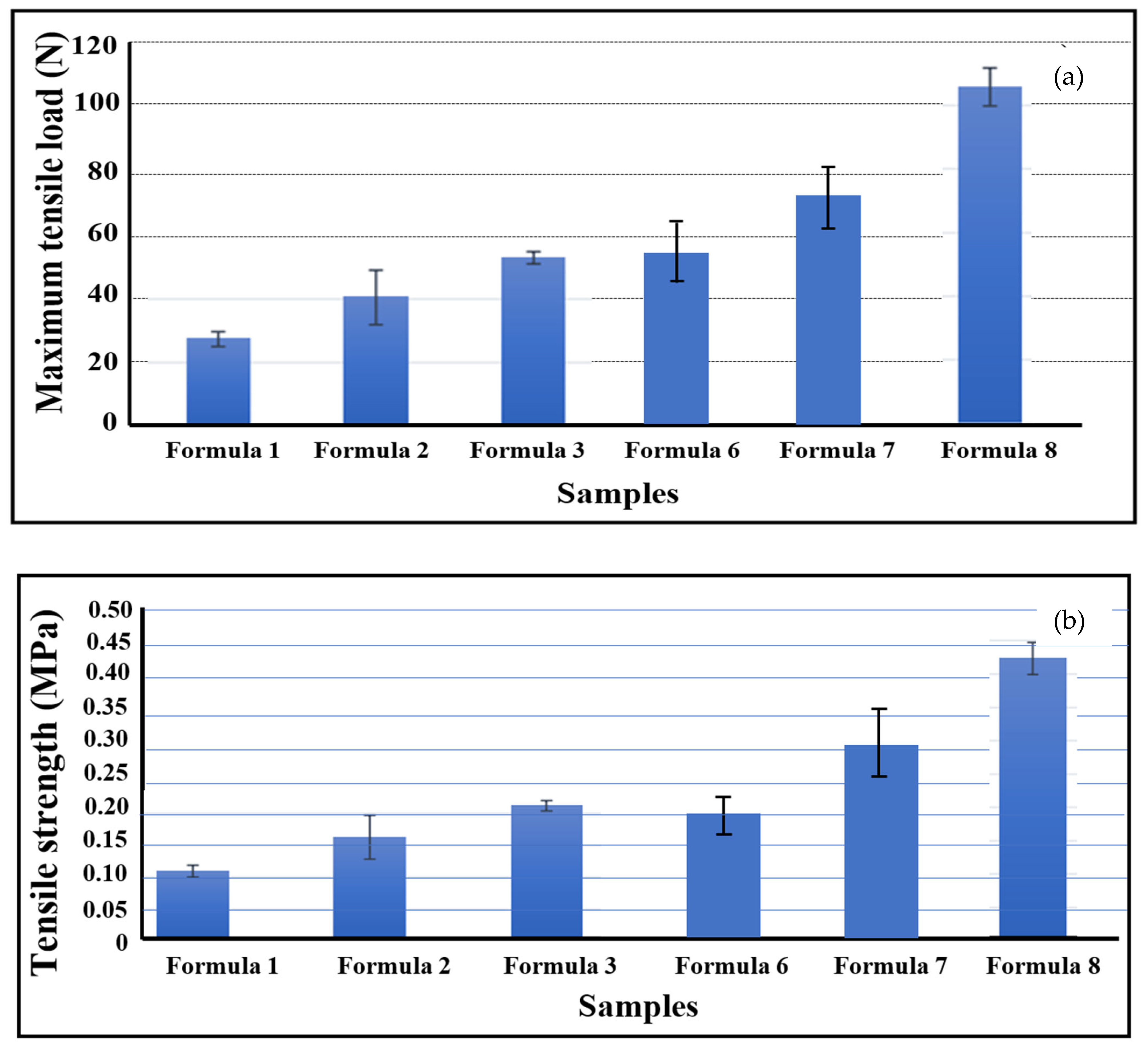
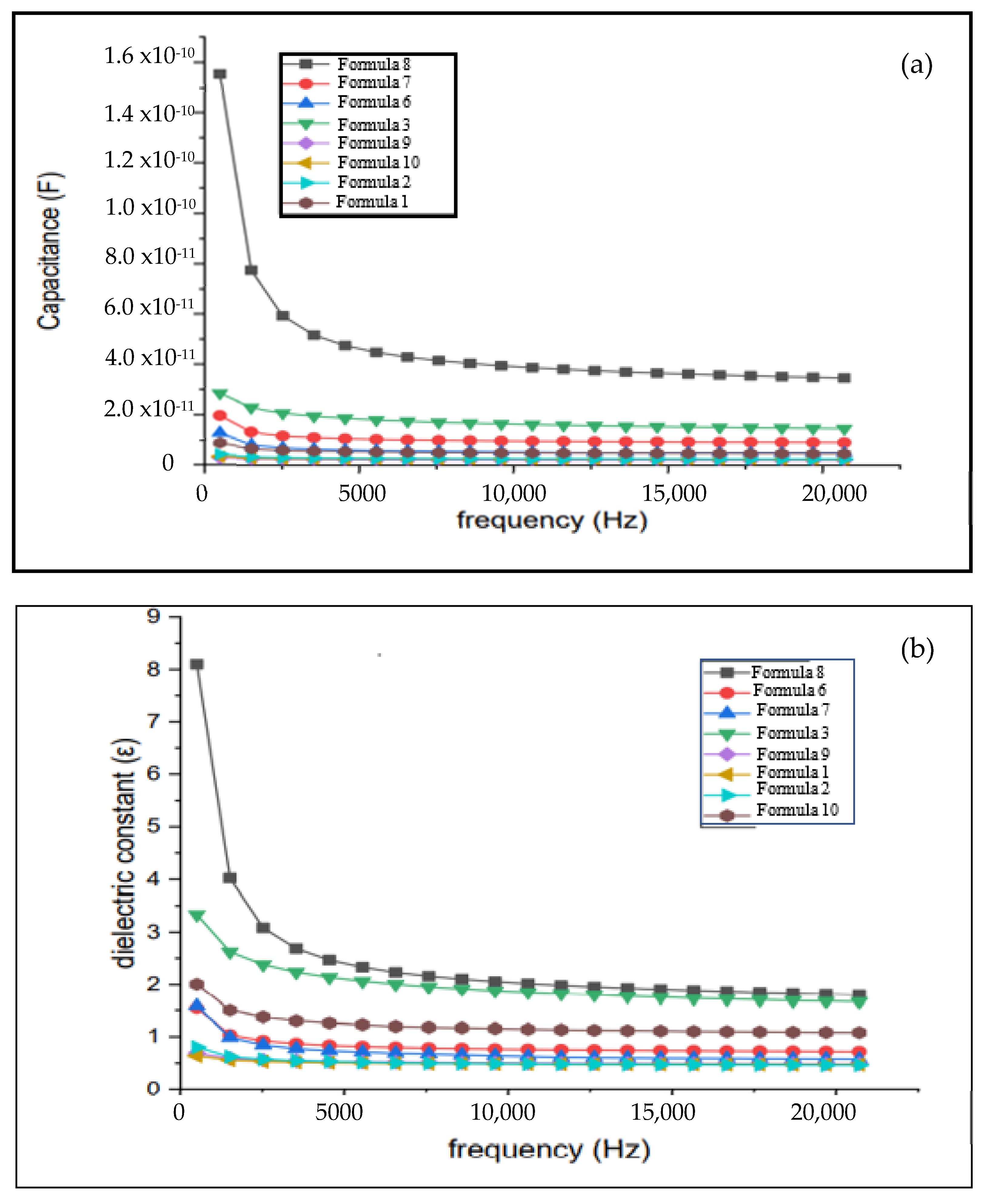
3.2. Mechanical, Optical, and Electrical Properties of Bio-Chitosan Composite Films
4. Conclusions
Author Contributions
Funding
Institutional Review Board Statement
Informed Consent Statement
Data Availability Statement
Acknowledgments
Conflicts of Interest
References
- Paulino, A.T.; Simionato, J.I.; Garcia, J.C.; Nozaki, J. Characterization of chitosan and chitin produced from silkworm crysalides. Carbohydr. Polym. 2006, 64, 98. [Google Scholar] [CrossRef]
- Guo, Y.; Chen, X.; Yang, F.; Wang, T.; Ni, M.; Chen, Y.; Yang, F.; Huang, D.; Fu, C.; Wang, S. Preparation and Characterization of Chitosan-Based Ternary Blend Edible Films with Efficient Antimicrobial Activities for Food Packaging Applications. J. Food Sci. 2019, 84, 1411. [Google Scholar] [CrossRef] [PubMed]
- Chung, J.H.Y.; Kade, J.; Jeiranikhameneh, A.; Yue, Z.; Mukherjee, P.; Wallace, G.G. A Bioprinting printing approach to regenerate cartilage for microtia treatment. Bioprinting 2018, 12, e00031. [Google Scholar] [CrossRef]
- Li, H.; Hu, C.; Yu, H.; Chen, C. Chitosan composite scaffolds for articular cartilage defect repair: A review. RSC Adv. 2018, 8, 3736. [Google Scholar] [CrossRef] [PubMed]
- Ahmed, S.; Ali, A.; Sheikh, J. A review on chitosan centred scaffolds and their applications in tissue engineering. Int. J. Biol. Macromol. 2018, 116, 849. [Google Scholar] [CrossRef]
- Liu, Y.; Jiang, X.; Wu, J.; Le, X. Molecular interactions, characterization and antimicrobial activity of curcumin–chitosan blend films. Food Hydrocoll. 2016, 52, 564. [Google Scholar] [CrossRef]
- Kumar, S.; Mukherjee, A.; Dutta, J. Chitosan based nanocomposite films and coatings: Emerging antimicrobial food packaging alternatives. Trends Food Sci. Technol. 2020, 97, 196. [Google Scholar] [CrossRef]
- Hoseinnejad, M.; Jafari, S.M.; Katouzian, I. Review on chitosan-based nanoparticles and their applications in food and agriculture. Critic. Rev. Microbiol. 2018, 44, 161. [Google Scholar]
- Zheng, L.Y.; Zhu, J.F. Study on antimicrobial activity of chitosan with different molecular weights. Carbohydr. Polym. 2003, 54, 527–530. [Google Scholar] [CrossRef]
- Alderman, O.L.G.; Skinner, L.B.; Benmore, C.J.; Tamalonis, A.; Weber, J.K.R. Structural study of amorphous chitosan materials. Phys. Rev. B 2014, 90, 094204. [Google Scholar] [CrossRef]
- Abdou, E.S.; Nagy, K.S.; Elsabee, M.Z. Extraction and characterization of chitin and chitosan from local sources. Bioresour. Technol. 2008, 99, 1359. [Google Scholar] [CrossRef] [PubMed]
- Bajaj, M.; Winter, J.; Gallert, C. Chitosan production and applications in wastewater treatment. Biochem. Eng. J. 2011, 56, 51. [Google Scholar]
- Wang, H.; Gong, X.; Miao, Y.; Guo, X.; Liu, C.; Fan, Y.Y.; Zhang, J.; Niu, B.; Li, W. Development of chitosan-based active food packaging materials. Food Chem. 2019, 283, 397. [Google Scholar] [CrossRef] [PubMed]
- Bahal, M.; Kaur, N.; Sharotri, N.; Sud, D. Recent advancements in chitosan-based biopolymer composites. Adv. Polym. Technol. 2019, 1, 12. [Google Scholar]
- Roy, S.J.-W. Applications of chitosan in drug delivery and tissue engineering. Int. J. Biol. Macromol. 2020, 148, 666. [Google Scholar]
- Higashimoto, S.; Azuma, M. Chitosan-based catalysts and their applications. Appl. Catal. B Environ. 2009, 89, 557. [Google Scholar] [CrossRef]
- Priyadarshi, R.; Rhim, J.W. Chitosan-based biodegradable films for food packaging. Innov. Food Sci. Emerg. Technol. 2020, 62, 102346. [Google Scholar] [CrossRef]
- Mesgari, M.; Aalami, A.H.; Sahebkar, A. Biomedical applications of chitosan and its derivatives. Int. J. Biol. Macromol. 2021, 176, 530. [Google Scholar]
- Awwad, A.M.; Amer, M.W.; Salem, N.M.; Abdeen, A.O. Synthesis and characterization of chitosan-based hydrogels for biomedical applications. Chem. Int. 2020, 6, 151. [Google Scholar]
- Alsohaimi, H.I.; Nassar, A.M.; Elnasr, T.A.S.; Cheba, B.A. Chitosan composites for water purification. J. Clean. Prod. 2020, 248, 119274. [Google Scholar] [CrossRef]
- Pugazhendhi, A.; Prabhu, R.; Muruganantham, K.; Shanmuganathan, R.; Natarajan, S. Photocatalytic degradation using chitosan-based materials. Photochem. Photobiol. B Biol. 2019, 190, 86. [Google Scholar]
- Hassan, H.; Omoniyi, K.; Okibe, F.; Nuhu, A.; Echioba, E. Environmental applications of chitosan composites. J. Appl. Sci. Environ. Manag. 2019, 23, 1795. [Google Scholar]
- Daghrir, R.; Drogui, P.; Robert, D. Advanced oxidation processes for water treatment using chitosan composites. Ind. Eng. Chem. Res. 2013, 52, 3581. [Google Scholar] [CrossRef]
- Ali, A.; Ahmed, S. Chitosan-based nanomaterials: Synthesis and applications. Int. J. Biol. Macromol. 2018, 109, 273. [Google Scholar]
- Ullattil, S.G.; Narendranath, S.B.; Pillai, S.C.; Periyat, P. Chitosan-based catalysts in chemical engineering applications. Chem. Eng. J. 2018, 343, 708. [Google Scholar] [CrossRef]
- Zhang, X.; Liu, Y.; Yong, H.; Qin, Y.; Liu, J.; Liu, J. Preparation and properties of curcumin-loaded chitosan films. Food Hydrocoll. 2019, 94, 80. [Google Scholar] [CrossRef]
- Shafaee, M.; Goharshadi, E.K.; Mashreghi, M.; Sadeghinia, M.J. Photochemical properties of chitosan nanoparticles. Photochem. Photobiol. A Chem. 2018, 357, 90. [Google Scholar]
- Gumiero, M.; Peressini, D.; Pizzariello, A.; Sensidoni, A.; Iacumin, L.; Comi, G.; Toniolo, R. Effect of chitosan coating on food preservation. Food Chem. 2013, 138, 1633. [Google Scholar] [CrossRef]
- Joy, J.; Mathew, J.; George, S.C. Chitosan-based materials for hydrogen energy storage. Int. J. Hydrogen Energy. 2018, 43, 4804. [Google Scholar] [CrossRef]
- Youssef, A.M.; El-Sayed, S.M. Characterization of chitosan films and their antimicrobial activity. Carbohydr. Polym. 2018, 193, 19. [Google Scholar] [CrossRef]
- Jbeli, A.; Ferraria, A.M.; Rego, A.M.B.D.; Boufi, S.; Bouattour, S. Chitosan nanocomposites for biomedical applications. Int. J. Biol. Macromol. 2018, 116, 1098. [Google Scholar]
- Salarbashi, D.; Tafaghodi, M.; Bazzaz, B.S.F. Preparation and characterization of chitosan-based materials. Carbohydr. Polym. 2018, 186, 384. [Google Scholar] [CrossRef] [PubMed]
- Xing, Y.; Tang, J.; Li, X.; Huang, R.; Wu, L.; Xu, Q.; Liu, X.; Bi, X. Antimicrobial activity of chitosan films. J. Food Prot. 2022, 85, 597. [Google Scholar] [CrossRef]
- Lin, B.F.; Luo, Y.G.; Teng, Z.; Zhang, B.C.; Zhou, B.; Wang, Q. Chitosan and its derivatives for food packaging. LWT Food Sci. Technol. 2015, 63, 1206. [Google Scholar] [CrossRef]
- Kraśniewska, K.; Gniewosz, M. Chitosan-based antimicrobial films. J. Nutr. Sci. 2012, 62, 199. [Google Scholar]
- Vejdan, A.; Ojagh, S.M.; Adeli, A.; Abdollahi, M. Edible chitosan films and coatings. LWT Food Sci. Technol. 2016, 71, 88. [Google Scholar] [CrossRef]
- Peighambardoust, S.J.; Peighambardoust, S.H.; Pournasir, N.M.; Pakdel, P. Shelf life extension using chitosan coatings. Food Packag. Shelf Life 2019, 22, 100420. [Google Scholar] [CrossRef]
- Charles, S.; Jomini, S.; Fessard, V.; Bigorgne-Vizade, E.; Rousselle, C.; Michel, C. Nanotoxicology of chitosan nanoparticles. Nanotoxicology 2018, 12, 357. [Google Scholar] [CrossRef]
- Gea, M.; Bonetta, S.; Iannarelli, L.; Giovannozzi, A.M.; Maurino, V.; Bonetta, S.; Hodoroaba, V.-D.; Armato, C.; Rossi, A.M.; Schilirò, T. Toxicological aspects of chitosan nanomaterials. Food Chem. Toxicol. 2019, 127, 89. [Google Scholar] [CrossRef]
- Proquin, H.; Rodríguez-Ibarra, C.; Moonen, C.G.; Ortega, I.M.U.; Briedé, J.J.; de Kok, T.M.; Van Loveren, H.; Chirino, Y.I. Mutagenicity assessment of chitosan nanoparticles. Mutagenesis 2017, 32, 139. [Google Scholar] [CrossRef]
- de Fonseca, J.M.; Valencia, G.A.; Soares, L.S.; Dotto, M.E.R.; Campos, C.E.M.; de Moreira, R.F.P.M.; Fritz, A.R.M. Hydroxypropyl methylcellulose-TiO2 and gelatin-TiO2 nanocomposite films: Physicochemical and structural properties. Int. J. Biol. Macromol. 2020, 151, 944. [Google Scholar] [CrossRef] [PubMed]
- He, Q.; Zhang, Y.; Cai, X.; Wang, S. Toxicity studies of chitosan nanoparticles. Int. J. Biol. Macromol. 2016, 84, 153. [Google Scholar]
- Zolfi, M.; Khodaiyan, F.; Mousavi, M.; Hashemi, M. Development and characterization of the kefiran–whey protein isolate–TiO2 nanocomposite films. Carbohydr. Polym. 2014, 109, 118. [Google Scholar] [CrossRef]
- Mohd, H.R.; Nur, A.I.; Khairul, A.M.A. Recent advances in chitosan composites for biomedical applications. Int. J. Biol. Macromol. 2020, 153, 1117. [Google Scholar]
- Masutani, E.M.; Kinoshita, C.K.; Tanaka, T.T.; Ellison, A.K.D.; Yoza, B.A. Synthesis and characterization of chitosan composites for pharmaceutical applications. Int. J. Biomater. 2014, 979636. [Google Scholar]
- Khodaei, D.; Oltrogge, K.; Hamidi-Esfahani, Z. Chitosan-based polymer composites: Preparation and functional properties. Lebensm. Wiss. Technol. 2020, 117, 108. [Google Scholar]
- Luo, Q.; Hossen, M.A.; Zeng, Y.; Dai, J.; Li, S.; Qin, W.; Liu, Y.J. Efficient removal of contaminants using chitosan-based composites in food engineering. Food Eng. 2022, 313, 110762. [Google Scholar] [CrossRef]
- Duconseille, A.; Astruc, T.; Quintana, N.; Meersman, F.; Sante-Lhoutellier, V. Impact of chitosan coatings on meat properties. Food Hydrocoll. 2015, 43, 360. [Google Scholar] [CrossRef]
- Ahmad, T.; Ismail, A.; Ahmad, S.A.; Khalil, K.A.; Kumar, Y.; Adeyemi, K.D.; Sazili, A.Q. Characterization of edible chitosan-based films with enhanced mechanical properties. Food Hydrocoll. 2017, 63, 85. [Google Scholar] [CrossRef]
- Andreuccetti, C.; Galicia-García, T.; Gonzalez-Nunez, R.; Martínez-Bustos, F.; Grosso, C.R.F. Functional and renewable chitosan polymers for packaging applications. Polym. Renew. Resour. 2017, 8, 11. [Google Scholar]
- Hosseini, S.F.; Rezaei, M.; Zandi, M.; Farahmandghavi, F. Fabrication of bio-nanocomposite films based on fish gelatin reinforced with chitosan nanoparticles. Food Hydrocoll. 2015, 44, 172. [Google Scholar] [CrossRef]
- Kumar, B.; Castro, M.; Feller, J.F. Chitosan-based micro- and nanocomposites: A review. J. Mater. Chem. 2012, 22, 10656. [Google Scholar] [CrossRef]
- Halawa, A.A.; Elshopakey, G.E.; Elmetwally, M.A.; El-Adl, M.; Lashen, S.; Shalaby, N.; Eldomany, E.; Farghali, A.; Sayed-Ahmed, M.Z.; Alam, N.; et al. Advanced chitosan-based nanocomposites for biomedical applications. Sci. Rep. 2022, 12, 19667. [Google Scholar]
- Knidri, H.E.L.; Belaabed, R.; Khalfaouy, R.E.L.; Laajeb, A.; Addaou, A.; Lahsin, A.J. Characterization and applications of chitosan composites in environmental science. Mater. Environ. Sci. 2017, 8, 3648. [Google Scholar]
- Laaraibi, A.; Charhouf, I.; Bennamara, A.; Abourriche, A.; Berrada, M.J. Extraction and characterization of chitosan from crab shell waste and its use in removal of heavy metals. Mater. Environ. Sci. 2015, 6, 3511. [Google Scholar]
- Teli, M.D.; Sheikh, J. Preparation and characterization of chitosan nanoparticles using ionic gelation method. Int. J. Biol. Macromol. 2012, 50, 1195. [Google Scholar]
- Bahrami Miyanji, P.; Semnani, D.; Hossein Ravandi, A.; Karbasi, S.; Fakhrali, A.; Mohammadi, S. Fabrication and characterization of chitosan-gelatin/single-walled carbon nanotubes electrospun composite scaffolds for cartilage tissue engineering applications. Polym. Adv. Technol. 2022, 33, 81–95. [Google Scholar] [CrossRef]
- Qian, Y.-F.; Zhang, K.-H.; Chen, F.; Ke, Q.-F.; Mo, X.-M. Fabrication and evaluation of chitosan/gelatin scaffold reinforced by nanoparticles. J. Biomater. Sci. Polym. Ed. 2011, 22, 1099. [Google Scholar]
- Gharaie, S.S.; Habibi, S.; Nazockdast, H.J. Development of chitosan/hemp fiber nonwoven textiles for sustainable applications. Text. Fibrous Mater. 2018, 1, 1. [Google Scholar]
- Spoială, A.; Ilie, C.I.; Dolete, G.; Croitoru, A.M.; Surdu, V.A.; Truşcă, R.D.; Motelica, L.; Oprea, O.C.; Ficai, D.; Ficai, A.; et al. Preparation and Characterization of Chitosan/TiO2 Composite Membranes as Adsorbent Materials for Water Purification. Membranes 2022, 12, 804. [Google Scholar] [CrossRef]
- Li, W.; He, D.; Hu, G.; Li, X.; Banerjee, G.; Li, J.; Lee, S.H.; Dong, Q.; Gao, T.; Brudvig, G.W.; et al. Visible-light-driven photocatalytic hydrogen evolution using chitosan-templated semiconductor nanocrystals. ACS Cent. Sci. 2018, 4, 631. [Google Scholar] [CrossRef] [PubMed]
- Kucukgulmez, A.; Celik, M.; Yanar, Y.; Sen, D.; Polat, H.; Kadak, E. Physicochemical characterization of chitosan extracted from Metapenaeus stebbingi shells. Food Chem. 2011, 126, 1144. [Google Scholar] [CrossRef]
- Naghibzadeh, M.; Amani, A.; Amini, M.; Esmaeilzadeh, E.; Mottaghi-Dastjerdi, N.; Faramarzi, M.A. An insight into the interactions between α-tocopherol and chitosan in ultrasound-prepared nanoparticles. J. Nanomater. 2010, 1, 18717. [Google Scholar] [CrossRef]
- Islam Md, A.A.; Mustafizur Rahman, A.F.M.; Iftekhar, S.; Salem, K.S.; Sultana, N.; Bari, M.L. Morphology, Thermal Stability, Electrical, and Mechanical Properties of Graphene Incorporated Poly (vinyl alcohol)-Gelatin Nanocomposites. J. Compos. Mater. 2016, 6, 172. [Google Scholar]
- Cui, Z.; Beach, E.S.; Anastas, P.T. Evaluation of the greenness of chemical processes for chemical and materials production. Green Chem. Lett. Rev. 2011, 4, 35. [Google Scholar] [CrossRef]
- Ling, Y.; Pan, X.; Wang, X.; Sun, R. Facile fabrication of chitosan active film with xylan via direct immersion. Cellulose 2014, 21, 1873. [Google Scholar]
- Roy, S.; Zhai, L.; Kim, H.C.; Pham, D.H.; Alrobei, H.; Kim, J. Tannic-Acid-Cross-Linked and TiO2-Nanoparticle-Reinforced Chitosan-Based Nanocomposite Film. Polymers 2021, 13, 228. [Google Scholar] [CrossRef]
- Vilela, C.; Pinto, R.J.B.; Coelho, J.; Domingues, M.R.M.; Daina, S.; Sadocco, P.; Santos, S.A.O.; Freire, C.S.R. Bioactive chitosan/ellagic acid films with UV-light protection for active food packaging. Food Hydrocoll. 2017, 73, 120. [Google Scholar] [CrossRef]
- Gaabour, L.H. Effect of addition of TiO2 nanoparticles on structural and dielectric properties of polystyrene/polyvinyl chloride polymer blend. AIP Adv. 2011, 11, 105120. [Google Scholar] [CrossRef]
- Anandhavelu, S.; Dhansekaran, V.; Sethuraman, V.; Park, H.J. Chitin and Chitosan Based Hybrid Nanocomposites for Super Capacitor Applications. J. Nanosci. Nanotechnol. 2017, 17, 1321. [Google Scholar] [CrossRef]
- Zhang, W.; Rhim, J.-W. Titanium dioxide (TiO2) for the manufacture of multifunctional active food packaging films. Food Packag. 2022, 31, 100806. [Google Scholar] [CrossRef]
- Castillo, B.C.; Prokhorov, E.; GLuna-Bárcenas, G.; Kovalenko, Y. Potential Use of Chitosan-TiO2 Nanocomposites for the Electroanalytical Detection of Imidacloprid. Polymers 2022, 14, 1686. [Google Scholar] [CrossRef] [PubMed]
- Sivanesan, I.; Gopal, J.; Muthu, M.; Shin, J.; Oh, J.W. Reviewing Chitin/Chitosan Nanofibers and Associated Nanocomposites and Their Attained Medical Milestones. Polymers 2021, 13, 2330. [Google Scholar] [CrossRef] [PubMed]
- Callister, W.D.; Rethwisch, D.G. Materials Science and Engineering, 10th ed.; Wiley: Hoboken, NJ, USA, 2018; ISBN 978-1-119-40549-8. [Google Scholar]


| Sample | Formula 1 1Chi-0Gel-0Ti | Formula 2 1CH-0Gel-0.5Ti | Formula 3 1Chi-0Gel-1Ti | Formula 4 1Chi-0Gel-2Ti | Formula 5 1Chi-0Gel-5Ti | Formula 6 1Chi-1Gel-0Ti | Formula 7 1Chi-1Gel-0.5Ti | Formula 8 1Chi-1Gel-1Ti | Formula 9 1Chi-1Gel-2Ti | Formula 10 1Chi-1Gel-5Ti |
|---|---|---|---|---|---|---|---|---|---|---|
| Chitosan powder (g) | 1 | 1 | 1 | 1 | 1 | 1 | 1 | 1 | 1 | 1 |
| Gelatin powder (g) | 0 | 0 | 0 | 0 | 0 | 1 | 1 | 1 | 1 | 1 |
| TiO2 (wt%) | 0 | 0.5 | 1.0 | 2.0 | 5.0 | 0 | 0.5 | 1.0 | 2.0 | 5.0 |
| Acetic acid 1% (mL) | 80 | 80 | 80 | 80 | 80 | 80 | 80 | 80 | 80 | 80 |
| Sample | Film Thickness (mm) | Swelling in Water | Swelling in Ethyl Alcohol | Percentage of Shrinkage after Drying (%) | Bulk Density (g/cm3) | Characteristics |
|---|---|---|---|---|---|---|
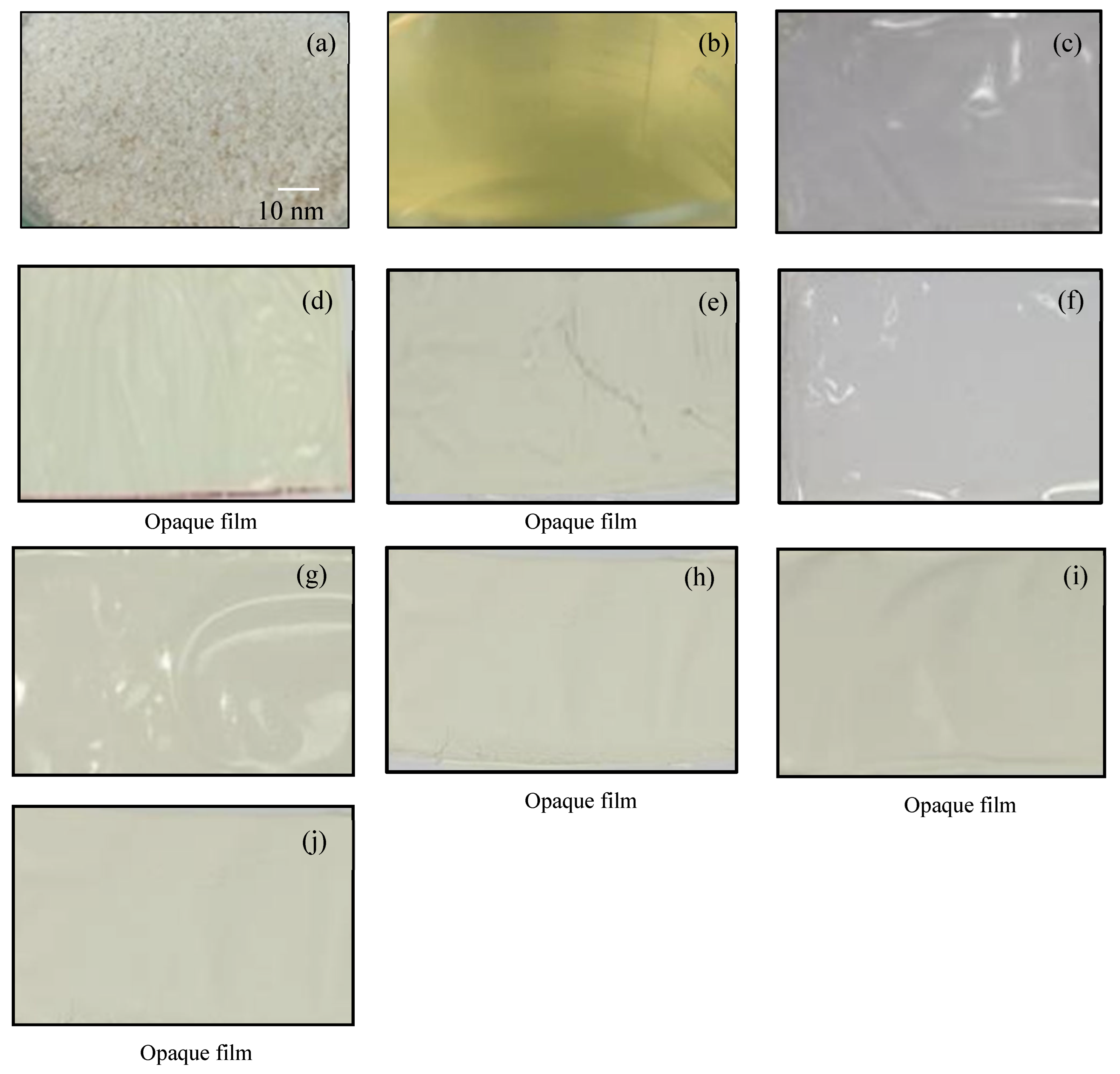
| Formula 1 1Chi-0Gel-0Ti | 0.03 ± 0.01 | Excellent swelling | No swelling | 88.25 ± 2.15 | 1.48 ± 0.32 | Transparent, with a smooth surface and no visible defects |
| Formula 2 1CH-0Gel-0.5Ti | 0.08 ± 0.02 | Excellent swelling | No swelling | 68.97 ± 7.99 | 0.86 ± 0.15 | Opaque in appearance, with a smooth surface and no visible tears |
| Formula 3 1Chi-0Gel-1Ti | 0.08 ± 0.01 | Excellent swelling | No swelling | 67.60 ± 3.58 | 0.89 ± 0.18 | Opaque in appearance, with a smooth surface and no visible tears |
| Formula 6 1Chi-1Gel-0Ti | 0.06 ± 0.01 | Excellent swelling | No swelling | 77.22 ± 4.02 | 1.38 ± 0.13 | Visually clear, uniform in appearance, and free of tear marks |
| Formula 7 1Chi-1Gel-0.5Ti | 0.09 ± 0.01 | Excellent swelling | No swelling | 59.95 ± 3.59 | 0.91 ± 0.04 | Opaque in appearance, with a smooth surface and no visible tears |
| Formula 8 1Chi-1Gel-1Ti | 0.09 ± 0.03 | Excellent swelling | No swelling | 54.17 ± 9.28 | 0.85 ± 0.18 | Opaque in appearance, with a smooth surface and no visible tears |
| Formula 9 1Chi-1Gel-2Ti | 0.16 ± 0.02 | Moderate swelling | No swelling | 31.63 ± 2.37 | 0.83 ± 0.09 | Opaque in appearance, with a smooth surface and no visible tears |
| Formula 10 1Chi-1Gel-5Ti | 0.17 ± 0.01 | Moderate swelling | No swelling | 28.50 ± 2.35 | 0.90 ± 0.03 | Opaque in appearance, with a smooth surface and no visible tears |
| Sample | Contact Angle at t = 0 ms (Degree, °) | Contact Angle at t = 2000 ms (Degree, °) | Decreasing Contact Angle Over Time (Degree/ms or °/ms) |
|---|---|---|---|
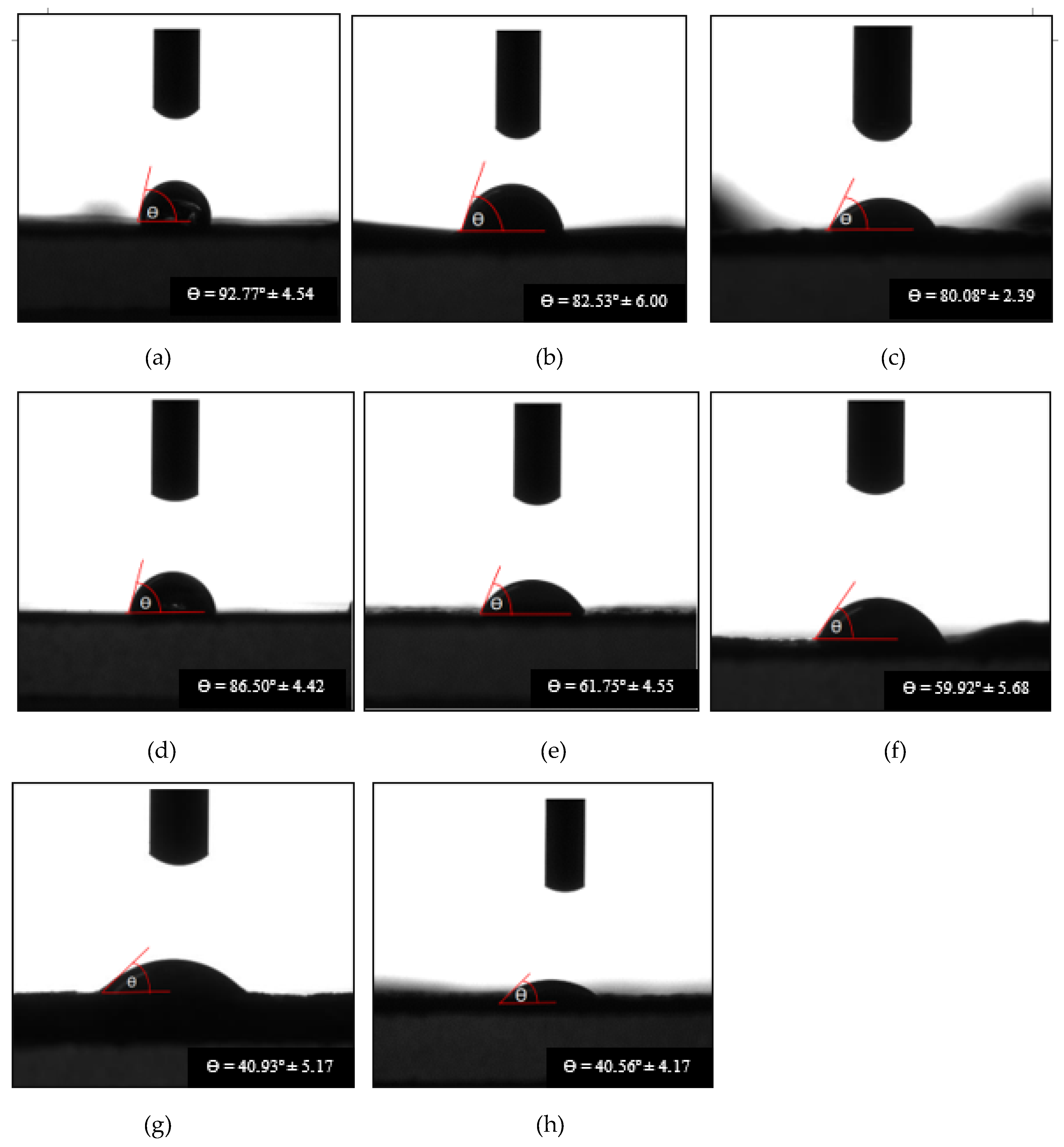
| Formula 1 1Chi-0Gel-0Ti | 103.98 ± 6.35 | 92.77 ± 4.54 | 0.58 ± 0.21 |
| Formula 2 1CH-0Gel-0.5Ti | 91.32 ± 4.53 | 82.53 ± 6.00 | 0.43 ± 0.07 |
| Formula 3 1Chi-0Gel-1Ti | 88.24 ± 3.63 | 80.08 ± 2.39 | 0.43 ± 0.05 |
| Formula 6 1Chi-1Gel-0Ti | 93.34 ± 7.47 | 86.50 ± 4.42 | 0.54 ± 0.09 |
| Formula 7 1Chi-1Gel-0.5Ti | 80.34 ± 7.37 | 61.75 ± 4.55 | 1.44 ± 0.06 |
| Formula 8 1Chi-1Gel-1Ti | 79.23 ± 4.05 | 59.92 ± 5.68 | 1.47 ± 0.28 |
| Formula 9 1Chi-1Gel-2Ti | 73.16 ± 5.50 | 40.93 ± 5.17 | 1.48 ± 0.24 |
| Formula 10 1Chi-1Gel-5Ti | 69.72 ± 6.54 | 40.56 ± 4.17 | 1.51 ± 0.11 |
| Sample | Percentage of Shrinkage (%) | Bulk Density of Dry Films (g/cm3) | Weight of Dry Films (g) | Weight of Absorbed Solvent (g) | Vr | Crosslink Density × 10−2 (Ve, mol/cm3) |
|---|---|---|---|---|---|---|
| Formula 1 1Chi-0Gel-0Ti | 88.25 ± 2.15 | 1.48 ± 0.32 | 0.0265 ± 0.0025 | 0.0017 | 0.9013 | 2.263 |
| Formula 2 1CH-0Gel-0.5Ti | 68.97 ± 7.99 | 0.86 ± 0.15 | 0.0221 ± 0.0020 | 0.0023 | 0.9604 | 4.162 |
| Formula 3 1Chi-0Gel-1Ti | 67.60 ± 3.58 | 0.89 ± 0.18 | 0.0256 ± 0.0026 | 0.0008 | 0.9689 | 4.627 |
| Formula 6 1Chi-1Gel-0Ti | 77.22 ± 4.02 | 1.38 ± 0.13 | 0.0338 ± 0.0018 | 0.0010 | 0.9550 | 3.912 |
| Formula 7 1Chi-1Gel-0.5Ti | 59.95 ± 3.59 | 0.91 ± 0.04 | 0.0484 ± 0.0072 | 0.0018 | 0.9624 | 4.262 |
| Formula 8 1Chi-1Gel-1Ti | 54.17 ± 9.28 | 0.85 ± 0.18 | 0.0421 ± 0.0059 | 0.0013 | 0.9706 | 4.735 |
| Formula 9 1Chi-1Gel-2Ti | 31.63 ± 2.37 | 0.83 ± 0.09 | 0.0761 ± 0.0139 | 0.0005 | 0.9938 | 7.677 |
| Formula 10 1Chi-1Gel-5Ti | 28.50 ± 2.35 | 0.90 ± 0.03 | 0.0611 ± 0.0032 | 0.0006 | 0.9899 | 6.758 |
| Sample | Capacitance × 10−10 (F) | Dielectric Constant |
|---|---|---|
| Formula 1 1Chi-0Gel-0Ti | 0.029 ± 0.062 | 1.54 ± 0.22 |
| Formula 2 1CH-0Gel-0.5Ti | 0.033 ± 0.095 | 1.58 ± 0.54 |
| Formula 3 1Chi-0Gel-1Ti | 0.284 ± 0.011 | 3.33 ± 0.13 |
| Formula 6 1Chi-1Gel-0Ti | 0.129 ± 0.015 | 1.69 ± 0.17 |
| Formula 7 1Chi-1Gel-0.5Ti | 0.197 ± 0.039 | 1.80 ± 0.07 |
| Formula 8 1Chi-1Gel-1Ti | 1.550 ± 0.150 | 8.10 ± 0.73 |
| Formula 9 1Chi-1Gel-2Ti | 0.090 ± 0.005 | 1.64 ± 0.27 |
| Formula 10 1Chi-1Gel-5Ti | 0.043 ± 0.003 | 2.01 ± 0.70 |
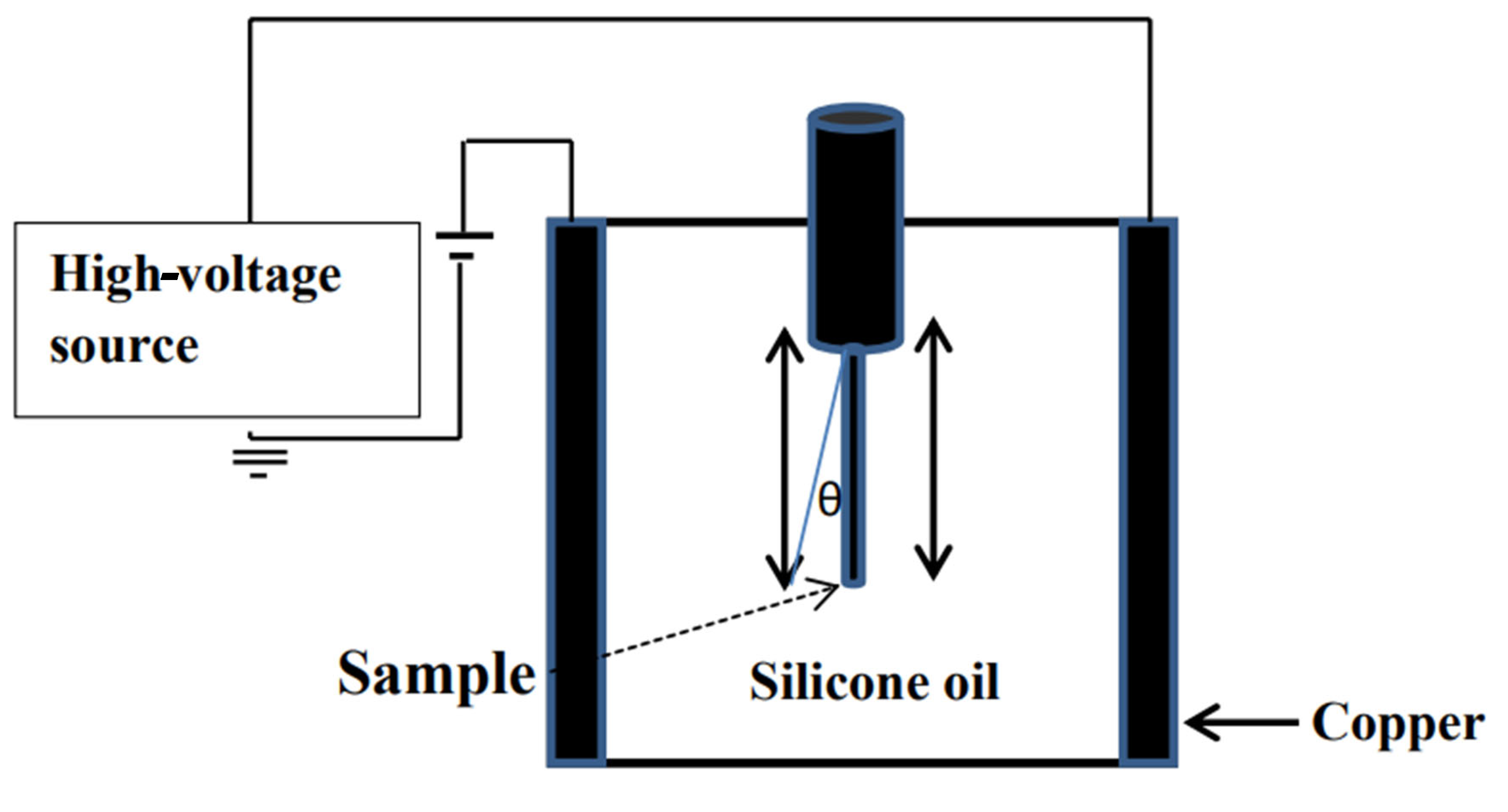
| Electric Field Strength (V/m) | Formula 1 1Chi-0Gel-0Ti (Degree, °) | Formula 2 1CH-0Gel-0.5Ti (Degree, °) | Formula 3 1Chi-0Gel-1Ti (Degree, °) | Formula 6 1Chi-1Gel-0Ti (Degree, °) | Formula 7 1Chi-1Gel-0.5Ti (Degree, °) | Formula 8 1Chi-1Gel-1Ti (Degree, °) | Formula 9 1Chi-1Gel-2Ti (Degree, °) | Formula 10 1Chi-1Gel-5Ti (Degree, °) |
|---|---|---|---|---|---|---|---|---|
| 0 | 0.00 ± 0.00 | 0.00 ± 0.00 | 0.00 ± 0.00 | 0.00 ± 0.00 | 0.00 ± 0.00 | 0.00 ± 0.00 | 0.00 ± 0.00 | 0.00 ± 0.00 |
| 25 | 0.00 ± 0.00 | 0.00 ± 0.00 | 0.00 ± 0.00 | 0.00 ± 0.00 | 0.00 ± 0.00 | 0.00 ± 0.00 | 0.00 ± 0.00 | 0.00 ± 0.00 |
| 50 | 0.00 ± 0.00 | 0.00 ± 0.00 | 0.00 ± 0.00 | 0.00 ± 0.00 | 0.00 ± 0.00 | 3.31 ± 0.17 | 0.00 ± 0.00 | 0.00 ± 0.00 |
| 100 | 0.00 ± 0.00 | 3.37 ± 1.04 | 5.92 ± 0.22 | 0.00 ± 0.00 | 4.64 ± 0.98 | 4.61 ± 2.58 | 4.65 ± 0.85 | 5.23 ± 1.76 |
| 150 | 2.64 ± 0.33 | 4.60 ± 1.65 | 9.07 ± 0.89 | 3.65 ± 0.01 | 6.70 ± 1.74 | 4.97 ± 0.65 | 5.16 ± 0.33 | 5.64 ± 3.33 |
| 200 | 4.02 ± 1.65 | 5.57 ± 1.50 | 10.44 ± 0.73 | 4.23 ± 0.76 | 9.00 ± 1.56 | 32.25 ± 3.56 | 6.21 ± 1.65 | 6.42 ± 3.65 |
| 250 | 4.03 ± 1.00 | 7.22 ± 2.30 | 18.96 ± 3.83 | 4.62 ± 0.83 | 17.85 ± 2.58 | 61.48 ± 6.97 | 6.40 ± 1.00 | 6.50 ± 2.00 |
| 300 | 4.17 ± 1.89 | 9.25 ± 3.12 | 43.67 ± 4.39 | 4.82 ± 0.65 | 38.87 ± 3.01 | 68.58 ± 7.36 | 42.07 ± 0.89 | 8.17 ± 1.89 |
| 350 | 4.91 ± 0.41 | 11.37 ± 3.71 | 54.63 ± 4.16 | 5.25 ± 0.01 | 59.06 ± 3.91 | 72.81 ± 5.57 | 50.09 ± 2.10 | 10.29 ± 2.41 |
| 400 | 5.28 ± 3.35 | 13.59 ± 4.97 | 58.15 ± 1.72 | 8.07 ± 0.89 | 64.77 ± 4.98 | 73.72 ± 5.27 | 52.54 ± 0.35 | 14.84 ± 5.35 |
| 450 | 8.70 ± 4.52 | 17.91 ± 3.93 | 59.34 ± 1.50 | 9.49 ± 0.41 | 67.62 ± 4.37 | 78.02 ± 2.56 | 56.70 ± 5.52 | 19.70 ± 7.52 |
| 500 | 13.58 ± 3.51 | 65.88 ± 5.02 | 62.61 ± 2.23 | 13.84 ± 2.35 | 70.05 ± 2.18 | 79.48 ± 3.28 | 59.34 ± 0.51 | 26.58 ± 6.51 |
| 550 | 22.03 ± 5.34 | 68.35 ± 2.33 | 64.04 ± 2.46 | 23.81 ± 4.51 | 70.03 ± 1.53 | 81.29 ± 0.75 | 60.05 ± 0.30 | 34.03 ± 7.34 |
Disclaimer/Publisher’s Note: The statements, opinions and data contained in all publications are solely those of the individual author(s) and contributor(s) and not of MDPI and/or the editor(s). MDPI and/or the editor(s) disclaim responsibility for any injury to people or property resulting from any ideas, methods, instructions or products referred to in the content. |
© 2025 by the authors. Licensee MDPI, Basel, Switzerland. This article is an open access article distributed under the terms and conditions of the Creative Commons Attribution (CC BY) license (https://creativecommons.org/licenses/by/4.0/).
Share and Cite
Tangboriboon, N.; Malichai, N.; Wantaha, G. Influence of Titanium Dioxide (TiO2) Nanocrystallinity on the Optoelectrical Properties of Chitosan Biocomposite Films Prepared via Sol–Gel Casting. J. Compos. Sci. 2025, 9, 334. https://doi.org/10.3390/jcs9070334
Tangboriboon N, Malichai N, Wantaha G. Influence of Titanium Dioxide (TiO2) Nanocrystallinity on the Optoelectrical Properties of Chitosan Biocomposite Films Prepared via Sol–Gel Casting. Journal of Composites Science. 2025; 9(7):334. https://doi.org/10.3390/jcs9070334
Chicago/Turabian StyleTangboriboon, Nuchnapa, Nitchakarn Malichai, and Guytawan Wantaha. 2025. "Influence of Titanium Dioxide (TiO2) Nanocrystallinity on the Optoelectrical Properties of Chitosan Biocomposite Films Prepared via Sol–Gel Casting" Journal of Composites Science 9, no. 7: 334. https://doi.org/10.3390/jcs9070334
APA StyleTangboriboon, N., Malichai, N., & Wantaha, G. (2025). Influence of Titanium Dioxide (TiO2) Nanocrystallinity on the Optoelectrical Properties of Chitosan Biocomposite Films Prepared via Sol–Gel Casting. Journal of Composites Science, 9(7), 334. https://doi.org/10.3390/jcs9070334