Article

Biomechanical Comparison of Titanium and CFR-PEEK Intramedullary Nails Using Finite Element Analysis

1
Department of Mechanics, Mathematics and Management, Polytechnic University of Bari, 70125 Bari, Italy
2
Orthopaedic & Trauma Unit, Department of Traslational Biomedicine and Neuroscience (DiBraiN), School of Medicine, University of Bari Aldo Moro, AOU Consorziale “Policlinico”, 70124 Bari, Italy
*
Author to whom correspondence should be addressed.
J. Compos. Sci. 2025, 9(11), 576; https://doi.org/10.3390/jcs9110576
Submission received: 22 September 2025 / Revised: 13 October 2025 / Accepted: 24 October 2025 / Published: 27 October 2025
(This article belongs to the Special Issue Polymer Composites and Fibers, 3rd Edition)

Abstract

This study analyzes the biomechanical performance of intramedullary nails made of titanium alloy (Ti-6Al-4V) and carbon fiber-reinforced polyetheretherketone (CFR-PEEK) for the treatment of proximal femoral fractures, with a focus on their effects under different bone density conditions representing young and osteoporotic bone. Using finite element models and analyses simulating mid-stance gait loading and incorporating muscle forces adjusted for age-related reduction, the load transfer and stress distribution were evaluated, along with the osteogenic index (OI) as a measure of biological stimulus for bone healing. Results showed that titanium nails produced lower bone stresses but caused significant proximal stress shielding, particularly in osteoporotic bone, which could impair healing. In contrast, CFR-PEEK nails exhibited higher and more uniformly distributed stresses along the femoral diaphysis and shifted the osteogenic stimulus into a range promoting more mature bone formation in both young and elderly femora. The composite material’s elastic modulus closer to bone and its orthotropic fiber arrangement contributed to these effects. The study concludes that CFR-PEEK nails offer a promising alternative to titanium by reducing stress shielding and enhancing the biomechanical environment favorable for fracture healing, especially in osteoporotic patients. Future work will include dynamic loading conditions and experimental validation to optimize implant design.

1. Introduction

Hip fracture is the most severe type of bone pathology in elderly people, due to its high rates of morbidity and mortality, and the considerable economic burden it places on patients and healthcare systems [1,2,3]. Current projections suggest that hip fractures could affect up to 6.3 million individuals worldwide by 2050, with 3.25 million cases expected to occur in Asia [4]. Intertrochanteric fractures (ITFs) are among the most common types of hip fracture and are associated with poorer clinical outcomes in terms of mortality and disability. From a surgical standpoint, ITFs are typically treated using intramedullary implants, either on their own or alongside osteosynthesis devices, such as plates [5].
The concept of intramedullary nailing was introduced in the 1930s by Gerhard Küntscher, who developed a V-shaped nail to treat femoral fractures [6,7,8]. This innovation exploited the medullary canal to provide internal stabilization, reducing the need for prolonged immobilization in a cast and allowing for earlier mobilization. Over the following decades, improvements in design and materials were introduced, from initial stainless-steel nails to nails with more complex cross-sections (e.g., hourglass and cloverleaf shapes) to provide rotational stability in the 1950s. Subsequently, proximal and distal locking was introduced in the 1970s and 1980s, extending the range of applications to unstable fractures [9,10]. Meanwhile, titanium became established due to its biocompatibility and corrosion resistance [11,12,13].
Despite their widespread use, metallic nails present limitations related to their high axial stiffness. Metallic implants absorb an excessive portion of the load, thereby reducing the stress transferred to the surrounding bone (stress shielding), which may lead to osteopenia, decreased bone density, and an increased risk of delayed union or loss of fixation [14,15]. For effective healing, the load transmitted to the bone must be sufficient to activate the biological repair responses.
Recent studies emphasize the importance of a favorable mechanical environment in the consolidation process [16,17,18,19]. Controlled axial micromovements have been shown to stimulate callus formation and shorten healing times, confirming the role of moderate compressive stresses in tissue regeneration [20,21,22].
To overcome the limitations of traditional metallic implants, research has explored composite materials and novel implant design strategies [23,24,25]. Examples include porous magnesium-based scaffolds coated with polymers/bioglasses, and devices incorporating hydroxyapatite with shape-memory polymers that allow morphological adaptation and targeted compression [26]. CAD/CAM technologies and 3D printing now enable customized implants that optimize load transfer and bone integration [27,28,29].
In this context, carbon fiber-reinforced polyetheretherketone (CFR-PEEK) has emerged as an alternative to titanium for intramedullary nails. This composite material has an elastic modulus (≈3.5 GPa) closer to that of cortical bone (1–20 GPa) compared to titanium alloys (≈106 GPa), thereby promoting a more physiological load transfer and potentially reducing stress shielding [30,31]. Moreover, its radiolucency and minimal CT/MRI artifacts facilitate postoperative monitoring [32,33,34,35].
Over the past decades, the biomechanical study of fractures and fixation devices has increasingly relied on non-invasive numerical tools [36,37,38,39]. Among these, finite element analysis (FEA) has proven to be effective for assessing stresses and strains at the bone–implant interface [39,40,41]. FEA enables the analysis of realistic 3D models of the fractured segment and the simulation of physiological or critical conditions, which are often difficult to reproduce experimentally. Recent evidence indicates that nails made of PEEK or functionally graded materials can reduce stress shielding and improve stress distribution compared to traditional metals [42,43].
Despite the growing interest in alternatives to titanium, the literature still includes few systematic FEA studies on the behavior of CFR-PEEK intramedullary nails in femoral fractures. Moreover, analyses that link nail material selection to the patient’s bone density remain lacking.
An intertrochanteric fracture (ITF) is closely related to osteoporosis. As people age, reductions in bone mineral density (BMD) and cortical thickness are observed. For example, studies report a decrease of around 14% per decade after the age of 50, which leads to reduced mechanical strength [44]. ITFs may not respond as well to treatment with cortical support if these changes occur. Nevertheless, many numerical simulations use bone parameters derived from younger individuals, which underestimates the deterioration typical of elderly patients. Furthermore, clinical protocols often fail to adequately tailor strategies according to age and bone fragility.
Considering these issues, the present study uses Finite Element Analysis (FEA) to compare two types of intramedullary nail for treating proximal femoral fractures: one made of titanium and the other made of carbon fiber reinforced PEEK (CFR-PEEK). This comparison will be conducted under two different bone density conditions to simulate the effect of osteoporosis on treatment outcomes. The objective is to quantify the influence of the materials’ mechanical properties on stress transferred to the surrounding bone, paying particular attention to the fracture site and the risk of stress shielding.
The results may provide practical insights into implant material selection based on bone density, contributing to strategies that ensure adequate mechanical stability and a more physiological biomechanical environment. This could reduce the risk of delayed healing and long-term complications. Furthermore, FEA-based evidence may guide the development of new-generation devices capable of effectively balancing structural strength with the biological stimulation of bone healing.

2. Materials and Methods

2.1. Modeling

Three-dimensional geometric modeling was initially performed using Autodesk Inventor 2024 (Autodesk Inc., San Francisco, CA, USA) and then exported for processing in the finite element analysis environment. The femur geometry was obtained from the Sawbones database (https://sawbones.com/), while the intramedullary nail was modeled according to technical data available in the literature. The 3D model was processed in Mimics 19.0 software, where an intertrochanteric fracture of type 31-A2, according to the AO/OTA classification, was reproduced (Figure 1). To simulate the displacement of the head–neck segment, a 4 mm gap was introduced [45].
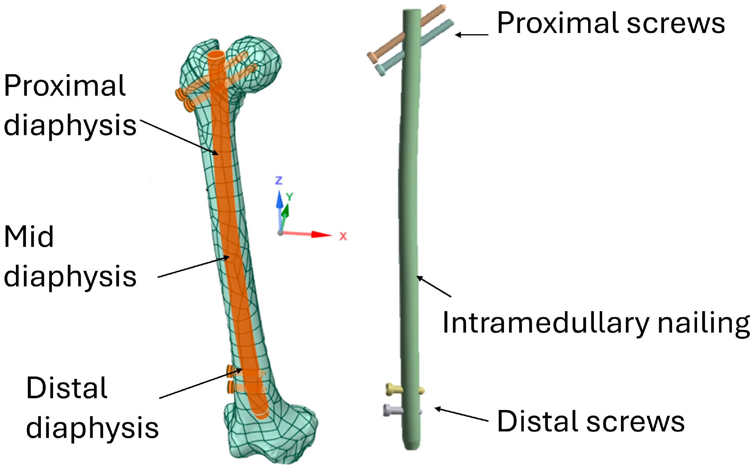
Subsequently, the nail was designed using the CAD software Autodesk Inventor and positioned within the intramedullary canal in accordance with surgical protocols [45,46] (Figure 2).
The intramedullary nail used in this study measured 410 mm in length and 17 mm in diameter. It was equipped with four locking screws: two proximal screws, each measuring 7 mm in diameter and 77 mm in length to ensure rotational stability, and two distal screws, each measuring 7 mm in diameter and 30 mm in length to ensure longitudinal stability.
Two materials were analyzed for the nail: a titanium alloy (Ti-6Al-4V) and CFR-PEEK (see Figure 3). For the CFR-PEEK nail, the fiber arrangement was defined according to the recommendations of Samiezadeh et al. [47]. Laminates composed of 10–20 plies with angular orientations of 0°, ±45° and 90° were considered to achieve an orthotropic and balanced configuration. This arrangement was found to be advantageous as it enables the intramedullary implant to achieve adequate axial flexibility whilst maintaining sufficient stiffness in bending and torsion. This ensures greater fracture stability and promotes the bone healing process. For sizing, adult femoral nails commonly use nominal outer diameters in the 9–12 mm range (cannulated designs), and structural CFR-PEEK laminates typically employ a fiber volume fraction V f of approximately 55–65%. Where needed for FEA, the longitudinal direction was aligned with the nail axis, and transverse/isotropic resin properties were assigned to the matrix-dominated directions.

2.2. Materials Properties

Bone was modeled as an orthotropic continuum with principal material axes aligned to the femoral anatomy [47,48,49,50]. The longitudinal modulus E33 and Poisson’s ratio ν were assigned as empirical functions of ash density ρ a s h   (g/cm3) taken from Cen et al. [48]. Consistently with that work, E33 follows a power-law form:
E 33 = a ρ a s h b
where a (scale) and b (exponent) are empirical coefficients calibrated against experimental and virtual testing data of proximal femoral bone. Specifically, (a) defines the overall stiffness level at a reference density, while (b) controls the sensitivity of stiffness to variations in mineralization. In addition, following quantitative computed tomography (QCT) data reported in the literature for both young and elderly patients [51,52,53], the elastic constants of cortical and trabecular bone (E11, E22, E33, G12, G13, G23) were expressed as functions of ash density (g/cm3) according to Cen et al. [48] (Table 1).
Within this framework, reductions in ρ a s h result in lower E33 (softer longitudinal response), higher strains for a given load, and modified implant–bone stress sharing, which directly impact stress shielding and osteogenic stimulus predictions.
To simulate the effects of aging on bone density, two values of ρ a s h were considered: 0.956 g/cm3 for a healthy patient and 0.82 g/cm3 for an elderly patient [54]. Ti-6Al-4V was modeled as an isotropic material, while CFR-PEEK, being a composite, was modeled as an orthotropic material (Table 2) [55,56,57,58]. The intramedullary nails in CFR-PEEK were designed according to the fiber configuration (0°, ±45°, 90°) reported in [56]. However, the explicit modeling of individual fiber orientations within the finite element framework is not computationally feasible. Therefore, a homogenization strategy was adopted, representing the composite as an equivalent orthotropic material whose elastic properties reproduce the macroscopic behavior of the CFR-PEEK laminate. This simplification preserves the anisotropy and stiffness characteristics of the composite while avoiding the high computational cost of ply-by-ply or fiber-level modeling, in line with common practices in biomechanical simulations.

2.3. FEA Modeling

The assembled CAD model was imported into ANSYS Workbench R2023 to generate a finite element (FE) model. For geometric discretization, tetrahedral SOLID 187 elements were employed, as they are particularly suitable for modeling complex geometries and simulating three-dimensional structural behavior [59].
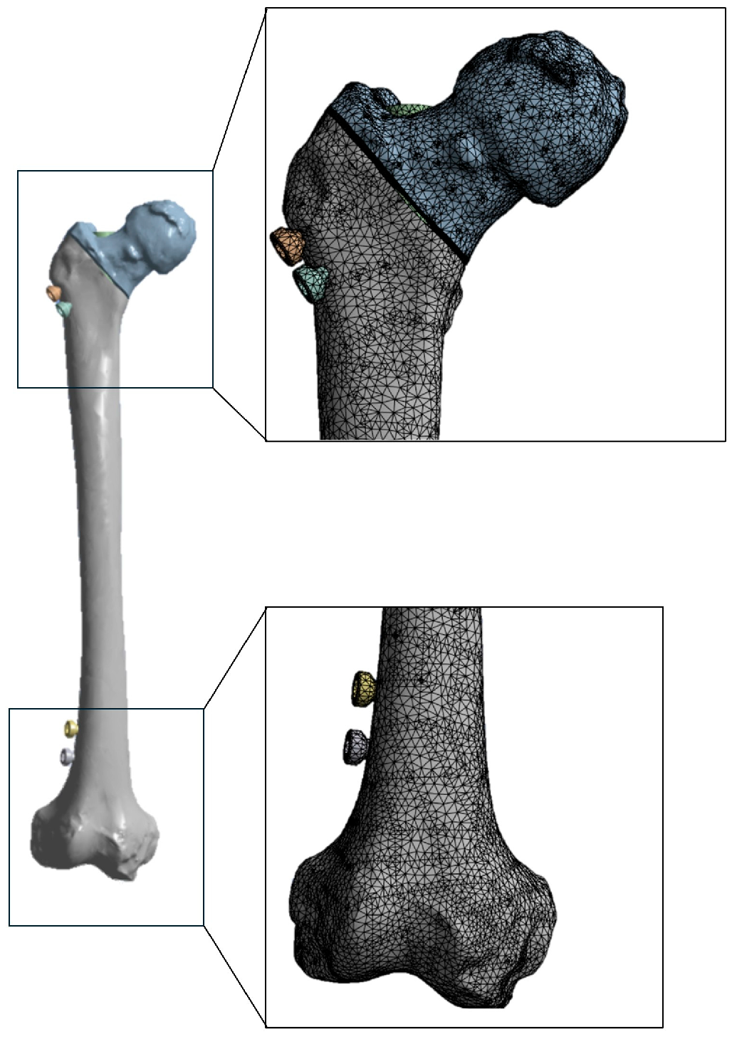
To optimize the balance between computational accuracy and processing time, a mesh size of 1 mm was assigned to the intramedullary nail and locking screws, while a coarser size of 2 mm was applied to the femur. This was in accordance with the findings of Toksoy et al. [60], who demonstrated that finer discretization of the implants and coarser discretization of the bone ensures reliable results while reducing computational cost. Additionally, a mesh convergence analysis was performed using the ‘relevance’ option in ANSYS. This confirmed that a 2 mm element size provided sufficient convergence, with deformation variations below 1% when the mesh was refined further (Figure 4).
The loading conditions applied to the finite element (FE) models of the femoral neck were carefully defined to simulate the mid-stance phase of gait under single-limb support [61]. Muscle forces were considered, including the three main groups of muscles: the abductors, the vastus lateralis and the iliopsoas, as well as the hip joint contact force. These muscle forces were modulated according to age, with a 40% reduction assumed for elderly subjects compared to younger ones. This is consistent with the loss of muscle mass and strength associated with aging [61,62,63].
The model accounted for the main joint and muscle forces, namely the joint reaction force (JRF), the abductor force (Fabd), the iliopsoas force (Flp), and the vastus lateralis force (Fvl) [64]. The numerical values of forces used for young and elderly subjects are summarized in Table 3.
Boundary conditions included nonlinear frictionless contact between the intramedullary nail and the endosteal canal; bonded contact at screw–bone interfaces; and a fully constrained distal femur (Figure 5). All contacts were implemented as surface-to-surface using the Augmented Lagrange formulation with program-controlled normal stiffness; separation was permitted at frictionless interfaces. Simulations were performed under quasi-static conditions with geometric/contact nonlinearity and automatic substepping.
The intramedullary nail–endosteal canal interface was modeled as nonlinear frictionless contact to represent the early postoperative, reamed canal condition with limited tangential load transfer, where construct stability is primarily provided by the locking screws.

2.4. Evaluation of the Osteogenic Response of Callus Based on Stress

To evaluate the osteogenic response of the bone callus, the principal stresses (r1, r2, r3) obtained at each callus node from the finite element analysis were considered. In accordance with the study conducted by Carter et al. [65], once the principal stresses are known, the octahedral shear stress (S) can be calculated as:
S = 1 3 r 1 r 2 2 + r 2 r 3 2 + r 3 r 1 2
Subsequently, the dilatational stress (D) can be evaluated as:
D = 1 3 r 1 + r 2 + r 3
These parameters are used to calculate the osteogenic stimulus index (I):
I = R n S + k D
where k is an empirical parameter modulating the influence of dilatational stress and Rn represents the number of loading cycles. Both parameters were set to 1 since excessively high k values (e.g., 2.0) may overestimate the inhibitory effect of dilatational pressure, as observed in the study conducted by Gardner et al. [66]. Rn = 1 corresponds to the quasi-static load case.
The osteogenic stimulus index (I) condenses the local mechanical environment into a single metric by combining deviatoric and dilatational (hydrostatic) stress components. Higher I values generally reflect a more osteogenic and mechanically stable environment conducive to bone formation, whereas lower values indicate insufficient stimulus.
According to the osteogenic stimulus index (I), the following tissue differentiations in the bone callus can be identified [65]:
I ≈ 5 MPa: strong osteogenic stimulus and robust ossification;
I ≈ 2–5 MPa: formation of maturing bone tissue;
I ≈ 0.5–2 MPa: fibrous and cartilaginous tissues with initial ossification;
I < 0.5 MPa: early stage of soft tissue or regions with impeded ossification.
For the comparative analysis of the biomechanical behavior of the two models (titanium nail vs. CFR-PEEK nail) regarding stress transfer, von Mises equivalent stress was adopted as the main parameter. This criterion is widely used in biomechanical and engineering studies and allows the three-dimensional stress state within a material to be represented by a single scalar value. Furthermore, to facilitate interpretation of the results, the von Mises equivalent stress values were displayed as color maps generated by the FEM analysis. These maps provide an intuitive visualization of stress distribution across the different components of the model, highlighting the most loaded areas through a progressive color scale (from minimum to maximum values).

3. Results

To evaluate the distribution of von Mises stresses within the femur, the intramedullary nail, and along the bone–nail interface, stress data were extracted from the finite elements located immediately adjacent to the contact surfaces on both the bone and nail sides. The finite element analyses were conducted considering different combinations of nail material (titanium and CFR-PEEK) and bone condition (young and elderly femur).

3.1. Distribution of Von Mises Stresses in the Femur

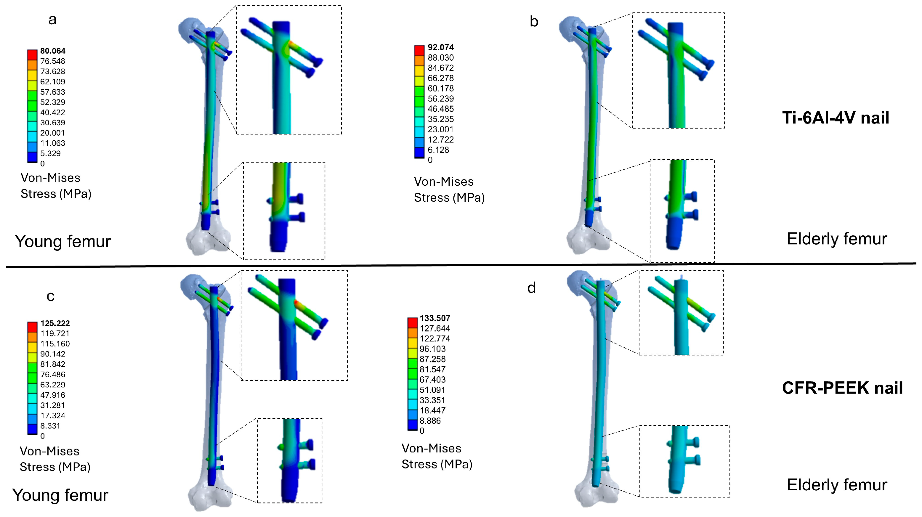
In a young femur stabilized with a Ti-6Al-4V nail (Figure 6a), von Mises stresses are primarily distributed along the diaphyseal cortex, reaching a maximum value of 46 MPa. In the elderly femur with a titanium nail (Figure 6b), stress increases to a peak of 57 MPa with a marked concentration in the distal region of the femur.
Higher stress values are observed with CFR-PEEK compared to titanium, but they are more evenly distributed between the proximal and distal portions. In the young femur (Figure 6c), maximum stress reaches 75 MPa with a more homogeneous distribution along the diaphysis. In the elderly femur (Figure 6d), stresses increase to 84 MPa with peak values concentrated mainly at the proximal screw holes.
The sectional views illustrate the differences in load transfer. Marked stress shielding is evident with the titanium nail. In the proximal area of the young femur (Figure 6a), for example, bone stress is approximately 5.5 MPa, which is lower than the 22 MPa measured in the nail. In the distal area, the bone stress increases to 37.85 MPa, compared to 43 MPa in the nail. This illustrates the uneven distribution of stress between the proximal and distal areas. This phenomenon is even more pronounced with reduced bone density, such as in an elderly femur (Figure 6b).
With CFR-PEEK, bone stress levels are higher and more widespread, which is compatible with a more physiological mechanical environment and a clear reduction in stress shielding. In both the proximal and distal regions, bone stress tends to exceed nail stress (see Figure 6c,d). However, in the elderly femur (Figure 6d), these levels may exceed the capacity of osteoporotic tissue. Therefore, it is appropriate to modulate or customize the stiffness of the composite according to the quality of the host bone to maintain stress distribution within a safe range for tissue health.

3.2. Distribution of Von Mises Stresses in the Intramedullary Nail

Figure 7a–d illustrates the von Mises stress distribution in the nails across the four analyzed configurations.
In the titanium nail model with a young femur (Figure 7a), peak stress reached approximately 77 MPa, concentrated around the transverse holes for the locking screws. In the elderly femur (Figure 7b), maximum stress increased to 84 MPa, with a wider distribution along the nail due to the reduced mechanical support of the bone.
In the case of CFR-PEEK, stress levels were higher than for titanium but more evenly distributed along the nail’s length. The highest values remained localized around the proximal holes, reaching 115 MPa in the young femur and 131 MPa in the elderly one. This behavior results from the closer elastic modulus of CFR-PEEK to that of bone, which promotes a more balanced load transfer at the bone–implant interface, mitigates stress shielding, and ensures effective mechanical stimulation of the surrounding bone tissue without compromising implant integrity.

3.3. Evaluation of the Osteogenic Stimulus Index

Figure 8 presents a top (axial) sectional view of the proximal femur–nail assembly, centered on the proximal locking holes and screws and oriented along the femoral axis to depict load transfer and stress distribution in the most critical region for construct stability and fracture healing. Within this fracture section, the osteogenic index (OI) shows clear differences between materials and age groups, consistent with Carter’s thresholds [65].
For model A (Ti-6Al-4V nail), the average OI value is 1.8 MPa for young femur and 1.6 MPa for elderly femur. Both values fall within the 0.5–2 MPa range, which is indicative of initial ossification (i.e., prevalence of fibrous or cartilaginous tissue).
For the CFR-PEEK nail, an average OI of 2.7 MPa is recorded at the fracture site for a young femur and 3.7 MPa for an elderly femur. These values fall within the 2–5 MPa range, which indicates maturing bone formation.
Therefore, based on these results, CFR-PEEK provides a higher OI than titanium at the same age, shifting the stimulus from the “initial” range (0.5–2 MPa) to the maturation window (2–5 MPa). With titanium, age reduces OI (A→B), while with CFR-PEEK, OI increases (C→D). This suggests that the composite material has a greater ability to maintain or increase the stimulus, even in osteoporotic bone.

4. Discussion

Femoral fractures are commonly treated with intramedullary nails or adjunct osteosynthesis devices [67,68,69]. The implant’s material and geometry are critical design variables, as they govern load transfer, construct stability, and the mechanobiological stimulus required for timely healing [70]. Recently, alternatives to conventional metals (e.g., Ti-6Al-4V) such as carbon fiber-reinforced PEEK (CFR-PEEK) and functionally graded materials with tailored stiffness have garnered increasing interest [71]. These options aim to reduce the bone–implant stiffness mismatch, limit stress shielding, and where radiolucent improve radiographic assessment [72]. In a prospective study, Takashima et al. [73] evaluated a CFR-PEEK intramedullary nail for proximal femoral fractures: among 20 patients, 95% achieved union at a mean of 3.7 months, with no significant complications and no reoperations.
Consistently, Sacchetti et al. [74] showed that the radiolucency of CFR-PEEK allows for a more objective evaluation of the callus, with higher inter-observer reliability and without an increase in complications.
The present finite element analyses (FEA) study compared Ti-6Al-4V and carbon-fiber-reinforced PEEK (CFR-PEEK) intramedullary nails implanted in femoral models representing young and elderly/osteoporotic bone. Two principal findings emerged: (1) with the titanium nail, the femur experienced lower overall stresses but exhibited marked proximal stress shielding; and (2) with the CFR-PEEK nail, femoral stresses were higher and more uniformly distributed along the diaphysis. Regarding healing potential, the osteogenic index (OI) showed that titanium concentrated the mechanical stimulus within the 0.5–2 MPa range (early ossification), whereas CFR-PEEK shifted the stimulus toward 2–5 MPa (maturing bone) in both bone phenotypes. Overall, a nail with lower stiffness than titanium and mechanical properties closer to bone promotes greater load transfer to the surrounding bone, in agreement with prior literature.
Similarly, Samiezadeh et al. [75] showed that a hybrid composite nail produced controlled increases in stresses at the fracture site and reduced stress shielding at equivalent fragment stability. In terms of durability, Samiezadeh et al. [76] documented for a carbon/epoxy composite nail a high-cycle fatigue strength of approximately 70% of UTS (HCFS ≈ 70% UTS) with no modulus degradation (≈42 GPa), supporting the composite’s performance under repeated loading. Likewise, Steinberg et al. [77] found that CF-PEEK nails/plates offer stiffness and fatigue resistance comparable to metallic devices, with lower debris generation at metal–polymer interfaces.
Along the same line of time-modulated stress shielding, the absorbable-sheath nail concept tested via FE by Dong et al. [78] proposes a programmed transition of construct stiffness, with potential benefits across different healing phases.
A seemingly discrepant point relative to some FEM studies that model (C)PEEK as isotropic regards stresses within the nail: in those works [31,79], PEEK nail stresses are sometimes lower than in metals, whereas in our study—where CFR-PEEK was modeled as orthotropic peak stresses in the nail were higher (115–131 MPa). This difference can be explained by material assumptions (isotropic PEEK vs. laminated orthotropic CFR-PEEK), local geometric factors (proximal holes, screw layout), and boundary conditions. Even so, our peak values remain well below the fatigue limits reported for composite nails [80].

Limitations

This study has several limitations. Analyses were conducted under a quasi-static mid-stance load case with simplified, age-adjusted boundary conditions; dynamic effects, subject-specific muscle and ligament forces, and fatigue life were not considered. Bone–implant interfaces were idealized as fully bonded, precluding micromotion and contact-mediated stress redistribution. Bone properties were assigned via ash-density relations at two representative density classes (young vs. elderly) without extended sensitivity analysis or QCT-based continuous mapping. No experimental validation was performed, and only a single CFR-PEEK configuration was examined. Future work will incorporate subject-specific gait cycles, variable-amplitude cyclic/fatigue simulations with S–N-based cumulative damage, experimental validation, sensitivity analysis on OI model parameters, and optimization of CFR-PEEK lay-ups and graded stiffness to reduce peak nail stresses while maintaining an osteogenic index within the 2–5 MPa window.

5. Conclusions

This biomechanical study demonstrated that the material choice for intramedullary nails significantly affects stress distribution and the biological stimulus for bone healing in proximal femoral fractures. Finite element analysis results showed that CFR-PEEK nails, with an elastic modulus closer to that of cortical bone compared to titanium, promote more uniform load transfer and higher osteogenic stimulus in both young and osteoporotic bone. Conversely, titanium nails, while offering greater stiffness, lead to pronounced stress shielding especially in the proximal femur, which may impair the biological response necessary for healing. Moreover, the radiolucency of CFR-PEEK facilitates postoperative imaging by minimizing artifacts, improving fracture monitoring. These findings support the use of composite materials such as CFR-PEEK in the design of innovative intramedullary implants that outperform traditional metallic devices, particularly in elderly patients with decreased bone density. In conclusion, using CFR-PEEK nails presents a promising strategy to enhance clinical outcomes in hip fractures, combining adequate mechanical stability with optimized biological stimulation to accelerate and improve the healing process.

Author Contributions

Conceptualization, M.C., G.S. and B.T.; methodology, M.C. and L.L.; software, M.C. and B.T.; validation, L.L., G.S. and E.P.; formal analysis, E.P., L.L. and B.T.; investigation, M.C., E.P., G.S. and L.L.; resources, B.T.; data curation, M.C. and L.L.; writing—original draft preparation, M.C. and L.L.; writing—review and editing, E.P., L.L. and B.T.; visualization, L.L., G.S. and E.P.; supervision, M.C. and B.T.; project administration, B.T.; funding acquisition, M.C. and B.T. All authors have read and agreed to the published version of the manuscript.

Funding

This research received no external funding.

Institutional Review Board Statement

Not applicable.

Informed Consent Statement

Not applicable.

Data Availability Statement

The original contributions presented in this study are included in the article. Further inquiries can be directed to the corresponding author.

Conflicts of Interest

The authors declare no conflicts of interest.

References

  1. Lindskog, D.M.; Baumgaertner, M.R. Unstable intertrochanteric hip fractures in the elderly. JAAOS—J. Am. Acad. Orthop. Surg. 2004, 12, 179–190. [Google Scholar] [CrossRef]
  2. Parker, M.J.; Gillespie, W.J.; Gillespie, L.D. Effectiveness of hip protectors for preventing hip fractures in elderly people: Systematic review. BMJ 2006, 332, 571–574. [Google Scholar] [CrossRef] [PubMed]
  3. Cianferotti, L.; Bifolco, G.; Caffarelli, C.; Mazziotti, G.; Migliaccio, S.; Napoli, N.; Ruggiero, C.; Cipriani, C. Nutrition, vitamin D, and calcium in elderly patients before and after a hip fracture and their impact on the musculoskeletal system: A narrative review. Nutrients 2024, 16, 1773. [Google Scholar] [CrossRef]
  4. Shen, Y.; Tan, B.; Zhang, J.; Zhang, N.; Wang, Z. Epidemiology and disease burden of fractures in Asia, 1990–2021: An analysis for the Global Burden of Disease Study 2021. J. Orthop. Transl. 2025, 52, 281–290. [Google Scholar] [CrossRef]
  5. Gonzalez-Martin, D.; Hernández-Castillejo, L.E.; Herrera-Perez, M.; Pais-Brito, J.L.; Gonzalez-Casamayor, S.; Garrido-Miguel, M. Osteosynthesis versus revision arthroplasty in Vancouver B2 periprosthetic hip fractures: A systematic review and meta-analysis. Eur. J. Trauma Emerg. Surg. 2023, 49, 87–106. [Google Scholar] [CrossRef] [PubMed]
  6. Yang, K.H. Brief History of Evolution of Femoral Nail. In The Art of Intramedullary Nailing for Femoral Fracture; Springer Nature: Singapore, 2022; pp. 1–2. [Google Scholar]
  7. Kanakamedala, A.C.; Schoof, L.H.; Schultz, B.J.; Kyriakides, P.W.; Ganta, A.; Konda, S.R. A review and critical analysis of the history of intramedullary nailing. Curr. Orthop. Pract. 2024, 35, 135–144. [Google Scholar] [CrossRef]
  8. Roy, S.; Sekhar, S.; Pal, J. Comparative Study of Outcome of Treatment of Fracture Shaft of Femur by Open Intramedullary Kuntscher’s Nail and Closed Intramedullary Interlocking Nail. J. Indian Med. Assoc. 2022, 120, 44–48. [Google Scholar]
  9. Bekos, A.; Sioutis, S.; Kostroglou, A.; Saranteas, T.; Mavrogenis, A.F. The history of intramedullary nailing. Int. Orthop. 2021, 45, 1355–1361. [Google Scholar] [CrossRef]
  10. Sigterman, T.; Verbruggen, J. Proximal humeral fractures in the elderly. Treatment with an intramedullary nail. Eur. J. Trauma Emerg. Surg. 2025, 51, 266. [Google Scholar] [CrossRef]
  11. Barber, C.C.; Burnham, M.; Ojameruaye, O.; McKee, M.D. A systematic review of the use of titanium versus stainless steel implants for fracture fixation. OTA Int. 2021, 4, e138. [Google Scholar] [CrossRef]
  12. Dávid, Á.L.; Mucsina, F.; Antal, E.; Lamberti, A.G.; Lőrincz, A.; Józsa, G. Comparison of titanium versus resorbable intramedullary nailing in pediatric forearm fractures. Children 2024, 11, 942. [Google Scholar] [CrossRef] [PubMed]
  13. Chen, Y.N.; Lee, P.Y. Mechanical behaviors of titanium, nickel–titanium, and stainless elastic intramedullary nail in fixation of tibial diaphyseal fractures. Injury 2023, 54, 111097. [Google Scholar] [CrossRef]
  14. Yaqoob, K.; Amjad, I.; Munir Awan, M.A.; Liaqat, U.; Zahoor, M.; Kashif, M. Novel method for the production of titanium foams to reduce stress shielding in implants. ACS Omega 2023, 8, 1876–1884. [Google Scholar] [CrossRef] [PubMed]
  15. Al Zoubi, N.F.; Tarlochan, F.; Mehboob, H.; Jarrar, F. Design of titanium alloy femoral stem cellular structure for stress shielding and stem stability: Computational analysis. Appl. Sci. 2022, 12, 1548. [Google Scholar] [CrossRef]
  16. Wu, W.; Zhao, Z.; Wang, Y.; Zhu, G.; Tan, K.; Liu, M.; Li, L. Biomechanical effects of mechanical stress on cells involved in fracture healing. Orthop. Surg. 2024, 16, 811–820. [Google Scholar] [CrossRef] [PubMed]
  17. Cheng, C.; Zhang, J.; Jia, J.; Li, X. Influence of knee flexion on early femoral fracture healing: A combined analysis of musculoskeletal dynamics and finite elements. Comput. Methods Programs Biomed. 2023, 241, 107757. [Google Scholar] [CrossRef]
  18. Ma, Q.; Miri, Z.; Haugen, H.J.; Moghanian, A.; Loca, D. Significance of mechanical loading in bone fracture healing, bone regeneration, and vascularization. J. Tissue Eng. 2023, 14, 20417314231172573. [Google Scholar] [CrossRef]
  19. Pan, X.; Gencturk, B.; Alnaggar, M.; Sohail, M.G.; Kahraman, R.; Al Nuaimi, N.; Rodrigues, D.F.; Yildirim, Y. Numerical simulation of the fracture and compression response of self-healing concrete containing engineered aggregates. Cem. Concr. Compos. 2023, 136, 104858. [Google Scholar] [CrossRef]
  20. Yang, Y.; Zhao, Z.; Qi, X.; Hu, Y.; Li, B.; Zhang, L. Computational modeling of Bone Fracture Healing under different initial conditions and mechanical load. IEEE Trans. Biomed. Eng. 2024, 71, 2105–2118. [Google Scholar] [CrossRef]
  21. Schmidt, I.; Albert, J.; Ritthaler, M.; Papastavrou, A.; Steinmann, P. Bone fracture healing within a continuum bone remodelling framework. Comput. Methods Biomech. Biomed. Eng. 2022, 25, 1040–1050. [Google Scholar] [CrossRef]
  22. Wang, M.; Jiang, G.; Yang, H.; Jin, X. Computational models of bone fracture healing and applications: A review. Biomed. Eng./Biomed. Tech. 2024, 69, 219–239. [Google Scholar] [CrossRef]
  23. Kang, J.H.; Sakthiabirami, K.; Jang, K.J.; Jang, J.G.; Oh, G.J.; Park, C.; Fisher, J.G.; Park, S.W. Mechanical and biological evaluation of lattice structured hydroxyapatite scaffolds produced via stereolithography additive manufacturing. Mater. Des. 2022, 214, 110372. [Google Scholar] [CrossRef]
  24. Song, C.; Liu, L.; Deng, Z.; Lei, H.; Yuan, F.; Yang, Y.; Li, Y.; Yu, J. Research progress on the design and performance of porous titanium alloy bone implants. J. Mater. Res. Technol. 2023, 23, 2626–2641. [Google Scholar] [CrossRef]
  25. Zhang, T.; Wei, Q.; Zhou, H.; Jing, Z.; Liu, X.; Zheng, Y.; Cai, H.; Wei, F.; Jiang, L.; Yu, M.; et al. Three-dimensional-printed individualized porous implants: A new “implant-bone” interface fusion concept for large bone defect treatment. Bioact. Mater. 2021, 6, 3659–3670. [Google Scholar] [CrossRef] [PubMed]
  26. Ceddia, M.; Montesani, L.; Comuzzi, L.; Cipollina, A.; Deporter, D.A.; Di Pietro, N.; Trentadue, B. The Influence of Insertion Torque on Stress Distribution in Peri-Implant Bones Around Ultra-Short Implants: An FEA Study. J. Funct. Biomater. 2025, 16, 260. [Google Scholar] [CrossRef]
  27. Sokac, M.; Milosevic, A.; Santosi, Z.; Vukelic, D.; Budak, I. Design and Verification of a New Fixture for Machining of Porous Blocks for Medical CAD/CAM Systems. Appl. Sci. 2025, 15, 794. [Google Scholar] [CrossRef]
  28. Feng, J.; Li, A.; Safaei, B.; Qin, Z.; Chu, F. Revolutionary coatings: Unlocking the full potential of energy dissipation and mechanical properties in nickel foam. Chem. Eng. J. 2025, 505, 159461. [Google Scholar] [CrossRef]
  29. Ceddia, M.; Morizio, A.; Solarino, G.; Trentadue, B. Reduction of Ceramic Wear by Concave Dimples on the Bearing Surface in CoC Hip Implants: A Finite Element Analysis. Ceramics 2025, 8, 51. [Google Scholar] [CrossRef]
  30. Moharil, S.; Reche, A.; Durge, K.; Moharil, S.S. Polyetheretherketone (PEEK) as a biomaterial: An overview. Cureus 2023, 15, e44307. [Google Scholar] [CrossRef]
  31. de Ruiter, L.; Rankin, K.; Browne, M.; Briscoe, A.; Janssen, D.; Verdonschot, N. Decreased stress shielding with a PEEK femoral total knee prosthesis measured in validated computational models. J. Biomech. 2021, 118, 110270. [Google Scholar] [CrossRef]
  32. Dallal, S.; Eslami, B.; Tiari, S. Recent Advances in PEEK for Biomedical Applications: A Comprehensive Review of Material Properties, Processing, and Additive Manufacturing. Polymers 2025, 17, 1968. [Google Scholar] [CrossRef] [PubMed]
  33. Ziran, B.H.; O’Pry, E.K.; Harris, R.M. Carbon Fiber-Reinforced PEEK Versus Titanium Tibial Intramedullary Nailing: A Preliminary Analysis and Results. J. Orthop. Trauma 2020, 34, 8429–8433. [Google Scholar] [CrossRef]
  34. Wang, Y.Q.; Hu, Y.C.; Xu, Z.M.; Zhao, Y.W.; Wu, J.M. An intramedullary nail with multifunctional interlocking for all types of fracture in both femurs. Orthop. Surg. 2009, 1, 121–126. [Google Scholar] [CrossRef]
  35. Giannoudis, V.P.; Rodham, P.; Antypas, A.; Mofori, N.; Chloros, G.; Giannoudis, P.V. Patient perspective on the use of carbon fibre plates for extremity fracture fixation. Eur. J. Orthop. Surg. Traumatol. 2023, 33, 2573–2577. [Google Scholar] [CrossRef]
  36. Lewis, G.S.; Mischler, D.; Wee, H.; Reid, J.S.; Varga, P. Finite element analysis of fracture fixation. Curr. Osteoporos. Rep. 2021, 19, 403–416. [Google Scholar] [CrossRef]
  37. Verma, A.; Jain, A.; Sethy, S.S.; Verma, V.; Goyal, N.; Vathulya, M.; Kandwal, P. Finite element analysis and its application in Orthopaedics: A narrative review. J. Clin. Orthop. Trauma 2024, 58, 102803. [Google Scholar] [CrossRef]
  38. Wang, K.Y.; Farid, A.R.; Comtesse, S.; von Keudell, A.G. Segmentation and finite element analysis in orthopaedic trauma. 3D Print. Med. 2025, 11, 39. [Google Scholar] [CrossRef]
  39. Kim, C.J.; Lee, J.S.; Goh, T.S.; Shin, W.C.; Lee, C. Finite element analysis of fixation stability according to reduction position for internal fixation of intertrochanteric fractures. Sci. Rep. 2024, 14, 19214. [Google Scholar] [CrossRef] [PubMed]
  40. Tang, Z.; Lv, Y.; Zhu, Z.; Lu, Y.; Zhou, H.; Zhang, Y.; Liao, Y.; Wang, B. Biomechanical characteristic differences of two new types of intramedullary nail devices in the treatment of comminuted intertrochanteric fractures of femur: A comparative study based on finite element analysis. J. Orthop. Surg. Res. 2024, 19, 583. [Google Scholar] [CrossRef]
  41. Ceddia, M.; Solarino, G.; Giannini, G.; De Giosa, G.; Tucci, M.; Trentadue, B. A Finite Element Analysis Study of Influence of Femoral Stem Material in Stress Shielding in a Model of Uncemented Total Hip Arthroplasty: Ti-6Al-4V versus Carbon Fibre-Reinforced PEEK Composite. J. Compos. Sci. 2024, 8, 254. [Google Scholar] [CrossRef]
  42. Ceddia, M.; Romasco, T.; Comuzzi, L.; Specchiulli, A.; Piattelli, A.; Lamberti, L.; Di Pietro, N.; Trentadue, B. Finite-element analysis study comparing titanium and polyetheretherketone caps in a conometric connection between implant and prosthesis. Adv. Eng. Mater. 2024, 26, 2400198. [Google Scholar] [CrossRef]
  43. Ceddia, M.; Solarino, G.; Pulcrano, A.; Benedetto, A.; Trentadue, B. Finite Element Analysis of a 3D-Printed Acetabular Prosthesis for an Acetabular Defect According to the Paprosky Classification. Materials 2025, 18, 1295. [Google Scholar] [CrossRef] [PubMed]
  44. Zhuang, H.F.; Li, Y.; Lin, J.; Cai, D.; Cai, S.; Yan, L.; Yao, X. Cortical thickness in the intertrochanteric region may be relevant to hip fracture type. BMC Musculoskelet. Disord. 2017, 18, 305. [Google Scholar] [CrossRef]
  45. Yamamoto, N.; Yamakawa, Y.; Inokuchi, T.; Iwamoto, Y.; Inoue, T.; Noda, T.; Kawasaki, K.; Ozaki, T. Hip fractures following intramedullary nailing fixation for femoral fractures. Injury 2022, 53, 1190–1195. [Google Scholar] [CrossRef]
  46. Bonfiglio, N.; Smimmo, A.; Carosini, A.; Perna, A.; Ruberto, P.; Minutillo, F.; De Santis, V.; Malerba, G. Subtrochanteric fractures in elderly people: Functional and radiographic outcomes after intramedullary locked nail fixation with or without cerclage. Eur. Rev. Med. Pharmacol. Sci. 2022, 26, 127–137. [Google Scholar]
  47. Samiezadeh, S.; Avval, P.T.; Fawaz, Z.; Bougherara, H. An Effective Approach for Optimization of a Composite Intramedullary Nail for Treating Femoral Shaft Fractures. J. Biomech. Eng. 2015, 137, 121001. [Google Scholar] [CrossRef]
  48. Cen, H.; Gong, H.; Liu, H.; Jia, S.; Wu, X.; Fan, Y. A Comparative Study on the Multiscale Mechanical Responses of Human Femoral Neck Between the Young and the Elderly Using Finite Element Method. Front. Bioeng. Biotechnol. 2022, 10, 893337. [Google Scholar] [CrossRef]
  49. Bazyar, P.; Baumgart, A.; Altenbach, H.; Usbeck, A. An overview of selected material properties in finite element modeling of the human femur. Biomechanics 2023, 3, 124–135. [Google Scholar] [CrossRef]
  50. Mathai, B.; Dhara, S.; Gupta, S. Orthotropic bone remodelling around uncemented femoral implant: A comparison with isotropic formulation. Biomech. Model. Mechanobiol. 2021, 20, 1115–1134. [Google Scholar] [CrossRef]
  51. Chytła, P.; Wojnarowska, W. Finite Element Analysis of Mechanical Behavior in Human Femoral Bone: A Comparative Study of Homogeneous Isotropic and Heterogeneous Isotropic Material Model. Authorea Prepr. 2025; ahead of print. [Google Scholar]
  52. Levy, M.; Yosibash, Z. Heterogeneous fracture toughness of human cortical bone tissue. Int. J. Fract. 2025, 249, 17. [Google Scholar] [CrossRef]
  53. Katz, Y.; Dahan, G.; Sosna, J.; Shelef, I.; Cherniavsky, E.; Yosibash, Z. Scanner influence on the mechanical response of QCT-based finite element analysis of long bones. J. Biomech. 2019, 86, 149–159. [Google Scholar] [CrossRef]
  54. Schileo, E.; Dall’Ara, E.; Taddei, F.; Malandrino, A.; Schotkamp, T.; Baleani, M.; Viceconti, M. An accurate estimation of bone density improves the accuracy of subject-specific finite element models. J. Biomech. 2008, 41, 2483–2491. [Google Scholar] [CrossRef]
  55. Knowles, N.K.; Whittier, D.E.; Besler, B.A.; Boyd, S.K. Proximal tibia bone stiffness and strength in HR-pQCT-and QCT-based finite element models. Ann. Biomed. Eng. 2021, 49, 2389–2398. [Google Scholar] [CrossRef] [PubMed]
  56. Ye, Y.; Yang, Z.; Qu, D.; Hu, B.; Li, L. Orthotropic Constitutive Modeling and Tsai-Wu Failure Criterion for Carbon Fiber-Reinforced PEEK Composites. Polymers 2025, 17, 1076. [Google Scholar] [CrossRef]
  57. Tseng, J.-C.; Huang, W.-C.; Chang, W.; Jeromin, A.; Keller, T.F.; Shen, J.; Chuang, A.C.; Wang, C.-C.; Lin, B.-H.; Amalia, L.; et al. Deformations of Ti-6Al-4V additive-manufacturing-induced isotropic and anisotropic columnar structures: Insitu measurements and underlying mechanisms. Addit. Manuf. 2020, 35, 101322. [Google Scholar] [CrossRef]
  58. Shash, Y.H.; El-Wakad, M.T.; El-Dosoky, M.A.A.; Dohiem, M.M. Evaluation of stresses on mandible bone and prosthetic parts in fixed prosthesis by utilizing CFR-PEEK, PEKK and PEEK frameworks. Sci. Rep. 2023, 13, 11542. [Google Scholar] [CrossRef]
  59. Pala, E.; Ozdemir, I.; Grund, T.; Lampke, T. The Influence of Design on Stress Concentration Reduction in Dental Implant Systems Using the Finite Element Method. Crystals 2023, 14, 20. [Google Scholar] [CrossRef]
  60. Toksoy, S.; Demirtaş, İ.; Güngörürler, M.; Öztuna, V. Finite element analysis of intramedullary nailing and nail-plate combinations for treating unstable proximal tibial metaphyseal fractures. Turk. J. Trauma Emerg. Surg./Ulus. Travma Acil Cerrahi Derg. 2024, 30, 861–867. [Google Scholar] [CrossRef] [PubMed]
  61. Keller, K.; Engelhardt, M. Strength and muscle mass loss with aging process. Age and strength loss. Muscles Ligaments Tendons J. 2014, 3, 346–350. [Google Scholar] [CrossRef]
  62. Lunn, D.E.; Redmond, A.C.; Chapman, G.J.; Lund, M.E.; Ferguson, S.J.; De Pieri, E. Hip contact force pathways in total hip replacement differ between patients and activities of daily living. J. Biomech. 2024, 176, 112309. [Google Scholar] [CrossRef]
  63. Amiri, P.; Bull, A.M. Prediction of in vivo hip contact forces during common activities of daily living using a segment-based musculoskeletal model. Front. Bioeng. Biotechnol. 2022, 10, 995279. [Google Scholar] [CrossRef] [PubMed]
  64. Bergmann, G.; Deuretzbacher, G.; Heller, M.; Graichen, F.; Rohlmann, A.; Strauss, J.; Duda, G.N. Hip contact forces and gait patterns from routine activities. J. Biomech. 2001, 34, 859–871. [Google Scholar] [CrossRef]
  65. Wong, M.; Carter, D.R. Mechanical stress and morphogenetic endochondral ossification of the sternum. J. Bone Jt. Surg. Am. 1988, 70, 992–1000. [Google Scholar] [CrossRef]
  66. Gardner, T.N.; Mishra, S.; Marks, L. The role of osteogenic index, octahedral shear stress and dilatational stress in the ossification of a fracture callus. Med. Eng. Phys. 2004, 26, 493–501. [Google Scholar] [CrossRef]
  67. O’Connor, M.I.; Switzer, J.A. AAOS clinical practice guideline summary: Management of hip fractures in older adults. JAAOS–J. Am. Acad. Orthop. Surg. 2022, 30, e1291–e1296. [Google Scholar] [CrossRef]
  68. Hu, L.; Xiong, Y.; Mi, B.; Panayi, A.C.; Zhou, W.; Liu, Y.; Liu, J.; Xue, H.; Yan, C.; Abududilibaier, A.; et al. Comparison of intramedullary nailing and plate fixation in distal tibial fractures with metaphyseal damage: A meta-analysis of randomized controlled trials. J. Orthop. Surg. Res. 2019, 14, 30. [Google Scholar] [CrossRef]
  69. Schemitsch, E.H.; Nowak, L.L.; Schulz, A.P.; Brink, O.; Poolman, R.W.; Mehta, S.; Stengel, D.; Zhang, C.Q.; Martinez, S.; Kinner, B.; et al. Intramedullary nailing vs sliding hip screw in trochanteric fracture management: The INSITE randomized clinical trial. JAMA Netw. Open 2023, 6, e2317164. [Google Scholar] [CrossRef]
  70. Kenedi, P.P.; de Oliveira e Silva Neto, J.R. Evaluating the bone load share of a femur-nail set—An analytical stiffness model. Res. Biomed. Eng. 2024, 40, 213–223. [Google Scholar] [CrossRef]
  71. Chen, P.; Fan, Z.; Xu, N.; Wang, H. A biomechanical investigation of a novel intramedullary nail used to salvage failed internal fixations in intertrochanteric fractures. J. Orthop. Surg. Res. 2023, 18, 632. [Google Scholar] [CrossRef] [PubMed]
  72. Vles, G.F.; Brodermann, M.H.; Roussot, M.A.; Youngman, J. Carbon-fiber-reinforced PEEK intramedullary nails defining the niche. Case Rep. Orthop. 2019, 2019, 1538158. [Google Scholar] [CrossRef] [PubMed]
  73. Takashima, K.; Nakahara, I.; Uemura, K.; Hamada, H.; Ando, W.; Takao, M.; Sugano, N. Clinical outcomes of proximal femoral fractures treated with a novel carbon fiber-reinforced polyetheretherketone intramedullary nail. Injury 2020, 51, 678–682. [Google Scholar] [CrossRef]
  74. Sacchetti, F.; Andreani, L.; Palazzuolo, M.; Cherix, S.; Bonicoli, E.; Neri, E.; Capanna, R. Carbon/PEEK nails: A case–control study of 22 cases. Eur. J. Orthop. Surg. Traumatol. 2020, 30, 643–651. [Google Scholar] [CrossRef]
  75. Samiezadeh, S.; Avval, P.T.; Fawaz, Z.; Bougherara, H. Biomechanical assessment of composite versus metallic intramedullary nailing system in femoral shaft fractures: A finite element study. Clin. Biomech. 2014, 29, 803–810. [Google Scholar] [CrossRef] [PubMed]
  76. Samiezadeh, S.; Schemitsch, E.H.; Zdero, R.; Bougherara, H. Biomechanical response under stress-controlled tension-tension fatigue of a novel carbon fiber/epoxy intramedullary nail for femur fractures. Med. Eng. Phys. 2020, 80, 26–32. [Google Scholar] [CrossRef] [PubMed]
  77. Steinberg, E.L.; Rath, E.; Shlaifer, A.; Chechik, O.; Maman, E.; Salai, M. Carbon fiber reinforced PEEK Optima—A composite material biomechanical properties and wear/debris characteristics of CF-PEEK composites for orthopedic trauma implants. J. Mech. Behav. Biomed. Mater. 2013, 17, 221–228. [Google Scholar] [CrossRef]
  78. Dong, Y.; Wang, Y.; Dong, L.; Jia, P.; Lu, F. Finite Element Analysis of Absorbable Sheath to Prevent Stress Shielding of Tibial Interlocking Intramedullary Nail. IOP Conf. Ser. Mater. Sci. Eng. 2017, 224, 012052. [Google Scholar] [CrossRef]
  79. Chen, L.; Gao, L.; Cui, H.; Guo, X.; Han, J.; Liu, J.; Yao, Y. Finite element comparison of titanium and polyetheretherketone materials for mandibular defect reconstruction. Am. J. Transl. Res. 2024, 16, 6097–6105. [Google Scholar] [CrossRef]
  80. Potter, B.K. From Bench to Bedside: Radiolucent Implants—Better Visualization or Camouflaged Gimmick? Clin. Orthop. Relat. Res. 2022, 480, 461–463. [Google Scholar] [CrossRef]
Figure 1. 3D model of the proximal femur fracture.
Jcs 09 00576 g001
Figure 2. 3D model of the nail inserted into the intramedullary canal of the femur.
Jcs 09 00576 g002
Figure 3. Nail configurations: Ti-6Al-4V and CFR-PEEK. The CFR-PEEK nail was modeled with fiber layers oriented at 0°, ±45°, and 90°.
Jcs 09 00576 g003
Figure 4. FEM model with a 2 mm mesh on the femur and 1 mm on the nail.
Jcs 09 00576 g004
Figure 5. Boundary and loading conditions applied to the finite element model.
Jcs 09 00576 g005
Figure 6. Von Mises stress distribution in the femur for young and elderly patients, comparing Ti-6Al-4V and CFR-PEEK intramedullary nails: (a) young femur with Ti-6Al-4V nail, (b) elderly femur with Ti-6Al-4V nail, (c) young femur with CFR-PEEK nail, and (d) elderly femur with CFR-PEEK nail.
Jcs 09 00576 g006
Figure 7. Von Mises stress analysis on the intramedullary nail for young and elderly patients, comparing Ti-6Al-4V and CFR-PEEK materials: (a) young femur with Ti-6Al-4V nail, (b) elderly femur with Ti-6Al-4V nail, (c) young femur with CFR-PEEK nail, and (d) elderly femur with CFR-PEEK nail.
Jcs 09 00576 g007
Figure 8. Top (axial) sectional view of the proximal femur–nail assembly, highlighting stress distribution around the proximal locking holes and screws: (a) young femur with Ti-6Al-4V nail, (b) elderly femur with Ti-6Al-4V nail, (c) young femur with CFR-PEEK nail, and (d) elderly femur with CFR-PEEK nail.
Jcs 09 00576 g008
Table 1. Mechanical properties of cortical and trabecular bone of the femur.
MaterialsYoung Modulus [GPa]Shear Modulus [GPa]Poisson’s Ratio
E 11 E 22 E 33 G 12 G 13 G 23 ν 12 ν 13 ν 23
Cortical bone 0.57 E 33 0.57 E 33 10500 ρ a s h 2.29 0.29 E 33 0.2 E 33 0.2 E 33 0.370.30.3
Trabecular bone E 33 × 0.47   ρ a s h 0.12 E 33 × 0.76   ρ a s h 0.09 10500 ρ a s h 2.29 E 33 × 0.26   ρ a s h 0.24 E 33 × 0.45   ρ a s h 0.18 E 33 × 0.29 ρ a s h 0.17 0.27 ρ a s h 0.09 0.14 ρ a s h 0.14 ρ a s h 0.07
Table 2. Mechanical properties of nail materials.
MaterialsYoung Modulus [GPa]Shear Modulus [GPa]Poisson’s Ratio
E 11 E 22 E 33 G 12 G 13 G 23 ν 12 ν 13 ν 23
CFR-PEEK18183.64.84.83.00.300.300.30
Ti-6Al-4V11011011041.241.241.20.340.340.34
Table 3. Forces acting on Hip Joint.
ForceYoung (N)Elderly (N)
JRF (Joint Reaction Force)11701100
Fabd (Abductors)300180
Flp (Iliopsoas)188112.8
Fvl (Vastus lateralis)292175.2
Disclaimer/Publisher’s Note: The statements, opinions and data contained in all publications are solely those of the individual author(s) and contributor(s) and not of MDPI and/or the editor(s). MDPI and/or the editor(s) disclaim responsibility for any injury to people or property resulting from any ideas, methods, instructions or products referred to in the content.