Article

Synthesis of Chitosan Capped Zinc Sulphide Nanoparticle Composites as an Antibacterial Agent for Liquid Handwash Disinfectant Applications

by
Eny Kusrini
1,*,
Lee D. Wilson
2,*,
Kanya Maharani Padmosoedarso
1,
Dias Puspitaning Mawarni
3,
Muhammad Sufyan
4 and
Anwar Usman
5
1
Department of Chemical Engineering, Faculty of Engineering, Universitas Indonesia, Kampus Baru UI, Depok 16424, Indonesia
2
Department of Chemistry, University of Saskatchewan, 110 Science Place, Room 156 Thorvaldson Building, Saskatoon, SK S7N 5C9, Canada
3
Faculty of Medical, Universitas Pembangunan Nasional Jakarta, Jl. Rs Fatmawati Raya, Depok 12450, Indonesia
4
Department of Management, Faculty of Economics and Business, Universitas Indonesia, Jl. Salemba Raya No. 4, Depok 16424, Indonesia
5
Department of Chemistry, Faculty of Science, Universiti Brunei Darussalam, Gadong BE1410, Brunei
*
Authors to whom correspondence should be addressed.
J. Compos. Sci. 2023, 7(2), 52; https://doi.org/10.3390/jcs7020052
Submission received: 5 December 2022 / Revised: 28 December 2022 / Accepted: 29 January 2023 / Published: 2 February 2023
(This article belongs to the Section Composites Applications)

Abstract

:
There is a need to develop alternative disinfectants that differ from conventional antibiotics to address antibacterial resistance, along with specialized materials for biomedical applications. Herein, we report on the synthesis of zinc sulfide (ZnS) capped with chitosan (CS) to produce CS-ZnS nanocomposites (NCs), which were assayed for antibacterial activity in liquid handwash formulations. The CS-ZnS NCs were prepared using the bottom-up wet-chemical method. The role of CS as the capping agent was investigated by varying the ratio of CS with respect to the ZnS precursor. The prepared CS-ZnS NCs were characterized using complementary spectral methods: scanning electron microscopy–energy dispersive X-ray spectroscopy, Fourier transform infrared spectroscopy, and X-ray diffraction. The antibacterial activities of liquid handwash (LH) formulations containing 1% (w/w) CS-ZnS NCs were tested against Staphylococcus aureus and Escherichia coli using the agar diffusion test method. This LH formulation displayed antibacterial activity against S. aureus with an average inhibition zone diameter in the range of 16.9–19.1 mm, and met the quality standards set by the National Standardization Agency. The formulated LH solutions containing CS-ZnS NCs showed antibacterial activity, which suggests that the CS-ZnS NCs have potential as an alternative active ingredient for tailored and non-irritant antibacterial LH detergents.

Graphical Abstract

1. Introduction

Liquid handwash (LH) formulations generally contain synthetic detergents as active ingredients, with or without any additional substances, which should not cause skin irritation [1]. Triclosan is a commonly used additive that acts as an all-purpose antibacterial agent in LH formulations [2]. The U.S. Food and Drug Administration (US-FDA) prohibits the use of triclosan as an additive due to its long-term health hazards because it causes bacterial resistance and hormonal imbalance [3]. Thus, there is a need to develop alternative antibacterial and unique antimicrobial agents to replace triclosan, and a need to address emerging issues related to antimicrobial resistance [3].
Metal sulfides with dimensions between 101 and 102 nm have unique properties that have attracted increasing attention for a wide range of applications. Among the various metal oxides of interest, terbium sulfide (TbS) [4] or cadmium sulfide (CdS) [5] capped with biopolymers, such as chitosan or chitin, display antimicrobial activity toward various bacterial strains, where they have potential for biomedical applications [6]. In addition, zinc sulfide (ZnS) and silver sulfide (Ag2S) nanoparticles (NPs) have been known to be active ingredients due to their potential antimicrobial activity against various bacterial strains [6,7]. Moreover, both ZnS and Ag2S NPs are non-cytotoxic, and are currently recognized as safe for use as antibacterial agents in liquid formulations [8,9]. According to Geng et al. [10] and Wang et al. [11], ZnS NPs are semiconductor materials that have a wide band gap energy in the range of 3.5–3.7 eV (cubic zinc blended structure) and 3.7–3.8 eV (hexagonal wurtzite structure), respectively. The high chemical stability of ZnS NPs have led to their potential utility in many fields, such as encapsulation, catalysts, molecular recognition systems, electronics, optoelectronics, and drug delivery systems [12,13,14,15,16].
ZnS NPs can be synthesized by various approaches: top-down and bottom-up synthesis methods. For the top-down method, the bulk material precursor is converted into NPs by physical treatments, whereas in the bottom-up method, chemical or biological processes are used to synthesize NPs via the self-assembly of atoms or molecules [12]. In this regard, ZnS NPs have been prepared using various physical and chemical treatments [17], such as microemulsions [18], hydrothermal [19], gas-phase condensation [20], precipitation [21], electrospray pyrolysis [22], electrochemical synthesis [23], wet-chemical synthesis [24], solid-state synthesis [25], laser ablation [26], solvothermal [27], sonochemical [28], mechanochemical [29], and microwave irradiation methods [30]. On the other hand, ZnS NPs have been synthesized using a template-free hydrothermal process using Zn(CH3COO)2 and Na2S2O3·5H2O in aqueous solution [31], while ZnS-chitosan hybrid NPs can be prepared through chemical deposition [32]. It is noteworthy that the bottom-up wet-chemical method has shown several advantages over the top-down method, including facile processes that do not require any specialized equipment [33], which offer external control over the physicochemical properties of resulting NPs, via control over various synthetic parameters, for specific end-use applications [34].
In general, nanomaterials have a propensity to undergo agglomeration due their high surface energy. In turn, capping agents are employed to enhance their stability by inhibiting the effects of uncontrolled growth and agglomeration [35]. Nanocompositing is one of the methods employed to tailor the physical properties and field of application of nanocomposites (NCs). NCs can be obtained by embedding NPs within polymers, such as chitosan (CS), to improve their performance. This includes their mechanical, water barrier, thermal, and oxidative properties, as well as their applications in many fields such as adsorption, photocatalysts, purification technology, separation, biomedical, environmental engineering, electrical engineering, and clean technology. Generally, the use of NCs contributes to notable improvements over their traditional bulk composites, as evidenced by their widened scope of technical applications.
It is noteworthy that CS is a versatile biopolymer derived from chitin, which can serve as a capping agent for the preparation and stabilization of NPs [5]. CS has abundant amine and hydroxyl groups, which are advantageous for complex formation and passivating metallic atoms at the surface sites of NCs. Moreover, CS is well known for its biocompatibility, biodegradability, and biological activity [36,37]. CS has potential utility for diverse applications in biomedical, separation, adsorption, purification, and the food packaging industry [32]. Therefore, the objective of this study was to prepare ZnS NPs with CS as the capping agent and to explore their antibacterial properties with different bacterial strains (Staphylococcus aureus, S. aureus and Escherichia coli, E. coli) as model systems. CS-ZnS NCs were evaluated as unique antibacterial agents for liquid handwash applications, according to the SNI 2588:2017 test method.

2. Materials and Methods

2.1. Materials

Zinc nitrate hexahydrate (Zn(NO3)2·6H2O), sodium sulfide (Na2S), and petroleum ether (boiling point; 40–60 °C) were purchased from PT. Smart Lab, Tangerang, Indonesia. Commercial chitosan (CS) powder with deacetylation degree of 92.3% was purchased from PT. Biotech Surindo, Cirebon, Indonesia. Ammonium lauryl sulfate (ALS) and lauryl glucoside were purchased from PT. BASF, Jakarta, Indonesia. Sodium dodecyl sulfate (SDS), ammonium hydroxide (NH4OH), acetic acid (CH3COOH), acetone (C3H6O), ethanol (C2H5OH), potassium hydroxide (KOH), phenolphthalein (C20H14O4), sodium sulfate anhydrous (Na2SO4), and buffer solution were purchased from Merck, Darmstadt, Germany. Ultrapure water (UPW) was obtained from the Indonesia Medical Education and Research Institute, Jakarta, Indonesia. All materials were of analytical reagent grade and used without any further purification unless specified otherwise. In this study, tetracycline and or chloramphenicol were used as general-purpose antibiotics for the treatment of various bacterial infections and as a positive antibacterial control systems.

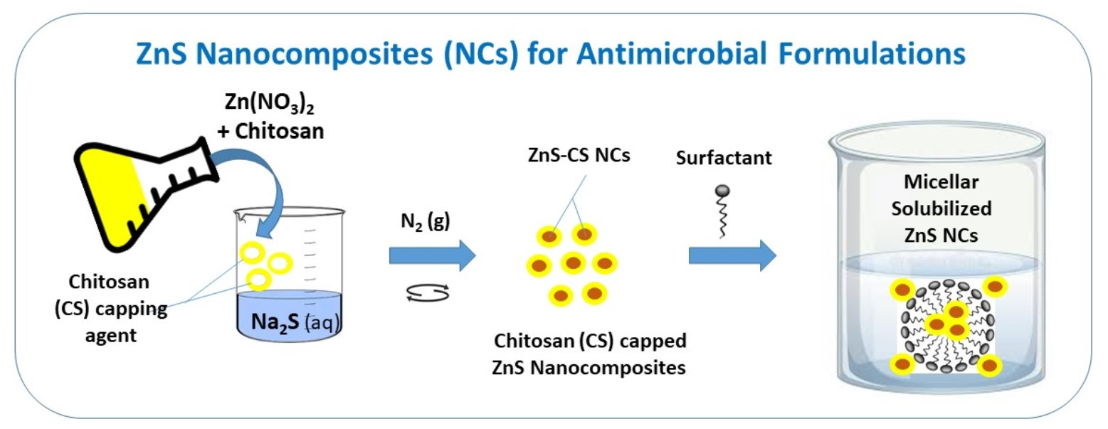
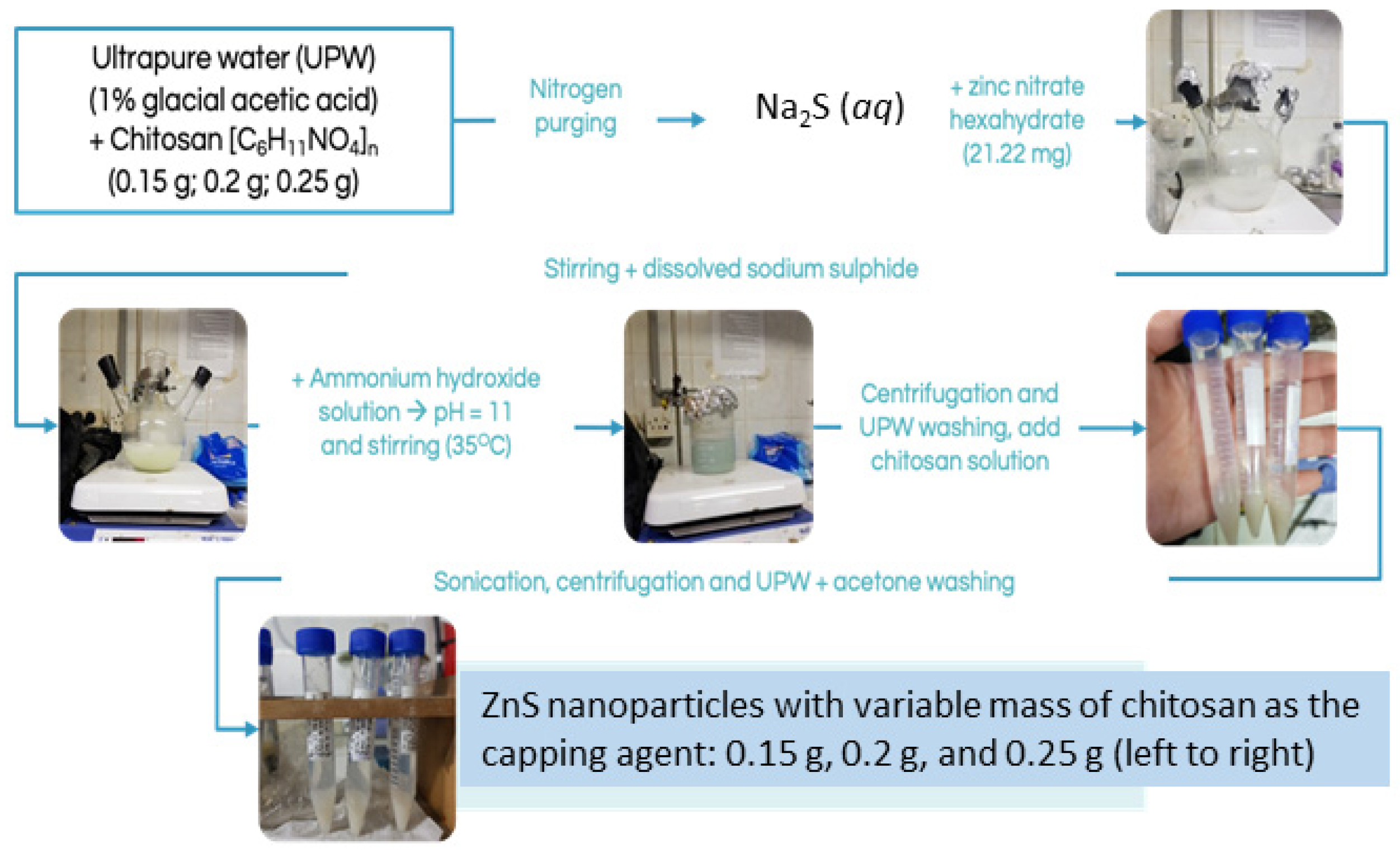
2.2. Synthesis of the Nanocomposites

CS-ZnS NCs were prepared according to an adapted method reported by Lim et al. [5]. CS (0.2 g) was dissolved in 40 mL UPW with 1% glacial acetic acid. The resulting CS solution was stirred while purging with nitrogen gas for a minimum of 30 min. Then, 21.2 mg of Zn(NO3)6H2O was added to the CS solution, followed by stirring for 150 min at 25 °C. The NPs precursor was prepared by dissolving Zn(NO3)2 in Na2S solution in UPW. The Zn(NO3)2 solution was added drop-wise until the color of the mixture changed from yellow to dark brown. The pH of this binary mixture was adjusted to pH 11 by addition of NH4OH, which was then stirred for 60 min at 35 °C. Then, the mixture was centrifuged at 4000 rpm for 15 min and washed several times with UPW before sonication for 90 min. The CS solution was added to the centrifuged mixture of NPs, which was then centrifuged at 4000 rpm for 10 min, followed by washing with UPW and C3H6O, respectively. The solid precipitate of CS-ZnS NCs was stored as a colloidal solution prior to characterization. A similar procedure was employed to prepare NCs with variable amounts of CS (0.15 g and 0.25 g), where the preparative route for the nanocomposites is outlined schematically in Figure 1.

2.3. Characterization

Scanning electron microscopy–energy dispersive X-ray (SEM-EDX) profiles of CS-ZnS NCs were obtained with a Fei Quanta FEG 650 at 15 kV with a fixed magnification (15,000×). Fourier transform infrared (FTIR) spectra were recorded on a Shimadzu IR Prestige-21 over a fixed spectral range (4000–400 cm−1), and X-ray diffraction (XRD) profiles were recorded on a Malvern Panalytical Aeris with Cu Kα radiation (λ = 0.15418 nm).

2.4. Formulation of a Liquid Handwash System

The method for formulation of LH systems employed an adapted method reported by Longe [2], where CS-ZnS NPs were used to produce the CS-ZnS NCs. Here, ALS (10 g) and SDS (10 g) were added to 39 g of UPW, followed by addition of lauryl glucoside (1 g). The mixture was stirred at 35 °C until homogeneous. Then, CS-ZnS NCs (1 g) was added to this mixture, which was stirred to yield a CS-ZnS LH solution, and stored at 25 °C for subsequent characterization. The total surfactant used in the LH formulation was 21%, which includes primary surfactants (ALS, 10% and SDS, 10%), and a secondary surfactant (lauryl glucoside, 1%), as listed in Table 1 below.

2.5. Antibacterial Activity Test

Antibacterial activity tests of LHs employed the agar diffusion test method. E. coli and S. aureus were cultured for 24 h, then 1 dosage of each bacterium was placed into a sterile trypticase soy broth. The resulting suspensions were homogenized, and their transmittance (%T) was measured by using a UV-Vis spectrophotometer (λ = 580 nm) that was adjusted to 25%T. Suspensions of bacteria (0.2 mL) were added drop-wise into each Petri dish. Tryptic soy agar medium (ca. 20–25 mL) was added and then homogenized, and the media were allowed to solidify. The CS-ZnS LH and antibiotic control systems (tetracycline and chloramphenicol) were respectively applied onto the surface of the solidified media, and then allowed to undergo absorption for 1 h. Then, the media were incubated for 18–24 h at 30–35 °C aerobically. The corresponding antibacterial activities of the LH systems were measured and evaluated by observing the clear zone of inhibition formed on the solidified media surface.

2.6. Quality Assessment

An assessment of quality control for the CS-ZnS LH systems was made based on a standard method (SNI 2588:2017) [1], where the roles of various parameters were examined: pH, ethanol-insoluble matter, total active ingredients, and free fatty acids. The LH systems were deemed compliant with the quality standard protocol when the following requirements were met: (a) pH between 4–10; (b) ethanol-insoluble matter > 10 wt.%; (c) total active ingredients < 0.5 wt.%; and (d) free fatty acid < 1 wt.%. The calculation of yield (%Y) and mole formula were estimated according to Equations (1) and (2). By combining Equations (1) and (2) into Equation (3), the yield (%) of CS-ZnS NCs was obtained.
% Y = T o t a l   o f   e l e m e n t   m a s s T o t a l   o f   m a s s s   p r e c u r s o r + c h i t o s a n   × 100 %
m e l e m e n t = A r e l e m e n t M r p r e k u r s o r × m p r e c u r s o r
% Y = m Z n + m S m c h i t o s a n + m Z i n c   s u l p h i d e + m s o d i u m   s u l p h i d e × 100 %
The mass of CS-ZnS NCs (before and after drying) enabled estimation of the water content, Equation (4).
W a t e r   c o n t e n t = M a s s   a f t e r   d r y i n g M a s s   b e f o r e   d r y i n g   M a s s   b e f o r e   d r y i n g
The amount of ethanol-insoluble material (wt.%) was calculated using Equation (5).
U n d i s s o l v e d   m a t e r i a l s   i n   e t h a n o l = b 2 b 0 b 1 × 100
Here, b0 is the weight of the filter paper (g), b1 is the weight of the test sample (g), and b2 is the weight of the filter paper and residue (g).
The free fatty acid was then calculated according to Equation (6):
F r e e   f a t t y   a c i d   ( % ) = 282 × V × N b × 100
where V is the volume of KOH or HCl (mL), N is the normality of KOH or HCl, b is the weight of the test sample (mg), and the value 282 indicates the equivalent weight of oleic acid.

3. Results and Discussion

As outlined above, the objective of this study was to prepare ZnS NPs with CS as the capping agent, along with an evaluation of the utility of the resulting CS-ZnS NCs as unique antibacterial agents for LH applications. In the sections below, the preparation and the structural characterization of the nanocomposites are described based on the FTIR, SEM-EDX, and XRD spectral data. The composition and antibacterial properties of the CS-ZnS NCs are evaluated in the presence of two model bacterial strains, S. aureus and E. coli.

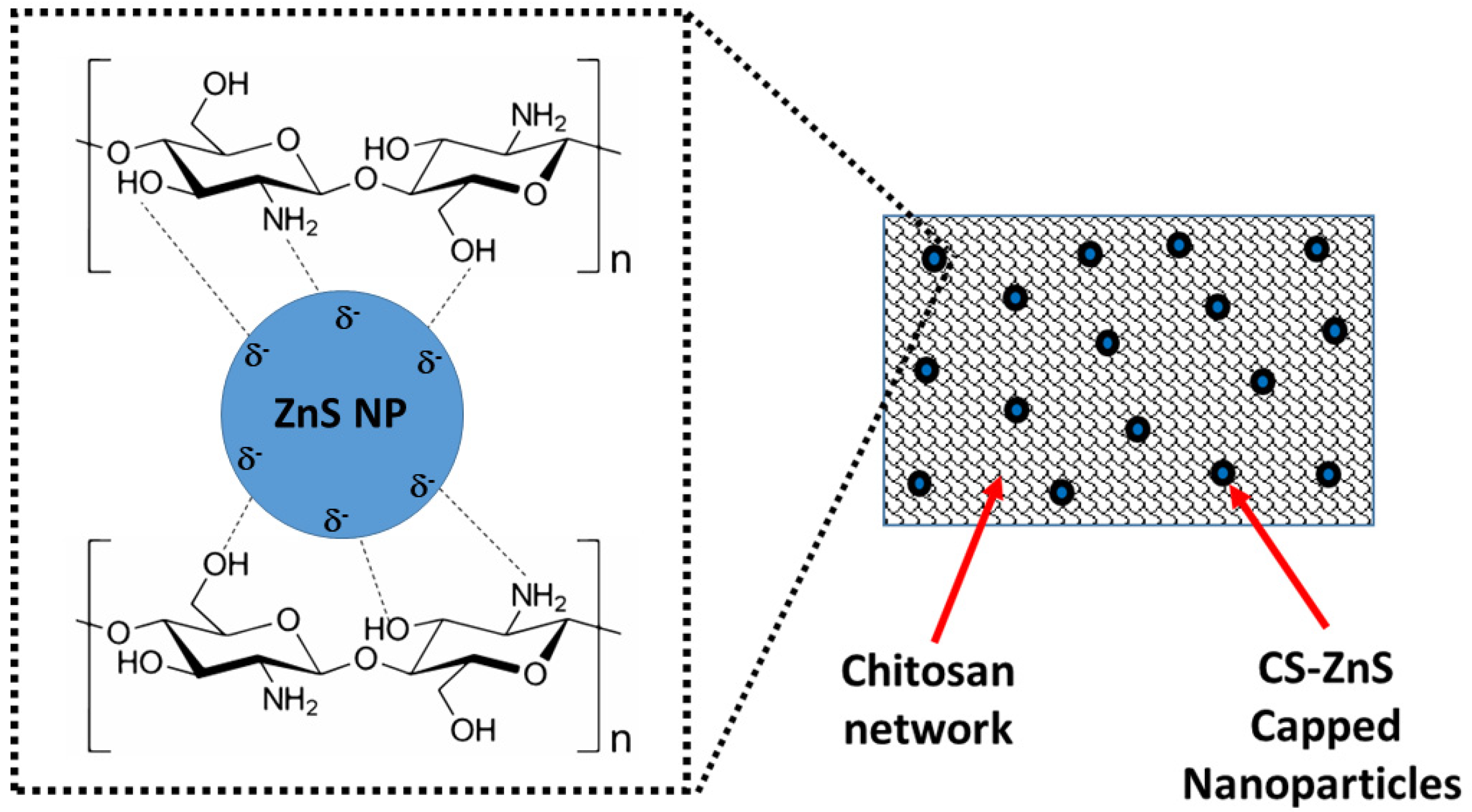
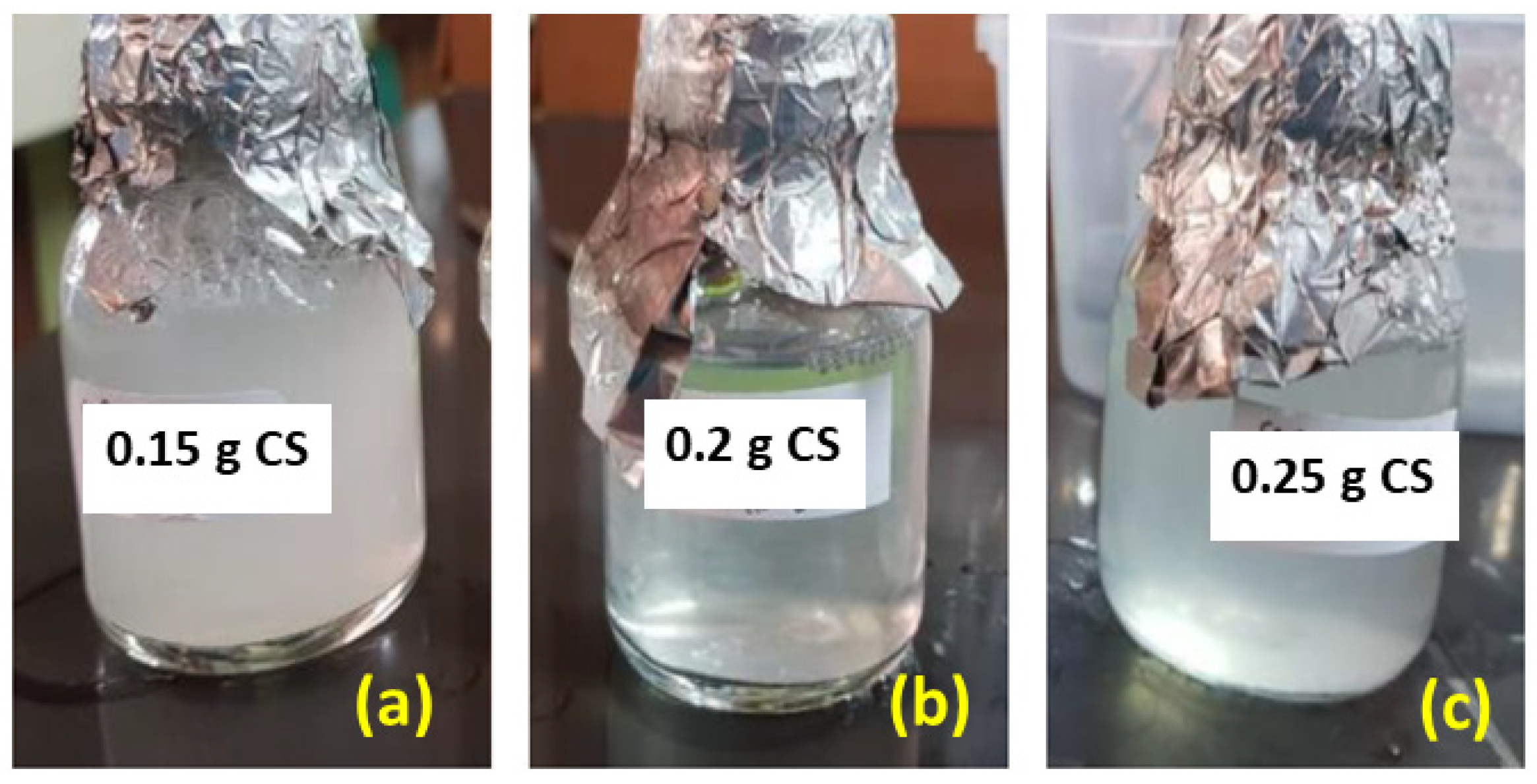
3.1. Preparation of CS-ZnS Nanocomposites

A schematic illustration of the preparation method of CS-ZnS NCs is shown in Figure 1. Based on the estimates according to Equation (3), the yield of CS-ZnS NCs with variable wt.% of the CS capping agent (0.15 to 0.25 g) varied from 27.24% to 31.30%.
The weight of CS-ZnS NCs before and after drying was calculated according to Equation (4), where the water content of CS-ZnS NCs was near 95%. The colloids of CS-ZnS NCs with CS mass variations (a) 0.15 g, (b) 0.2 g, and (c) 0.25 g were kept in the ultrapure water at 25 °C. The dried CS-ZnS NCs are shown in the right hand panels of Figure 2. It should be noted that prior to SEM, XRD, and FTIR characterization, the CS-ZnS NC samples were dried in a vacuum oven at 50–60 °C for 5 to 6 h to enable removal of water, since the presence of residual water content can contribute to variations in the measured spectral parameters for the samples such as crystallinity.

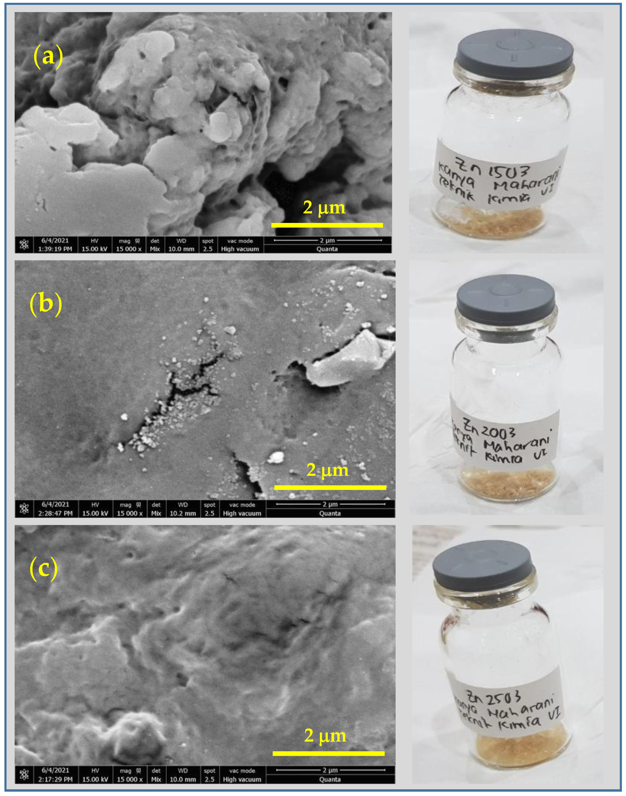
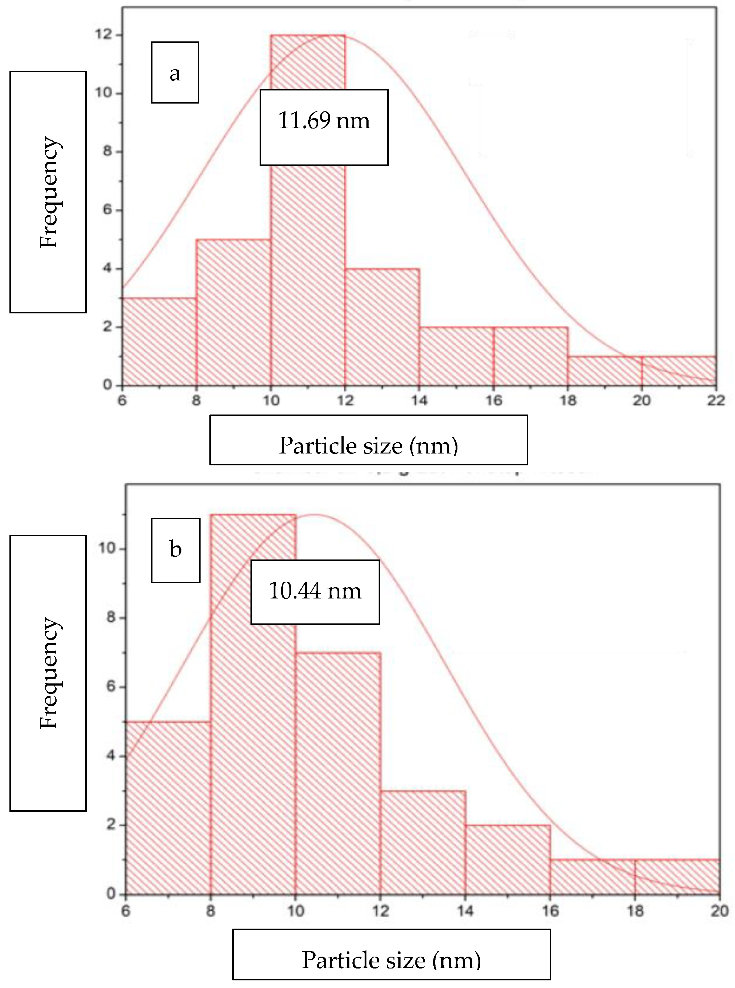
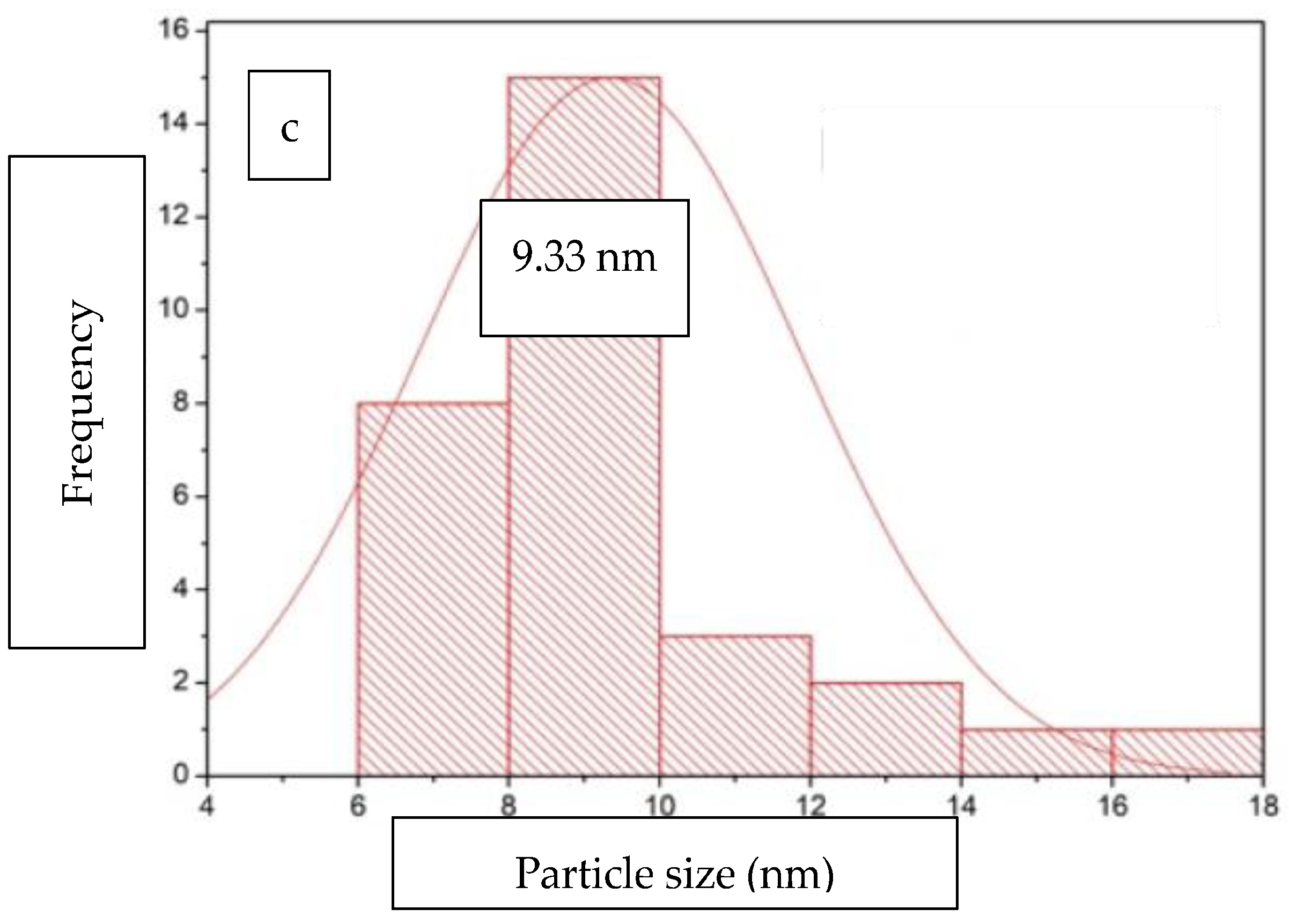
3.2. SEM-EDS Results of CS-ZnS NCs

SEM was used to evaluate the morphology of the prepared CS-ZnS NCs, and the SEM images are shown in the left-hand panel of Figure 2. The corresponding SEM images of CS-ZnS NCs with variable CS content from 0.15 to 0.25 g are shown in Figure 2a–c, where the right-hand panels correspond to digital images of the colloidal NCs after drying. According to the SEM images, the shapes of CS-ZnS NCs are spherical within the layered assembly of chitosan (CS).
The SEM images reveal various textural properties and surface topology of the CS-ZnS NCs. The spherical shape of Cs-ZnS NCs is consistent with the enclathration of the ZnS NPs within the layered network of chitosan (cf. Section 3.4 vide infra). These findings are similar to the morphologies of ZnS nanoparticles reported by Parvaneh et al. [38] and Afsheen et al. [39]. The average particle size and size distribution of CS-ZnS NCs were calculated to determine the effect of the capping agent content on the size parameters. Based on the SEM images, the sizes of selected particles were estimated using ImageJ software and their particle size distribution was deduced using Origin software. The histograms for the particle size distribution show CS-ZnS NCs containing variable chitosan content (0.15 g, 0.20 g, and 0.25 g). The corresponding results reveal that the average particle size was 11.69 nm, 10.44 nm, and 9.33 nm, respectively (cf. Figure 3a–c). The SEM imaging results indicate that the increased content of the capping agent led to a decrease in the particle size with a more even size distribution. Similar effects were observed by Tamrakar et al. [40] for synthetic ZnS NCs with variable levels of capping agent.
EDX elemental analysis of CS-ZnS NCs and their composition (wt.%) are listed in Table 2 (vide infra). The data reveal that the metal precursors in the NCs and elemental species of the capping agent were detected by EDX. For CS-ZnS NCs, the largest elemental content was up to 48–56% for C, followed by a lower content (ca. 29–34%) for O. The N-content ranged between 7–11%, while Zn had the lowest abundance (ca. 6–8%) in the NCs.
However, the presence of elemental S could not be quantitatively measured reliably for both types of NCs, due to the use of Au sputter coating, which was used to produce high-magnification SEM images. The L and M lines of Au coating can interfere with the EDX results by replacing and reducing the content (wt.%) of elements such as sulfur in CS-ZnS NCs [41,42].
Additional SEM images were obtained at greater magnification for the chitosan-capped ZnS nanocomposite, which were prepared at pH 10 (cf. Figure S1, Supplementary Material). Several images of different sample regions (1, 2, and 3) were obtained at variable magnification (60,000× and 150,000×), as compared with the results shown in Figure 2. With reference to Figure S1 (Supplementary Material), the surface of the sample reveals a number of pseudo-spherical structures that range in size from ca. 10 nm to 100 nm across the surface of the sample of regions (1, 2, and 3). The variable particles sizes inferred from the SEM results relate to dispersed NPs and aggregated structures at the sample surface. The light-colored regions reveal the emission of secondary electrons, which are attributed to the ZnS domains near the sample interface. The variation of light- and dark-colored regions is attributed to thinner vs. thicker chitosan shells surrounding the ZnS NPs, in agreement with the loading levels of CS employed in the capping process. This trend concurs with the use of chromium films for contrast image enhancement, along with evidence of Zn species (ca. 0–5 wt.%) based on the EDX analysis for regions 1–3 of the NP domains in Figure S1 (cf. Supplementary Material) and the results listed in Table 2 above.

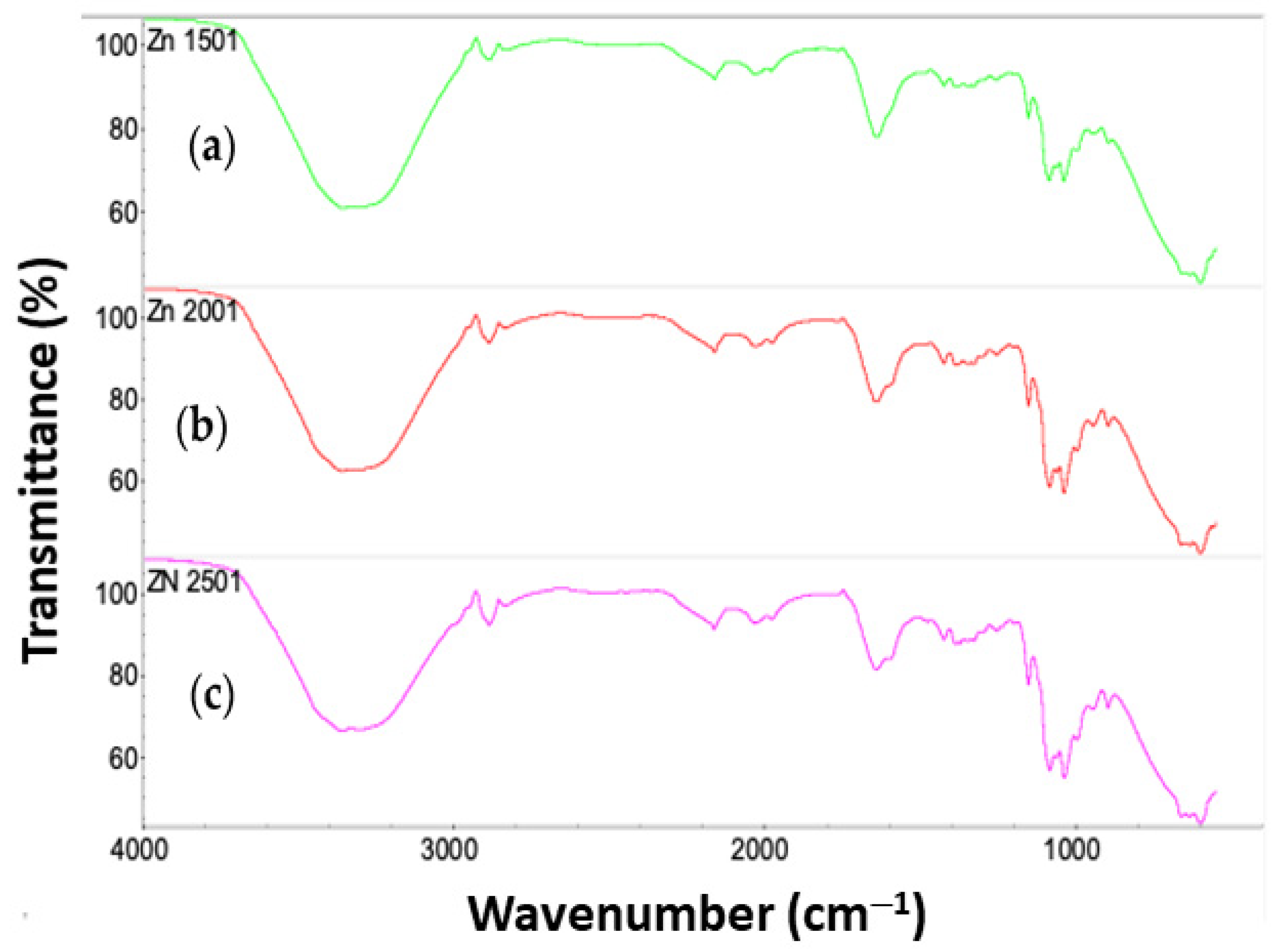
3.3. FTIR Analysis of CS-ZnS NCs

FTIR spectroscopy provides a convenient method to evaluate the presence of functional groups that arise from multicomponent systems, such as metal sulfide NCs that contain chitosan as the capping agent, according to Kusrini et al. [4]. In Figure 4, the FTIR spectra of CS-ZnS NCs were compared to the FTIR spectrum of pure CS to determine whether CS was successfully capped onto the ZnS NPs. The FTIR spectrum of pure CS was compared with the results reported by Abdelhady [43], whereas IR spectral results obtained here for the CS-ZnS NCs are shown in Figure 4.
New IR bands were observed at 655–664 cm−1, 631–633 cm−1, and 594–597 cm−1, which were assigned as Zn-S bonds. These claims were supported by Liu et al. and Zamiri et al., which support that the bands for Zn-S bonds are noted at 450–1000 cm−1 [44,45]. Moreover, the IR bands at 3357–3361 cm−1 were shifted to lower wavenumbers, which validated the interaction between the CS functional groups (–NH2 and –OH) and the donor-acceptor groups of the ZnS NPs [46]. Following that, the maximum intensity of the CS band occurred at 0.25 g, which was the optimum dose to maintain stability and prevent aggregation of CS-ZnS NCs.

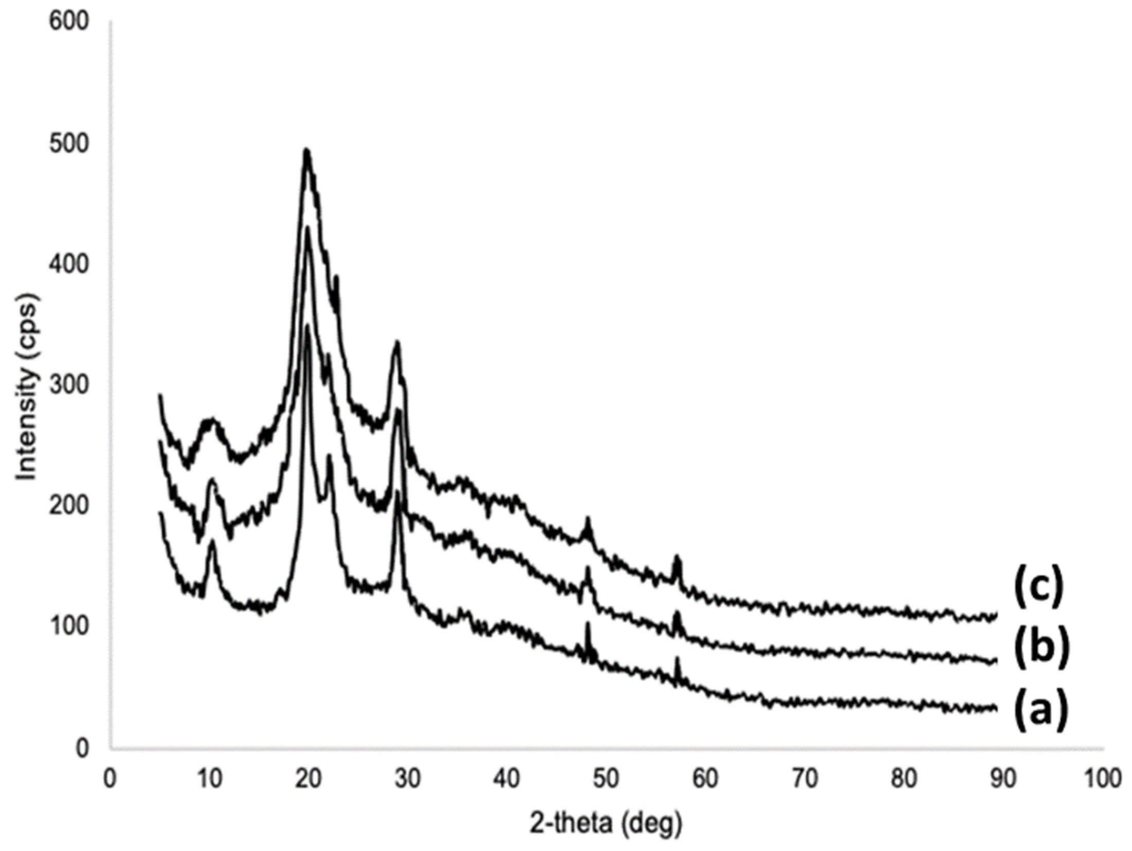
3.4. XRD Analysis of CS-ZnS NCs

XRD provides useful insight for the presence of crystalline domains within multicomponent composites. As indicated above, the CS-ZnS NCs are comprised of an organic biopolymer (chitosan), which is semi-crystalline in nature, along with a crystalline ZnS fraction. The XRD spectra of the NCs are shown in Figure 5. The corresponding XRD spectral analysis of CS-ZnS NCs was referenced to the XRD spectra of ZnS and CS from the ICDD database [47] and the XRD results of Kumar et al. [48]. The XRD peaks at 28.9°, 48.1°, and 57.1° show the presence of ZnS with a cubic crystal structure (cf. Figure 5a). Miller indices of the assigned XRD lines are listed in parentheses: 28.9° (111), 48.1° (220), and 57.1° (311) [47]. The XRD profile shows similar XRD lines near 10° and 20° that are assigned to the signatures of the CS capping agent for the ZnS NCs.
The crystallite size (L) of each NP sample was calculated using the Scherrer equation, as presented in Equation (7). The crystallite size is generally measured by the full-width-at-half-maximum (FWHM) at a smaller peak angle. Therefore, the FWHM for CS-ZnS NPs was taken at an angle of 28.9°. The corresponding XRD patterns of CS-ZnS NCs are presented in Figure 5a–c.
L = K × λ β × cos θ
In Equation (7), K is the Scherrer constant (shape factor of nanoparticles = 0.89), λ is the wavelength = 0.15418 nm (for the copper anode), β is the corrected FWHM, and θ is the Bragg angle. The crystallite sizes of CS-ZnS NPs that contain chitosan as the capping agent were 9.7 nm and 8.5 nm, when CS was 0.15 g and 0.2 g, respectively. The smallest crystallite size was 7.3 nm from ZnS NPs when CS was 0.25 g.
Based on the structural characterization of the NCs reported above, the structure of the CS-ZnS NCs and the corresponding interactions between the groups of the CS capping agent with the ZnS NPs can be inferred from the FTIR and XRD results presented. A conceptual illustration of the interaction between the ZnS NPs and the capping agent is shown in Figure 6.

3.5. Preparation of Liquid Handwash Containing CS-ZnS NCs

Liquid soaps used for hand washing typically contain various components, such as (i) water (40–80%), (ii) surfactant (20–40%), (iii) thickening agent (0.1–1%), (iv) fragrance ingredients (0.1–1%), (v) colorant (<0.01%), pearlizing agent (<1%), and (vi) antibacterial agent(s) (0.5–1%), along with a preservative (<1%). Water is the principal component for the manufacture of liquid soaps, acting as a carrier and solvent for other component materials. The type of water used is generally deionized or distilled water. The presence of positively charged ions (cations) may affect the surface activity and performance of the surfactant. Surfactants or surface-active agents generally are comprised of a hydrophobic hydrocarbon moiety and a hydrophilic functional group. The variation of the hydrophilic and hydrophobic groups of a surfactant serves to alter its properties such as the wettability, emulsifier ability, dispersive ability, or foaming ability. The surfactant is a key component in hand sanitizer because it can increase the water wettability and detergency since surfactants play a key role in contaminant removal during washing. In addition, surfactants can also emulsify, dissolve, or suspend impurities in wastewater. The type of surfactant used in the manufacture of liquid hand sanitizer is often the same as surfactants that are commonly used in household products. In general, liquid soap used for hand washing contains at least two types of surfactants: (a) a primary surfactant (20–40%) that plays a role in foam formation and cleansing of contaminants; and (b) a secondary surfactant (1–10%) that functions to increase the softness of the foam formed so that the texture has greater appeal during its application. The addition of a thickening agent, better known as a thickener, serves to increase the viscosity of liquid hand soap and also plays a role in controlling the flow rate (viscosity) of the detergent formulation. Thickening agents include anionic surfactants, gums, starches, or polymeric materials. Fragrance ingredients are used to provide natural and/or synthetic chemical fragrant compounds for liquid hand soap. The addition of these additives is important for increasing the consumer appeal of the final product. The fragrant selection is based on the level of compatibility with the surfactant employed. Colorants and pearlizing agents are two useful additives for improving the physical appearance of liquid hand soap. The difference between the two is that dyes are added to alter the color, whereas pearlizing agents vary the shine of the resulting formulation. The types of dyes that are commonly used are similar to those employed in cosmetics or medicines. The pearlizing agent often used is a fatty alcohol or a titanium coated with mica. Components such as the antibacterial agent(s) and preservative(s) are also very important in the formulation of liquid soap, which are used to combat pathogenic bacteria by reducing metabolic activity and/or neutralization of bacteria [4]. The addition of antibacterial agents to liquid soap imparts disinfectant properties because they prevent bacterial growth or kill bacteria upon contact with the skin. In general, triclosan is a commonly employed antibacterial agent in liquid hand soap, which may also contain other auxiliary antibacterial agents [3]. Since these compounds are not able to protect the product from other microbes such as fungi, preservatives are added to liquid soap to prevent the growth of these microbes and to extend the shelf life of hand soap products.
LH solutions with the addition of CS-ZnS NCs as antibacterial agents were prepared using a mixture of distilled water, surfactant, and antibacterial agent. The largest composition of this formula was dominated by water, up to 78% (wet state) or 78.95% (dry state), followed by surfactants, as much as 21%, and antibacterial agents, 1% (wet) or 0.05% (dry). This composition was adapted to the general formulation of LH solutions. The procedure for making such formulations started with mixing various surfactants into distilled water, where ALS and SDS were the primary surfactants added to remove dirt and foam. Then, the secondary surfactant (lauryl glucoside) was used to increase the softness of the foam. The selection of anionic surfactants as primary surfactants in this category is related to their wide use in commercial cleaning products and their cleaning effectiveness. The nonionic surfactant lauryl glucoside was used as a secondary surfactant because it has a synergistic effect with anionic surfactants and favorable biocompatibility due to its low irritation potential. Finally, CS-ZnS NCs were added as antibacterial agents in the formulation of LH solutions. An illustration of the prepared LH solutions with CS-ZnS containing variable CS (0.15 g, 0.2 g, and 0.25 g) is shown in Figure 7.

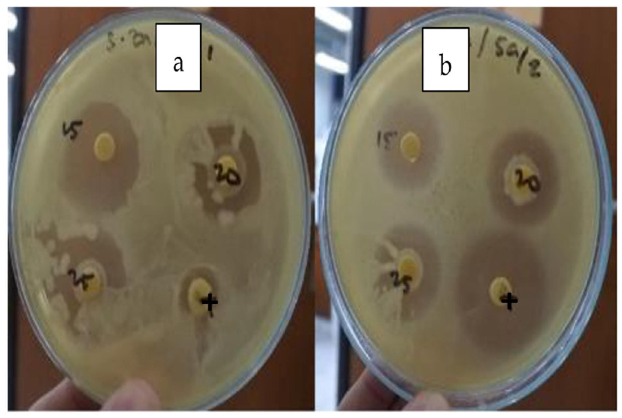
3.6. Antibacterial Activity of CS-ZnS LHs

The inhibition zone is a region of medium where bacteria are unable to grow in the agar diffusion test due to the presence of agents that display antimicrobial activity. Zones of inhibition diameters with less than 10 mm are referred to as weak, zones of inhibition ranging from 10 to 16 mm are considered moderate, and zones of inhibition higher than 16 mm are considered as active [49]. The zone of inhibition diameters of CS-ZnS NCs are shown in Figure 8.
Based on the data obtained from agar diffusion tests, all of the formulated CS-ZnS NCs in LH solutions had strong antibacterial activity against S. aureus with an inhibition diameter zone between 16.9 to 19.1 mm. According to Gul et al. [49], all LH formulations for the CS-ZnS systems are active as antibacterial agents. The detailed average zones of inhibition for the CS-ZnS LH systems are presented in Table 2 and Figure 8a,b. Furthermore, the average inhibition zone results reveal that the antibacterial activities of these CS-ZnS LH systems increased as the content of the capping agent increased. This trend concurs with the results reported by Giridhar et al. [50]. By comparison, the LH systems that contain CS-ZnS NCs did not inhibit the growth of E. coli bacteria. Omar et al. [51] reported that the ZnS NPs with a Schiff-base capping agent did not have antibacterial activity against E. coli due to the high minimum concentration required. The average inhibition zones of CS-ZnS LHs against S. aureus and E. coli are summarized in Table 3.
By comparison, Table 4 shows the inhibition zone results for Eu(NO3)3, Tb(NO3)3, Zn(NO3)2, CS, and tetracycline. Tetracycline is an antibiotic that serves as a positive control herein against E. coli and S. aureus [4]. It is important to note that the inhibition zone diameters for Zn(NO3)2 against E. coli and S. aureus are 22.1 mm and 32 mm, respectively, which are higher than the rare earth metal nitrate salts or even the antibiotic control (tetracycline). The inhibition zones of Zn(NO3)2 are higher than 16 mm [49], revealing that the Zn salt is a strong antibacterial agent against E. coli and S. aureus. As summarized in Table 3, the formulated CS-ZnS LH systems are mainly active against S. aureus, and are largely inactive against E. coli. This is reasonable, as CS only shows antibacterial activity against S. aureus, with the inhibition zone being 6.8 mm, which is categorized as a weak antibacterial agent [4].

3.7. Formulation of Liquid Handwash and Quality Assessment of the CS-ZnS Systems

The compositions of the LH systems are presented in Table 1 (cf. Section 2.4), where the SNI 2588:2017 method was used as the standard for quality assessment of the handwash solutions. pH is a necessary parameter to consider since irritation and skin dehydration can happen if the pH conditions of CS-ZnS LHs are too basic [52]. Ethanol-insoluble matter (>10%) was used to determine the amount of additives added to CS-ZnS LHs. The total active ingredients indicate the total substances that produce chemical or biological effects for CS-ZnS LH systems. Eventually, if free fatty acid content in CS-ZnS LHs becomes too high, the handwash formulations would become rancid due to oxidation.
The quality assessment showed that the pH of the CS-ZnS LH system was neutral, and covered a narrow pH range from 6.6 to 7. There was only 0.01% ethanol-insoluble material in CS-ZnS LHs, where the only additive was CS as the antibacterial agent. The total active ingredients in the CS-ZnS LH systems exceeded 99% due to the presence of surfactants as active ingredients. CS-ZnS LHs had a very small free fatty acid content (ca. 0.01%) when comparing the results with the quality requirements of liquid hand soap set forth by the National Standardization Agency. In addition, all of the formulated CS-ZnS LHs meet the SNI 2588:2017 standard requirements (cf. Table 5), which reveals their suitability for future applications. The detailed results regarding the quality assessment of LH systems that contain CS-ZnS NCs are summarized in Table 5. The difference in the content of the capping agent that was added to the NPs from 0.15 to 0.25 g did not yield a significant effect on the total active ingredients. This finding also indicated that CS can be used as a capping agent due to its unique ability to undergo a complex formation with metal sulfide NPs [53,54]. Since CS has an established acceptance for its biocompatibility [55,56], the CS-ZnS NCs would be suitable for biomedical applications.

4. Conclusions

In summary, chitosan-capped zinc sulfide (CS-ZnS) NCs were successfully prepared and used as antibacterial agents for the formulation of liquid handwash systems. ZnS NPs were synthesized through a wet-chemical method with chitosan (CS) as the capping agent at variable CS composition. The formation of ZnS NPs and CS-ZnS NCs were supported by SEM, XRD, and FTIR spectral results. In CS-ZnS NCs, the ZnS NPs were spherical with an average ZnS NP size in the range of 9.33 nm to 11.9 nm, deposited in layers of CS, as further evidenced by SEM images at higher resolution. The ZnS NPs were cubic with a nanoscale crystallite size (ca. 7.3–9.7 nm). CS-ZnS LH systems were successfully formulated by combining surfactants with the CS-capped ZnS NPs. The antibacterial activity of the formulated CS-ZnS LH systems were screened against E. coli and S. aureus strains. All of the formulated CS-ZnS LHs with different amounts of CS showed favorable antibacterial activity against S. aureus with the diameter of the inhibition zone in the range of 16.9 mm to 19.1 mm. The quality of formulated CS-ZnS LHs was assessed based on their agar diffusion test characteristics, which met the standards set by the National Standardization Agency. Further research on CS-ZnS NCs is proposed to develop advanced formulations with antimicrobial properties for other types of microorganisms to address other disinfectant applications.

Supplementary Materials

The following supporting information can be downloaded at: https://www.mdpi.com/article/10.3390/jcs7020052/s1. Figure S1 outlines SEM results at variable magnification in various sample domains (region 1, 2, and 3) for the CS-ZnS NPs (pH 10).

Author Contributions

Conceptualization, E.K. and K.M.P.; methodology, E.K.; formal analysis, D.P.M., E.K. and A.U.; investigation and validation, A.U.; resources, L.D.W.; data curation, M.S.; writing—original draft preparation, K.M.P.; writing—review paper, L.D.W.; visualization, M.S.; supervision, E.K.; project administration, E.K.; funding acquisition, E.K. All authors have read and agreed to the published version of the manuscript.

Funding

The authors greatly acknowledge the World Class Professor (WCP) Program 2022, No. 3252/E4/DT.04.03/2022 and Publikasi Terindeks Internasional (PUTI) Q2 grant 2022—2023 No. NKB-688/UN2.RST/HKP.05.00/2022.

Institutional Review Board Statement

Not applicable.

Informed Consent Statement

Not applicable.

Data Availability Statement

Not applicable.

Acknowledgments

The authors greatly acknowledge Kementerian Pendidikan, Kebudayaan, Riset, Dan Teknologi Direktorat Jenderal Pendidikan Tinggi Riset Dan Teknologi through the World Class Professor (WCP) Program 2022, No. 3252/E4/DT.04.03/2022. The authors also thank to Universitas Indonesia through the Publikasi Terindeks Internasional (PUTI) Q2 grant 2022–2023 No: NKB-688/UN2.RST/HKP.05.00/2022 for research funding and providing facilities to conduct this research. LDW acknowledges Eiko Kawamura for technical support with SEM imaging at the WCV Image Centre (University of Saskatchewan).

Conflicts of Interest

The authors declare no conflict of interest.

References

  1. SNI 2588:2017; Sabun Cair Pembersih Tangan (Liquid Handwash). Badan Standardisasi Nasional (National Standardization Agency): Jakarta, Indonesia, 2017.
  2. Longe, J. How Products Are Made: An Illustrated Guide to Product Manufacturing, 4th ed.; Gale Research: Detroit, MI, USA, 1998. [Google Scholar]
  3. Ullah, H.; Ali, S. Classification of Antibacterial Agents and Their Functions. In Antibacterial Agents; Kumavath, R.N., Ed.; IntechOpen: Kerala, India, 2017. [Google Scholar] [CrossRef]
  4. Kusrini, E.; Safira, A.I.; Usman, A.; Prasetyanto, E.A.; Nugrahaningtyas, K.D.; Santosa, S.J.; Wilson, L.D. Nanocomposites of Terbium Sulfide Nanoparticles with a Chitosan Capping Agent for Antibacterial Applications. J. Compos. Sci. 2023, 7, 39. [Google Scholar] [CrossRef]
  5. Lim, M.; Shahri, N.; Taha, H.; Mahadi, A.; Kusrini, E.; Lim, J.; Usman, A. Biocompatible chitin-encapsulated CdS quantum dots: Fabrication and antibacterial screening. Carbohydr. Polym. 2021, 260, 117806. [Google Scholar] [CrossRef] [PubMed]
  6. Thomas, D.; Lee, H.O.; Santiago, K.C.; Pelzer, M.; Kuti, A.; Jenrette, E.; Bahoura, M. Rapid Microwave Synthesis of Tunable Cadmium Selenide (CdSe) Quantum Dots for Optoelectronic Applications. J. Nanomater. 2020, 2020, 5056875. [Google Scholar] [CrossRef]
  7. Li, G.P.; Zhai, J.F.; Li, D.; Fang, X.N.; Jiang, H.; Dong, Q.Z.; Wang, E.K. One-pot synthesis of monodispersed ZnS nanospheres with high antibacterial activity. J. Mater. Chem. 2010, 20, 9215–9219. [Google Scholar] [CrossRef]
  8. Neelgund, G.M.; Oki, A.; Luo, Z. Antimicrobial activity of CdS and Ag2S quantum dots immobilized on poly(amidoamine) grafted carbon nanotubes. Colloids Surf. B: Biointerfaces 2012, 100, 215–221. [Google Scholar] [CrossRef]
  9. Li, H.; Li, M.; Wan, Y.S.; Lelkes, P.I.; Shih, W.H. Cytotoxicity Test of Water Soluble ZnS and CdS Quantum Dots. J. Nanosci. Nanotechnol. 2011, 11, 3543–3551. [Google Scholar] [CrossRef] [PubMed]
  10. Geng, B.Y.; Liu, X.W.; Du, O.B.; Wei, X.W.; Zhang, L.D. Structure and optical properties of periodically twinned ZnS nanowires. Appl. Phys. Lett. 2006, 88, 163104. [Google Scholar] [CrossRef]
  11. Wang, L.; Dai, J.; Liu, X.; Zhu, Z.; Huang, X.; Wu, P. Morphology-controlling synthesis of ZnS through a hydrothermal/solvothermal method. Ceram. Int. 2012, 38, 1873–1878. [Google Scholar] [CrossRef]
  12. Li, Z.; Chen, P.; Xu, X.; Ye, X.; Wang, J. Preparation of chitosan–sodium alginate microcapsules containing ZnS nanoparticles and its effect on the drug release. Mater. Sci. Eng. C 2009, 7, 2250–2253. [Google Scholar] [CrossRef]
  13. Mamiyev, Z.Q.; Balayeva, N.O. Optical and structural studies of ZnS nanoparticles synthesized via chemical in situ technique. Chem. Phys. Lett. 2016, 646, 69–74. [Google Scholar] [CrossRef]
  14. Donne, A.L.; Jana, S.K.; Banerjee, S.; Basu, S.; Binetti, S. Optimized luminescence properties of Mn doped ZnS nanoparticles for photovoltaic applications. J. Appl. Phys. 2013, 113, 014903. [Google Scholar] [CrossRef]
  15. La Porta, F.D.A.; Nogueira, A.E.; Gracia, L.; Pereira, W.S.; Botelho, G.; Mulinari, T.A.; Andrés, J.; Longo, E. An experimental and theoretical investigation on the optical and photocatalytic properties of ZnS nanoparticles. J. Phys. Chem. Solids 2017, 103, 179–189. [Google Scholar] [CrossRef]
  16. Mamiyev, Z.; Balayeva, N.O. Metal Sulfide Photocatalysts for Hydrogen Generation: A Review of Recent Advances. Catalysts 2022, 12, 1316. [Google Scholar] [CrossRef]
  17. Sarangi, B.; Mishra, S.P.; Behera, N. Advances in green synthesis of ZnS nanoparticles: An overview. Mater. Sci. Semicond. Process. 2022, 147, 106723. [Google Scholar] [CrossRef]
  18. Xu, J.; Li, Y. Formation of zinc sulfide nanorods and nanoparticles in ternary W/O microemulsions. J. Colloid Interface Sci. 2003, 259, 275–281. [Google Scholar] [CrossRef]
  19. Hoa, T.T.Q.; Vu, L.V.; Canh, T.D.; Long, N.N. Preparation of ZnS nanoparticles by hydrothermal method. J. Phys. Conf. Ser. 2009, 187, 012081. [Google Scholar] [CrossRef]
  20. Sánchez-Lopez, J.C.; Fernández, A. The gas-phase condensation method for the preparation of quantum-sized ZnS nanoparticles. Thin Solid Film. 1998, 317, 497–499. [Google Scholar] [CrossRef]
  21. Hedayati, K.; Zendehnam, A.; Hassanpour, F. Fabrication and characterization of zinc sulfide nanoparticles and nanocomposites prepared via a simple chemical precipitation method. J. Nanostruct. 2016, 6, 207–212. [Google Scholar] [CrossRef]
  22. Lenggoro, I.W.; Okuyama, K.; de la Morat, J.F.; Tohge, N. Preparation of ZnS nanoparticles by electrospray pyrolysis. J. Aerosol Sci. 2000, 31, 121–136. [Google Scholar] [CrossRef]
  23. Rahimi-Nasarabadi, M. Electrochemical synthesis and characterization of zinc sulfide nanoparticles. J. Nanostruct. 2014, 4, 211–216. [Google Scholar] [CrossRef]
  24. Thottoli, A.K.; Achuthanunni, A.K. Effect of polyvinyl alcohol concentration on the ZnS nanoparticles and wet chemical synthesis of wurtzite ZnS nanoparticles. J. Nanostruct. Chem. 2013, 3, 31. [Google Scholar] [CrossRef]
  25. Wang, L.P.; Hong, G.Y. A new preparation of zinc sulfide nanoparticles by solid state method at low temperature. Mater. Res. Bull. 2000, 35, 695–701. [Google Scholar] [CrossRef]
  26. Gan, Y.; Wang, L.; Wang, R. Creation of ZnS nanoparticles by laser ablation in water. Appl. Phys. A 2016, 122, 104. [Google Scholar] [CrossRef]
  27. Biswas, S.; Kar, S.; Chaudhuri, S. Synthesis and characterization of zinc sulfide nanostructures. Synth. React. Inorg. Metal-Org. Nano-Metal Chem. 2006, 36, 33–36. [Google Scholar] [CrossRef]
  28. Wang, G.Z.; Geng, B.Y.; Huang, X.M.; Wang, Y.W.; Li, G.H.; Zhang, L.D. A convenient ultrasonic irradiation technique for in situ synthesis of zinc sulfide nanocrystallites at room temperature. Appl. Phys. Mater. Sci. Process. 2003, 77, 933–936. [Google Scholar] [CrossRef]
  29. Tolia, J.V.; Chakraborty, M.; Murthy, Z.V.P. Photocatalytic degradation of malachite green dye using doped and undoped ZnS nanoparticles. Pol. J. Chem. Technol. 2012, 14, 16–21. [Google Scholar] [CrossRef]
  30. Lalithadevi, B.; Rao, K.M.; Ramananda, D. Investigations on structural and optical properties of starch capped ZnS nanoparticles synthesized by microwave irradiation method. Chem. Phys. Lett. 2018, 700, 74–79. [Google Scholar] [CrossRef]
  31. Yun, Y.-H.; Youn, H.-G.; Shin, J.-Y.; Yoon, S.-D. Preparation of functional chitosan-based nanocomposite films containing ZnS nanoparticles. Int. J. Biol. Macromol. 2017, 104, 1150–1157. [Google Scholar] [CrossRef]
  32. Bahri, S.; Homaei, A.; Mosaddegh, E. Zinc sulfide-chitosan hybrid nanoparticles as a robust surface for immobilization of Sillago sihama α-amylase. Colloids Surf. B Biointerfaces 2022, 218, 112754. [Google Scholar] [CrossRef]
  33. Zhang, X.; Liu, M.; Liu, H.; Zhang, S. Low-toxic Ag2S quantum dots for photoelectrochemical detection glucose and cancer cells. Biosens. Bioelectron. 2014, 56, 307–312. [Google Scholar] [CrossRef]
  34. Kumar, D.S.; Kumar, B.J.; Mahesh, H.M. Chapter 3: Quantum Nanostructures (QDs): An Overview. Synth. Inorg. Nanomater. 2018, 59–88. [Google Scholar] [CrossRef]
  35. Valizadeh, A.; Mikaeili, H.; Samiei, M.; Farkhani, S.; Zarghami, N.; Kouhi, M. Quantum dots: Synthesis, bioapplications, and toxicity. Nanoscale Res. Lett. 2012, 7, 480. [Google Scholar] [CrossRef]
  36. Green, M. The nature of quantum dot capping ligands. J. Mater. Chem. 2010, 20, 5797. [Google Scholar]
  37. Şenel, S.; McClure, S. Potential applications of chitosan in veterinary medicine. Adv. Drug Deliv. Rev. 2004, 56, 1467–1480. [Google Scholar] [CrossRef] [PubMed]
  38. Parvaneh, I.; Samira, S.; Mohsen, N. Characterization of ZnS nanoparticles synthesized by co-precipitation method. Chin. Phys. B 2015, 24, 046104. [Google Scholar]
  39. Afsheen, S.; Naseer, H.; Iqbal, T.; Abrar, M.; Bashir, A.; Ijaz, M. Synthesis and characterization of metal sulphide nanoparticles to investigate the effect of nanoparticles on germination of soybean and wheat seeds. Mater. Chem. Phys. 2020, 252, 123216. [Google Scholar] [CrossRef]
  40. Tamrakar, R.; Ramrakhiani, M.; Chandra, B. Effect of Capping Agent Concentration on Photophysical Properties of Zinc Sulfide Nanocrystals. Open Nanosci. J. 2008, 2, 12–16. [Google Scholar] [CrossRef]
  41. Luyk, E. Sputter Coating for SEM: How This Sample Preparation Technique Assists Your Imaging [Internet]. ThermoFisher. 2019. Available online: https://www.thermofisher.com/blog/microscopy/sputter-coating-for-sem-how-this-sample-preparation-technique-assists-your-imaging/ (accessed on 6 November 2022).
  42. Rave Scientific. Proper Target Material Selection When Coating Samples Using an SEM Sputter Coater [Internet]. Rave Scientific. 2021. Available online: https://ravescientific.com/education/37-proper-target-material-selection-when-coating-samples-using-an-sem-sputter-coater (accessed on 6 November 2022).
  43. AbdElhady, M.M. Preparation and Characterization of Chitosan/Zinc Oxide Nanoparticles for Imparting Antimicrobial and UV Protection to Cotton Fabric. Int. J. Carbohydr. Chem. 2012, 2012, 840591. [Google Scholar] [CrossRef]
  44. Liu, L.; Dai, J.; Zhao, T.; Guo, S.; Hou, D.; Zhang, P. A novel Zn(II) dithiocarbamate/ZnS nanocomposite for highly efficient Cr6+ removal from aqueous solutions. RSC Adv. 2017, 7, 35075–35085. [Google Scholar] [CrossRef]
  45. Zamiri, R.; Abbastabar Ahangar, H.; Zakaria, A.; Zamiri, G.; Shabani, M.; Singh, B.; Ferreira, J. The structural and optical constants of Ag2S semiconductor nanostructure in the Far-Infrared. Chem. Cent. J. 2015, 9, 28. [Google Scholar] [CrossRef]
  46. Mansur, H.; Mansur, A. Nano-photocatalysts based on ZnS quantum dots/chitosan for the photodegradation of dye pollutants. IOP Conf. Ser. Mater. Sci. Eng. 2015, 76, 012003. [Google Scholar] [CrossRef] [Green Version]
  47. Gates-Rector, S.; Blanton, T. The Powder Diffraction File: A quality materials characterization database. Powder Diffr. 2019, 34, 352–360. [Google Scholar] [CrossRef]
  48. Kumar, S.; Dutta, P.; Koh, J. A physico-chemical and biological study of novel chitosan–chloroquinoline derivative for biomedical applications. Int. J. Biol. Macromol. 2011, 49, 356–361. [Google Scholar] [CrossRef] [PubMed]
  49. Gul, Z.; Akbar, A.; Leghari, S.K. Elucidating therapeutic and biological potential of berberis baluchistanica ahrendt bark, leaf, and root extracts. Front. Microbiol. 2022, 13, 823673. [Google Scholar] [CrossRef]
  50. Giridhar, M.; Naik, H.S.B.; Prabhakar, M.C.; Naik, M.M.; Ballesh, N.; Mahesh, M.C. Synthesis, characterization and antibacterial activity of water-soluble dye-capped zinc sulphide nanoparticles from waste Zn–C battery. Bull. Mater. Sci. 2021, 44, 6. [Google Scholar] [CrossRef]
  51. Omar, M.; Sanif, M.; Ali, N.; Hamid, M.; Taha, H.; Mahadi, A. Synthesis of Schiff Base Encapsulated ZnS Nanoparticles: Characterization and Antibacterial Screening. Int. J. Technol. 2020, 11, 1309. [Google Scholar] [CrossRef]
  52. Tarun, J.; Susan, V.; Susan, J.; Suria, J.; Criton, S. Evaluation of pH of bathing soaps and shampoos for skin and hair care. Indian J. Dermatol. 2014, 59, 442. [Google Scholar] [CrossRef]
  53. Usman, A.; Kusrini, E.; Widiantoro, A.B.; Hardiya, E.; Abdullah, N.A.; Yulizar, Y. Fabrication of Chitosan Nanoparticles Containing Samarium Ion Potentially Applicable for Fluorescence Detection and Energy Transfer. Int. J. Technol. 2018, 9, 1112–1120. [Google Scholar] [CrossRef]
  54. Kusrini, E.; Shiong, N.S.; Harahap, Y.; Yulizar, Y.; Dianursanti Arbianti, R.; Pudjiastuti, A.R. Effects of Monocarboxylic Acids and Potassium Persulfate on Preparation of Chitosan Nanoparticles. Int. J. Technol. 2015, 6, 11–21. [Google Scholar] [CrossRef]
  55. Kusrini, E.; Ayuningtyas, K.; Mawarni, D.P.; Wilson, L.D.; Sufyan, M.; Rahman, A.; Prasetyanto, Y.E.A.; Usman, A. Micro-structured Materials for the Removal of Heavy Metals using a Natural Polymer Composite. Int. J. Technol. 2021, 12, 275–286. [Google Scholar] [CrossRef]
  56. Kusrini, E.; Wu, S.; Susanto, B.H.; Lukita, M.; Gozan, M.; Hans, M.D.; Rahman, A.; Degirmenci, V.; Usman, A. Simultaneous Absorption and Adsorption Processes for Biogas Purification using Ca(OH)2 Solution and Activated Clinoptilolite Zeolite/Chitosan Composites. Int. J. Technol. 2019, 10, 1243–1250. [Google Scholar] [CrossRef] [Green Version]
Figure 1. The preparation method of CS-ZnS NCs with variable CS content from 0.15 to 0.25 g using the wet-chemical method with bottom-up approach.
Jcs 07 00052 g001
Figure 2. SEM images of CS-ZnS nanocomposites with chitosan (a) 0.15 g, (b) 0.2 g, and (c) 0.25 g. The image magnification is 15,000×. The right-hand panel corresponds to a digital image of the dried of CS-ZnS NCs.
Jcs 07 00052 g002
Figure 3. The corresponding particle size distribution plots were obtained from ImageJ software (n = 30 samples) analysis of SEM results for CS-ZnS NCs that contain variable CS loading: (a) 0.15 g, (b) 0.2 g, and (c) and 0.25 g.
Jcs 07 00052 g003aJcs 07 00052 g003b
Figure 4. FTIR spectra of CS-ZnS NCs with variable chitosan content: (a) 0.15 g (Zn 1501), (b) 0.2 g (Zn 2001), and (c)0.25 g (Zn 2501).
Jcs 07 00052 g004
Figure 5. XRD patterns of CS-ZnS NCs with variable weight of chitosan: (a) 0.15 g CS, (b) 0.2 g CS, and (c) 0.25 g CS.
Jcs 07 00052 g005
Figure 6. Conceptual illustration of the structure and electrostatic interactions between the polar functional groups of chitosan (CS) and the donor-acceptor groups of a zinc sulfide nanoparticle (ZnS NP) with the capping agent (CS). The CS shell surrounding the ZnS NPs (blue spheres) is shown by the use of bold black lines, whereas the microporous CS network is shown by the narrow black wavy lines.
Jcs 07 00052 g006
Figure 7. Illustration of liquid handwash solutions for CS-ZnS NCs in formulations with variable CS content: (a) 0.15 g CS, (b) 0.2 g CS, and (c) 0.25 g CS.
Jcs 07 00052 g007
Figure 8. Antibacterial activities of CS-ZnS NCs in LH solutions toward S. aureus, where 15 denotes CS with 0.15 g, 20 denotes CS with 0.20 g, 25 denotes CS with 0.25 g, and “+” is tetracycline as a positive control. (a,b) are duplicate measurements for the determination of antibacterial activities of CS-ZnS LH solutions.
Jcs 07 00052 g008
Table 1. Composition of CS-ZnS liquid handwash solutions.
ComponentComposition
(wt.%)
Solvent (distilled water)78
Primary surfactant (ALS)10
Primary surfactant (SDS)10
Secondary surfactant (LG)1
Antibacterial agent (CS-ZnS NCs)1
Abbreviations: ammonium lauryl sulfate (ALS); sodium dodecyl sulfate (SDS); and lauryl glucoside (LG).
Table 2. Weight content of each element based on EDX measurements in CS-ZnS nanocomposites with different weight content of capping agent chitosan (CS varies at 0.15 g, 0.2g, and 0.25 g).
Weight Content (%)
ElementCS-ZnS NCs
0.15 g CS0.2 g CS0.25 g CS
C48.6%52.3%55.4%
O34.0%29.6%30.0%
N11.1%10.5%7.6%
Zn 6.3%7.6%7.0%
S≈0%≈0%≈0%
Table 3. Average inhibition zone of CS-ZnS LHs against S. aureus and E. coli.
Liquid Handwash
Solutions
Average Zone of Inhibition Diameter (mm)
S. aureusE. coli
CS-ZnS LHs with 0.15 g CS16.90.00
CS-ZnS LHs with 0.2 g CS18.30.00
CS-ZnS LHs with 0.25 g CS19.10.00
Tetracycline as control22.717.6
Table 4. The inhibition zone of metal nitrate salts, CS, and tetracycline.
SampleInhibition Zone Diameter (mm)
E. coliS. aureus
Zn(NO3)222.132
Eu(NO3)39.414.2
Tb(NO3)311.08.2
CS-6.8
Tetracycline
(positive control)
18.39.4
Table 5. Quality assessment of the liquid handwash formulations that contain CS-ZnS NCs.
. CS-ZnS Liquid Handwash Formulations
ParameterRequirement0.15 g CS0.2 g CS 0.25 g CS
pH4–106.976.956.90
Ethanol-insoluble
matter
>10%0.011%0.01%0.01%
Total active
ingredients
<0.5%99.33%99.32%99.31
Free fatty acid<1%0.009%0.01%0.01%
Disclaimer/Publisher’s Note: The statements, opinions and data contained in all publications are solely those of the individual author(s) and contributor(s) and not of MDPI and/or the editor(s). MDPI and/or the editor(s) disclaim responsibility for any injury to people or property resulting from any ideas, methods, instructions or products referred to in the content.

