Article

Impact of Dialysis Clinical Operating Conditions on Human Serum Protein-Mediated Inflammatory Biomarkers Released in Patients Using Polyarylethersulfone Membranes

1
Department of Chemical and Biological Engineering, University of Saskatchewan, 57 Campus Drive, Saskatoon, SK S7N 5A9, Canada
2
Division of Biomedical Engineering, College of Engineering, University of Saskatchewan, 57 Campus Drive, Saskatoon, SK S7N 5A9, Canada
3
College of Kinesiology, University of Saskatchewan, 87 Campus Dr, Saskatoon, SK S7N 5B, Canada
4
Chemical Engineering Department, Toronto Metropolitan University, 350 Victoria St., Toronto, ON M5B 2K3, Canada
5
Nephrology Division, College of Medicine, University of Saskatchewan, 107 Wiggins Rd, Saskatoon, SK S7N 5E5, Canada
6
Saskatchewan Transplant Program, St. Paul’s Hospital, 1702 20th Street West, Saskatoon, SK S7M 0Z9, Canada
*
Author to whom correspondence should be addressed.
J. Compos. Sci. 2022, 6(8), 226; https://doi.org/10.3390/jcs6080226
Submission received: 8 July 2022 / Revised: 23 July 2022 / Accepted: 2 August 2022 / Published: 4 August 2022
(This article belongs to the Special Issue Progress in Polymer Composites, Volume II)

Abstract

:
Hemodialysis (HD) is a life-sustaining treatment of crucial importance in managing end-stage renal disease (ESRD). However, this membrane-based therapy is associated with acute side-effects due to bioincompatibility issues and limitations on the removal of uremic toxins. The present study assessed the influence of hydrodynamic conditions applied during HD treatment on protein-mediated inflammatory and thrombotic responses. The membrane modules considered are commonly used in Canadian hospitals and are comprised of a polymer blend of polyarylether sulfone-polyvinylpyrrolidone (PAES). The membranes morphology and hydrophilicity were assessed using SEM, AFM, BET, and zeta potential. An in vitro study evaluated the adsorptive behavior of fibrinogen (FB) to the membrane under different flow conditions. Lower rates of 200 mL/min promoted slower and significant FB adsorption, leading to more severe inflammatory and thrombotic responses. Hydrodynamic conditions also affected the concentration of all inflammatory biomarkers. Lower flow rates triggered more complement activation as well as coagulation, clotting, and inflammatory responses compared to higher flow rates. At the end of the dialysis session, patients treated with a Qb of 200 mL/min presented a significant increase in the concentration of C5a (232%), properdin (114%), serpin (545%), IL-1α (50%), IL-6 (450%), and vWF (212%). IL-1β and TNF-α concentrations declined by 12.5 and 35.5%, respectively. Male patients experienced more severe inflammatory responses than female patients at the operating conditions considered. Comparing the pre- and post-dialysis levels of female and male patients, female patients experienced significantly higher levels of IL-6 and properdin, while male patients presented higher levels of C5a, IL-1α, and IL-6. The results of this study will help clinical doctors evaluate the impact of HD operating conditions on blood activations before prescribing treatment and inform expectations for outcomes in female and male patients.

1. Introduction

The annual increase in the global prevalence of chronic kidney disease (CKD) is a concerning reality. In 2017, it was estimated that almost 700 million people were diagnosed with CKD [1,2]. The projections point to an upward trend in the CKD incidence, which has been aggravated with the COVID-19 pandemic [3,4]. With the decreased renal function, over 2.5 million patients worldwide require renal replacement therapy to manage the disease, and hemodialysis (HD) is the most prescribed treatment. Hemodialysis consists of an extracorporeal blood purifying method that allows the removal of metabolic wastes and excess fluids. HD is a membrane-based process and is hindered by protein adsorption in many ways. Protein adsorption leads to membrane fouling, which corresponds to the deposition of protein molecules within the porous structure and the gradual formation of a secondary filtration layer on the membrane surface. This phenomenon results in decreased therapeutic usefulness by limiting the diffusive and convective capacity of toxin removal and other complications associated with changes in osmolarity and ultrafiltration rate during treatment [5,6]. Additionally, protein adsorption triggers the activation of biochemical cascades that result in increased inflammation as well as coagulatory and thrombotic responses that can lead to life-threatening short- and long-term side-effects [7,8]. Patients undergoing chronic treatment can face long-term complications such as amyloidosis, malnutrition, accelerated atherosclerosis, and thrombotic diathesis, neutropenia, and hypoxemia [5,7]. The high mortality and morbidity rate in HD patients have been associated with the increased incidence of cardiovascular disease and hypoalbuminemia [9,10,11,12,13,14]. All these consequences that can jeopardize patient health and increase the risk of morbidity and mortality are related to the instantaneous protein adsorption that takes place when blood encounters the dialysis membrane [15].
The adsorption of proteins to the dialyzer membrane is a nonspecific process that can lead to a wide range of outcomes. This chain of intricate reactions has been extensively investigated and, although not fully understood, it has been observed to involve platelets, leukocytes, and the complement and coagulation systems [16]. Additionally, the extent of these undesired reactions is related to the bioincompatibility of dialysis membranes [16,17,18,19].
Cell activation is initiated by the contact between the patient’s blood and the HD membrane [14]. During HD, neutrophils, macrophages and monocytes are recruited and activated upon contact with the dialyzer membrane, then inflammatory cytokines (IL-6, IL-1, and TNF-α) and complement activators are released [20]. The complement system has a critical role in immunoprotection and is able to distinguish self and nonself [21]. The activation of the complement system can occur via different pathways depending on the nature of the interactions between blood components and foreign surfaces. All pathways converge to the cleavage of complement component 3 (C3) into C3a and C3b. The increase in C3a levels promotes the generation of C5 convertase, which breaks C5, forming C5a and C5b. C5a is a powerful anaphylatoxin and promotes a procoagulant state in hemodialysis patients, whereas C5b binds to the surface and interacts with C6-C9 to form the membrane complex attack (MAC/C5b-9) [22]. The coagulation and complement systems take on essential functions throughout the inflammatory response. Acute inflammation begins quickly, becoming severe within 10 and 30 min with symptoms lasting for multiple days. Coagulation cascade triggering is traditionally hindered by the administration of heparin [23]. However, the coagulation cascade enhances thrombin generation through the intrinsic pathway where the cascade aims to ensure hemostatic maintenance [24]. In the activation of both cascades, factor X initiates prothrombin using a series of sequential factors. Active thrombin can then catalyze the polymerization of fibrin and succeeding fibrin polymers create a clot together with activated platelets [25]. This leads to the conclusion that this polymerization is related to the fibrinogen (FB) function.
The present study aims to: (i) characterize the morphology and hydrophilicity of membrane modules commonly used in Canadian hospitals; (ii) evaluate the adsorptive behavior of FB to membrane fibers under different in vitro flow conditions; (iii) evaluate the influence of hydrodynamic conditions during HD treatment on FB adsorption and protein-mediated inflammatory and thrombotic responses; (iv) measure inflammatory biomarker release, before, during, and after dialysis in samples collected from dialysis patients; (v) assess the influence of biological sex on inflammatory and thrombotic responses.

2. Materials and Methods

2.1. Materials and Chemicals

In our present study, we used Ravenclear 400 dialyzer membranes, which are composed of a polymeric blend of polyarylether sulfone-polyvinylpyrrolidone (PAES). These clinical-grade membranes were supplied by St Paul’s Hospital, Saskatoon, Canada. This medical-grade membrane is widely prescribed in Canadian Hospitals based on the reported optimal filtration flux, solute removal, and hemocompatibility. For our in vitro protein adsorption investigation, we utilized human serum fibrinogen (2 mg/mL; Sigma-Aldrich, ON, Canada), saline (0.9% NaCl Injection USP, Baxter), and phosphate-buffered solution (1.0 M, pH 7.4 at 25 °C, Sigma-Aldrich) to simulate the patient’s blood. For the dialysate solution, we acquired the following reagents (Sigma Aldrich, ON, Canada): sodium bicarbonate (NaHCO3, ≥99.7%), sodium acetate (CH3COONa, ≥99.0%), sodium chloride (NaCl, ≥99.0%), potassium chloride (KCl, ≥99.0%), calcium chloride (CaCl2, ≥93.0%), glucose (anhydrous, 96%), and magnesium chloride (MgCl2, ≥98.0%).

2.2. Membrane Characterization

We utilized Atomic Force Microscopy (AFM, Model 4500 AFM instrument, Keysight Technologies, Chandler, AZ, USA) and Scanning Electron Microscopy (SEM, Hitachi SU8010, Hitachi High-Tech, Tokyo, Japan) to determine the morphological properties of the membrane including surface roughness, porous structure, fiber diameter, and thickness. SEM was also utilized to visualize the change in membrane morphology upon contact with fibrinogen. Zeta potential (Zetasizer-Nano Series, Malvern Instruments Ltd., Malvern, UK, ± 0.01 mV) and Brunauer–Emmett–Teller (BET, ASAP 2020 system, Micromeritics, Norcross, GA, USA) analyses were performed to assess membrane surface charge, surface area, and pore size distribution. The detailed methodology of each technique can be found in our recent report [26].

2.3. In Vitro FB Adsorption

To evaluate the adsorption of FB and consequent fouling under similar hydrodynamic conditions of HD operation, we simulated HD sessions in vitro. To that end, we passed an aqueous solution of FB of a physiological concentration of 2 mg/mL and pH 7.2 through the PAES membrane. The concentration value was selected to accommodate the FB concentration observed in female and male patients. The dialysate was created from the following reagents (Sigma Aldrich) dissolved in distilled water: NaCl, CaCl2, KCl, NaHCO3, MgCl2, CH3COONa, and glucose at a similar composition to that reported in [26]. In each simulated HD experiment, flow rates of the FB solution (Qb) and dialysate solution (Qd) were kept constant and the pressure was monitored in all inlets and outlets. Operating conditions were set to match common clinical conditions (Qb = 200–400 mL/min; Qd = 400–500 mL/min). The adsorption of FB was closely monitored by measuring the change in FB concentration in the stock solution using UV/Vis spectrometry (Flame, Ocean Optics, Dunedin, FL, USA). The detailed UV/Vis spectrometry methodology is reported in our recent study [25] where we constructed a calibration curve for the concentration of the protein as a function of the UV/Vis absorbance at 278.9 nm. The schematic presentation of our experimental setup is shown in Figure S1 and experiments were conducted in triplicate. Experiments were conducted until the level of adsorption was constant and indicating that equilibrium was achieved. Samples of 1.5 mL were collected from the protein solution every minute and analyzed at room temperature (22 °C). The FB concentration in solution at a given time ( C s , i ) was used to calculate the concentration adsorbed to the membrane surface ( C i ) according to Equation (1), where C 0 is the initial FB concentration in solution:
N o r m a l i z e d   c o n c e n t r a t i o n = C i C 0 = 1 C s , i C 0

2.4. Clinical Studies

2.4.1. Clinical Study of Inflammatory Biomarkers

We assessed the influence of prescribed HD operating conditions on the release of inflammatory biomarkers in HD patients treated with the PAES dialyzer. Following the Research Ethics Approval, we recruited twelve HD patients and two healthy controls from St. Paul’s Hospital dialysis center. For the dialysis patients, the inclusion criteria included a diagnosed kidney disease, male or female, and under 60 years of age. For the healthy controls, the criteria were the same for age and biological sex but normal kidney function. The studied population characteristics are presented in Table 1.
Blood samples were collected from the dialysis patients before dialysis, at 30 min, 1 h, and 4 h (end of dialysis treatment). The samples were analyzed using Luminex assays and the Bio-Plex-200 system (Bio-Rad, Hercules, CA, USA) for complement component 5a (C5a), properdin, tumor necrosis factor (TNF)-α, interleukin (IL)-1α, IL-1β, IL-6, serpin/antithrombin-III, and Von Willebrand factor (vWF). Detailed information on these analyses is reported in our previous studies by Westphalen et al., (2020) [27] and Saadati et al., (2020) [25]. Statistical analysis was performed using t-test. Data were expressed as mean values ± standard error of the mean. A p-value of less than 0.05 was considered as indicating a significant difference. The normality of the data was tested using the Shapiro–Wilk normality test and its results are presented in Table S1. Triplicate measurements were performed for all samples and controls. Here, we report the mean, standard deviation, and p-values from those measurements. Those data are also in agreement with the biomarkers models developed by Abdelrasoul et al., (2021) [28].

2.4.2. In Vitro Incubation of PAES Membrane

These tests were conducted to determine the effect of clinical practices on inflammatory responses associated with FB adsorption. Small aliquots of uremic serum were obtained from the blood samples collected pre-dialysis and were incubated with clean PAES fibers for 30 min at 37 °C, as reported in [29]. Using Luminex assays, the levels of properdin, serpin/antithrombin III, TNF-α, IL-1β, IL-6, and vWF were measured and compared to those observed in the blood samples collected during the HD session (at 30 min). The samples and controls were run in triplicate, with the mean, standard deviation, and p-values reported.

3. Results

3.1. Membrane Morphology and Surface Charge

The AFM micrographs (Figure 1) were utilized to determine the membrane average roughness (Ra) and root-mean-square roughness (RRMS), and the results are summarized in Table S2. The SEM images (Figure 2) revealed an interconnected porous structure within the membrane fibers. The PAES microfibers presented an average diameter of 266 μm and uniform thickness varying sightly between 21.7 and 24.0 μm along the measured length. The BET surface area was 1.99 ± 0.32 m2/g, the average pore size was 27.84 ± 6.32 nm, and the zeta potential measured was −68 mV. The PAES membrane presented a wide pore size distribution that facilitates the clearance of a broad spectrum of uremic toxins. The PES membrane proved to have a good mechanical behavior between −50 °C and 180 °C and high dimensional stability with a tensile strength of 85.55–109.25 ± 6.00 M.Pa. The zeta potential (−68 mV) is indicative of the fouling capacity of the membrane due to its surface hydrophilicity. Surface hydrophilicity has been observed to hinder protein adsorption, hence reducing deposition and fouling of the membrane [30].

3.2. In Vitro Fibrinogen Adsorption

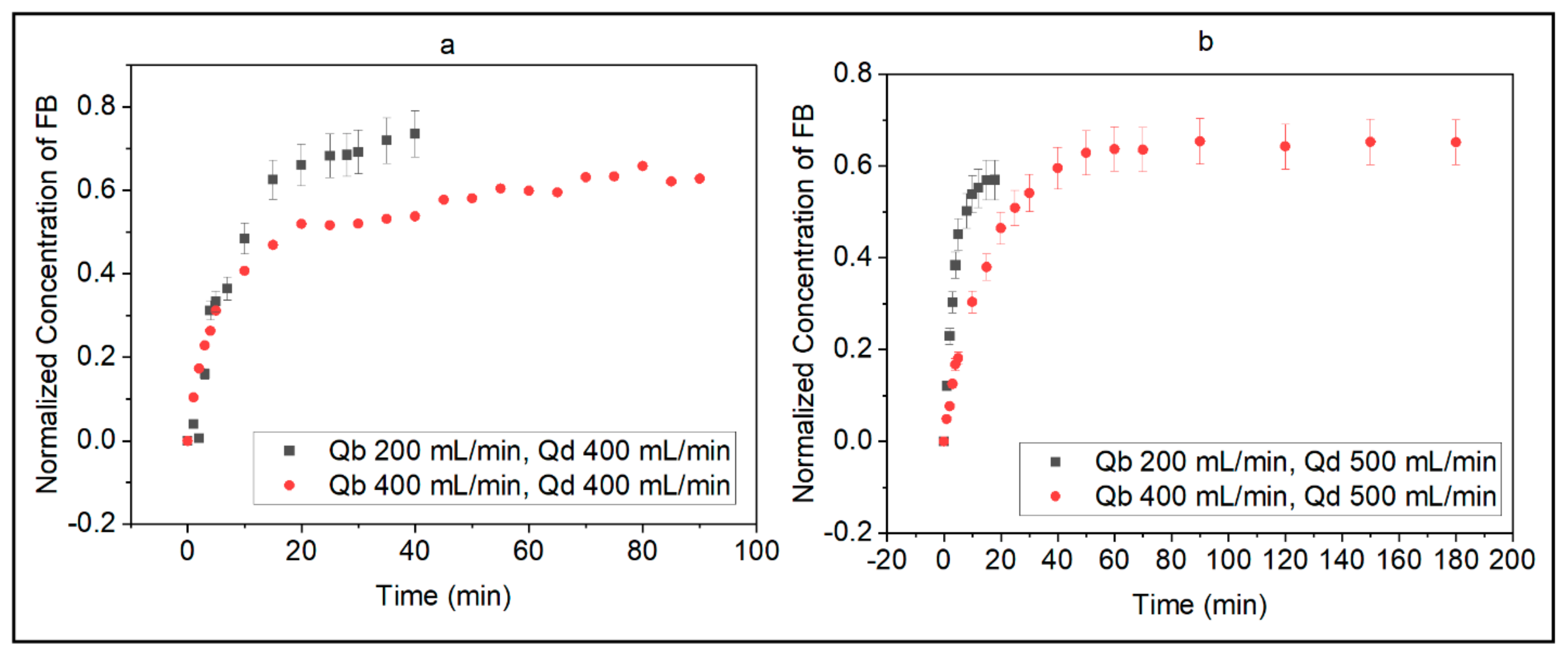
The adsorptive behavior of FB was evaluated using various values of Qb and Qd similar to those prescribed to HD patients (Figure 3a,b). The results indicated that Qd impacted the adsorption of FB when operating at a lower Qb. Adsorption occurred faster at a lower Qb for both Qd values, especially during the first 10 min. Specifically, the difference between the amount of adsorbed FB at Qb values of 200 and 400 mL/min was 19.5 and 77.6% at Qd values of 400 and 500 mL/min, respectively. Moreover, the time to achieve a normalized concentration of adsorbed FB of 0.50 for a Qd of 400 mL/min was 10 and 20 min and for a Qd of 500 mL/min was 8 and 25 min for Qb values of 200 and 400 mL/min, respectively. The in vitro adsorption of FB showed that at a lower Qb (200 mL/min), adsorption occurred faster, and equilibrium was achieved in approximately 35 and 15 min for Qd values of 400 and 500 mL/min, respectively. At a higher Qb (400 mL/min), the adsorption of FB approached equilibrium more slowly but to similar final normalized values of 0.63 and 0.65 for Qd values of 400 and 500 mL/min, respectively; however, at a lower Qb (200 mL/min), final normalized values were quite different (0.73 and 0.57 for Qd values of 400 and 500 mL/min, respectively). At a Qb of 200 mL/min, the pressure on the blood side of the membrane was 0.85 psi and on the dialysate side was 1.15 and 1.0 psi for Qd values of 400 and 500 mL/min, respectively.
Figure 4 shows the surface of a PAES membrane before and after contact with the FB solution. The transmembrane pressure (TMP) varied from −0.08 to −0.23 for Qb = 200 mL/min, −0.03 to −0.13 for Qb = 400 mL/min (Figure 5a), −0.10 to −0.30 for Qb = 200 mL/min, and −0.08 to −0.13 for Qb = 400 mL/min (Figure 5b). As a consequence of the negative TMP, back-filtration was observed in all the in vitro cases under study, and the back-filtration rates (BF) are listed in Table 2. The impact of different levels of Qd is depicted in Figure 6, which shows that a higher Qd leads to faster FB adsorption and a smaller amount is adsorbed at equilibrium.

3.3. Clinical Study of Inflammatory Biomarkers

3.3.1. Influence of Hydrodynamic Conditions on the Release of Inflammatory Biomarkers

The hydrodynamic conditions were observed to affect the release of all biomarkers. Figure 7 shows the concentration of inflammatory biomarkers in samples collected 30 min into a PAES dialysis session and in samples incubated in patients’ uremic serum with PAES membrane for 30 min. Concentrations of properdin, vWF, IL-1β, TNF-α, and IL-6 are higher and concentrations of serpin/antithrombin III are significantly lower in the dialysis vs. incubated samples.

3.3.2. Levels of Inflammatory Biomarkers at Various Operating Conditions

Complement Activation Factors

Figure 8A,B display the change in concentration of C5a and properdin in blood samples before dialysis, at 30 min, 1 h, and 4 h (end of treatment) from HD patients subjected to different operating conditions. Before the HD session, HD patients and healthy controls had similar levels of C5a (p < 0.0579) and properdin (p < 0.544). Similar trends were observed for both factors during the first hour of treatment at Qb = 200 or 300 mL/min, but properdin levels exhibited opposing trends at Qb = 400 mL/min (decrease) vs. 500 mL/min (increase). C5a concentrations in HD patients were higher than in healthy controls throughout HD treatment. After 4 h, the highest and lowest levels of C5a were observed in patients treated at Qb = 400 or 500 mL/min and Qb = 200 mL/min, respectively; the highest and lowest levels of properdin were observed for Qb = 200 and 500 mL/min, respectively. Figure S2A displays the overall percent change in C5a and properdin concentrations in patients treated at different Qb values. Patients treated at Qb = 200 mL/min experienced the highest increases in both biomarkers, with C5a and properdin increasing by 232 and 114%, respectively; smaller changes were observed at higher Qb values. C5a concentrations were above initial values for all flow rates, whereas properdin concentrations declined by 17 and 10% at Qb = 400 and 500 mL/min, respectively.

Coagulation and Clotting Factor

Figure 8C shows changes in serpin/antithrombin III concentration at different time points in blood samples from HD patients treated at Qb values of 200–500 mL/min. Before the HD session, serpin levels in HD patients were significantly higher than healthy controls (p < 0.02116). For all treatment conditions, the serpin concentration significantly decreased (p < 0.0150) in the first 30 min. At the end of the session, serpin concentrations were significantly higher in patients treated at 200 mL/min than other flow rates (p < 0.00228). Furthermore, serpin levels of HD patients treated at higher Qb values did not differ significantly from healthy controls. Figure S2B shows the overall percent change in serpin/antithrombin III concentration experienced by patients treated at different Qb values. Patients treated at Qb = 200 mL/min experienced a 545% increase in serpin concentrations compared to pre-dialysis, while patients treated at higher Qb values (300, 400, and 500 mL/min) presented overall decreases (83, 92.5, and 64%, respectively).

Pro-Inflammatory and Pro-Thrombotic Factors

Figure 8D–H show changes in the concentration of pro-inflammatory cytokines (TNF-α, IL-1α, vWF, IL-1β, and IL-6) in blood samples from patients subjected to different Qb values (200, 300, 400, or 500 mL/min). Before HD, TNF-α (p < 0.00371), vWF (p < 0.0218), and IL-6 (p < 0.0803) concentrations were higher in patients than in healthy controls, whereas IL-1α (p < 0.00642) and IL-1β (p < 0.03994) concentrations were lower. After 1 h, IL-1β, TNF-α, IL-1α, and vWF concentrations declined but the IL-6 concentration remained elevated. After 4 h, patients treated at Qb = 200 mL/min had IL-1α, IL-6, and vWF concentrations that had increased by 50, 450, and 212% compared to pre-dialysis levels; IL-1β and TNF-α concentrations declined by 12.5 and 35.5%, respectively. For patients undergoing HD treatment at Qb = 300 mL/min, TNF-α, IL-1α, IL-6, and vWF concentrations declined while IL-1β concentrations increased in the first 30 min. Similar trends were observed for patients treated at Qb = 400 or 500 mL/min. After 1 h of treatment, patients treated at Qb = 300 mL/min experienced a further decrease in TNF-α, IL-1α, IL-6, and vWF concentrations while IL-1β concentrations remained elevated. Similar results were observed for patients treated at Qb = 400 or 500 mL/min, but with increased IL-1α concentrations at 400 mL/min and IL-6 concentrations at 500 mL/min. However, after 4 h, the concentrations of vWF, IL-1α, and IL-6 had significantly increased in patients treated at a blood flow rate of 200 mL/min. On the other hand, almost all pro-inflammatory and pro-thrombotic factors had declined in patients treated at higher flow rates. The IL-1β concentrations declined in all patients, except those treated at Qb = 400 mL/min. TNF-α concentrations significantly decreased for all patients, with those treated at Qb = 400 or 500 mL/min. IL-1α concentrations increased in patients treated at 200 mL/min but declined in patients treated at higher flow rates. For IL-6, patients treated at the lowest and highest flow rates experienced an overall increase and those treated at intermediate flow rates experienced a reduction. For vWF, patients treated at Qb = 200 or 400 mL/min had, respectively, overall increases of 212 and 38% compared to pre-HD concentrations, while patients treated at Qb = 300 or 500 mL/min experienced a decrease. Figure S2C shows the overall changes in inflammatory biomarker concentrations.

3.3.3. Inflammatory Responses in Male and Female Patients

Figure 9 shows the concentration of inflammatory biomarkers of male and female patients before, during, and after HD. Female patients presented higher pre-dialysis C5a, vWF, and IL-1α concentrations, whereas male patients presented higher properdin, serpin, TNF-α, IL-6, and IL-1β concentrations. During the first 30 min of treatment, C5a, properdin, serpin, and vWF concentrations converged to similar levels for both female and male patients. After 1 h, female patients presented higher levels of C5a, properdin, serpin, and vWF, whereas male patients experienced higher levels of TNF-α, IL-1β, IL-1α, and IL-6. Different trends are noted thereafter. Notably, female patients experienced an increase in the level of all biomarkers except properdin and IL-6 between 1 and 4 h of treatment. Figure S3 shows the overall change in biomarker concentrations experienced by female and male patients, calculated by comparing concentrations at 4 h to pre-dialysis levels. Female patients experienced significantly higher levels of IL-6 and properdin, corresponding to overall increases of 182 and 79%, respectively, while male patients presented higher levels of C5a, IL-1α, and IL-6, corresponding to overall increases of 80, 76.5, and 10%, respectively.

4. Discussion

The in vitro adsorption of fibrinogen demonstrated that Qd impacts the adsorption of the protein when operating at a lower Qb. Adsorption occurs faster at a lower Qb for both Qd values, especially during the first 10 min. When a lower Qb is used, the protein molecules take longer to pass through the dialyzer, enhancing the contact between protein molecules and the membrane surface. The attachment of more FB molecules to the membrane in the first few minutes of contact corroborates findings from our theoretical model with respect to the strong affinity between them.
In crossflow filtration mode, a high feed flow rate is expected to create higher shear near the membrane surface, promoting the continuous washing of particles away from the surface, lower particle attachment, and less fouling. In HD operation, a higher Qb increases the pressure on the simulated blood side, which, in some cases, overcomes the increase in shear near the surface. As seen in Figure 5, for both cases, a lower Qb leads to significantly lower TMP during operation. For a fixed value of Qd but higher Qb, the shear stress near the surface is higher and may detach particles from the cake layer, and the TMP is higher compared to cases with a lower flow rate. As such, a higher TMP promotes convective transport and a higher removal of toxins, while a lower Qb may not allow the same removal efficacy. A negative TMP value indicates that the pressure applied on the dialysate side is higher than on the blood side and is evidence of back-filtration (BF). As the difference between Qb and Qd increases, a higher BF rate is observed (Table 2). In all cases, BF took place after the first 5 min. A higher BF rate is expected to result in some protein molecules detaching from the cake layer, resulting in a lower adsorption rate. These observations indicate that patients undergoing HD treatment might experience less FB adsorption at higher Qd values, which can lead to less inflammatory responses. At higher Qb values, the removal of small toxins is also enhanced due to the increase in TMP. Furthermore, less BF is observed due to a smaller pressure difference across the membrane.
To further evaluate the effect of Qd, Figure 6 plots normalized concentrations of FB adsorbed for a fixed Qb of 200 mL/min and Qd values of 300, 400, and 500 mL/min. As Qd increases, FB adsorption occurs faster, and a smaller amount is adsorbed at equilibrium. Thus, a higher Qd is expected to lead to less FB adsorption, which can reduce inflammatory responses in HD patients and enhance the removal of small-molecular-weight toxins. These data align with the common clinical practice of Canadian hospitals to use a Qd of 500 mL/min. However, monitoring patients closely and evaluating the occurrence of BF in clinical practice is important to avoid undesired side-effects.
Hydrodynamic conditions within the HD membrane module can impact protein adsorption and lead to RBC rupture, enhance protein adsorption and platelet adhesion, and lead to coagulation pathways and thrombus formation. Figure 7 shows the concentration of inflammatory biomarkers in samples collected 30 min into a PAES dialysis session and in samples incubated in patients’ uremic serum with the PAES membrane for 30 min. Concentrations of properdin, vWF, IL-1β, TNF-α, and IL-6 are higher and concentrations of serpin/antithrombin III are significantly lower in the dialysis vs. incubated samples. This is expected because serpin is a clotting factor, and, hence, the absence of flow would lead to an increase in concentration. These results indicate that hydrodynamic conditions established by Qb and Qd influence the level of inflammation experienced by dialysis patients, with serpin concentration more affected than the other components.
Before the HD session, HD patients and healthy controls presented similar levels of C5a (p < 0.0579) and properdin (p < 0.544). The levels of serpin (p < 0.02116), TNF-α (p < 0.00371), vWF (p < 0.0218), and IL-6 (p < 0.0803) were higher in HD patients and the levels of IL-1α (p < 0.00642) and IL-1β (p < 0.03994) were lower. Elevated levels of vWF are commonly observed in uremic patients and are associated with recurrent platelet activation promoted by HD; elevated levels are related to long-term, low-grade endothelial damage and recurrent platelet activation [31]. At the end of the dialysis session, patients treated with a Qb of 200 mL/min presented a significant increase in the concentration of C5a (232%), properdin (114%), serpin (545%), IL-1α (50%), IL-6 (450%), and vWF (212%). IL-1β and TNF-α concentrations declined by 12.5 and 35.5%, respectively.
Comparing the change in concentration levels of C5a and properdin, the results of this study suggest that lower flow rates trigger more complement activation than higher flow rates. Analyzing the pre- and post-HD levels of the coagulation factor serpin, it was observed that patients prescribed a lower Qb presented increased levels, whereas the other patients demonstrated a decreased tendency. This is attributed to the lower clearance promoted by the lower Qb and the longer contact between the blood and membrane surface and suggests lower flow rates promote coagulation and clotting pathways. These results align with observations that high flux interventions are associated with a reduced risk of cardiac-related events [32]. Regarding the levels of pro-inflammatory and pro-thrombotic factors, at Qb = 200 mL/min, vWF, IL-6, and TNF-α concentrations increased in the first 30 min, while IL-1β and IL-1α concentrations declined. The first minutes of contact between blood components and the membrane surface are critical to the development of inflammatory responses as more surface area is available for contact. More severe interactions are expected at a lower Qb and a consequent longer residence time of the blood in the membrane module [26]. These results indicate less activation during the first 30 min at higher flow rates. Overall, most patients experienced reduced levels of pro-inflammatory and pro-thrombotic factors after 1 h.
The results obtained from the clinical study can be related to observations above on FB adsorption to PAES membranes. At increased blood flow rates, the hydrodynamic conditions developed inside the polymeric fibers lead to faster adsorption of FB, but at a lower cumulative amount. Consequently, this leads to a lower activation of inflammatory and thrombotic responses and a better clearance of toxins. For low blood flow rates, the time of contact between blood components and the dialyzer membrane is prolonged, leading to more activation. The impact of the hydrophilic nature of the PAES membrane surface is also important; dehydration of RBCs is expected at the zeta potential of −68 mV, which favors rupture and hemolysis [33,34,35]. Hemolysis leads to negative effects on the immune system, promoting the release of cytokine and leukocytes such as IL-6, TNF-α, IL-1α, and IL-1β [36,37,38].
Comparing the pre- and post-dialysis levels of female and male patients, female patients experienced significantly higher levels of IL-6 and properdin, while male patients presented higher levels of C5a, IL-1α, and IL-6. These results indicate that male patients experience more severe inflammatory responses than female patients when using the PAES dialysis membrane at the specified operating conditions. Overall, the sex of HD patients is a biological variable factor and it differs in innate and adaptive immunological responses against foreign and self-antigens. Many genes on the X chromosome [39] and levels of hormonal mediators, for example, estrogen [40], progesterone [41], and androgen [40,41], regulate immune functions. They play an important role in modulating sex differences in the development of the immune response. The molecular mechanisms of mediating these factors have been reported in detail [41]. Another study showed the relation of hormonal factors influencing propensity, progression, and biochemical and psychological aspects of CKD [41].

5. Conclusions

The results of this study lead us to conclude that at fixed Qd, a higher Qb results in higher TMP; this effect overrides the effect of increased shear stress near the surface that may detach particles from the cake layer. Less back-filtration occurs due to a smaller pressure difference across the membrane. A higher Qd leads to the faster adsorption of FB but lower overall levels due to the hydrodynamic factor, whereas a low Qb results in prolonged contact between the blood and membrane surface, which promotes more activation. At the end of the dialysis session, patients treated with a Qb of 200 mL/min presented a significant increase in the concentration of C5a (232%), properdin (114%), serpin (545%), IL-1α (50%), IL-6 (450%), and vWF (212%). IL-1β and TNF-α concentrations declined by 12.5 and 35.5%, respectively. Hydrodynamic conditions increase the concentration of all inflammatory biomarkers except serpin, which is attributed to its role as a clotting factor. The release of pro-inflammatory and pro-thrombotic factors in patients’ serum confirms that lower flow rates trigger more adverse responses compared to higher flow rates. Male patients experience more severe inflammatory responses than female patients when using the PAES dialysis membrane at the specified operating conditions. The results of this study will help clinical doctors evaluate the impact of HD operating conditions on blood activations before prescribing treatment and inform expectations for outcomes in female and male patients.

Supplementary Materials

The following supporting information can be downloaded at: https://www.mdpi.com/article/10.3390/jcs6080226/s1, Figure S1: Experimental setup for in-vitro FB adsorption; Figure S2: Overall percent change in concentration of (A) C5a, properdin; (B) Serpin and (C) TNF-α, IL-1α, IL-1β, IL-6, and vWF in patients treated with different values of Qb (Qd = 500 mL/min); Figure S3: Overall percent change in concentration of inflammatory biomarkers for female and male patients (Qb = 500 mL/min); Table S1: Shapiro-Wilk normality test results; Table S2: Surface roughness measurements for hollow polymer membrane fibers.

Author Contributions

Conceptualization, A.A.; methodology, A.A.; formal analysis, H.W., S.S., and J.B.; investigation, A.A. and A.S.; data curation, A.A.; writing—original draft preparation, H.W., S.S., J.B. and A.A.; writing—review and editing, H.D., A.S. and A.A.; visualization, H.W. and J.B. supervision, A.A.; project administration, A.A.; funding acquisition, A.A. All authors have read and agreed to the published version of the manuscript.

Funding

Saskatchewan Research Health Foundation (SHRF)-Establishment Grant.

Institutional Review Board Statement

Amira Abdelrasoul, the principal investigator of the project, obtained Research Ethics Approval and Operational Approval to conduct this research in the Saskatchewan Health Authority. She is responsible for regulatory approvals that pertain to this project, and for ensuring the project was conducted according to the governing law. All participants in this study signed a written informed consent, approved by the University of Saskatchewan Biomedical Research Ethics Board (Bio-REB).

Informed Consent Statement

Informed consent was obtained from all participants involved in the study.

Data Availability Statement

The raw/processed data required to reproduce these findings cannot be shared at this time, as the data are critical to ongoing research.

Acknowledgments

The authors acknowledge funding support from the Saskatchewan Research Health Foundation (SHRF) and are grateful to the University of Saskatchewan (U of S) for all facilities provided to conduct this research in collaboration with the Saskatchewan Transplant Program at St. Paul’s Hospital. The authors also acknowledge the Health Sciences Laboratories, Saskatchewan Structural Sciences Centre (SSSC), and Western College of Veterinary Medicine (WCVM) Imaging Centre at the U of S for the services and facilities provided. The authors also acknowledge help received from Brooke Thompson at Health Science Laboratories; Jason Maley at the SSSC; Eiko Kawamuraand at WCVM.

Conflicts of Interest

The authors have no conflict of interest to declare.

References

  1. Bikbov, B.; Purcell, C.A.; Levey, A.S.; Smith, M.; Abdoli, A.; Abebe, M.; Adebayo, O.M.; Afarideh, M.; Agarwal, S.K.; Agudelo-Botero, M.; et al. Global, regional, and national burden of chronic kidney disease, 1990–2017: A systematic analysis for the Global Burden of Disease Study 2017. Lancet 2020, 395, 709–733. [Google Scholar] [CrossRef] [Green Version]
  2. Joshi, P.K.; Rao, P.P. Global pulses scenario: Status and outlook. Ann. N. Y. Acad. Sci. 2016, 1392, 6–17. [Google Scholar] [CrossRef] [Green Version]
  3. Kooman, J.P.; van der Sande, F.M. COVID-19 in ESRD and Acute Kidney Injury. Blood Purif. 2020, 50, 610–620. [Google Scholar] [CrossRef]
  4. McAdams, M.; Ostrosky-Frid, M.; Rajora, N.; Hedayati, S.S. Effect of COVID-19 on Kidney Disease Incidence and Management. Kidney360 2020, 2, 141–153. [Google Scholar] [CrossRef]
  5. Urbani, A.; Sirolli, V.; Lupisella, S.; Levi-Mortera, S.; Pavone, B.; Pieroni, L.; Bonomini, M. Proteomic investigations on the effect of different membrane materials on blood protein adsorption during haemodialysis. Blood Transfus. 2012, 10, s101–s112. [Google Scholar] [CrossRef]
  6. Urbani, A.; Lupisella, S.; Sirolli, V.; Bucci, S.; Amoroso, L.; Pavone, B.; Pieroni, L.; Sacchetta, P.; Bonomini, M. Proteomic analysis of protein adsorption capacity of different haemodialysis membranes. Mol. BioSyst. 2012, 8, 1029–1039. [Google Scholar] [CrossRef]
  7. Hakim, R.M. Clinical implications of hemodialysis membrane biocompatibility. Kidney Int. 1993, 44, 484–494. [Google Scholar] [CrossRef] [Green Version]
  8. Westphalen, H.; Abdelrasoul, A.; Shoker, A. Protein adsorption phenomena in hemodialysis membranes: Mechanisms, influences of clinical practices, modeling, and challenges. Colloid Interface Sci. Commun. 2020, 40, 100348. [Google Scholar] [CrossRef]
  9. Cozzolino, M.; Mangano, M.; Stucchi, A.; Ciceri, P.; Conte, F.; Galassi, A. Cardiovascular disease in dialysis patients. Nephrol. Dial. Transplant. 2018, 33, iii28–iii34. [Google Scholar] [CrossRef]
  10. Meijers, B.K.I.; De Loor, H.; Bammens, B.; Verbeke, K.; Vanrenterghem, Y.; Evenepoel, P. p-Cresyl Sulfate and Indoxyl Sulfate in Hemodialysis Patients. Clin. J. Am. Soc. Nephrol. 2009, 4, 1932–1938. [Google Scholar] [CrossRef] [Green Version]
  11. Burmeister, J.E.; Mosmann, C.B.; Costa, V.B.; Saraiva, R.T.; Grandi, R.R.; Bastos, J.P.; Gonçalves, L.F.; Rosito, G.A. Prevalence of Cardiovascular Risk Factors in Hemodialysis Patients—The CORDIAL Study. Arq. Bras. Cardiol. 2014, 102, 473–480. [Google Scholar] [CrossRef] [PubMed]
  12. Gatta, A.; Verardo, A.; Bolognesi, M. Hypoalbuminemia. Intern. Emerg. Med. 2012, 7, S193–S199. [Google Scholar] [CrossRef]
  13. Kirschbaum, B. Hypotransferrinemia of Chronically Hemodialyzed Patients. Artif. Organs 1999, 23, 1047–1054. [Google Scholar] [CrossRef]
  14. Koga, Y.; Fujieda, H.; Meguro, H.; Ueno, Y.; Aoki, T.; Miwa, K.; Kainoh, M. Biocompatibility of Polysulfone Hemodialysis Membranes and Its Mechanisms: Involvement of Fibrinogen and Its Integrin Receptors in Activation of Platelets and Neutrophils. Artif. Organs 2018, 42, E246–E258. [Google Scholar] [CrossRef]
  15. Abdelrasoul, A.; Shoker, A. Influence of Hydration Shell of Hemodialysis Clinical Membranes on Surrogate Biomarkers Activation in Uremic Serum of Dialysis Patients. Biomed. Eng. Adv. 2022. [Google Scholar] [CrossRef]
  16. Nazari, S.; Abdelrasoul, A. Surface Zwitterionization of HemodialysisMembranesfor Hemocompatibility Enhancement and Protein-mediated anti-adhesion: A Critical Review. Biomed. Eng. Adv. 2022, 3, 100026. [Google Scholar] [CrossRef]
  17. Mollahosseini, A.; Abdelrasoul, A.; Shoker, A. Advances in Membrane Technologies; IntechOpen: Rijeka, Croatia, 2020. [Google Scholar] [CrossRef] [Green Version]
  18. Mollahosseini, A.; Abdelrasoul, A.; Shoker, A. A critical review of recent advances in hemodialysis membranes hemocompatibility and guidelines for future development. Mater. Chem. Phys. 2020, 248, 122911. [Google Scholar] [CrossRef]
  19. Mollahosseini, A.; Abdelrasoul, A.; Shoker, A. Latest advances in zwitterionic structures modified dialysis membranes. Mater. Today Chem. 2020, 15, 100227. [Google Scholar] [CrossRef]
  20. Merle, N.S.; Noé, R.; Halbwachs-Mecarelli, L.; Fremeaux-Bacchi, V.; Roumenina, L.T. Complement System Part II: Role in Immunity. Front. Immunol. 2015, 6, 257. [Google Scholar] [CrossRef] [Green Version]
  21. Tetta, C.; Roy, T.; Gatti, E.; Cerutti, S. The rise of hemodialysis machines: New technologies in minimizing cardiovascular complications. Expert Rev. Cardiovasc. Ther. 2011, 9, 155–164. [Google Scholar] [CrossRef]
  22. Adams, R.L.C.; Bird, R.J. Review article: Coagulation cascade and therapeutics update: Relevance to nephrology. Part 1: Overview of coagulation, thrombophilias and history of anticoagulants. Nephrology 2009, 14, 462–470. [Google Scholar] [CrossRef] [PubMed]
  23. Oikonomopoulou, K.; Ricklin, D.; Ward, P.A.; Lambris, J.D. Interactions between coagulation and complement—Their role in inflammation. In Seminars in Immunopathology; Springer: Berlin/Heidelberg, Germany, 2012; Volume 34, pp. 151–165. [Google Scholar] [CrossRef] [Green Version]
  24. Westphalen, H.; Saadati, S.; Eduok, U.; Abdelrasoul, A.; Shoker, A.; Choi, P.; Doan, H.; Ein-Mozaffari, F. Case studies of clinical hemodialysis membranes: Influences of membrane morphology and biocompatibility on uremic blood-membrane interactions and inflammatory biomarkers. Sci. Rep. 2020, 10, 14808. [Google Scholar] [CrossRef] [PubMed]
  25. Westphalen, H.; Abdelrasoul, A.; Shoker, A.; Zhu, N. Assessment of hemodialysis clinical practices using polyaryl ether sulfone-polyvinylpyrrolidone (PAES: PVP) clinical membrane: Modeling of in vitro fibrinogen adsorption, in situ synchrotron-based imaging, and clinical inflammatory biomarkers investigations. Sep. Purif. Technol. 2021, 259, 118136. [Google Scholar] [CrossRef]
  26. Saadati, S.; Westphalen, H.; Eduok, U.; Abdelrasoul, A.; Shoker, A.; Choi, P.; Doan, H.; Ein-Mozaffari, F.; Zhu, N. Biocompatibility enhancement of hemodialysis membranes using a novel zwitterionic copolymer: Experimental, in situ synchrotron imaging, molecular docking, and clinical inflammatory biomarkers investigations. Mater. Sci. Eng. C 2020, 117, 111301. [Google Scholar] [CrossRef]
  27. Tanaka, M.; Mochizuki, A. Effect of water structure on blood compatibility—Thermal analysis of water in poly(meth)acrylate. J. Biomed. Mater. Res. 2004, 68A, 684–695. [Google Scholar] [CrossRef]
  28. Abdelrasoul, A.; Westphalen, H.; Saadati, S.; Shoker, A. Hemodialysis biocompatibility mathematical models to predict the inflammatory biomarkers released in dialysis patients based on hemodialysis membrane characteristics and clinical practices. Sci. Rep. 2021, 11, 23080. [Google Scholar] [CrossRef]
  29. Eberst, M.E.; Berkowitz, L.R. Hemostasis in renal disease: Pathophysiology and management. Am. J. Med. 1994, 96, 168–179. [Google Scholar] [CrossRef]
  30. Cheung, A.K.; Levin, N.W.; Greene, T.; Agodoa, L.; Bailey, J.; Beck, G.; Clark, W.; Levey, A.S.; Leypoldt, J.K.; Ornt, D.B.; et al. Effects of High-Flux Hemodialysis on Clinical Outcomes: Results of the HEMO Study. J. Am. Soc. Nephrol. 2003, 14, 3251–3263. [Google Scholar] [CrossRef] [Green Version]
  31. Abdelrasoul, A.; Shoker, A. Induced hemocompatibility of polyethersulfone (PES) hemodialysis membrane using polyvinylpyrrolidone: Investigation on human serum fibrinogen adsorption and inflammatory biomarkers released. Chem. Eng. Res. Des. 2022, 177, 615–624. [Google Scholar] [CrossRef]
  32. Straub, R.H. The Complex Role of Estrogens in Inflammation. Endocr. Rev. 2007, 28, 521–574. [Google Scholar] [CrossRef] [Green Version]
  33. Abdelrasoul, A.; Shoker, A. Investigations on the Impact of Hemodialysis Clinical Practices on Human Plasma Proteins Loss and von Willebrand factor. Kidney Int. Rep. 2022, 7, S258. [Google Scholar] [CrossRef]
  34. Westphalen, H.; Kalugin, D.; Abdelrasoul, A. Structure, Function, and Adsorption of Highly Abundant Blood Proteins and its Critical Influence on Hemodialysis Patients: A critical Review. Biomed. Eng. Adv. 2021, 2, 100021. [Google Scholar] [CrossRef]
  35. Eduok, U.; Westphalen, H.; Abdelrasoul, A.; Shoker, A. Influence of UV-irradiation intensity and exposure duration on the hemobiocompatibility enhancement of a novel synthesized phosphobetaine zwitterions polyether sulfone clinical hemodialysis membranes. J. Biomed. Mater. Res. Part B Appl. Biomater. 2022, 110, 573–586. [Google Scholar] [CrossRef]
  36. Saadati, S.; Eduok, U.; Westphalen, H.; Abdelrasoul, A.; Shoker, A.; Choi, P.; Doan, H.; Ein-Mozaffari, F.; Zhu, N. Assessment of Polyethersulfone and Polyacrylonitrile Hemodialysis Clinical Membranes: In situ Synchrotron-based Imaging of Human Serum Proteins Adsorption, Interaction Analyses, Molecular Docking and Clinical Inflammatory Biomarkers Investigations. Mater. Today Commun. 2021, 29, 102928. [Google Scholar] [CrossRef]
  37. Mollahosseini, A.; Argumeedi, S.; Abdelrasoul, A.; Shoker, A. A Case Study of Poly(aryl ether sulfone) Hemodialysis Membrane Interactions with Human Blood: Molecular Dynamics Simulation and Experimental Analyses. Comput. Methods Programs Biomed. 2020, 197, 105742. [Google Scholar] [CrossRef]
  38. Saadati, S.; Eduok, U.; Westphalen, H.; Abdelrasoul, A.; Shoker, A.; Choi, P.; Doan, H.; Ein-Mozaffari, F.; Zhu, N. In situ Synchrotron Imaging of Human Serum Proteins Interactions, Molecular Docking and Inflammatory Biomarkers of Hemocompatible Synthesized Zwitterionic Polymer Coated-Polyvinylidene Fluoride (PVDF) Dialysis Membranes. Surf. Interfaces 2021, 27, 101505. [Google Scholar] [CrossRef]
  39. Jones, L.A.; Kreem, S.; Shweash, M.; Paul, A.; Alexander, J.; Roberts, C.W. Differential Modulation of TLR3- and TLR4-Mediated Dendritic Cell Maturation and Function by Progesterone. J. Immunol. 2010, 185, 4525–4534. [Google Scholar] [CrossRef]
  40. Dunn, S.E.; Ousman, S.S.; Sobel, R.A.; Zuniga, L.; Baranzini, S.E.; Youssef, S.; Crowell, A.; Loh, J.; Oksenberg, J.; Steinman, L. Peroxisome proliferator–activated receptor (PPAR)α expression in T cells mediates gender differences in development of T cell–mediated autoimmunity. J. Exp. Med. 2007, 204, 321–330. [Google Scholar] [CrossRef] [Green Version]
  41. Carrero, J.J. Gender Differences in Chronic Kidney Disease: Underpinnings and Therapeutic Implications. Kidney Blood Press. Res. 2010, 33, 383–392. [Google Scholar] [CrossRef]
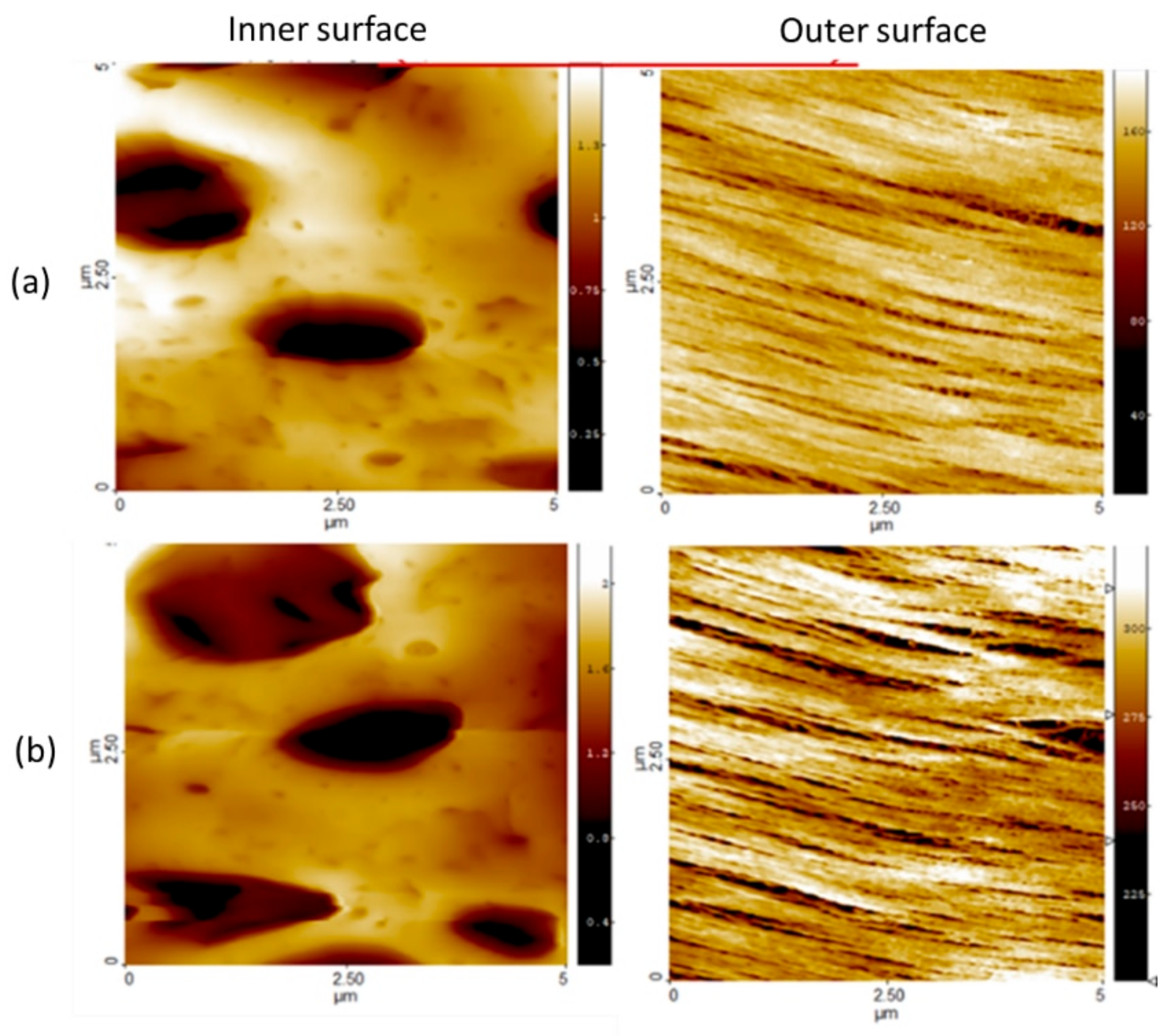
Figure 1. AFM micrographs of neat PAES hemodialysis membrane fibers collected at two different spots of closed proximity ((a) first and (b) second rows).
Jcs 06 00226 g001
Figure 2. SEM micrographs of neat PAES hemodialysis membrane fiber surface: (a) whole microtubes; (b) the magnified surface image; (c) membrane diameter and thickness.
Jcs 06 00226 g002
Figure 3. (a,b) Change in the normalized concentration of FB adsorbed (Ci/C0; Qb = 200 or 400 mL/min; Qd = 400 or 500 mL/min.
Jcs 06 00226 g003
Figure 4. SEM images of the PAES membrane surface (a) before and (b) after contact with FB solution.
Jcs 06 00226 g004
Figure 5. (a,b) Variation in TMP during simulated dialysis session (Qb = 200 or 400 mL/min; Qd = 400 or 500 mL/min).
Jcs 06 00226 g005
Figure 6. Changes in the normalized concentration of FB adsorbed (Qb = 200 mL/min; Qd = 300, 400, or 500 mL/min).
Jcs 06 00226 g006
Figure 7. Concentration of (a) properdin and serpin/antithrombin III, (b) vWF, and (c) IL-1β, TNF-α, and IL-6 from dialysis (Qb = 200 mL/min; Qd = 500 mL/min) and incubated samples.
Jcs 06 00226 g007
Figure 8. The concentration (pg/ml) of (A) C5, (B) properdin, (C) Serpin, (D) TNF-α, (E) IL-1α, (F) IL-1β, (G) IL-6, and (H) vWF in blood samples collected during dialysis sessions from patients treated with different Qb levels (Qd = 500 mL/min).
Jcs 06 00226 g008
Figure 9. Concentrations of (a) C5a, (b) properdin, (c) serpin, (d) TNF-α, (e) IL-1β, (f) IL-1α, (g) IL-6, and (h) vWF in blood samples collected during dialysis session from female and male patients (Qb = 200 mL/min; Qd = 500 mL/min).
Jcs 06 00226 g009
Table 1. Characteristics of the population included in the clinical study.
Healthy ControlHemodialysis Patients
VariablesValuesN%N%
GenderFemale150541.66
Male150758.33
Age<50 years00541.66
≥50 years2100758.33
RaceNon-black210012100
Black0000
BMI a<27 kg/m20000
≥27 kg/m2210012100
CAD bYes00541.66
NO2100758.33
DiabetesYes00650.00
No2100650.00
HypertensionYes1501083.33
No150216.66
PVD cYes00433.33
No2100866.67
ESRD duration<1 yearNANA18.33
1–5 yearsNANA1083.34
>5 yearsNANA18.33
BMI a: body mass index (kg/m2); CAD b: coronary artery disease; PVD c: peripheral vascular disease.
Table 2. Back-filtration rate at different operating conditions.
Qb (mL/min)Qd (mL/min)BF Rate (mL/min)
2004009.6
4004002.8
20050016.7
4005008.1
Publisher’s Note: MDPI stays neutral with regard to jurisdictional claims in published maps and institutional affiliations.