PHBV/MWCNT Films: Hydrophobicity, Thermal and Mechanical Properties as a Function of MWCNT Concentration

 ,
,
Abstract
:1. Introduction
2. Materials and Methods
3. Results
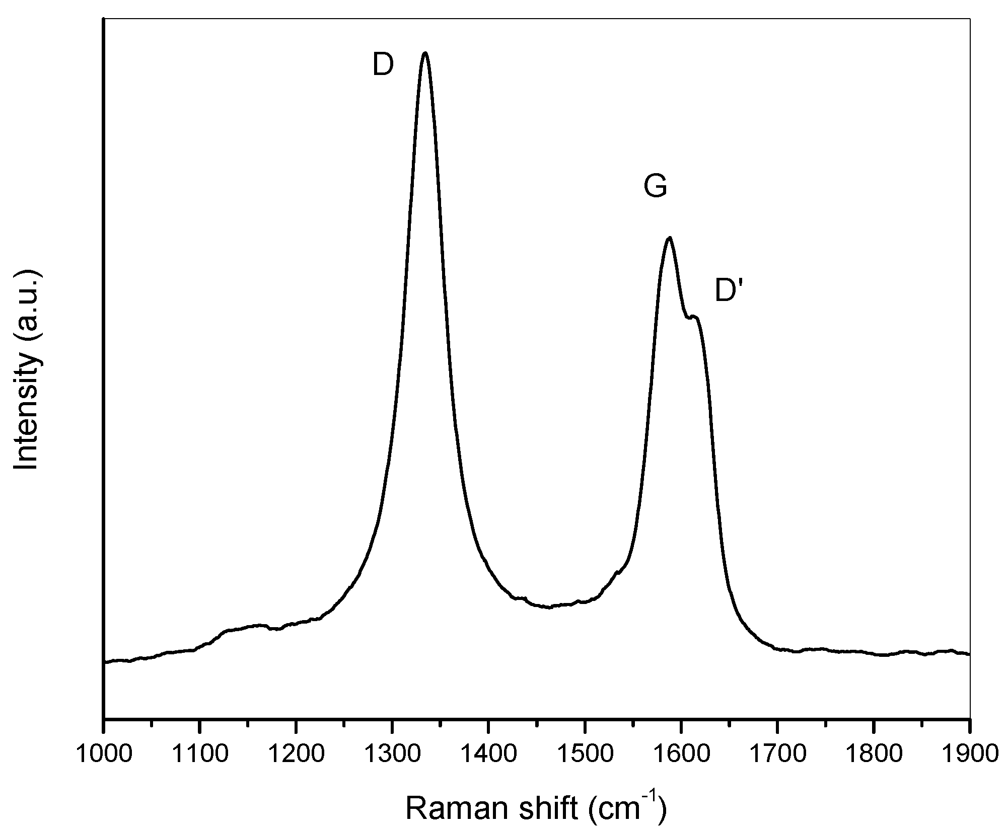
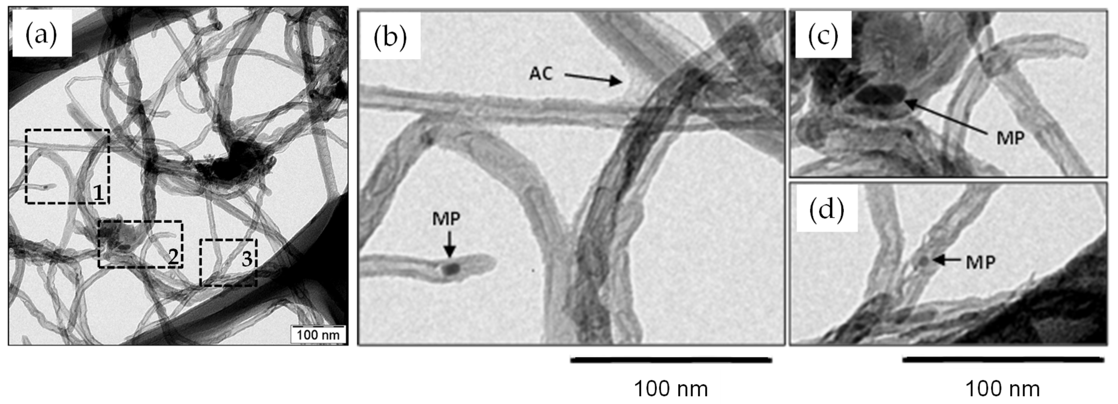
3.1. Characterization of MWCNTs
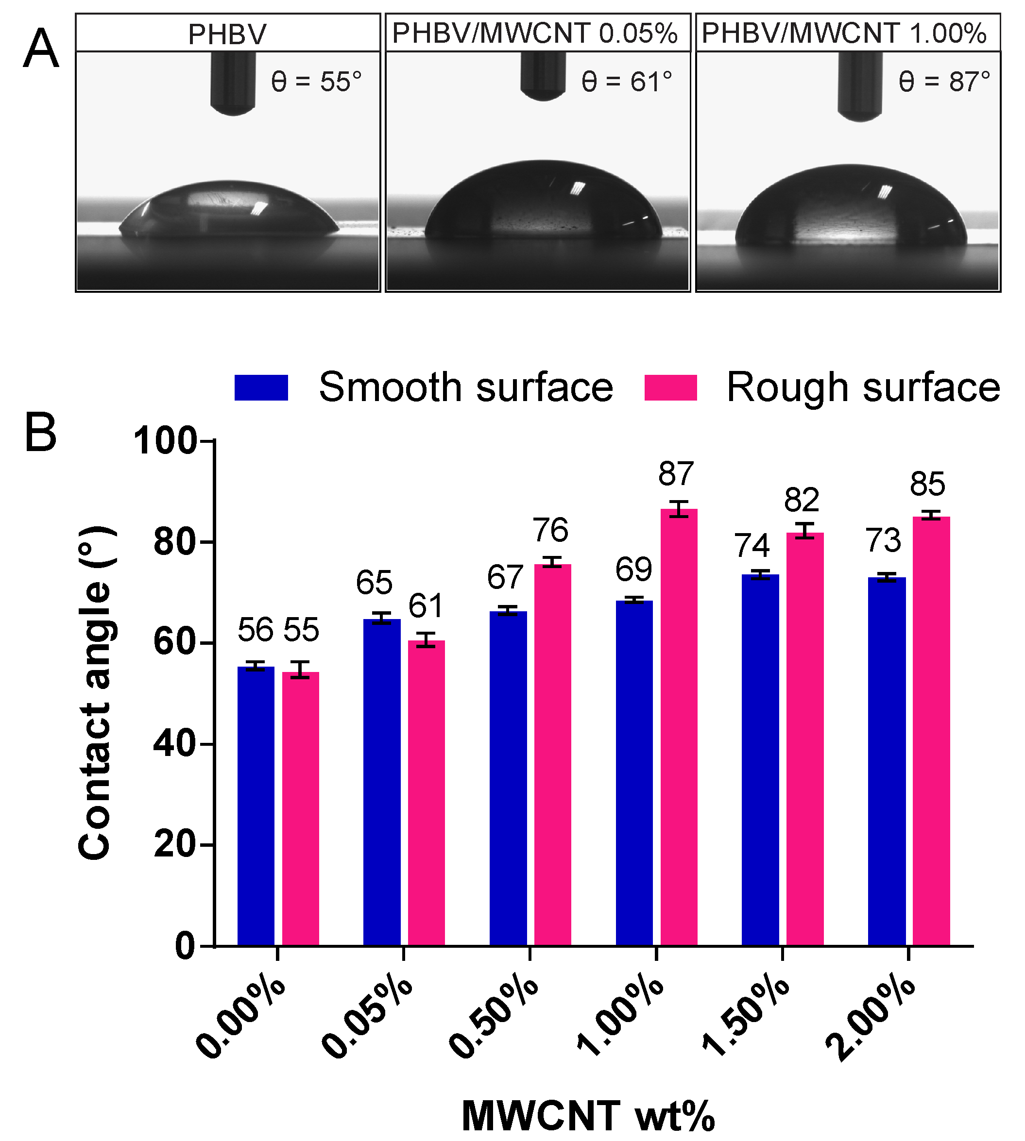
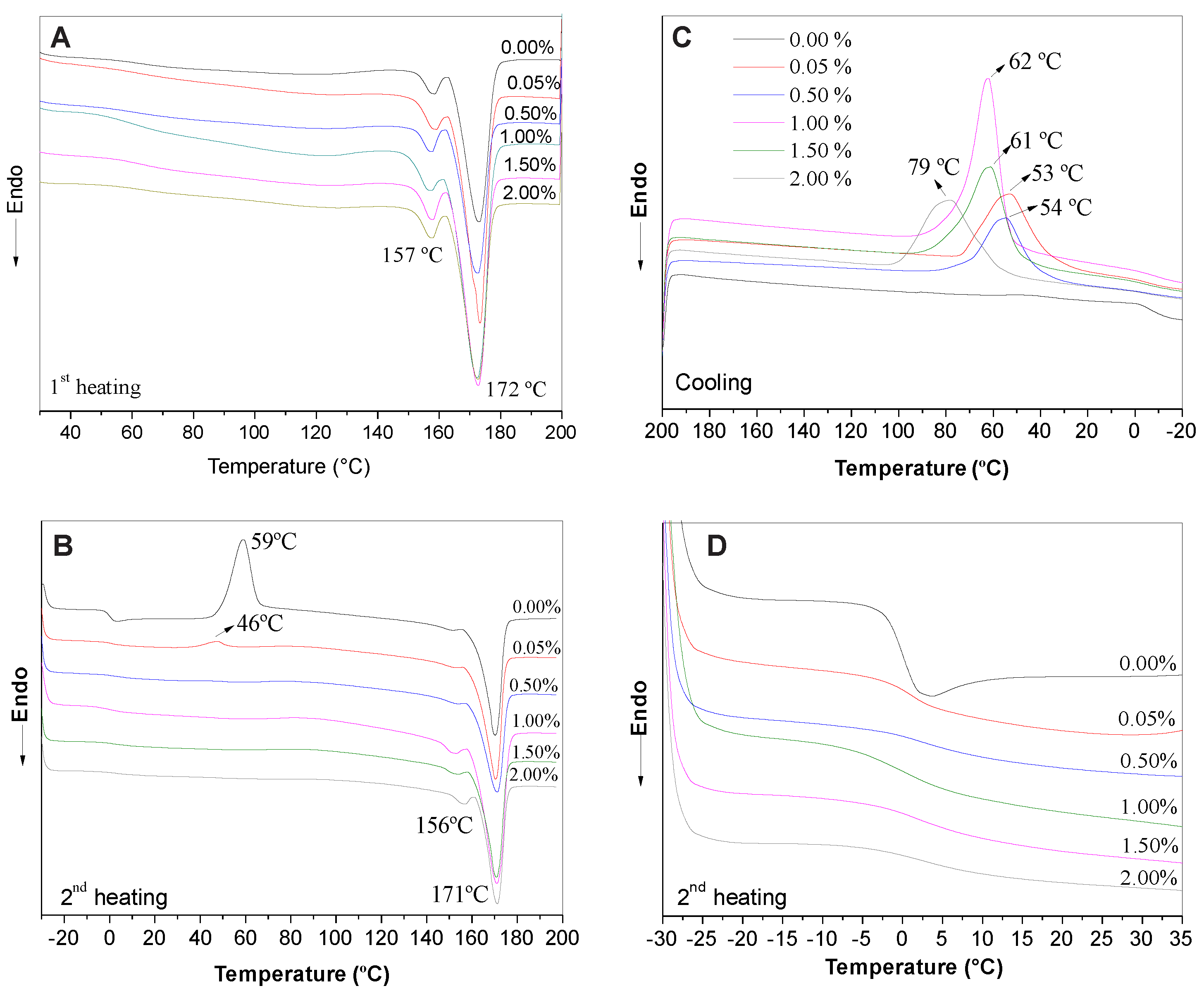
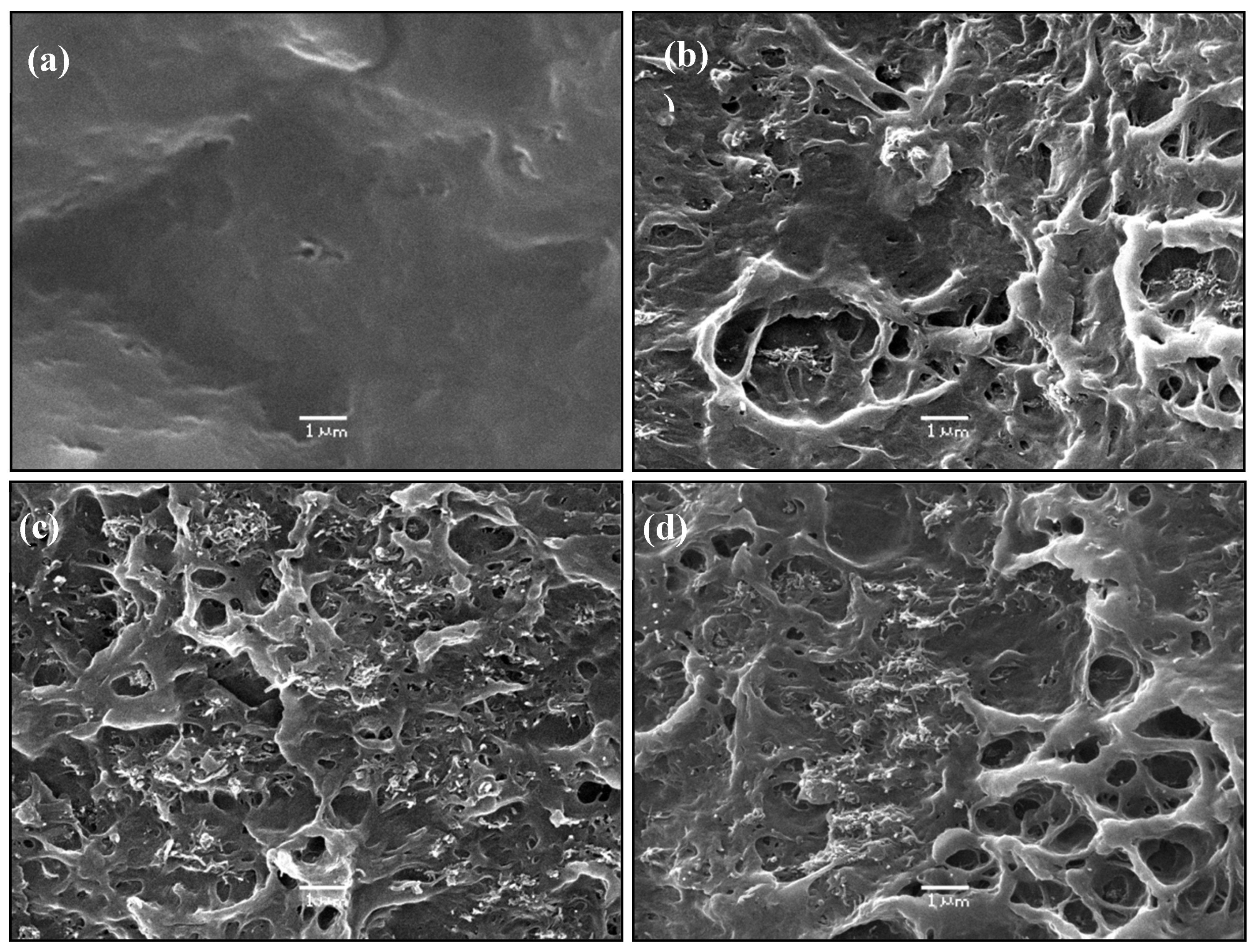
3.2. Characterization of Nanocomposite Films
4. Discussion
5. Conclusions
Author Contributions
Funding
Conflicts of Interest
References
- Alshehrei, F. Biodegradation of Synthetic and Natural Plastic by Microorganisms. J. Appl. Environ. Microbiol. 2017, 5, 8–19. [Google Scholar]
- Raza, Z.A.; Abid, S.; Banat, I.M. Polyhydroxyalkanoates: Characteristics, production, recent developments and applications. Int. Biodeterior. Biodegrad. 2018, 126, 45–56. [Google Scholar] [CrossRef]
- Li, Z.; Yang, J.; Loh, X.J. Polyhydroxyalkanoates: Opening doors for a sustainable future. NPG Asia Mater. 2016, 8, 265–285. [Google Scholar] [CrossRef]
- Rivera-Briso, A.; Serrano-Aroca, Á. Poly(3-Hydroxybutyrate-co-3-Hydroxyvalerate): Enhancement Strategies for Advanced Applications. Polymers 2018, 10, 732. [Google Scholar] [CrossRef]
- Philip, S.; Keshavarz, T.; Roy, I. Polyhydroxyalkanoates: Biodegradable polymers with a range of applications. J. Chem. Technol. Biotechnol. 2007, 82, 233–247. [Google Scholar] [CrossRef]
- Yu, H.-Y.; Qin, Z.-Y.; Sun, B.; Yang, X.-G.; Yao, J.-M. Reinforcement of transparent poly(3-hydroxybutyrate-co-3-hydroxyvalerate) by incorporation of functionalized carbon nanotubes as a novel bionanocomposite for food packaging. Compos. Sci. Technol. 2014, 94, 96–104. [Google Scholar] [CrossRef]
- Masood, F.; Yasin, T.; Hameed, A. Comparative oxo-biodegradation study of poly-3-hydroxybutyrate-co-3-hydroxyvalerate/polypropylene blend in controlled environments. Int. Biodeterior. Biodegrad. 2014, 87, 1–8. [Google Scholar] [CrossRef]
- Hazer, D.B.; Kiliçai, E.; Hazer, B. Poly(3-hydroxyalkanoate)s: Diversification and biomedical applications A state of the art review. Mater. Sci. Eng. C 2012, 32, 637–647. [Google Scholar] [CrossRef]
- Wu, Q.; Wang, Y.; Chen, G.-Q. Medical Application of Microbial Biopolyesters Polyhydroxyalkanoates. Artif Cells Blood Substit. Biotechnol. 2009, 37, 1–12. [Google Scholar] [CrossRef]
- Gorodzha, S.N.; Muslimov, A.R.; Syromotina, D.S.; Timin, A.S.; Tcvetkov, N.Y.; Lepik, K.V.; Petrova, A.V.; Surmeneva, M.A.; Gorin, D.A.; Sukhorukov, G.B.; et al. A comparison study between electrospun polycaprolactone and scaffolds for bone tissue engineering. Colloids Surf. B Biointerfaces 2017, 160, 48–59. [Google Scholar] [CrossRef]
- Deepthi, S.; Nivedhitha, S.M.; Vijayan, P.; Nair, S.V.; Jayakumar, R. Engineering poly(hydroxy butyrate-co-hydroxy valerate) based vascular scaffolds to mimic native artery. Int. J. Biol. Macromol. 2018, 109, 85–98. [Google Scholar] [CrossRef] [PubMed]
- Recco, M.S.; Floriano, A.C.; Tada, D.B.; Lemes, A.P.; Langa, R.; Cristovan, F.H. Poly(3-hydroxybutyrate-co-valerate)/Poly(3-thiophene ethyl acetate) blends as electroactive biomaterial substrate to tissue engineering application. RSC Adv. 2016, 6, 25330–25338. [Google Scholar] [CrossRef]
- Lemes, A.P.; Soto-Oviedo, M.A.; Innocentini-Mei, L.H.; Durán, N. Processing, chemical structure and morphology of poly(hydroxybutyrate-co-valerate)/lignosulfonate composites. In Proceedings of the ENPROMER-2005, 2nd Mercosur Congress on Chemical Engineering and 4th Mercosur Congress on Process Systems Engineering Enpromer, Rio de Janeiro, Brazil, 14–18 August 2005; pp. 1–6. [Google Scholar]
- Ten, E.; Bahr, D.F.; Li, B.; Jiang, L.; Wolcott, M.P. Effects of cellulose nanowhiskers on mechanical, dielectric, and rheological properties of poly(3-hydroxybutyrate-co-3-hydroxyvalerate)/cellulose nanowhisker composites. Ind. Eng. Chem. Res. 2012, 51, 2941–2951. [Google Scholar] [CrossRef]
- Du, J.; Zhao, G.; Pan, M.; Zhuang, L.; Li, D.; Zhang, R. Crystallization and mechanical properties of reinforced PHBV composites using melt compounding: Effect of CNCs and CNFs. Carbohydr. Polym. 2017, 168, 255–262. [Google Scholar]
- Castro-Mayorga, J.L.; Martínez-Abad, A.; Fabra, M.J.; Olivera, C.; Reis, M.; Lagarón, J.M. Stabilization of antimicrobial silver nanoparticles by a polyhydroxyalkanoate obtained from mixed bacterial culture. Int. J. Biol. Macromol. 2014, 71, 103–110. [Google Scholar] [CrossRef]
- Castro-Mayorga, J.L.; Fabra, M.J.; Lagaron, J.M. Stabilized nanosilver based antimicrobial poly(3-hydroxybutyrate-co-3-hydroxyvalerate) nanocomposites of interest in active food packaging. Innov. Food Sci. Emerg. Technol. 2016, 33, 524–533. [Google Scholar] [CrossRef]
- Montagna, L.S.; Montanheiro, T.L.; Machado, J.P.B.; Passador, F.R.; Lemes, A.P.; Rezende, M.C. Effect of Graphite Nanosheets on Properties of Poly(3-hydroxybutyrate-co-3-hydroxyvalerate). Int. J. Polym. Sci. 2017, 2017, 9316761. [Google Scholar] [CrossRef]
- Montagna, L.S.; Montanheiro, T.L.; Baldan, M.R.; Oliveira, A.P.S.; de Farias, M.A.; Hocevar, M.A.; Folgueras, L.C.; Passador, F.R.; Lemes, A.P.; Rezende, M.C. Effect of graphite nanosheets on electrical, electromagnetic, mechanical and morphological characteristics of PHBV/GNS nanocomposites. Adv. Mater. Lett. 2018, 9, 499–504. [Google Scholar] [CrossRef]
- Braga, N.F.; Vital, D.A.; Guerrini, L.M.; Lemes, A.P.; Formaggio, D.M.D.; Tada, D.B.; Arantes, T.M.; Cristovan, F.H. PHBV-TiO2 mats prepared by electrospinning technique: Physico-chemical properties and cytocompatibility. Biopolymers 2018, 109, e23120. [Google Scholar] [CrossRef]
- Montanheiro, T.L.A.; Cristóvan, F.H.; Machado, J.P.B.; Tada, D.B.; Durán, N.; Lemes, A.P. Effect of MWCNT functionalization on thermal and electrical properties of PHBV/MWCNT nanocomposites. J. Mater. Res. 2014, 30, 55–65. [Google Scholar] [CrossRef]
- Montanheiro, T.L.A.; Montagna, L.S.; Machado, J.P.B.; Lemes, A.P. Covalent functionalization of MWCNT with PHBV chains: Evaluation of the functionalization and production of nanocomposites. Polym. Compos. 2017, 40, 288–295. [Google Scholar] [CrossRef]
- Vidhate, S.; Innocentini-mei, L.; Souza, N.A.D. Mechanical and Electrical Multifunctional Poly(3-hydroxybutyrate-co-3-hydroxyvalerate)—Multiwall Carbon Nanotube Nanocomposites. Polym. Eng. Sci. 2012, 52, 1367–1374. [Google Scholar] [CrossRef]
- Yu, H.-Y.; Yao, J.-M.; Qin, Z.-Y.; Liu, L.; Yang, X.-G. Comparison of covalent and noncovalent interactions of carbon nanotubes on the crystallization behavior and thermal properties of poly(3-hydroxybutyrate-co-3-hydroxyvalerate). J. Appl. Polym. Sci. 2013, 130, 4299–4307. [Google Scholar] [CrossRef]
- Lemes, A.P.; Montanheiro, T.L.A.; Passador, F.R.; Durán, N. Nanocomposites of Polyhydroxyalkanoates Reinforced with Carbon Nanotubes: Chemical and Biological Properties. In Eco-Friendly Polymer Nanocomposites; Springer: New Delhi, India, 2015; pp. 79–108. [Google Scholar]
- Qiu, H.; Yang, J. Structure and Properties of Carbon Nanotubes. In Industrial Applications of Carbon Nanotubes; Elsevier Inc.: New York, NY, USA, 2016; pp. 47–69. [Google Scholar]
- Jian, M.Q.; Xie, H.H.; Xia, K.L.; Zhang, Y.Y. Challenge and Opportunities of Carbon Nanotubes. In Industrial Applications of Carbon Nanotubes; Elsevier Inc.: New York, NY, USA, 2016; pp. 433–476. [Google Scholar]
- Pan, L.; Pei, X.; He, R.; Wan, Q.; Wang, J. Multiwall carbon nanotubes/polycaprolactone composites for bone tissue engineering application. Colloids Surf. B Biointerfaces 2012, 93, 226–234. [Google Scholar] [CrossRef] [PubMed]
- Pinto, V.C.; Ramos, T.; Alves, A.S.F.; Xavier, J.; Tavares, P.J.; Moreira, P.M.G.P.; Guedes, R.M. Dispersion and failure analysis of PLA, PLA/GNP and PLA/CNT-COOH biodegradable nanocomposites by SEM and DIC inspection. Eng. Fail Anal. 2017, 71, 63–71. [Google Scholar] [CrossRef]
- Park, S.H.; Lee, S.G.; Kim, S.H. Isothermal crystallization behavior and mechanical properties of polylactide/carbon nanotube nanocomposites. Compos. Part A Appl. Sci. Manuf. 2013, 46, 11–18. [Google Scholar] [CrossRef]
- Ma, Y.; Zheng, Y.; Wei, G.; Song, W.; Hu, T.; Yang, H.; Xue, R. Processing, Structure, and Properties of Multiwalled Carbon Nanotube/Poly(hydroxybutyrate-co-valerate) Biopolymer Nanocomposites. J. Appl. Polym. Sci. 2012, 125, E620–E629. [Google Scholar] [CrossRef]
- Liu, L.; Qin, Y.; Guo, Z.; Zhu, D. Reduction of solubilized multi-walled carbon nanotubes. Carbon N. Y. 2003, 41, 331–335. [Google Scholar] [CrossRef]
- Antunes, E.F.; Lobo, A.O.; Corat, E.J.; Trava-Airoldi, V.J.; Martin, A.A.; Veríssimo, C. Comparative study of first- and second-order Raman spectra of MWCNT at visible and infrared laser excitation. Carbon N. Y. 2006, 44, 2202–2211. [Google Scholar] [CrossRef]
- Osswald, S.; Havel, M.; Gogotsi, Y. Monitoring oxidation of multiwalled carbon nanotubes by Raman spectroscopy. J. Raman Spectrosc. 2007, 38, 728–736. [Google Scholar] [CrossRef]
- de Menezes, B.R.C.; Ferreira, F.V.; Silva, B.C.; Simonetti, E.A.N.; Bastos, T.M.; Cividanes, L.S.; Thim, G.P. Effects of octadecylamine functionalization of carbon nanotubes on dispersion, polarity, and mechanical properties of CNT/HDPE nanocomposites. J. Mater. Sci. 2018, 53, 14311–14327. [Google Scholar] [CrossRef]
- Endo, M.; Takeuchi, K.; Kobori, K.; Takahashi, K.; Kroto, H.W.; Sarkar, A. Pyrolytic carbon nanotubes from vapor-grown carbon fibers. Carbon N. Y. 1995, 33, 873–881. [Google Scholar] [CrossRef]
- Gunaratne, L.M.W.K.; Shanks, R.A.; Amarasinghe, G. Thermal history effects on crystallisation and melting of poly(3-hydroxybutyrate). Thermochim. Acta 2004, 423, 127–135. [Google Scholar] [CrossRef]
- Lai, M.; Li, J.; Yang, J.; Liu, J.; Tong, X.; Cheng, H. The morphology and thermal properties of multi-walled carbon nanotube and poly(hydroxybutyrate-co-hydroxyvalerate) composite. Polym. Int. 2004, 53, 1479–1484. [Google Scholar] [CrossRef]
- Shaffer, M.S.P.; Windle, A.H. Fabrication and Characterization of Carbon Nanotube/Poly(vinyl alcohol) Composites. Adv. Mater. 1999, 11, 937–941. [Google Scholar] [CrossRef]
- Cai, Z.; Xu, Y.; Yang, H.; Jia, J.; Liu, Y. Poly(hydroxybutyrate)/cellulose acetate blend nanofiber scaffolds: Preparation, characterization and cytocompatibility. Mater. Sci. Eng. C 2016, 58, 757–767. [Google Scholar]
- Yu, H.; Qin, Z.; Zhou, Z. Cellulose nanocrystals as green fillers to improve crystallization and hydrophilic property of poly(3-hydroxybutyrate-co-3-hydroxyvalerate). Prog. Nat. Sci. Mater. Int. 2011, 21, 478–484. [Google Scholar] [CrossRef]
- Bauhofer, W.; Kovacs, J.Z. A review and analysis of electrical percolation in carbon nanotube polymer composites. Compos. Sci. Technol. 2009, 69, 1486–1498. [Google Scholar] [CrossRef]
- Grady, B.P. Effects of carbon nanotubes on polymer physics. J. Polym. Sci. Part B Polym. Phys. 2012, 50, 591–623. [Google Scholar] [CrossRef]


| MWCNT wt % | Tc (°C) | ΔHm (J·g−1) | Xc (%) |
|---|---|---|---|
| 0.00 | - | 61.2 | 56.1 |
| 0.05 | 53 | 69.6 | 63.9 |
| 0.50 | 54 | 59.3 | 54.7 |
| 1.00 | 62 | 59.0 | 54.7 |
| 1.50 | 61 | 69.5 | 64.7 |
| 2.00 | 79 | 74.4 | 69.6 |
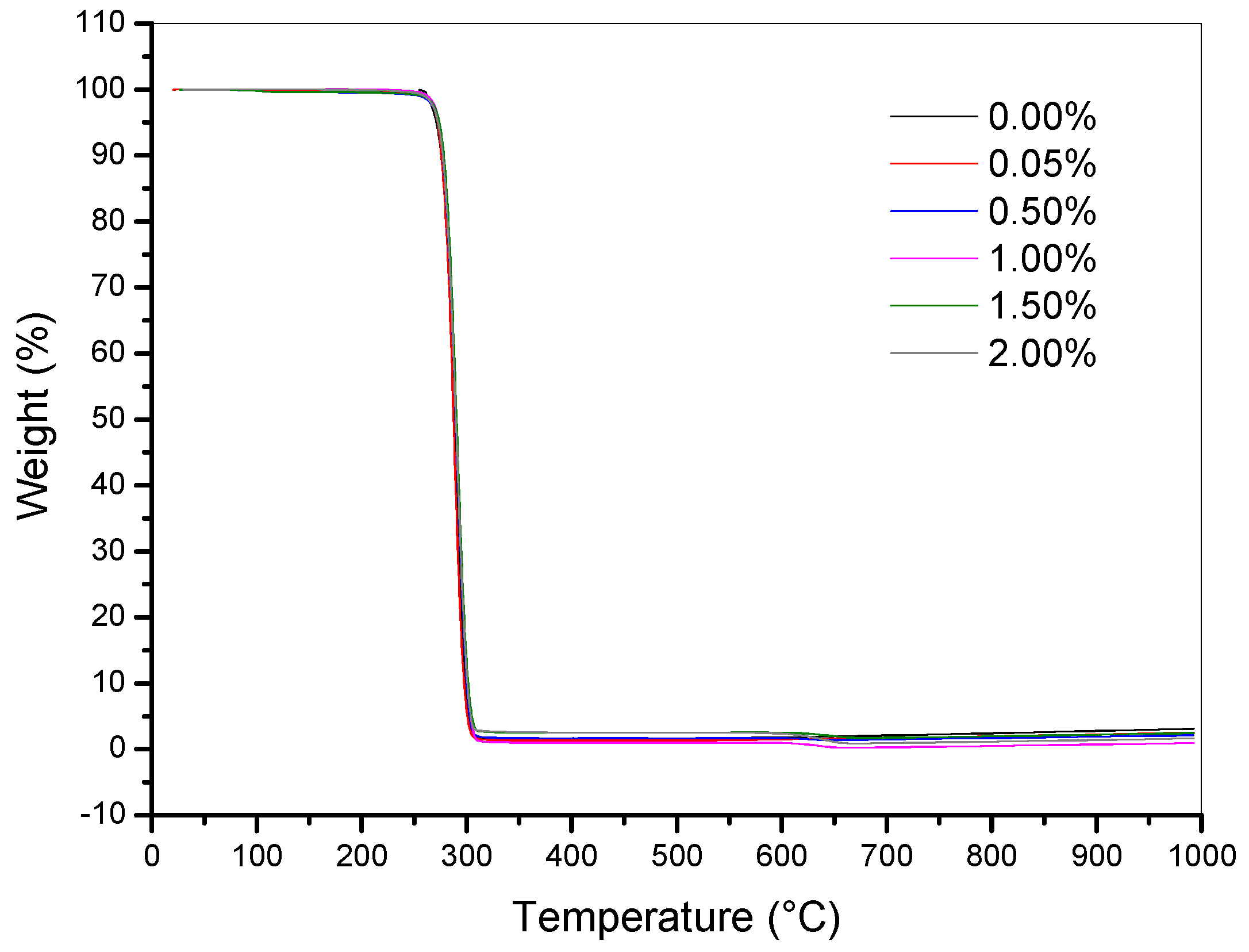
| MWCNT wt % | T1 (°C) | T5 (°C) | T10 (°C) | Tmax (°C) |
|---|---|---|---|---|
| 0.00 | 260 | 266 | 269 | 279 |
| 0.05 | 261 | 269 | 276 | 283 |
| 0.50 | 260 | 269 | 273 | 285 |
| 1.00 | 261 | 270 | 274 | 285 |
| 1.50 | 261 | 270 | 274 | 285 |
| 2.00 | 259 | 268 | 272 | 283 |
| MWCNT (%) | E (MPa) | σmax (MPa) | εmax (%) |
|---|---|---|---|
| 0.00 | 847 ± 94 | 15.59 ± 0.93 | 4.64 ± 0.50 |
| 0.05 | 1001 ± 36 | 18.13 ± 1.31 | 2.70 ± 0.22 |
| 0.50 | 998 ± 87 | 21.04 ± 0.80 | 2.45 ± 0.14 |
| 1.00 | 875 ± 87 | 19.67 ± 1.39 | 2.24 ± 0.24 |
| 1.50 | 1250 ± 62 | 23.23 ± 0.60 | 2.92 ± 0.12 |
| 2.00 | 1165 ± 116 | 19.21 ± 2.50 | 1.79 ± 0.21 |
| p* | 0.010 | 8.6 x 10⁻⁷ | 0.006 |
© 2019 by the authors. Licensee MDPI, Basel, Switzerland. This article is an open access article distributed under the terms and conditions of the Creative Commons Attribution (CC BY) license (http://creativecommons.org/licenses/by/4.0/).
Share and Cite
Lemes, A.P.; Montanheiro, T.L.d.A.; da Silva, A.P.; Durán, N. PHBV/MWCNT Films: Hydrophobicity, Thermal and Mechanical Properties as a Function of MWCNT Concentration. J. Compos. Sci. 2019, 3, 12. https://doi.org/10.3390/jcs3010012
Lemes AP, Montanheiro TLdA, da Silva AP, Durán N. PHBV/MWCNT Films: Hydrophobicity, Thermal and Mechanical Properties as a Function of MWCNT Concentration. Journal of Composites Science. 2019; 3(1):12. https://doi.org/10.3390/jcs3010012
Chicago/Turabian StyleLemes, Ana Paula, Thaís Larissa do Amaral Montanheiro, Ana Paula da Silva, and Nelson Durán. 2019. "PHBV/MWCNT Films: Hydrophobicity, Thermal and Mechanical Properties as a Function of MWCNT Concentration" Journal of Composites Science 3, no. 1: 12. https://doi.org/10.3390/jcs3010012
APA StyleLemes, A. P., Montanheiro, T. L. d. A., da Silva, A. P., & Durán, N. (2019). PHBV/MWCNT Films: Hydrophobicity, Thermal and Mechanical Properties as a Function of MWCNT Concentration. Journal of Composites Science, 3(1), 12. https://doi.org/10.3390/jcs3010012