Case Report

A Humanized Anti-IL-4Rα Monoclonal Antibody Improves Aural Fullness

1
Department of Otorhinolaryngology, Union Hospital, Tongji Medical College, Huazhong University of Science and Technology, Wuhan 430074, China
2
Allergy Center, Department of Otolaryngology, Affiliated Eye and ENT Hospital, Fudan University, Shanghai 200437, China
*
Authors to whom correspondence should be addressed.
These authors contributed equally to this work.
J. Otorhinolaryngol. Hear. Balance Med. 2025, 6(2), 21; https://doi.org/10.3390/ohbm6020021
Submission received: 8 September 2025 / Revised: 15 November 2025 / Accepted: 19 November 2025 / Published: 21 November 2025
(This article belongs to the Special Issue Etiology, Diagnosis, and Treatment of Congenital Hearing Loss)

Abstract

Background and Clinical Significance: Otitis media with effusion (OME) is characterized by persistent middle ear effusion without acute infection. Type 2 inflammation, mediated by IL-4 and IL-13 signaling via the IL-4Rα receptor, has been implicated in the pathogenesis of chronic rhinosinusitis with nasal polyps (CRSwNP), asthma, and possibly OME. Refractory OME in adults remains a therapeutic challenge, as conventional treatments often fail to achieve long-term resolution. Targeted biologic therapies that modulate type 2 inflammation may offer a novel treatment option. Case Presentation: We report the case of a 60-year-old man with a 15-year history of allergic rhinitis and CRSwNP, complicated by recurrent asthma exacerbations, who presented with bilateral aural fullness, hearing loss, and tinnitus. His symptoms persisted despite repeated tympanic punctures, Eustachian tube insufflation, and corticosteroid therapy. Otoscopy revealed dull tympanic membranes with effusion, and audiometry showed conductive hearing loss with a B-type tympanogram on the left. Laboratory findings demonstrated mild peripheral eosinophilia. The patient was diagnosed with OME, likely secondary to type 2 inflammation. After nine biweekly injections of Stapokibart (CM310)—a humanized monoclonal antibody targeting IL-4Rα—aural fullness completely resolved. Otoscopic findings and tympanograms normalized, and hearing thresholds improved significantly. Retrospective evaluation using Iino’s diagnostic framework suggested that the patient did not meet the full criteria for eosinophilic otitis media (EOM); nevertheless, marked symptomatic and functional improvement was achieved. No recurrence or adverse effects were observed during follow-up. Conclusions: This case suggests that IL-4Rα blockade with Stapokibart may be effective in treating refractory OME associated with type 2 inflammation, even in patients who do not fulfill the diagnostic criteria for EOM. These findings highlight the potential of anti-IL-4Rα biologics as a novel therapeutic option for middle ear diseases driven by type 2 inflammation.

1. Introduction

Otitis media with effusion (OME) is characterized by the persistent presence of non-purulent serous or mucoid fluid within the middle-ear cavity [1,2] and commonly presents with symptoms such as aural fullness and hearing impairment [3]. The potential etiologies of OME include local malignancies—most notably nasopharyngeal carcinoma—sinonasal diseases, laryngopharyngeal reflux, and Eustachian tube dysfunction [4]. The condition is also more frequently observed in allergic individuals compared with non-allergic populations [5]. Historically, atopic diseases were often underestimated as risk factors for recurrent or persistent otitis media, although accumulating evidence now indicates that the prevalence of OME is significantly higher in patients with allergic disorders than in healthy individuals [6].
Allergic rhinitis (AR) is a chronic, IgE-mediated type-2 inflammatory disease [7]. Inflammatory cytokines such as IL-4 and IL-13 may impair Eustachian tube function, leading to middle-ear effusion, aural fullness, and conductive hearing loss. Allergic inflammation of the nasal mucosa may influence the nasopharynx and middle ear through two major pathways: (1) by releasing mediators, cytokines, and other inflammatory factors that migrate into nasal secretions and subsequently reach the Eustachian tube orifice, and (2) through primary AR-related epithelial barrier defects, which are characterized by mucosal edema and excessive nasal secretions. These changes reduce the mucosa’s resistance to pathogen invasion and increase susceptibility to recurrent infections [8]. Consequently, patients with AR generally have a higher risk of developing Eustachian tube dysfunction [9]. Current AR management includes allergen avoidance, H1-antihistamines, intranasal corticosteroids, allergen-specific immunotherapy (AIT), and patient education. However, despite receiving standard-of-care (SoC) therapy, 62% of patients report dissatisfaction due to limited long-term efficacy and persistent residual symptoms [10]. For patients in this category, biologic therapy has emerged as a novel and more effective treatment option.
For patients with this refractory disease, biologic therapy was initiated as a novel and more effective therapeutic approach. Stapokibart (CM310) is a humanized antibody that targets IL-4Rα and effectively inhibits its interaction with both IL-4 and IL-13, which are key cytokines involved in type 2 inflammation [10,11]. The indications for this biologic agent are as follows: 1. Atopic dermatitis: For the treatment of moderate-to-severe atopic dermatitis in adults who have not responded adequately to topical therapy or for whom topical therapy is not suitable. 2. Chronic sinusitis with nasal polyps: For adults with chronic sinusitis with nasal polyps who have inadequate response to glucocorticoid therapy and/or surgery, used in combination with intranasal glucocorticoid therapy. 3. Seasonal allergic rhinitis: For adults with moderate to severe seasonal allergic rhinitis who have inadequate symptom control after treatment with intranasal glucocorticoids combined with antihistamines. In treating a patient with concurrent chronic sinusitis with nasal polyps and seasonal allergic rhinitis, we observed that Stapokibart not only alleviated nasal symptoms but also relieved the patient’s long-standing complaints of “aural fullness”. Since OME is not currently an approved indication for Stapokibart, we have compiled this case report to share this unexpected therapeutic effect and to explore the possible therapeutic value of Stapokibart in alleviating ear fullness secondary to type 2 inflammation. Written informed consent was obtained from the patient for publication of this case report and any accompanying images.

2. Case Presentation

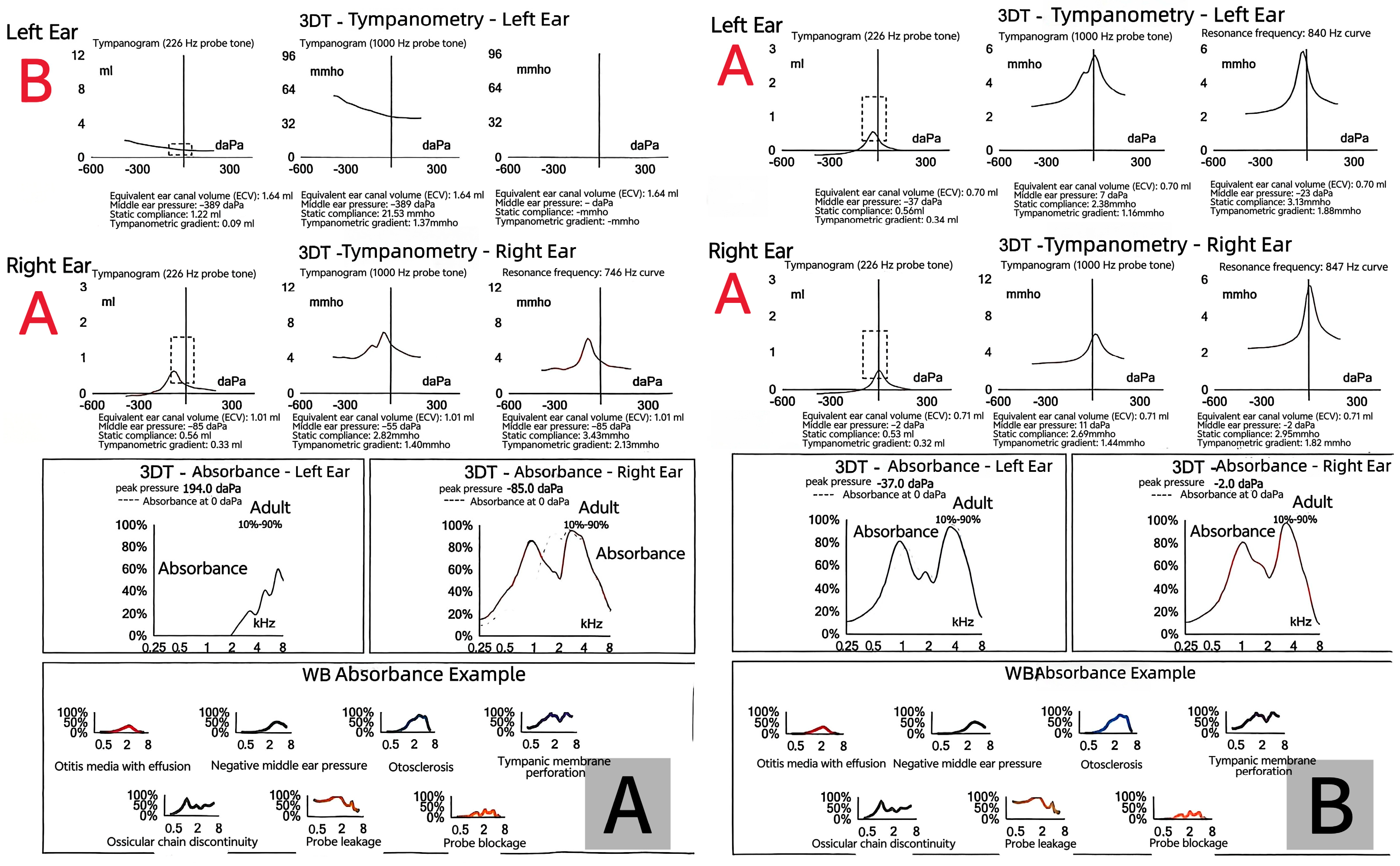
A 60-year-old man presented with recurrent bilateral aural fullness, hearing loss, and tinnitus. He had a 15-year history of allergic rhinitis and CRSwNP, and multiple asthma exacerbations. His symptoms had been recurrent and refractory to conventional therapies, including tympanic puncture, Eustachian tube insufflation, and corticosteroid treatment. On otoscopic examination, both tympanic membranes appeared dull with visible effusion (Figure 1A). Audiometry revealed conductive hearing loss with a B-type tympanogram in the left ear and A-type in the right ear(Figure 2A). His peripheral eosinophil count was mildly elevated (0.13 × 10⁹/L), and Phadiatop screening was negative. He was diagnosed with OME, likely secondary to type 2 inflammation.
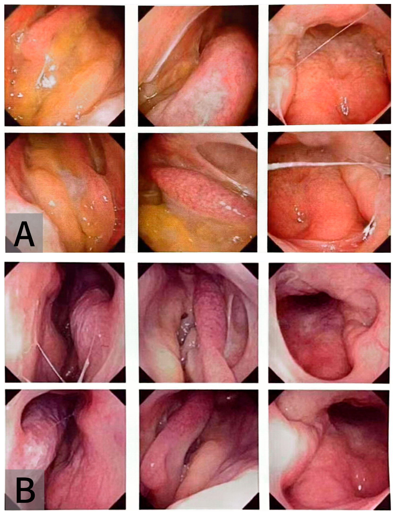
The patient was initiated on Stapokibart (CM310), a humanized anti-IL-4Rα monoclonal antibody, on 13 March 2025 (600 mg loading dose, followed by 300 mg biweekly). By June, his ear congestion symptoms had improved significantly. Otoscopic examination showed that both eardrums were intact with no obvious fluid accumulation (Figure 1B). Tympanometry showed A-type curves bilaterally (Figure 2B). Audiometric testing indicated an improvement in hearing thresholds, particularly in the left ear (Figure 3). At the most recent follow-up on 8 July 2025, he had received nine injections, with no recurrence of ear symptoms. CT imaging showed mild sinusitis, and the peripheral eosinophil count remained mildly elevated. The condition of the patient’s nasopharynx is shown in Figure 4. Before treatment, polyps and other inflammatory changes were observed in the nasal cavity. After treatment, these findings showed noticeable improvement, and the openings of both Eustachian tubes appeared normal and unobstructed. The patient has not experienced any drug-related adverse reactions since the start of treatment (possible side effects of Stapokibart are listed in Table 1).

3. Discussion

Otitis media (OM) arises from the intricate interaction between viral and bacterial pathogens and the host’s immune response, with all recognized contributing factors ultimately linked to these two central mechanisms [12]. The pathogenesis of otitis media with effusion (OME) is thought to involve the following process: after a viral or bacterial infection, congestion of the respiratory mucosa—including the nasopharynx and Eustachian tube—leads to Eustachian tube dysfunction, which compromises the middle ear’s capacity for mucociliary clearance and pressure equilibration. Persistent dysfunction may allow pathogenic microorganisms from the nasopharynx to enter the middle ear cavity, initiating inflammatory responses. Moreover, overproduction of mucin proteins disrupts normal mucociliary transport, resulting in the retention of viscous effusion within the middle ear space [12].
Based on the patient’s history and imaging, his ear symptoms were closely linked to Eustachian tube dysfunction, a well-recognized complication in upper airway inflammatory disorders such as CRSwNP and allergic rhinitis. The underlying mechanism is often attributable to edema and obstruction of the nasal and nasopharyngeal mucosa, which disrupts pressure regulation in the middle ear [13]. Aural fullness in the patient’s ear may be attributable to Eustachian tube dysfunction, a condition often associated with conductive hearing loss. In the present case, the patient’s bone-conduction thresholds improved after treatment. Similar findings have been reported in previous studies, where improvement in bone-conduction thresholds was observed in approximately 10% of patients who underwent surgery for chronic otitis media [14,15]. These observations suggest that bone-conduction impairment in such patients may be, at least in part, reversible. The underlying mechanisms are thought to involve both mechanical and biochemical factors. Specifically, the toxic effects of inflammatory mediators released during chronic middle-ear inflammation may transiently disrupt inner-ear physiology, while mechanical changes within the middle ear can also alter cochlear sound transmission. In many cases, these two mechanisms likely act synergistically. Therefore, it is generally considered that bone-conduction abnormalities associated with chronic otitis media may result from the chronic inflammatory process itself [16]. Eustachian tube dysfunction should be considered a diagnosis of exclusion. Sinonasal inflammation—often transient and reversible—can precipitate dysfunction of the Eustachian tube, particularly when triggered by allergic responses [17,18]. In addition, periostin has been detected within the thickened mucosa and is believed to be associated with the persistence of inflammation [19]. Takeshita Y et al. suggested that a reduction in IL-4 and/or IL-13 levels may suppress periostin production, thereby alleviating inflammation and subsequently improving Eustachian tube obstruction [20].
Importantly, this patient had multiple type 2 inflammation–related conditions, including CRSwNP, allergic rhinitis, and asthma. Previous studies have shown that IL-4 and IL-13 are key cytokines driving type 2 inflammation, leading to chronic mucosal inflammation in the nasal cavity, paranasal sinuses, and middle ear [10]. This inflammatory process can impair Eustachian tube patency and promote the development and persistence of OME. Conventional management—including polypectomy, anti-inflammatory medications, and Eustachian tube ventilation therapies—may provide only short-term relief, as they do not address the underlying, sustained inflammatory drive, resulting in a high recurrence rate.
In this case, following failure of standard therapy, the patient was started on Stapokibart. Although Stapokibart is currently approved in China for atopic dermatitis, CRSwNP, and seasonal allergic rhinitis, its efficacy in OME has not been reported [21]. In this patient, treatment was followed by marked resolution of bilateral aural fullness, normalization of tympanograms to A-type bilaterally, improvement in hearing thresholds, and sustained symptom control throughout follow-up without relapse.
In previous studies, biologic agents have been reported for the treatment of eosinophilic otitis media (EOM)—a rare, refractory, and chronic type of otitis media characterized by highly viscous middle ear effusion, elevated eosinophil counts in both the effusion and peripheral blood, and frequently associated comorbidities such as nasal polyps and asthma [22]. Favorable therapeutic outcomes have been achieved; for instance, Saori Kikuchi et al. retrospectively analyzed 29 patients with EOM and evaluated the efficacy of mepolizumab and dupilumab according to individual clinical characteristics. Both biologics demonstrated marked therapeutic effects within six months of administration, which remained stable for up to twelve months without recurrence [23]. Similarly, Daiki Nakashima et al. reported that dupilumab was effective in all EOM patients included in their study, suggesting that EOM may represent a distinct single endotype in which IL-4 and IL-13 play key roles in the pathogenesis [24]. It was further proposed that dupilumab may improve EOM by enhancing Eustachian tube function [20]. Although the patient in this case exhibited several features resembling eosinophilic otitis media (EOM), a retrospective assessment based on the diagnostic and scoring criteria proposed by Iino et al. indicated that the criteria for EOM were not fully met [25]. The condition was therefore classified as refractory OME rather than true EOM. Nevertheless, we cannot entirely exclude the possibility that this case represents an atypical or partial subtype within the EOM spectrum. However, treatment with Stapokibart resulted in a remarkable improvement in ear fullness and hearing function. This observation indicates that biologic therapy targeting IL-4Rα may also benefit patients with Eustachian tube dysfunction or OME secondary to type 2 inflammation, even in the absence of definitive EOM. This potentially expands the therapeutic applicability of anti-IL-4Rα biologics.
Given that Stapokibart shares a highly similar mechanism of action with these biologics, and considering the clinical course of our patient, we suggest that this agent not only controls sinonasal inflammation but also ameliorates middle ear conditions associated with Eustachian tube dysfunction, thereby providing a novel therapeutic option for recurrent and treatment-resistant OME. Although this indication is not yet approved, these findings provide preliminary support for its potential role and highlight the need for prospective clinical trials to confirm its efficacy and safety. This case also reinforces the concept of unified airway disease and the importance of comprehensive management across the upper and lower airways in type 2 inflammatory disorders.

4. Conclusions

OME is a common comorbidity in patients with CRSwNP [22]. Type 2 inflammation, driven by IL-4 and IL-13, plays a key role in chronic nasal and middle ear pathology [10]. This patient’s symptoms correlated with rhinitis exacerbations, indicating unified airway involvement. Traditional therapies failed to provide long-term relief. Following Stapokibart treatment, symptoms markedly improved, suggesting potential benefits of IL-4Rα inhibition in restoring Eustachian tube function and resolving middle ear effusion. Although not currently an approved indication, this case provides preliminary evidence supporting Stapokibart’s potential utility in managing OME symptoms in type 2 inflammatory conditions. Further clinical trials are needed to confirm its efficacy and safety in this context.

Author Contributions

Y.Z. (Yiyun Zhang), M.S. and Y.Z. (Yan Zhou): preparation, creation, and/or presentation of the published work, specifically writing the initial draft. J.C.: ideas; formulation or evolution of overarching research goals and aims. H.L. and Y.S.: ideas; formulation or evolution of overarching research goals and aims. H.L. and Y.S.: Oversight and leadership responsibility for the research activity planning, including mentorship external to the core team. All authors have read and agreed to the published version of the manuscript.

Funding

Foundation for Innovative Research Groups of the National Natural Science Foundation (No. 82521001).

Institutional Review Board Statement

The study was conducted in accordance with the Declaration of Helsinki. Our institution does not require ethics approval for reporting individual cases or case series.

Informed Consent Statement

Written informed consent was obtained from the patient for publication of this case report and any accompanying images.

Data Availability Statement

The data presented in this study are available on request from the corresponding author due to the privacy restriction.

Conflicts of Interest

The authors declare no conflict of interest.

Abbreviations

The following abbreviations are used in this manuscript:
OMEOtitis Media with Effusion
CRSwNPChronic Rhinosinusitis with Nasal Polyps
ADAtopic Dermatitis
SARSeasonal Allergic Rhinitis

References

  1. Lautermann, J.; Begall, K.; Hilger, G.; Wilhelm, T.; Mir-Salim, P.; Kaschke, O.; Zahnert, T. Leitlinie seromukotympanum—Langfassung. HNO 2012, 60, 540–543. [Google Scholar] [CrossRef]
  2. Klimek, L.; Brough, H.A.; Arasi, S.; Toppila-Salmi, S.; Bergmann, C.; Jutel, M.; Bousquet, J.; Hox, V.; Gevaert, P.; Tomazic, P.V.; et al. Otitis media with effusion (OME) and eustachian tube dysfunction: The role of allergy and immunity—An EAACI position paper. Allergy 2025, 80, 2429–2441. [Google Scholar] [CrossRef] [PubMed]
  3. Farboud, A.; Skinner, R.; Pratap, R. Otitis media with effusion (glue ear). BMJ 2011, 343, d3770. [Google Scholar] [CrossRef] [PubMed]
  4. Karyanta, M.; Satrowiyoto, S.; Wulandari, D.P. Prevalence ratio of otitis media with effusion in laryngopharyngeal reflux. Int. J. Otolaryngol. 2019, 2019, 7460891. [Google Scholar] [CrossRef] [PubMed]
  5. Oh, J.H.; Kim, W.J. Interaction between allergy and middle ear infection. Curr. Allergy Asthma Rep. 2016, 16, 66. [Google Scholar] [CrossRef]
  6. Chantzi, F.M.; Kafetzis, D.A.; Bairamis, T.; Avramidou, C.; Paleologou, N.; Grimani, I.; Apostolopoulos, N.; Papadopoulos, N.G. IgE sensitization, respiratory allergy symptoms, and heritability independently increase the risk of otitis media with effusion. Allergy 2006, 61, 332–336. [Google Scholar] [CrossRef]
  7. Greiner, A.N.; Hellings, P.W.; Rotiroti, G.; Scadding, G.K. Allergic rhinitis. Lancet 2011, 378, 2112–2122. [Google Scholar] [CrossRef]
  8. Pelikan, Z. Audiometric changes in chronic secretory otitis media due to nasal allergy. Otol. Neurotol. 2009, 30, 868–875. [Google Scholar] [CrossRef]
  9. Lazo-Sáenz, J.G.; Galván-Aguilera, A.A.; Martínez-Ordaz, V.A.; Velasco-Rodríguez, V.M.; Nieves-Rentería, A.; Rincón-Castañeda, C. Eustachian tube dysfunction in allergic rhinitis. Otolaryngol. Head Neck Surg. 2005, 132, 626–629. [Google Scholar] [CrossRef]
  10. Le Floc’h, A.; Allinne, J.; Nagashima, K.; Scott, G.; Birchard, D.; Asrat, S.; Bai, Y.; Lim, W.K.; Martin, J.; Huang, T.; et al. Dual blockade of IL-4 and IL-13 with dupilumab is required to broadly inhibit type 2 inflammation. Allergy 2020, 75, 1188–1204. [Google Scholar] [CrossRef]
  11. Zhang, Y.; Li, J.; Wang, M.; Li, X.; Yan, B.; Liu, J.; Shi, L.; Cao, Z.; Feng, Y.; Liu, W.; et al. Stapokibart for moderate-to-severe seasonal allergic rhinitis: A randomized phase 3 trial. Nat. Med. 2025, 31, 2213–2221. [Google Scholar] [CrossRef] [PubMed]
  12. Rovers, M.M.; Schilder, A.G.; Zielhuis, G.A.; Rosenfeld, R.M. Otitis media. Lancet 2004, 363, 465–473. [Google Scholar] [CrossRef]
  13. Maddineni, S.; Ahmad, I. Updates in eustachian tube dysfunction. Otolaryngol. Clin. N. Am. 2022, 55, 1151–1164. [Google Scholar] [CrossRef]
  14. Vartiainen, E.; Seppä, J. Results of bone conduction following surgery for chronic ear disease. Eur. Arch. Otorhinolaryngol. 1997, 254, 384–386. [Google Scholar] [CrossRef] [PubMed]
  15. Noordzij, J.P.; Dodson, E.E.; Ruth, R.A.; Arts, H.A.; Lambert, P.R. Chronic otitis media and sensorineural hearing loss: Is there a clinically significant relation? Am. J. Otol. 1995, 16, 420–423. [Google Scholar]
  16. Wiatr, M.; Składzień, J.; Wiatr, A.; Tomik, J.; Stręk, P.; Medoń, D. Postoperative bone conduction threshold changes in patients operated on for chronic otitis media: Analysis. Otolaryngol. Pol. 2015, 69, 1–6. [Google Scholar] [CrossRef]
  17. Sproat, R.; Burgess, C.; Lancaster, T.; Martinez-Devesa, P. Eustachian tube dysfunction in adults. BMJ 2014, 348, g1647. [Google Scholar] [CrossRef]
  18. Wang, E.; Liu, B.; Wang, Y.; Yao, W.; Sun, Y. Occlusion of the lateral semicircular canal through the external ear canal: A case report. Ear Nose Throat J. 2022, 101, NP447–NP450. [Google Scholar] [CrossRef]
  19. Nishizawa, H.; Matsubara, A.; Nakagawa, T.; Ohta, N.; Izuhara, K.; Shirasaki, T.; Abe, T.; Takeda, I.; Shinkawa, H. The role of periostin in eosinophilic otitis media. Acta Otolaryngol. 2012, 132, 838–844. [Google Scholar] [CrossRef] [PubMed]
  20. Takeshita, Y.; Tada, Y.; Okano, M.; To, M.; To, Y. Dupilumab remarkably improved eustachian tube obstruction: A case of mepolizumab-resistant eosinophilic otitis media. J. Investig. Allergol. Clin. Immunol. 2023, 33, 57–58. [Google Scholar] [CrossRef]
  21. Shirley, M. Stapokibart: First approval. Drugs 2025, 85, 715–720. [Google Scholar] [CrossRef] [PubMed]
  22. Lara-Sánchez, H.; Vallejo, L.A. Eosinophilic otitis media. N. Engl. J. Med. 2017, 376, e10. [Google Scholar] [CrossRef] [PubMed]
  23. Kikuchi, S.; Sugiyama, T.; Suzuki, S.; Iino, Y. Clinical efficacy of mepolizumab and dupilumab for eosinophilic otitis media: Analysis of patient clinical characteristics. Allergol. Int. 2025; in press. [Google Scholar] [CrossRef]
  24. Nakashima, D.; Nakayama, T.; Minagawa, S.; Adachi, T.; Mitsuyama, C.; Shida, Y.; Nakajima, T.; Haruna, S.I.; Matsuwaki, Y. Dupilumab improves eosinophilic otitis media associated with eosinophilic chronic rhinosinusitis. Allergol. Int. 2023, 72, 557–563. [Google Scholar] [CrossRef]
  25. Iino, Y. Eosinophilic otitis media: State-of-the-art diagnosis and treatment. Auris Nasus Larynx 2023, 50, 479–489. [Google Scholar] [CrossRef] [PubMed]
Figure 1. (A) Otoscopic image taken in February, demonstrating bilateral middle ear effusion. (B) Otoscopic images showing resolution of middle ear effusion in both ears, June 2025.
Ohbm 06 00021 g001
Figure 2. (A) shows the wideband impedance audiometry results prior to treatment, revealing a Type B curve (red font) in the left ear and a Type A curve (red font) in the right ear. (B) displays the follow-up wideband impedance audiometry results after treatment, indicating Type A curves (red font) in both ears. The multi-colored lines in the WB Absorbance Example panel are solely used to illustrate and distinguish the different pathological absorbance patterns. The dashed box is used to mark the peak region of the tympanogram.
Ohbm 06 00021 g002
Figure 3. (A) shows the pure-tone audiometry results before treatment. (B) shows the pure-tone audiometry results after treatment. The horizontal axis in the figure represents the test frequencies, and the vertical axis indicates sound intensity. Frequency is shown in hertz (Hz), and sound intensity is shown in decibels hearing level (dB HL). Adjacent air-conduction thresholds are connected with solid lines, while bone-conduction thresholds are connected with dashed lines or left unconnected. Typically, the right-ear audiogram curve is drawn in red and the left-ear curve in blue. The yellow-shaded area in the figure is the speech spectrum, also known as the “speech banana.” It represents the frequencies and intensities of vowels and consonants when spoken at everyday conversational levels (65–70 dB SPL).
Ohbm 06 00021 g003
Figure 4. (A) shows the pre-treatment nasopharyngoscopy findings, revealing polyps in both the middle nasal meatus and olfactory cleft, accompanied by purulent nasal discharge. (B) demonstrates the post-treatment nasopharyngoscopy findings, with smooth mucosa in the nasopharynx and normal openings of both eustachian tubes.
Ohbm 06 00021 g004
Table 1. Stapokibart Adverse Reaction List.
System Organ ClassFrequencyAdverse Reaction
Infections and infestationsCommonConjunctivitis
General disorders and administration site conditionsCommonInjection site reaction *
Musculoskeletal and connective tissue disordersCommonArthralgia (Joint pain)
Eye disordersCommonDry eye
UncommonBlepharitis (Eyelid inflammation)
UncommonKeratitis (Corneal inflammation)
Immune system disordersRareHypersensitivity reaction
* Injection site reactions include the following events: injection site reaction, injection site erythema, injection site pain, injection site swelling, and injection site induration.
Disclaimer/Publisher’s Note: The statements, opinions and data contained in all publications are solely those of the individual author(s) and contributor(s) and not of MDPI and/or the editor(s). MDPI and/or the editor(s) disclaim responsibility for any injury to people or property resulting from any ideas, methods, instructions or products referred to in the content.