Article

Effects of Retinoid Treatment on Cochlear Development, Connexin Expression and Hearing Thresholds in Mice

Department of Otolaryngology, Emory University School of Medicine, 615 Michael Street, Room# 543, Atlanta, GA 30322-3030, USA
*
Author to whom correspondence should be addressed.
J. Otorhinolaryngol. Hear. Balance Med. 2018, 1(1), 2; https://doi.org/10.3390/ohbm1010002
Submission received: 5 July 2017 / Revised: 10 August 2017 / Accepted: 17 October 2017 / Published: 23 October 2017

Abstract

:
Mutations in GJB2, gene coding for connexin 26 (Cx26), and GJB6, gene coding for connexin 30 (Cx30), are the most common genetic defects causing non-syndromic hereditary hearing loss. We previously reported that overexpression of Cx26 completely rescues the hearing in a mouse model of human GJB6 null mutations. The results suggest that therapeutic agents up-regulating the expression of Cx26 may potentially be a novel treatment for non-syndromic hereditary deafness caused by Cx30 null mutations. Retinoids are a family of vitamin A derivatives that exert broad and profound effects on cochlear protein expression including connexins. They are readily available and already utilized as therapeutic agents for recurrent otitis media and hearing loss due to noise exposure. In this study, we characterized the expression of Cx26 and Cx30 in the postnatal inner ear by different retinoids including retinyl palmitate (RP), the main source of vitamin A in over-the-counter (OTC) supplements, retinyl acetate (RAc) which is an isomer of RP, and all-trans-retinoic acid (ATRA), the most active retinoid derivative. The results revealed ATRA significantly increased cochlear Cx26 expression and improved hearing in Cx30 knockout (KO) mice by 10 dB suggesting its potential benefits as a therapeutic agent. In contrast, RP selectively reduced cochlear Cx30 expression and did not improve hearing thresholds at the dosages we tested.

1. Introduction

Found in a variety of animal (e.g., liver, eggs & milk) and plant (as provitamin beta-carotene) sources, retinoids or the family of vitamin A derivatives are essential nutrients for normal development [1]. Of its many functions, vitamin A plays a critical role in the development of the inner ear [2]. Animal models show that vitamin A deficiency during development predisposes fetuses with deleterious effects such as malformed otic vesicles, absent endolymphatic duct, atrophy of vestibular and cochlear epithelium, and hypertrophy of the otic capsule [3]. On the other hand, excessive exposure to vitamin A causes a multitude of congenital pathologies such as microtia, anotia, and inner ear malformations clinically known as Michel aplasia and Mondini dysplasia [4,5].
In the postnatal setting, however, vitamin A has been implicated as a treatment for hearing loss in recurrent otitis media and noise exposures [6,7,8]. Delineating benefits and potentially damaging developmental effects of vitamin A, for the treatment of hearing loss, are challenging tasks and continue to be a topic of investigation. The central mechanism for treating hearing loss appears to be vitamin A’s anti-inflammatory properties. However, it is also well recognized that vitamin A has the ability to regulate connexin (Cx) expression, which is essential in normal hearing [7,8,9,10].
Mutations in either GJB2 (gene coding for Cx26) or GJB6 (gene coding for Cx30) are the most common genetic defects known to cause congenital hearing loss in humans [11,12,13]. Most gap junctions (GJs) in the cochlea are co-assembled from Cx26 and Cx30, and overexpression of Cx26 will rescue hearing in the mouse model of human GJB6 null mutations [14]. These results suggest that therapeutic agents, such as vitamin A known to up-regulate the expression of Cx26, may be used as a novel drug treatment for non-syndromic hereditary deafness caused by Cx30 functionally null mutations in critical developmental periods.
Hence, the goal of this study was to characterize the modulation of Cx expression in the postnatal inner ear by different retinoids: retinyl palmitate (RP), the main source of vitamin A in over-the-counter (OTC) supplements, retinyl acetate (RAc), an isomer of RP, and all-trans-retinoic acid (ATRA), which is the most active form of retinoid, during the critical developmental period when normal Cx function is essential for the maturation of the cochlear sensory epithelium [15,16,17]. In addition, we assessed the effects of vitamin A on cochlear morphology and hearing thresholds to better characterize risks and benefits of retinoids as a potential therapeutic agent in non-syndromic hereditary deafness.

2. Materials and Methods

2.1. In Vivo Applications of Retinoids by Round Window Placement and by Intraperitoneal (IP) Injections

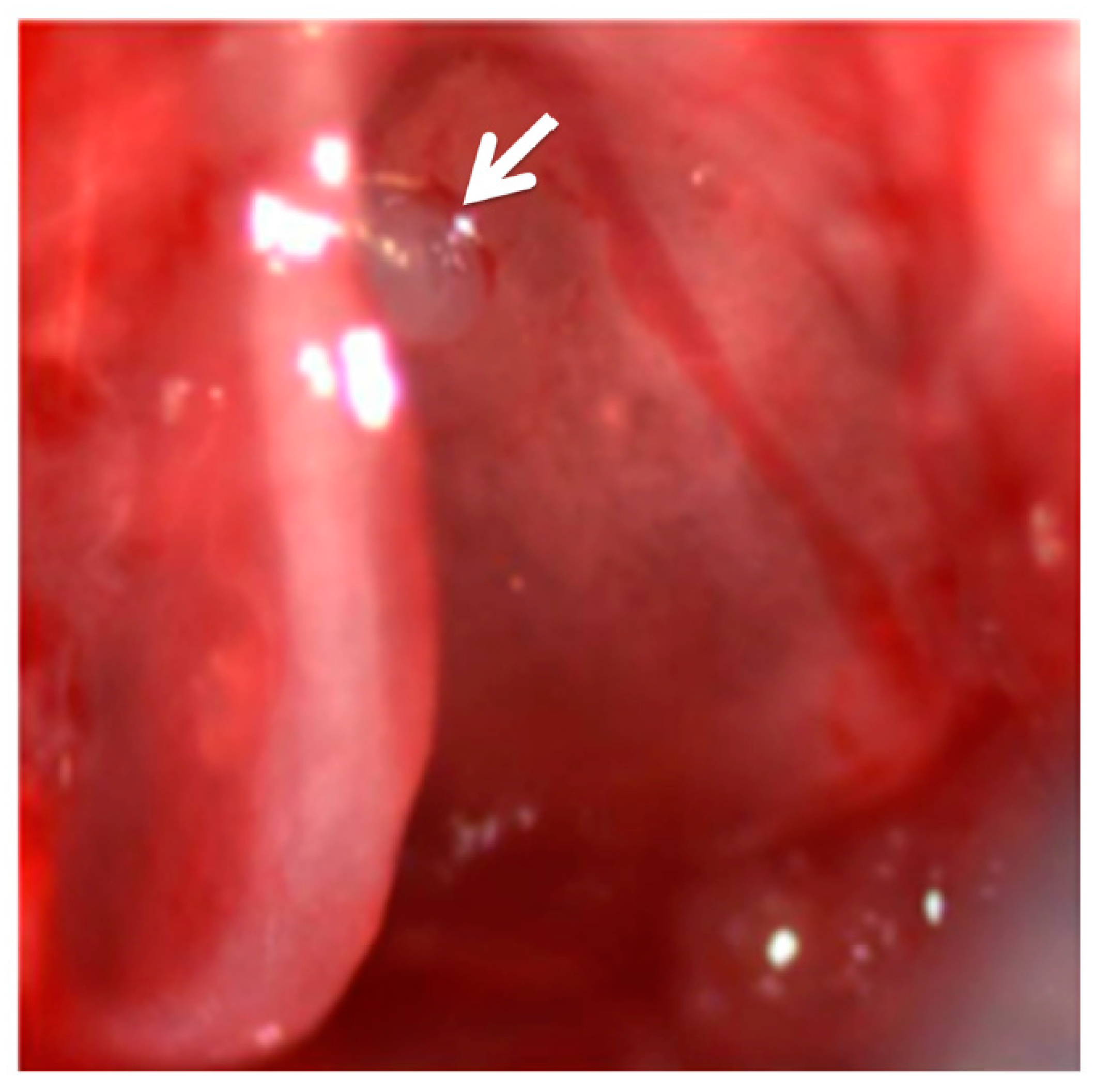
All animal experiments were approved by the Emory Institutional Animal Care and Use Committee and performed using accepted veterinary standards. C57BL/6 mice (Charles River Inc., Wilmington, MA, USA) and Gjb6−/− mice [18] were used in the studies. For drug applications through the round window, a retro-auricular approach was used to expose the tympanic membrane and tympanic ring. The tympanic ring with tympanic membrane was detached from posterior adjacent tissue to allow exposure of cochlea and round window niche. A small cube (3–4 mm3) of absorbable gelatin sponge (Gelfoam, Pfizer Inc., New York, NY, USA, CAT# 59-9863) soaked with one of the retinoid derivatives (retinyl palmitate (RP 30 μM), or retinyl acetate (RAc 30 μM), or all-trans-retinoic acid (ATRA) 30 μM and 300 μM) dissolved in ethanol and phosphate buffered saline (PBS) was placed onto the round window membrane (Figure A1). Drugs are supposedly to reach targeted cells in the cochlea by diffusion across the round window membrane, and also across the basilar membrane to enter the endolymph; therefore, the final concentration of the drugs should be much lower than the original concentration. RP, RAc, and ATRA were purchased from Sigma Aldrich (catalogue #: R3375, R4632, R2625, respectively). The surgery was performed only in the left ear for each mouse and the contralateral ear was used as a non-treatment control. However, previous report also showed that small molecules and viral particles are capable of escaping from one side of the ear to reach the contralateral side [19]. This was taken into the consideration for data interpretation in our studies. Post operatively, we placed mice on a heating pad set at 37 °C (model TR-100; Fine Science Tool Inc., Foster City, CA, USA) to allow animals to recover from the procedure before returning to the animal housing facility. In separate experiments, intraperitoneal (IP) injections were administered at 10 mg/kg at either P5 or P8 every other day until a total of 10 injections were given. Mice in the placebo group received the equivalent dose of PBS with the same amount of ethanol under the same injection time schedule.

2.2. Cochlear Resin Sectioning and Immunolabeling Assays

The cochleae were dissected and fixed in 4% paraformaldehyde (dissolved in PBS). Subsequently, a small opening was made at the apex of the cochlea to allow formation of a perfusion loop via the oval window with 2% PFA and 2.5% glutaraldehyde in PBS, and the samples were further postfixed at 4 °C overnight. Samples were then decalcified in 0.35 M EDTA (pH 7.5, in PBS) for 72 h at 4 °C, followed by gradual dehydration in alcohol of increasing grades, infiltrated, and embedded in epoxy resin (catalogue #: 18060; Ted Pella Inc., Redding, CA, USA) with the conventional protocols [15]. Consecutive cochlear sections (5 µm in thickness) were stained with toluidine blue. Morphology of the sensory epithelium was examined from midmodiolar cochlear resin sections (5 μm in thickness, cut with a Microm HM 335E microtome, Walldorf, Germany).
To determine expression of connexin proteins by immunolabeling, cryo-cochlear sections (8 μm in thickness, cut with a Leica CM1850, Nussloch, Germany) were used. Detail of immunolabeling protocol can be found in our published papers [20]. Briefly, samples were incubated in primary antibody (1:400) plus 5% goat serum and 0.1% Triton at 4 °C overnight. Primary antibody included antibodies against Cx26 (Invitrogen Inc., Carlsbad, CA, USA, catalog# 71–0500, 0.25 µg/mL) and Cx30 (Invitrogen Inc., catalog# 71–2200, 0.5 µg/mL). We also used hair cell markers, myosin6 (1:500) or phalloidin (1:1000), obtained from Proteus bioscience (Ramona, CA, USA) and Sigma-Aldrich Inc. (St. Louis, MO, USA) respectively, on some of the whole-mount cochlear samples as counterstains. Sections and whole-mount preparations were washed in PBS the following day, then incubated for 1 hour at room temperature with secondary antibodies (all purchased from Invitrogen) including: Alexa-555 goat anti rabbit IgG (1:500); Alexa-488 conjugated goat anti mouse IgG (1:500); or Alexa-488 goat anti rabbit IgG (1:500). Images were acquired by a conventional fluorescent microscope (Zeiss Axiovert 135TV, Carl Zeiss, Göttingen, Germany), and analyzed by ImageJ software package (NIH, http://rsbweb.nih.gov/ij/ features.html).

2.3. Auditory Brainstem Responses (ABRs)

ABR measures hearing sensitivity based on sound-evoked potentials and provides an objective measure of hearing thresholds [21]. ABR tests were performed from adult mice (at P26 or older) to allow for appropriate hearing maturation in mice. Tone burst sound stimuli were presented with a sound tube attached to a high-frequency speaker (Tucker Davis Technologies, Alachua, FL, USA), and the tube was inserted to each ear to deliver tone-burst at 4, 8, 12, 18, 24 and 32 kHz to test frequency-specific hearing thresholds. Details of the ABR are provided elsewhere in our published papers [14]. The unpaired student’s t-test was used to determine the significance of differences between data points at the same test frequencies among various groups. The data analysis and plotting software package Origin (ver. 8, Originlab, Northampton, MA, USA) was used for statistical analyses. We defined the criteria of “statistically significant” as p < 0.05.

2.4. Western Blot Analysis

Total proteins from the cochleae of at least two mice were extracted in radio-immunoprecipitation assay (RIPA) lysis buffer by following the manufacturer’s instructions (Upstate Biotechnology Cell Signaling Systems, Lake Placid, NY, USA). Protein concentrations were measured by using a bicinchoninic acid protein assay kit (Pierce, Rockford, IL, USA). Proteins were separated by electrophoresis on a 12% SDS polyacrylamide gel. After transferring to nitrocellulose membranes, we detected Cxs by Western blotting by using polyclonal antibodies against Cx26 (0.5 μg/mL) and/or Cx30 (1 μg/mL; Zymed, South San Francisco, CA, USA). An equal amount of protein (5 μg) was loaded in each lane and this was calibrated with Western blots of β-actin (β-actin, dilution factor 1:1000; Chemicon International, Temecula, CA, USA) as controls. Protein bands on the blots were visualized by enhanced chemiluminescence (SuperSignal; Pierce, Rockford, IL, USA) exposed to x-ray films (Hyper Film; Amersham Biosciences, Piscataway, NJ, USA). The unpaired student’s t-test was used to determine the significance of differences between various test conditions using the data analysis and plotting software package Origin (ver.8, Originlab, Northampton, MA, USA). We defined the criteria of “statistically significant” as p < 0.05.

3. Results

Placement of gel foam soaked with retinyl palmitate (RP, 30 μM) on the round window (RW) showed different expression patterns of Cx26 and Cx30 in the cochlea. Immunolabeling results obtained from adult mice (one month old) revealed that there is a reduction of Cx30 expression in the cochlea to below the detection sensitivity after application of 30 μM of RP on the RW (comparing Figure 1A,B), at both the treated (Figure 1B) and non-treated sides (Figure 1C). In contrast, Cx26 expression was clearly detected (comparing Figure 1D–F). As negative controls, we tested the effect of placing gel foams soaked in PBS without RP and containing the same amount of ethanol in the preparation of RP. Results showed that there is no effect on Cx30 expression level compared to the wild-type (WT) mice when RP was not applied (data not shown). The control data supported the notion that reduction in Cx30 expression in the non-treated ear (Figure 1C) was caused by cross-transfer of RP between the two ears. RP was probably leaked from the treated ear as previously reported by others in gene therapy tests for viral particles [19], and caused the Cx30 reduction in the non-treated ear.
Western blot results were consistent with findings from the immunolabeling. We first compared RP treatment applied at postnatal day 6 (P6) on Cx expressions in the lateral wall (LW) and the organ of Corti (OC) of the inner ear (Figure 2A). Both Cx26 and Cx30 expressions were reduced in the treated ears, and the reductions were more evident in the organ of Corti (p < 0.05) compared to the lateral wall (p > 0.05) (Figure 2A). In the non-treated ear, Cx30 levels were not statistically different from WT controls at P6, although there was some apparent increase in Cx26 expression (Figure 2A). When the Western blots were done at postnatal day 10 (P10), Cx30 expressions in both ears were greatly reduced to about 21% of the control levels (Figure 2B), confirming the immunolabeling results as shown in Figure 1. To further test the effects of retinoids in the cochlear Cx expressions, we tested another compound retinyl acetate (RAc), which is in the same family as RP and predicted to exert the similar effect in the inner ear. Application of RAc (30 μM) on the round window membrane again reduced Cx30 expression level at P10 to that below the detection sensitivity by immunolabeling (Figure 3A–C). Cx26 expression, in contrast, was still detectable after RAc treatment (Figure 3D–F). These results demonstrated that retinoids applied at P10 selectively reduced the Cx30 expressions while leaving the Cx26 relatively intact. When cochlear sections were examined, we found that RP (30 μM)-induced reduction of Cx expression did not result in any cell degeneration in the organ of Corti (Figure 2C, panels A–D).
We next tested RP effects on hearing and cochlear Cx expressions when systemically applied through intraperitoneal injections (IP given at 10 mg/kg, at P5, n = 6). When hearing thresholds were examined by ABR tests from adult mice (P26–28), we found severe hearing loss for mice injected at P5 (n = 6) at most frequencies we tested (Figure 4A, data points shown by hollow triangles, p < 0.05 for frequencies of 4, 8, 12, 18, 24 kHz, p > 0.05 at 32 kHz, when compared to WT controls). Mice (n = 5) received RP injections at P8, however, exhibited only a mild hearing loss when compared to controls (Figure 4A, data points shown by filled squares, p > 0.05 for all frequencies tested). Western blot analyses revealed a significant loss of Cx30 expression in mice injected at P5, while Cx26 expression was not significantly different in mice injected from P8 (Figure 4B, bands in the middle). It is interesting to note that, although given at greater than daily recommended dose, RP dose (10 mg/kg) we tested lies within the therapeutic range in adult mice [22]. Results also suggested that retinoid-induced Cx expression reduction in the cochlea (mostly Cx30, as demonstrated in Figure 1, Figure 2 and Figure 3) were severe enough to result in hearing loss in mice without causing permanent morphological degeneration in the cochlea (Figure 2(C1–C4)). Matured tunnel of Corti and nuel’s space, typical of wild-type mouse at this developmental stage, were observed.
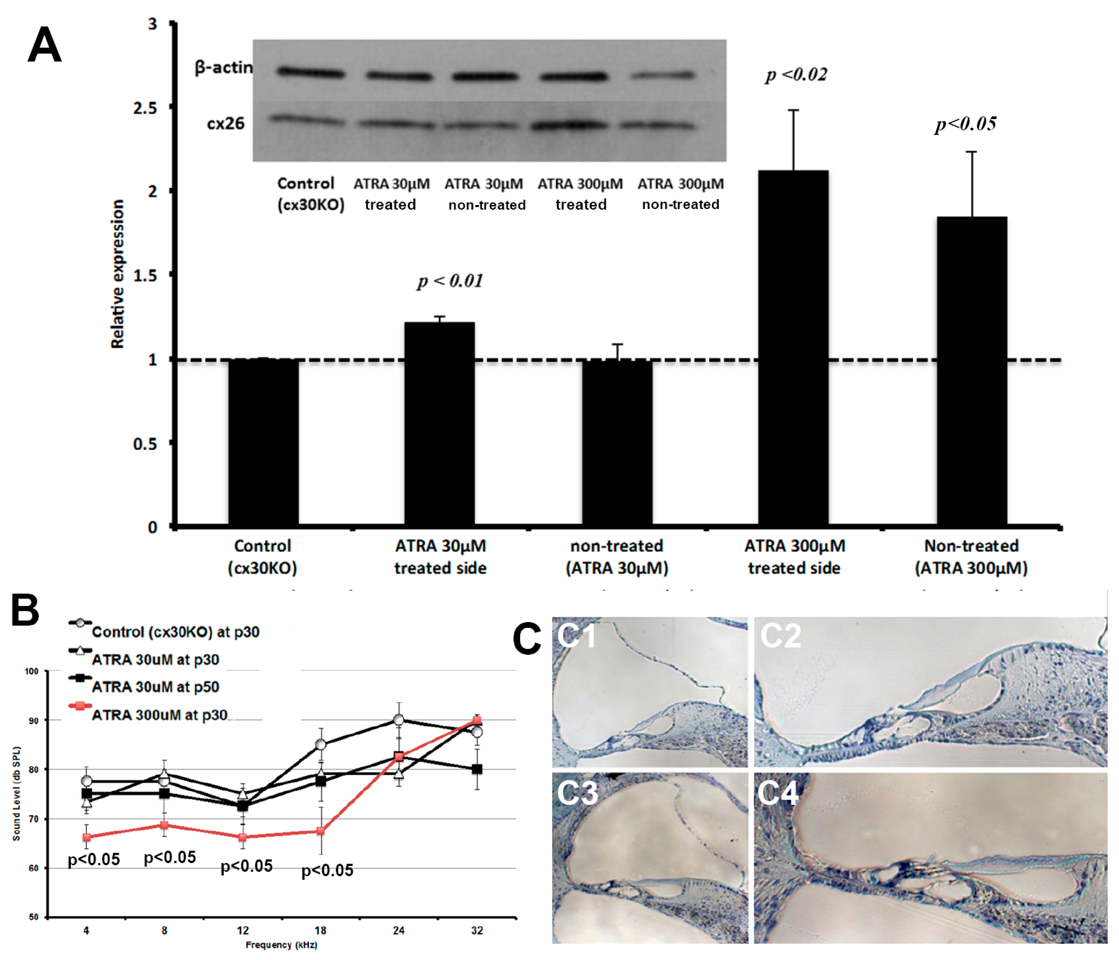
In the next series of experiments we tested the effect of retinoids using the Cx30 knockout (KO) mouse model [18] to examine its effects on Cx expression and hearing (Figure 5). We used all-trans-retinoic acid (ATRA), which was tested in previous studies by others and known to influence the Cx expression. It is also known as one of the most potent forms of retinoids [23,24,25]. Western blot analysis showed statistically significant and dose-dependent increases in Cx26 expression in the cochleae of 30 KO mice treated with 30 μM or 300 μM ATRA respectively (Figure 5A). With 300 μM ATRA applied, Cx26 expressions were nearly doubled when compared to controls (Figure 5A, last two bars). In addition, ABR test results obtained from adult mice (P30, n = 5) revealed approximately ~10 db improvement in hearing thresholds at testing frequencies of 4 kHz, 8 kHz, 12 kHz and 18 k Hz in mice treated with 300 μM ATRA (Figure 5B, red data curve). Morphology of the cochleae showed normal hair cells and no signs of cellular degeneration in other regions of the cochlea (Figure 5C).
Throughout our experiments, we monitored the mice for circling behavior, head tilt, and swimming ability, which are parameters indicative of vestibular functions [26]. We also monitored possible behavioral manifestations of hypervitaminosis A, which include some of the major symptoms in humans and primates such as malaise, drowsiness, nausea, vomiting, poor balance, and muscular incoordination [27]. None of these signs and symptoms was present in any of the experimental animals.

4. Discussion

Our study described the effects of retinoids on cochlear Cx expression during postnatal inner ear maturation as well as the effects on hearing function. Vitamin A has been used as a drug to treat ear diseases (e.g., recurrent otitis media) but has not been used for the treatment of non-syndrome hereditary deafness. Our rationale for studying vitamin A regulation of cochlear Cx expression was based on previous demonstration that most gap junctions in the cochlea are assembled from Cx26 and Cx30, and overexpressing Cx26 alone would sufficiently rescue hearing in Gjb6−/− Mice [14,28]. Our results strongly demonstrated a pattern of Cx expression regulated by retinoids, depending on dosage, route, and timing of injection or surgery.
Both immunolabeling and Western blot data revealed selective reduction of cochlear Cx30 expression by RP while Cx26 expression remain mostly unchanged. In contrast, a more potent retinoid (ATRA) at the dose tested, significantly increased cochlear Cx26 expression (while leaving Cx30 level relatively unchanged) and improved hearing in Cx30 knockout mice by about around 10 dB. The reason why ATRA selectively increased the Cx26 expression is unknown, and we can only speculate the underlying mechanism. Vitamin A impacts the expression of at least 500 known genes including Cx26 and Cx43 [29,30]. Retinoids generally exert their actions by binding to the retinoic acid receptor, which affects the functions of nearby DNA-binding proteins that are critically involved in the transcriptional control of gene expressions. One plausible explanation for the differential ATRA effects we observed for Cx26 and Cx30 could be that the accessory proteins to the retinoic acid receptor may be different near the two connexin genes. This may lead to very different downstream effect, that ultimately resulted in the upregulation of Cx26 expression while negatively affecting the level of Cx30. Ahmad et al. showed that transgenic overexpression of Cx26 completely preserves hearing in the Cx30 KO mice [14]. Similarly, in our study, surgical administration of 300 μM ATRA in Cx30 KO mice resulted in a two-fold increase in the expression of Cx26 (Figure 5A) and resulted in hearing improvement. Quantitatively, Ahmad et al. achieved about a three-fold increase in the Cx26 level in the Cx30 KO mouse model leading to improved hearing [14]. These quantitative comparisons suggest that higher dosage of ATRA may better enhance hearing.
It is important to note that vitamin A is not a known ototoxic agent and the relationship between vitamin A and the inner ear protein expressions in newborn mice are largely unknown. Studies suggest that the minimum daily requirement of vitamin A in mice is 0.72 mg/kg while the minimum teratogenic dose is 75 mg/kg/day by IP injections [22]. Our dose at 10 mg/kg every other day resulted in severe hearing loss when IP administration started at P5. The loss of Cx30 expression seen in Western blot analysis may account for the changes in hearing acuity although it may alter the expression of unaccounted genes.
Timing of retinoid administration appears to be critical in hearing development and Cx expression. This was supported by our data obtained for P8 injections (IP), which led to a mild hearing loss compared to severe hearing loss when injected at P5. In our studies, no systemic signs or symptoms of vitamin A toxicity were visible during our inspection of the treated animals. The underlying cellular and molecular mechanism regarding impact of vitamin A on Cx30 expression is unknown. However, even with a small dose of vitamin A, profound effects on hearing appear to be clinical detrimental while no significant morphological changes in the cochlea were observed (Figure 2C and Figure 5C). Thus, further studies are warranted to understand this relationship and determine the critical period for therapeutic intervention.
This is the first direct demonstration that early postnatal Vitamin A injections resulted in severe hearing loss. Our study provides a cautious tale for high-dose vitamin A exposure in newborn mice and direct impact on Cx expression may be the underlying mechanism of this effect, although it will need further verification. Moreover, the most active form of vitamin A, ATRA, may be a novel therapeutic agent when applied during cochlear development to aid in hearing by increasing Cx26 expression. Therapeutic benefit from an essential nutrient carries great potential for clinical adoption especially when there are no available treatments.
Undoubtedly, many studies have shown the various health benefits of vitamin A including lower risk of hearing loss and even reduced risk of death. However, vitamin A should be taken with caution as there is a significant potential for vitamin A toxicity. Especially in the developed world where 200,000 international units (IU) of vitamin A is commonly given for long-term maintenance dose and 25,000 IU capsules are easily available, the potential for toxic effects must be highlighted as a public health concern. In contradistinction, vitamin A deficiency is common in the developing world and efforts should focus on providing adequate daily allowance of 5000 IU for adults and 2500 IU in children [31]. In conclusion, vitamin A impacts hearing in the critical developmental period, and its impact on Cx expression may be the underlying mechanism. Especially, ATRA holds early promise in treating Cx30 null-induced deafness by overexpression of Cx26. Therefore, further research investigating the risk and benefits of ATRA and other vitamin A sub-types is critical for the development of possible therapeutics in non-syndrome hereditary deafness.

Acknowledgments

This work was funded by NIH (RO1 DC006483 and RO1 DC014496) grants to X.L.

Author Contributions

Both authors conceived and designed the experiments. Y.K. performed the experiments. Both authors analyzed the data and prepared the manuscript.

Conflicts of Interest

The authors declare no conflict of interest.

Appendix

Figure A1. Surgical placement of gel foam soaked in vitamin A derivative was done by opening up the tympanic ring and exposing the RW. Posterior to the above photograph is the RW (indicated by a white arrow) where the placement of the gel foam occurred.
Ohbm 01 00002 g006

References

  1. Frey, S.K.; Vogel, S. Vitamin a metabolism and adipose tissue biology. Nutrients 2011, 3, 27–39. [Google Scholar] [CrossRef] [PubMed]
  2. Finnell, R.H.; Shaw, G.M.; Lammer, E.J.; Brandl, K.L.; Carmichael, S.L.; Rosenquist, T.H. Gene-nutrient interactions: Importance of folates and retinoids during early embryogenesis. Toxicol. Appl. Pharmacol. 2004, 198, 75–85. [Google Scholar] [CrossRef] [PubMed]
  3. Frenz, D.A.; Liu, W.; Cvekl, A.; Xie, Q.; Wassef, L.; Quadro, L.; Niederreither, K.; Maconochie, M.; Shanske, A. Retinoid signaling in inner ear development: A “Goldilocks” phenomenon. Am. J. Med. Genet. A 2010, 152A, 2947–2961. [Google Scholar] [CrossRef] [PubMed]
  4. Lammer, E.J.; Chen, D.T.; Hoar, R.M.; Agnish, N.D.; Benke, P.J.; Braun, J.T.; Curry, C.J.; Fernhoff, P.M.; Grix, A.W., Jr.; Lott, I.T.; et al. Retinoic acid embryopathy. N. Engl. J. Med. 1985, 313, 837–841. [Google Scholar] [CrossRef] [PubMed]
  5. Burk, D.T.; Willhite, C.C. Inner ear malformations induced by isotretinoin in hamster fetuses. Teratology 1992, 46, 147–157. [Google Scholar] [CrossRef] [PubMed]
  6. Lobel, M.J. Clinical studies with parenteral vitamin A therapy in deafness; preliminary report. Eye Ear Nose Throat Mon. 1949, 28, 213–219. [Google Scholar] [PubMed]
  7. Schmitz, J.; West, K.P., Jr.; Khatry, S.K.; Wu, L.; LeClerq, S.C.; Karna, U.L.; Katz, J.; Sommer, A.; Pillion, J. Vitamin A supplementation in preschool children and risk of hearing loss as adolescents and young adults in rural Nepal: Randomised trial cohort follow-up study. BMJ 2012, 344, d7962. [Google Scholar] [CrossRef] [PubMed]
  8. Shim, H.J.; Kang, H.H.; Ahn, J.H.; Chung, J.W. Retinoic acid applied after noise exposure can recover the noise-induced hearing loss in mice. Acta Otolaryngol. 2009, 129, 233–238. [Google Scholar] [CrossRef] [PubMed]
  9. Aladag, I.; Kang, H.H.; Ahn, J.H.; Chung, J.W. Efficacy of vitamin A in experimentally induced acute otitis media. Int. J. Pediatr. Otorhinolaryngol. 2007, 71, 623–628. [Google Scholar] [CrossRef] [PubMed]
  10. Ünal, M.; Öztürk, C.; Aslan, G.; Aydin, Ö.; Görür, K. The effect of high single dose parenteral vitamin A in addition to antibiotic therapy on healing of maxillary sinusitis in experimental acute sinusitis. Int. J. Pediatr. Otorhinolaryngol. 2002, 65, 219–223. [Google Scholar] [CrossRef]
  11. Zelante, L.; Gasparini, P.; Estivill, X.; Melchionda, S.; D’Agruma, L.; Govea, N.; Milá, M.; Monica, M.D.; Lutfi, J.; Shohat, M.; et al. Connexin26 mutations associated with the most common form of non-syndromic neurosensory autosomal recessive deafness (DFNB1) in Mediterraneans. Hum. Mol. Genet. 1997, 6, 1605–1609. [Google Scholar] [CrossRef] [PubMed]
  12. Denoyelle, F.; Weil, D.; Maw, M.A.; Wilcox, S.A.; Lench, N.J.; Allen-Powell, D.R.; Osborn, A.H.; Dahl, H.H.; Middleton, A.; Houseman, M.J.; et al. Prelingual deafness: High prevalence of a 30delG mutation in the connexin 26 gene. Hum. Mol. Genet. 1997, 6, 2173–2177. [Google Scholar] [CrossRef] [PubMed]
  13. Del Castillo, I.; Villamar, M.; Moreno-Pelayo, M.A.; del Castillo, F.J.; Alvarez, A.; Tellería, D.; Menéndez, I.; Moreno, F. A deletion involving the connexin 30 gene in nonsyndromic hearing impairment. N. Engl. J. Med. 2002, 346, 243–249. [Google Scholar] [CrossRef] [PubMed]
  14. Ahmad, S.; Tang, W.; Chang, Q.; Qu, Y.; Hibshman, J.; Li, Y.; Söhl, G.; Willecke, K.; Chen, P.; Lin, X. Restoration of connexin26 protein level in the cochlea completely rescues hearing in a mouse model of human connexin30-linked deafness. Proc. Natl. Acad. Sci. USA 2007, 104, 1337–1341. [Google Scholar] [CrossRef] [PubMed]
  15. Sun, Y.; Tang, W.; Chang, Q.; Wang, Y.; Kong, W.; Lin, X. Connexin30 null and conditional connexin26 null mice display distinct pattern and time course of cellular degeneration in the cochlea. J. Comp. Neurol. 2009, 516, 569–579. [Google Scholar] [CrossRef] [PubMed]
  16. Wang, Y.; Chang, Q.; Tang, W.; Sun, Y.; Zhou, B.; Li, H.; Lin, X. Targeted connexin26 ablation arrests postnatal development of the organ of Corti. Biochem. Biophys. Res. Commun. 2009, 385, 33–37. [Google Scholar] [CrossRef] [PubMed]
  17. Chang, Q.; Tang, W.; Kim, Y.; Lin, X. Timed conditional null of connexin26 in mice reveals temporary requirements of connexin26 in key cochlear developmental events before the onset of hearing. Neurobiol. Dis. 2015, 73, 418–427. [Google Scholar] [CrossRef] [PubMed]
  18. Teubner, B.; Michel, V.; Pesch, J.; Lautermann, J.; Cohen-Salmon, M.; Söhl, G.; Jahnke, K.; Winterhager, E.; Herberhold, C.; Hardelin, J.P. Connexin30 (Gjb6)-deficiency causes severe hearing impairment and lack of endocochlear potential. Hum. Mol. Genet. 2003, 12, 13–21. [Google Scholar] [CrossRef] [PubMed]
  19. Lalwani, A.K.; Jero, J.; Mhatre, A.N. Current issues in cochlear gene transfer. Audiol. Neurootol. 2002, 7, 146–151. [Google Scholar] [CrossRef] [PubMed]
  20. Sun, J.; Ahmad, S.; Chen, S.; Tang, W.; Zhang, Y.; Chen, P.; Lin, X. Cochlear gap junctions coassembled from Cx26 and 30 show faster intercellular Ca2+ signaling than homomeric counterparts. Am. J. Physiol. Cell. Physiol. 2005, 288, C613–C623. [Google Scholar] [CrossRef] [PubMed]
  21. Thompson, D.C.; McPhillips, H.; Davis, R.L.; Lieu, T.L.; Homer, C.J.; Helfand, M. Universal newborn hearing screening: Summary of evidence. J. Am. Med. Assoc. USA 2001, 286, 2000–2010. [Google Scholar] [CrossRef]
  22. Kamm, J.; Ashenfelter, K.; Ehmann, C.W. Preclinical and clinical toxicology of selected retinoids. In The Retinoids; Sporn, M., Roberts, A., Goodman, D., Eds.; Academic Press: New York, NY, USA, 1984; pp. 287–326. [Google Scholar]
  23. Chen, W.; Yan, C.; Hou, J.; Pu, J.; Ouyang, J.; Wen, D. ATRA enhances bystander effect of suicide gene therapy in the treatment of prostate cancer. Urol. Oncol. 2008, 26, 397–405. [Google Scholar] [CrossRef] [PubMed]
  24. Hatakeyama, S.; Mikami, T.; Habano, W.; Takeda, Y. Expression of connexins and the effect of retinoic acid in oral keratinocytes. J. Oral. Sci. 2011, 53, 327–332. [Google Scholar] [CrossRef] [PubMed]
  25. Liu, Y.B.; Xu, B.; Wang, J.W.; Fang, H.Q.; Li, J.T.; Li, H.J.; Tang, Z.; Qian, H.R.; Feng, X.D.; Peng, S.Y. Effects of all-trans retinoic acid on expression of connexin genes and gap junction communication in hepatocellular carcinoma cell lines. Zhonghua Yi Xue Za Zhi 2005, 85, 1414–1418. [Google Scholar] [PubMed]
  26. Steel, K.; Hardisty, R. Assessing Hearing, Vision and Balance in Mice, in What’s Wrong with My Mouse? New Interplays Between Mouse Genes and Behaviour; Society of Neuroscience Short Course Syllabus: Washington, DC, USA, 1996; pp. 26–28. [Google Scholar]
  27. Macapinlac, M.P.; Olson, J.A. A lethal hypervitaminosis A syndrome in young monkeys (Macacus fascicularis) following a single intramuscular dose of a water-miscible preparation containing vitamins A, D2 and E. Int. J. Vitam. Nutr. Res. 1981, 51, 331–341. [Google Scholar] [PubMed]
  28. Ahmad, S.; Chen, S.; Sun, J.; Lin, X. Connexins 26 and 30 are co-assembled to form gap junctions in the cochlea of mice. Biochem. Biophys. Res. Commun. 2003, 307, 362–368. [Google Scholar] [CrossRef]
  29. Balmer, J.E.; Blomhoff, R. Gene expression regulation by retinoic acid. J. Lipid Res. 2002, 43, 1773–1808. [Google Scholar] [CrossRef] [PubMed]
  30. Masgrau-Peya, E.; Salomon, D.; Saurat, J.H.; Meda, P. In vivo modulation of connexins 43 and 26 of human epidermis by topical retinoic acid treatment. J. Histochem. Cytochem. 1997, 45, 1207–1215. [Google Scholar] [CrossRef] [PubMed]
  31. US Department of Agriculture, Agricultural Research Service. USDA National Nutrient Database for Standard Reference, Release 25; Agricultural Research Service: Ames, IA, USA, 2011.
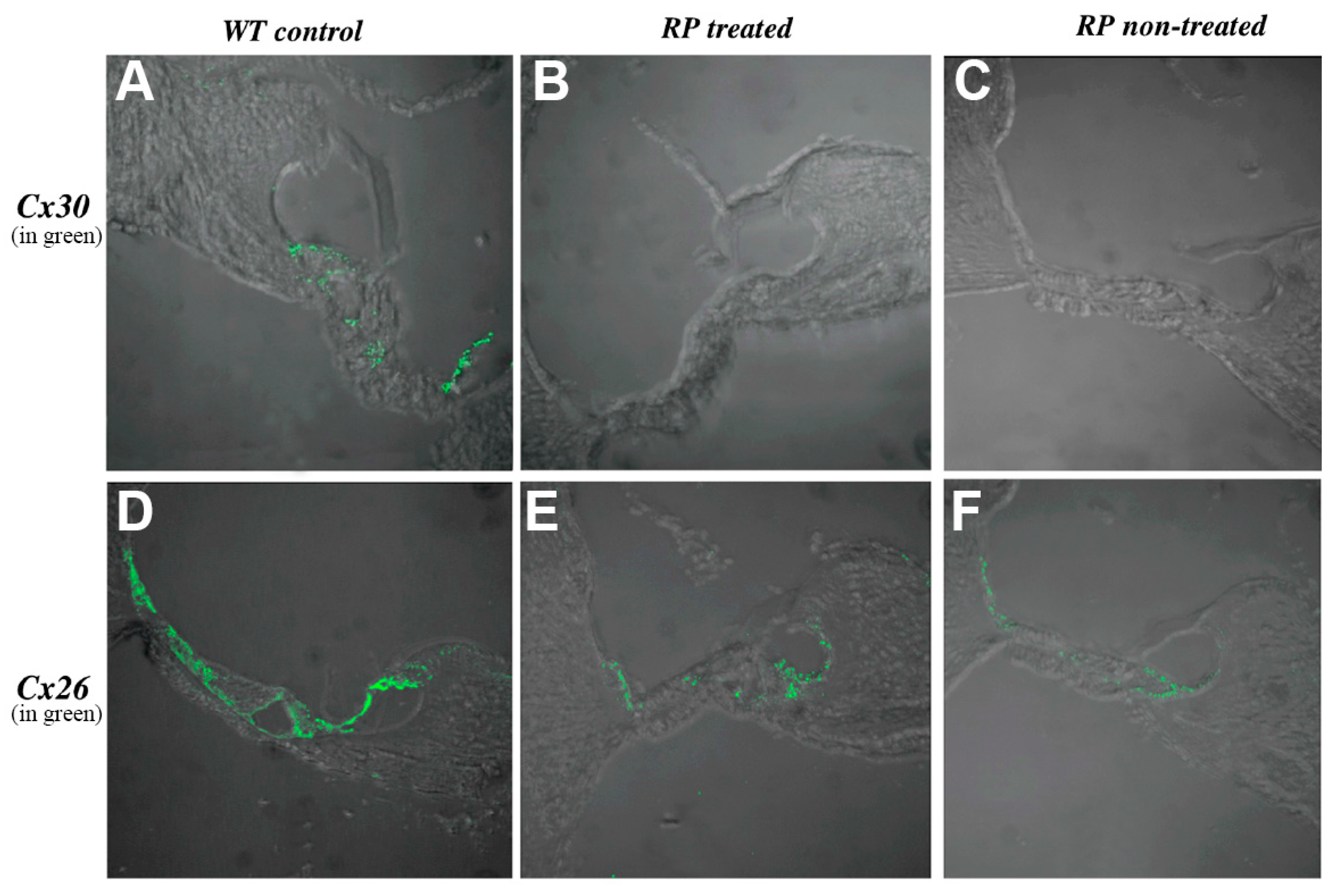
Figure 1. Treatment of retinyl palmitate (RP) differentially affects Cx26 and Cx30 expressions in the cochlea. Immunolabeling results with Cx30 (panels in the upper row, (AC)) and Cx26 (panels in the lower row; (DF)) obtained from adult cochlear sections (one-month-old mice) are given. Experiments were repeated three times and similar results were obtained. Data are shown for results with RP (30 μM) obtained from the treated ear (panel B); the non-treated ear (panel C) and the wild-type (WT) control (panel A); RP was given by placing soaked gel foam at the round window (RW). Results show that Cx30 was below the level of detection 10 days after treatment (panels B,C); In contrast, Cx26 was detected positively in the cochlea (panels E,F) after RP treatment. Panel D is the WT control result.
Ohbm 01 00002 g001
Figure 2. Western blot analysis on RP effect of Cx expressions in the inner ear and cochlear morphology. (A) Cx26 (filled bars) and Cx30 (dashed bars) expressions at P6 in the lateral wall (LW) and organ of Corti (OC), as quantified by Western blots and normalized to appropriate controls; (B) Western blot analysis results of Cx26 (filled bars) and Cx30 (dashed bars) obtained from P10 cochleae after treatment with RP (30 μM)-soaked gel foam, and compared to controls; (C) Epoxy resin cochlear sections reveal normal morphology after implantation of RP at 30 μM. Panels in the left column (C1,C3) show the view of whole cochlear section. (C2,C4) depict magnified views of the organ of Corti in (C1,C3) respectively. Matured tunnel of Corti and nuel’s space, typical of wild-type mouse at this developmental stage, were observed.
Ohbm 01 00002 g002
Figure 3. The effect of applying retinyl acetate (RAc) on the round window membrane for Cx30 (upper panels, (AC)) and Cx26 (lower panels, (DF)) expressions in the cochlea. Experiments were repeated three times and similar results were obtained. RAc is an isomer of RP. Immunolabeling showed that treatment with RAc 30 μM led to marked decrease in Cx30 (B,C) expression in the cochlea; while Cx26 expression was still detected (E,F).
Ohbm 01 00002 g003
Figure 4. (A) Chronic intraperitoneal (IP) injections of RP (10 mg/kg) resulted in a noticeable change in hearing thresholds as measured by auditory brainstem responses (ABRs). When injections are introduced at P5, severe hearing loss results were observed at P26–28. The severity of hearing loss was mild when IP injections are started at P8; (B) Western blot reveals undetectable levels of Cx30 in P26 mice injected with RP (10 mg/kg) at P5 (bands in the middle). This reduction in protein expression was only apparent in mice injected with RP at P5, and not at P8 (lower bands) or with controls injected with PBS.
Ohbm 01 00002 g004
Figure 5. (A) Western blot analysis reveals statistically significant increase in the expression of Cx26 in mice treated with all-trans-retinoic acid (ATRA) 30 μM (p < 0.01) and ATRA 300 μM (p < 0.02) when compared to normalized controls; (B) Hearing thresholds as measured by auditory brainstem responses (ABRs) in adult Cx30 KO mice (P30) with and without treatment (see panel legend in the inset); (C) Epoxy resin sections show no obvious morphological changes at P10 in Cx30 KO mice after implantation of ATRA 30 μM at the RW. Right column present magnified views of the tunnel of Corti and nuel’s space.
Ohbm 01 00002 g005