Proceeding Paper

Determination of Expression Signature and Proportion of mtDNA in Plasma Fractions in Patients with Renal Cell Carcinoma †

by
Elena Arance-Criado
1,*,
Fernando Vázquez-Alonso
2,
Mª Yarmila García-Iglesias
3,
Rocío López-Cintas
3,
Sara Martín-Esteban
4,
Ginesa López-Torres
5,
Ana Isabel Cortés-Valverde
6,
María Jesús Alvarez-Cubero
7 and
Luis Javier Martínez-Gonzalez
1,*
1
GENYO, Centre for Genomics and Oncological Research, Pfizer/University of Granada/Andalusian Regional Government, Av. de la Ilustración 114, 18016 Granada, Spain
2
Urology Department, Hospital Virgen de las Nieves, Av. De las Fuerzas Armadas 2, 18014 Granada, Spain
3
Gran Capitán Health Center, Calle Gran Capitán 10, 18002 Granada, Spain
4
Peligros Health Center, Calle Valencia, s/n, 18210 Peligros, Spain
5
Salvador Caballero Health Center, Calle Dr. Azpitarte, 6, 18012 Granada, Spain
6
Casería Montijo Health Center, Carr. de Jaén, s/n, 18013 Granada, Spain
7
Department of Biochemistry and Molecular Biology III and Inmunology, Faculty of Medicine, University of Granada, Av. de la Investigación, 11, 18016 Granada, Spain
*
Authors to whom correspondence should be addressed.
Presented at the First International Electronic Conference on Genes: Theoretical and Applied Genomics, 2–30 November 2020; Available online: https://iecge.sciforum.net/.
Proceedings 2021, 76(1), 9; https://doi.org/10.3390/IECGE-07148
Published: 2 November 2020

Abstract

:
Renal Cell Carcinoma (RCC) is the third most common urologic malignancy, remaining one of the most lethal urological malignancies, preferably in developed countries. The incidence and mortality rates differ significantly according to sex, race, age and external factors such as smoking, obesity and hypertension increasing RCC risk. The use of novel predictive biomarkers is currently being increased as these improve the diagnosis, progression and prognosis of RCC. Since recent studies have demonstrated a promising association between mitochondrial DNA (mtDNA) copy number alteration in peripheral blood and the risk of developing RCC, we conducted a case-control study into a cohort of 15 controls and 13 patients to determine exosomes mtDNA content in plasma fractions as a potential novel non-invasive biomarker in liquid biopsy in order to monitor the RCC status in patients. In this way, plasma fractions highly purified in exosomes were obtained from blood samples from controls and RCC cases, and relative mtDNA content was measured by quantitative real-time polymerase chain reaction (qPCR). Our results show fragment size distribution profile and we observed that in phase F; with a higher content of exosomal mtDNA; p value shows statistically significant differences in mitochondrial genes HV long and CYB long.

1. Introduction

Renal cell carcinoma (RCC) is the third most common urologic malignancy, remains being one of the most lethal urological malignancies [1]. The incidence of RCC is increasing globally, with rates varying by country, age, race, and sex. Main troubles in managing this disease are unspecific symptoms and incidental detection of RCC by abdominal imaging techniques causing that stages in diagnosis are high. These aspects reinforce the need of identifying novel predictive biomarkers for diagnosis, progression and prognosis of RCC what is fundamental [2]. RCC is not a single entity but includes various tumor subtypes that have been identified on the basis of either characteristic pathologic features or distinctive molecular changes [3].
One of the major challenges of personalized oncology lies in identifying predictive biomarkers of response to therapy that are practical in the clinical setting [4]. For that reason, there are many efforts in searching biomarkers for a proper stratification that will help to an accurate treatment by the differentiation of different subtypes [5].
mtDNA exists as a circular, double-stranded nucleic acid with a high copy number. There are some studies that found that mitochondrial DNA (mtDNA) copy number in peripheral blood has been found to be associated with risk of developing several cancers, like breast, renal, ovarian or lung cancer [6,7,8]. That is the case of the relationship between mtDNA copy number (mtDNAcn) and risk of colorectal cancer [8]. It is known the role of mitochondrial DNA (mtDNA) and its susceptibility to oxidative stress and mutation [9], for that reason, there are many new researches focus on this molecule.
Here we reinforce the role of non-invasive biomarkers in liquid biopsy, mainly in exosomes and mtDNA as promise biomarkers that could improve current ones in RCC. The main objective is to determine the concentration of exosomal mtDNA present in plasma fractions of controls and patients, so that it can be used as a potencial non-invasive biomarker of CRC.

2. Methods

2.1. Patients and Samples

Participants enrolled in this study were recruited by urologists of “Virgen de las Nieves University Hospital”, Granada, Spain. A control group recruited from clinics “Gran Capitán, Salvador Caballero and Caseria de Montijo” was also included. The study protocol was approved by the Ethics Committee (CEI) and informed written consent from all participants was obtained in accordance with the tenets of the Declaration of Helsinki.
A cohort of 28 blood samples collected in EDTA tubes was used, 13 considered cases and the other 15 without cancer, constituting the control group. Samples were processed 4 h later from the collection at collaborators hospitals. To obtain plasma, peripheral whole blood samples were centrifuged for 10 min at 1400× g and at 4 °C and stored at −80 °C.

2.2. Isolation and Extraction of Exosomes from Plasma Samples

To carry out exosomes collection we started with 1 mL of plasma. In order to obtain different plasma fractions of interest, successive centrifugations were carried out with the main aim of eliminating free part of exosomes containing apoptotic bodies and macrovesicles (Table A1). Afterwards, an ultracentrifugation was performed (160,000 g at 4 °C for 2 h) with the purpose of obtaining purified fraction rich in exosomes (phase E). With the purpose of analyzing different plasma fractions, aliquots of 200 µL were taken from supernatants obtained after centrifugations (phases A and C). Sediment from the second centrifugation (phase B) was also analyzed, as well as the supernatant after ultracentrifugation (D phase). Therefore, a total of 80 samples were collected, all of which were kept at −20 °C until their subsequent extraction. All these phases were concentrated at 43 °C (Thermo Scientific™ Savant™ DNA 120SpeedVac™ Concentrator), and then DNA extraction was carried out according to the method detailed by Freeman B et al. [10], a non-organic (proteinase K and salting out) protocol with some modifications described in Gomez-Martín et al. 2015 [11]; subsequently quantified by Qubit Fluorometer and NanoDrop2000c systems (Thermo Fisher Scientific, Waltham, MA, USA).

2.3. Determination of the Relative Concentration of mtDNA by qPCR

Quantification of mtDNA copy number was performed by QuantStudioTM 12K Flex Real-Time PCR System (qPCR) (ThermoScientific) according to the manufacturer’s protocol (iTaq™ Universal SYBR® Green One-Step Kit, Bio-Rad) [12].
Primers were designed for three different regions mitochondrial hypervariable region HV1, apocytochrome b of complex III (MT-CYB) and the hemoglobin subunit beta gene (HBB) gene as reference gene. In addition, two fragments of different length were designed; one of short size between 75–100 bp and another one amplicon with range from 175–200 bp. All these primers were used at a final concentration of 10 µM.
All reactions were incubated in a 96-well plate at 95 °C for 10 min, followed by 40 cycles of 95 °C for 15 s, and 60 °C for 1 min. All qPCR reactions were done in triplicate and the negative controls were included with every qPCR assay. No amplification of the signal was observed when water was added instead of cDNA sample.

3. Results and Discussion

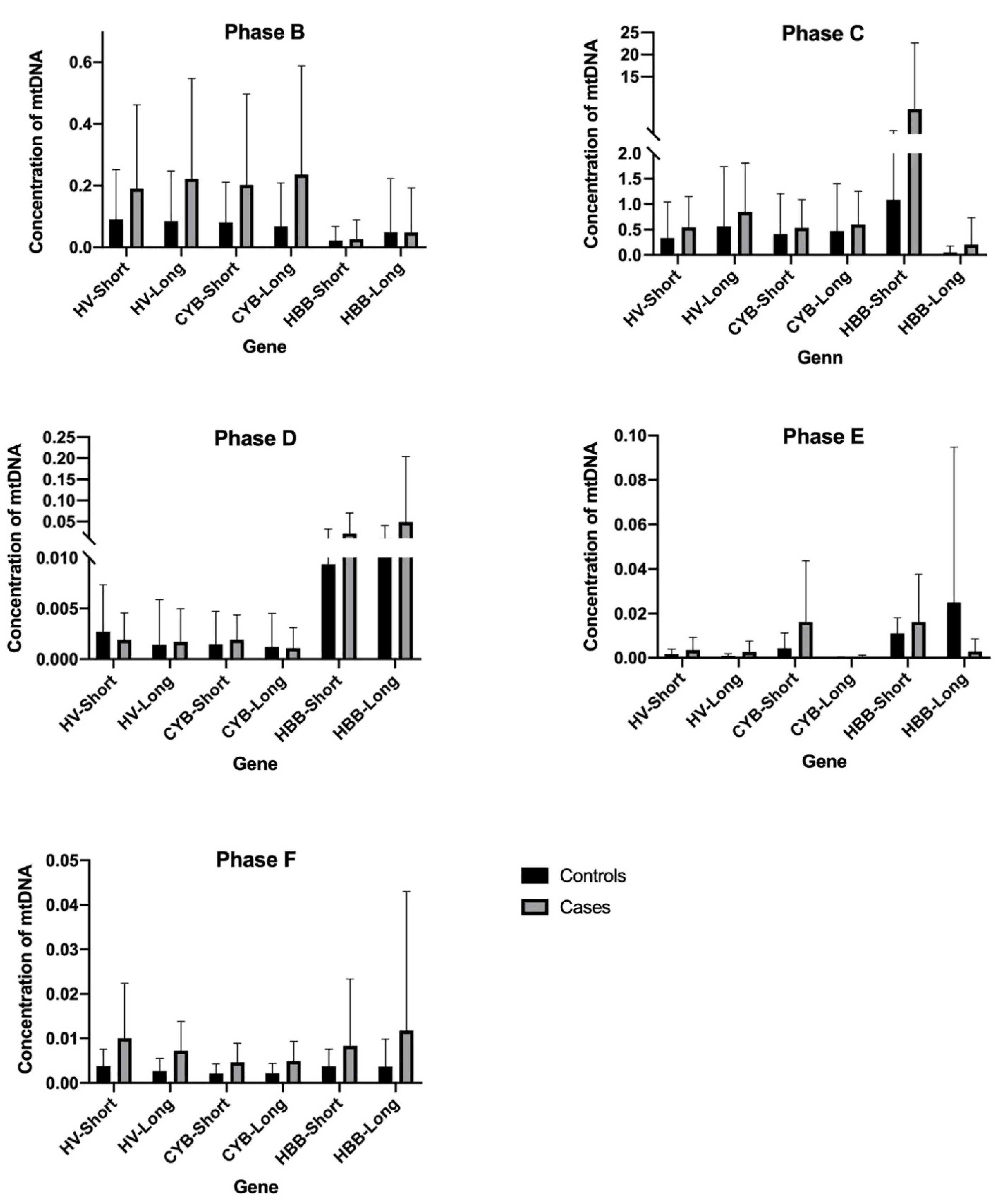
It can be observed that de amount of DNA both nuclear DNA and mtDNA are higher in cases compared with controls, regardless of the phase of the protocol studied.
Analyzing DNA from the first phase to the last one, there is a decrease in the concentration of both mtDNA and nuclear DNA as the exosomes isolation process is followed.
On the one hand, in phase B (plasmatic fraction) there is a high concentration of circulating free DNA and DNA contained in all type of extracellular vesicles (EVs): apoptotic bodies, microvesicles and exosomes. In this fraction, the amount of mtDNA is higher than the nuclear DNA (Figure A1).
On the other hand, phase C (sediment of the centrifugation) is only rich in apoptotic bodies and microvesicles but also contain an elevated quantity of DNA even more than in phase B. This DNA is a product of the amplification of mtDNA derived from the vesicles (Figure A1).
Fractions D and E correspond to the supernatant of the second centrifugation and ultracentrifugation, respectively. These have a high quantity of nuclear DNA and mtDNA, containing fraction D more copies of nuclear DNA than fraction E. In both phases, shorter fragments have a bigger proportion than the larger ones, resulting from free mitochondrial DNA. This DNA presents a small size (180 pb) and normally at a low concentration in biological fluids [13,14,15]. In healthy subjects, circulating DNA derives principally from apoptotic processes from myeloid and lymphoid cells. This DNA circulates in the peripheral blood like nucleosomes or chromatosomes, protected from the action of nucleases. The peak found at 167 pb principally corresponded to a chromatosome form [16]. Due to mtDNA does not have histones nor mechanisms to repair DNA, it is susceptible to fragmentation. This explains the higher proportion of HV-short compared with HV-long, and CYB-short and CYB-long (Figure A1).
Finally, with respect to exosomes-rich phase F (our phase of interest), it shows an increment in concentration of mtDNA due to it is encapsulated in that vesicles, while nuclear DNA appear more represented in phase E. It is observed a little increase in the mitochondrial long genes of this study (HV and CYB), due to the protective role of the lipid bilayer of the exosomes (Figure A1). After making a statistical analysis of the samples and applying a t-test (t-student) to each of the study phases, we observed that in phase F appears the only significant data regarding the difference in DNA concentration between cases and controls. In that fraction, with a higher content of exosomal mtDNA, p value shows statistically significant differences in mitochondrial genes HV long and CYB long. While the nuclear gene (both short and long) shows high p values, short fragments for the mitochondrial genes (HV short and HV long) show interesting p values even though they are not significant (Table A2).

4. Conclusions

This experiment allowed us to analyze the fragment size distribution pattern of different regions of interest (as cell-free DNA and content in EVs) in each plasma fraction, and we have confirmed the high mtDNA content in exosomes as a powerful biomarker. Therefore, application of liquid biopsy in the clinical scenario is a promising non-invasive technique for prediction, early diagnosis and monitoring of cancer treatment. We affirm that it would be quite interesting to study how the amount of mitochondrial DNA varies in controls and patients with RCC in fractions rich in exosomal content, allowing the preservation of mtDNA thanks to its lipid bilayer structure.

Institutional Review Board Statement

The study was conducted according to the guidelines of the Declaration of Helsinki, and approved by Research Ethics Committee of Granada Centre (CEI-Granada, internal code 0165-N-19, 22/04/2019).

Informed Consent Statement

Informed consent was obtained from all subjects involved in the study.

Data Availability Statement

The data presented in this study are available on request from the corresponding author. The data are not publicly available due to ethical issues.

Appendix A

Table A1. Fractions obtained during exosomes isolation.
Fraction or PhaseSampleObtainingObjective
A200 µL plasmaPlasma obtained after centrifugation (1400× g, 4 °C, 10 min)Determination of the relative concentration of mtDNA
BPelletPellet obtained after centrifugation with DTT + PBS (16,000× g, 4 °C, 20 min)
C200 µL supernatantSupernatant obtained after centrifugation (15,000× g, 4 °C, 30 min)
D6 mL supernatantSupernatant obtained after ultracentrifugation (160,000× g, 4 °C, 2 h)
EPelletPellet obtained after ultracentrifugation (160,000× g, 4 °C, 2 h)
Table A2. Means of each gene fragment of controls and cases in each of the phase F. P value regarding the difference in DNA concentration between cases and controls applying a t-test (t-student).
Genep ValueMean of ControlsMean of Cases
HV-Short0.0748710.0038390.01005
HV-Long0.0216760.0026710.007255
CYB-Short0.0589730.0021890.004655
CYB-Long0.0499600.0022330.004892
HBB-Short0.2636380.0037720.008355
HBB-Long0.4101660.0036740.01176
Figure A1. Representation of the DNA concentration of each gene fragment of controls and patients in each of the phases. The black bars represent the group of the controls, and the grey ones the group of the patients.
Proceedings 76 00009 g0a1

References

  1. Capitanio, U.; Bensalah, K.; Bex, A.; Boorjian, S.A.; Bray, F.; Coleman, J.; Gore, J.L.; Sun, M.; Wood, C.; Russo, P. Epidemiology of Renal Cell Carcinoma [Figure presented]. Eur. Urol. 2019, 75, 74–84. [Google Scholar] [CrossRef] [PubMed]
  2. Oto, J.; Plana, E.; Sánchez-González, J.V.; García-Olaverri, J.; Fernández-Pardo, Á.; España, F.; Martínez-Sarmiento, M.; Vera-Donoso, C.D.; Navarro, S.; Medina, P. Urinary microRNAs: Looking for a New Tool in Diagnosis, Prognosis, and Monitoring of Renal Cancer. Curr. Urol. Rep. 2020, 21, 1–8. [Google Scholar] [CrossRef]
  3. Signoretti, S.; Flaifel, A.; Chen, Y.-B.; Reuter, V.E. Renal cell carcinoma in the era of precision medicine: From molecular pathology to tissue-based biomarkers. J. Clin. Oncol. 2018, 36, 3553–3559. [Google Scholar] [CrossRef]
  4. Dudani, S.; Savard, M.F.; Heng, D.Y. An Update on Predictive Biomarkers in Metastatic Renal Cell Carcinoma. Eur. Urol. Focus 2019, 6, 34–36. [Google Scholar] [CrossRef] [PubMed]
  5. Farber, N.J.; Kim, C.J.; Modi, P.K.; Hon, J.D.; Sadimin, E.T.; Singer, E.A. Renal cell carcinoma: The search for a reliable biomarker. Transl. Cancer Res. 2017, 6, 620–632. [Google Scholar] [CrossRef] [PubMed]
  6. Yu, M. Circulating cell-free mitochondrial DNA as a novel cancer biomarker: Opportunities and challenges. Mitochondrial DNA 2012, 23, 329–332. [Google Scholar] [CrossRef] [PubMed]
  7. Kohler, C.; Barekati, Z.; Radpour, R.; Zhong, X.Y. Cell-free DNA in the circulation as a potential cancer biomarker. Anticancer Res. 2011, 31, 2623–2628. [Google Scholar] [PubMed]
  8. Gentiluomo, M.; Katzke, V.; Kaaks, R.; Tjønneland, A.; Severi, G.; Perduca, V.; Boutron-Ruault, M.-C.; Weiderpass, E.; Ferrari, P.; Johnson, T.; et al. Mitochondrial DNA copy-number variation and pancreatic cancer risk in the prospective EPIC cohort. Cancer Epidemiol. Biomark. Prev. 2020, 29, 681–686. [Google Scholar] [CrossRef] [PubMed]
  9. Yang, K.; Li, X.; Forman, M.R.; Monahan, P.O.; Graham, B.H.; Joshi, A.; Song, M.; Hang, D.; Ogino, S.; Giovannucci, E.L.; et al. Pre-diagnostic leukocyte mitochondrial DNA copy number and colorectal cancer risk. Carcinogenesis 2019, 40, 1462–1468. [Google Scholar] [CrossRef] [PubMed]
  10. Freeman, B.; Smith, N.R.; Curtis, C.J.; Huckett, L.; Mill, J.; Craig, I.W. DNA from buccal swabs recruited by mail: Evaluation of storage effects on long-term stability and suitability for multiplex polymerase chain reaction genotyping. Behav. Genet. 2003, 33, 67–72. [Google Scholar] [CrossRef] [PubMed]
  11. Gómez-Martín, A.; Hernández, A.F.; Martínez-González, L.J.; González-Alzaga, B.; Rodríguez-Barranco, M.; López-Flores, I.; Aguilar-Garduno, C.; Lacasana, M. Polymorphisms of pesticide-metabolizing genes in children living in intensive farming communities. Chemosphere 2015, 139, 534–540. [Google Scholar] [CrossRef] [PubMed]
  12. Bio-Rad. iTaq TM. Universal Probes One-Step Kit. pp. 1–2. Available online: https://www.bio-rad.com/webroot/web/pdf/lsr/literature/10032046.pdf.
  13. Yamamoto, Y.; Uemura, M.; Fujita, M.; Maejima, K.; Koh, Y.; Matsushita, M.; Nakano, K.; Hayashi, Y.; Wang, C.; Ishizuya, Y.; et al. Clinical significance of the mutational landscape and fragmentation of circulating tumor DNA in renal cell carcinoma. Cancer Sci. 2019, 110, 617–628. [Google Scholar] [CrossRef]
  14. Mouliere, F.; Robert, B.; Peyrotte, E.A.; Del Rio, M.; Ychou, M.; Molina, F.; Gongora, C.; Thierry, A.R. High Fragmentation Characterizes Tumour-Derived Circulating DNA. PLoS ONE 2011, 6, e23418. [Google Scholar] [CrossRef] [PubMed]
  15. Fernando, M.R.; Jiang, C.; Krzyzanowski, G.D.; Ryan, W.L. Analysis of human blood plasma cell-free DNA fragment size distribution using EvaGreen chemistry based droplet digital PCR assays. Clin. Chim. Acta 2018, 483, 39–47. [Google Scholar] [CrossRef] [PubMed]
  16. Snyder, M.W.; Kircher, M.; Hill, A.J.; Daza, R.M.; Shendure, J. Cell-free DNA Comprises an in Vivo Nucleosome Footprint that Informs Its Tissues-Of-Origin. Cell 2016, 164, 57–68. [Google Scholar] [CrossRef] [PubMed]
Publisher’s Note: MDPI stays neutral with regard to jurisdictional claims in published maps and institutional affiliations.