Proceeding Paper

Polyproline-Rich Peptides Organize Four Cholinesterase Subunits into a Tetramer; BChE and AChE Scavenge Polyproline Peptides Released during Metabolic Turnover †

by
Oksana Lockridge
* and
Lawrence M. Schopfer
Eppley Institute, University of Nebraska Medical Center, Omaha, NE 68198, USA
*
Author to whom correspondence should be addressed.
Presented at the 2nd International Electronic Conference on Crystals, 10–20 November 2020; Available online: https://iocc_2020.sciforum.net/.
Proceedings 2020, 62(1), 5; https://doi.org/10.3390/proceedings2020062005
Published: 31 December 2020
(This article belongs to the Proceedings of The 2nd International Online Conference on Crystals)

Abstract

:
The genes for acetylcholinesterase (AChE) and butyrylcholinesterase (BChE) encode the proteins responsible for enzyme activity. Additional gene products, PRiMA and ColQ, anchor AChE and BChE proteins into membranes. Soluble AChE and BChE tetramers are composed of four identical subunits plus one polyproline-rich peptide. Dilution does not release the polyproline-rich peptide from tetramers. However, protein denaturation, for example, heating in a boiling water bath, dissociates the polyproline-rich peptide. Using mass spectrometry to sequence peptides released from soluble AChE and BChE tetramers, we find sequences that correspond to proline-rich regions from a variety of proteins. A typical peptide sequence contains 20 consecutive prolines in a 23-residue peptide, LPPPPPPPPPPPPPPPPPPPPLP. There is no single, common consensus sequence, i.e., no specific gene appears to be responsible for the polyproline-rich peptides found in soluble AChE and BChE tetramers. We propose that during metabolic turnover, protein fragments containing polyproline-rich sequences are scavenged by AChE and BChE dimers, to make stable AChE and BChE tetramers. The 40-residue, alpha-helical C-terminus of AChE or BChE is the tetramerization domain that binds the polyproline-rich peptide. Four parallel alpha helices wrap around a single antiparallel polyproline peptide to lock the tetramer in place. This organization was established by classical X-ray crystallography for isolated C-termini in complex with a proline-rich peptide. The organization was confirmed for intact, tetrameric human BChE using cryoelectron microscopy. When 40 amino acids are deleted from the carboxy terminus, monomeric enzymes are created that retain full enzymatic activity.

Graphical Abstract

1. Introduction

Butyrylcholinesterase (P06276) in human plasma is stable in the circulation with a half-life of 11 days [1]. Its stability is attributed to several factors including (a) its large size of 340 kDa, (b) the fact that it is sugar coated with 36 N-linked glycans per tetramer [2,3], (c) it is resistant to proteolysis, and d) it is a tetramer. The focus of this review is the tetramer organization of butyrylcholinesterase (BChE). Soluble BChE and acetylcholinesterase (AChE) are assembled into tetramers through the interaction of four tetramerization domains with one polyproline-rich peptide [4,5]. As of the year 2020, this motif for tetramerization is unique for the cholinesterases, but future studies may find it in other protein tetramers.

2. Tetramers Are the Product of More Than One Gene

The coding sequence for the 85 kDa monomer of human BChE (P06276) is on chromosome 3q26 [6], and the 70 kDa monomer of human AChE (P22303) is on chromosome 7q22 [7]. Monomeric proteins with these sequences have full enzyme activity, but they are unstable in the circulation because they are not tetramers. Assembly into tetramers requires additional gene products. The membrane bound forms of BChE and AChE use polyproline-rich regions of ColQ and PRiMA to assemble into tetramers. The tail end of these polyproline-rich proteins anchor BChE and AChE into the basal lamina at neuromuscular junctions or to membranes in the brain [8,9]. In contrast, no specific gene encodes the polyproline-rich peptides found in soluble BChE and AChE tetramers. The soluble BChE and AChE tetramers assemble around any polyproline-rich peptide, regardless of its origin or length as long as the peptide has at least 12 residues. An example is the 15-residue LLTPPPPPLFPPPFF of ColQ [10]. Polyproline peptides purchased from Sigma-Aldrich with molecular weights from 2000 to 5000 convert recombinant BChE monomers and dimers into tetramers [11,12].

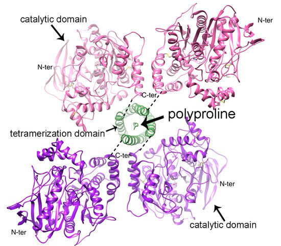
3. Tetramerization Domain

The tetramerization domain of soluble BChE and AChE tetramers is located at the C-terminus and is encoded by a separate exon. The sequence of the 40-residue BChE tetramerization domain is NIDEAEWEWKAGFHRWNNYMMDWLNQFNDYTSKKESC571VGL. The tetramerization domain forms an alpha helix [4,13]. Two alpha helices are linked through a disulfide bond at Cysteine 599 (C571 in the mature secreted BChE). This disulfide bond is the only disulfide bond between subunits [14]. The BChE tetramer is a dimer of two disulfide-linked dimers containing a 4-helix bundle at the interface between two monomers [4]. Four tetramerization domains assemble in a superhelical, coiled-coil structure around a central polyproline II helix, as in Figure 1. The polyproline peptide is tightly bound via hydrophobic stacking with tryptophans and by hydrogen bonds [4,13].

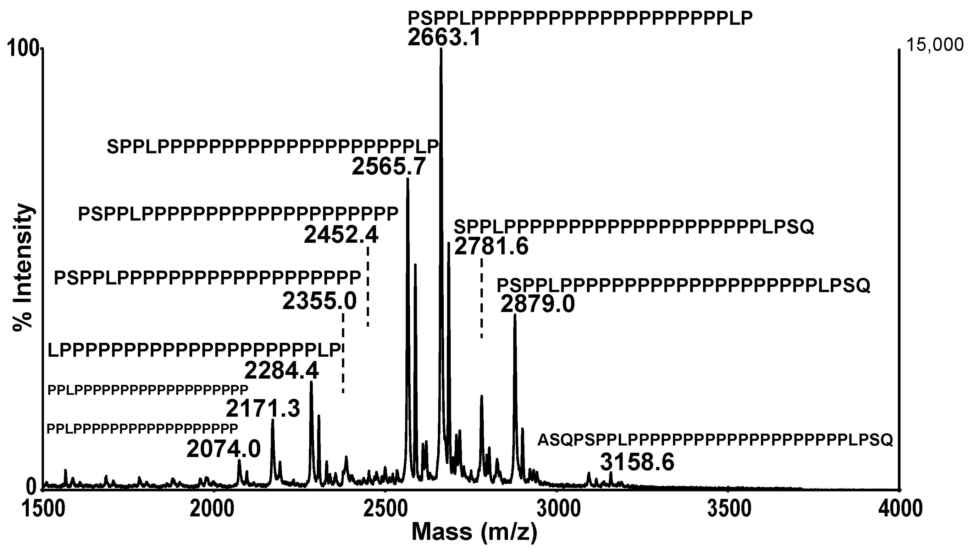
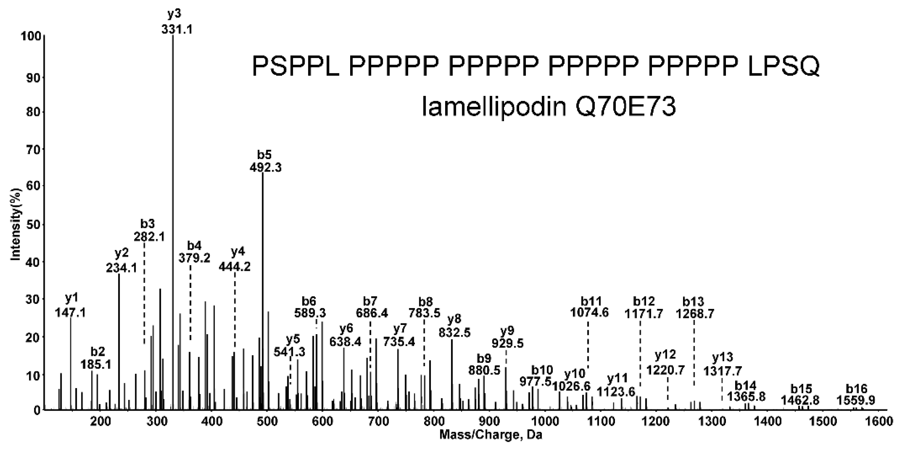
4. Mass Spectrometry Identification of Tetramer Organizing Peptides

We have identified polyproline-rich peptides in BChE tetramers isolated from human plasma, equine plasma, porcine milk, and from recombinant human BChE expressed in Chinese Hamster Ovary Cells [15,16,17,18,19]. In all cases the polyproline peptides were bound noncovalently. Polyproline peptides remained tightly bound in dilute protein solutions but were released when the proteins were denatured in a boiling water bath. The sequences of the released polyproline peptides were determined by mass spectrometry. Figure 2 shows the masses and sequences of 10 polyproline-rich peptides released from human BChE tetramers.
Peptides were separated by high pressure liquid chromatography followed by electrospray ionization mass spectrometry (LC–MS/MS). Fragmentation of the 29-residue lamellipodin peptide in the 5600 Triple-TOF mass spectrometer yielded the MS/MS spectrum in Figure 3. Masses of the b-ion and y-ion series support the amino acid sequence PSPPL PPPPP PPPPP PPPPP PPPPP LPSQ. Peptides released from equine plasma BChE tetramers, porcine milk BChE tetramers, fetal bovine serum AChE, and recombinant human BChE tetramers expressed in Chinese Hamster Ovary cells were also separated and sequenced by LC–MS/MS in the 5600 Triple-TOF mass spectrometer.

5. Polyproline-Rich Peptides in Soluble BChE Tetramers

Table 1 shows that the polyproline peptides in human plasma BChE tetramers originate from 13 different proteins [18]. Lamellipodin contributes 70% of the polyproline peptides. Lamellipodin donates 39 polyproline peptides, ranging in length from 11 to 29 residues, to human plasma BChE tetramers. Short peptides can be derived from longer peptides by losing amino acids through the action of N- and C-terminal aminopeptidases and carboxypeptidase [20]. The longest observed peptide associated with a specific donor protein is listed in Table 1. In some cases, polyproline peptides could be matched to more than one donor protein. The short LPPPP PPPPP P peptide was matched to 27 different proteins. Polyproline-rich peptides consist predominantly of prolines and often include leucine, alanine, serine or glutamine, but never tryptophan.
Polyproline peptides in equine plasma BChE tetramers originate from 12 proteins, of which eight proteins have a match in the mammalian taxonomy, but four have no perfect match [16]. Some polyproline sequences could be matched to more than one protein. For example, a string of 21 contiguous prolines fits both UDP-N-acetylglucosamine transferase subunit ALG13 homolog and formin-like protein 2-like in the Equus caballus taxonomy. Polyproline peptides originating from lamellipodin were present in both equine and human plasma BChE tetramers.
Human plasma BChE and equine plasma BChE tetramers have four polyproline peptide donor proteins in common: UDP-N-acetylglucosamine transferase subunit ALG13 homolog, lamellipodin, leimodin-2, and formin-binding protein 4.
Table 2 lists 12 proteins that donate polyproline peptides to BChE tetramers in porcine milk [19,21]. The most frequent donors are lysine-specific demethylase 6B, acrosin, proline-rich protein 12, and homeobox protein hox-B4. No polyproline peptides from lamellipodin were found in BChE tetramers of porcine milk. The protein donors of polyproline-rich peptides in BChE tetramers from porcine milk are not identical to those in BChE tetramers from human plasma, though three protein donors appear in both Table 1 and Table 2. These are homeobox protein HoxB4, Zinc finger homeobox protein 4, and Zinc finger CCCH domain-containing protein 4.
Recombinant human BChE tetramers expressed in Chinese Hamster Ovary cells (Cricetulus griseus) were purified and analyzed for polyproline peptides. The goal was to determine whether polyproline peptide sequences are specific to the BChE protein or to the cells that synthesize BChE. We identified 60 protein donors of the polyproline peptides in recombinant BChE tetramers [15]. The 60 donor proteins are all Chinese Hamster Ovary (Cricetulus griseus) proteins. Despite their origin from a nonhuman species, the polyproline peptides were incorporated into recombinant human BChE. Five donor proteins from Chinese Hamster Ovary cells were also donor proteins for human plasma BChE synthesized in the liver. The names and accession numbers of the five common donor proteins are listed in Table 3.
Two proteins have accession numbers for Mus musculus because the Cricetulus griseus database is incomplete.
No donor protein contributed the majority of polyproline-rich peptides to recombinant human BChE tetramers expressed in Chinese Hamster Ovary cells. This contrasts with BChE tetramers purified from human plasma, where 70% of the tetramer-organizing peptides were traced to lamellipodin. It was concluded that polyproline peptide sequences in human BChE tetramers are specific to the cells that synthesize BChE and are not specific to the BChE protein.

6. Polyproline-Rich Peptides in Soluble AChE Tetramers

Purified fetal bovine serum AChE tetramers released polyproline-rich peptides [22] from the five donor proteins listed in Table 4. All five of these proteins are also donors for the peptides in human plasma BChE tetramers.
Accession numbers for Homo sapiens proteins are listed because the Bos Taurus database is incomplete.

7. BChE and AChE Scavenge Polyproline Peptides Released from Proteins in the Cytoplasm, Nucleus, Endoplasmic Reticulum, Extracellular Space, and Cell Membrane

Tetramer-organizing polyproline-rich peptides derive from a large number of proteins that reside in a variety of cell compartments including the cytoplasm, nucleus, endoplasmic reticulum, extracellular space, and cell membrane. For example, lamellipodin resides on the cytoplasm side of the cell membrane. Homeobox protein Hox-B4 resides in the nucleus. BChE is secreted through the Golgi apparatus and is never in the cytoplasm or the nucleus. Another fact to consider is that human BChE dimers are converted to human BChE tetramers upon addition of polyproline peptides from Sigma-Aldrich [23]. This was demonstrated for mouse plasma. The human BChE dimers had been produced in mouse plasma by injecting mice with an adenovirus vector encoding human BChE [23]. Exogenously added polyproline peptides became incorporated to form BChE tetramers.
The AChE tetramer in fetal bovine serum, similar to the BChE tetramer in human serum, incorporates polyproline peptides from a variety of protein donors. These observations lead to the conclusion that polyproline peptides are released from cellular proteins during metabolic turnover. The peptides circulate in the blood. Before the peptides reach the kidney, they are taken up by newly synthesized BChE and AChE subunits. This process defines a new function for BChE and AChE, that of scavenging polyproline-rich peptides.

8. Conclusions

Soluble BChE and AChE are peptide scavengers. They scavenge polyproline-rich peptides that are released during cell degradation. This is a newly defined function of soluble BChE and AChE. If excess polyproline-rich peptides are toxic to cells, then scavenging activity protects the cells.
Polyproline-rich peptides in BChE and AChE tetramers originate from a variety of proteins that reside in the cytoplasm, nucleus, endoplasmic reticulum, and cell membrane. Secreted BChE and AChE have no access to proteins in the cytoplasm and nucleus. During cell degradation, peptides are released into the circulation, where they are scavenged by newly synthesized BChE and AChE monomers.
Soluble BChE and AChE tetramers are not degradation products of membrane-bound BChE and AChE. The evidence for this statement is that their polyproline peptides derive primarily from lamellipodin and not from ColQ and PRiMA polyproline peptides.
The BChE tetramer incorporates not only short polyproline-rich peptides, but also long protein fragments that contain a polyproline-rich region. An example is the C5 variant of human BChE whose tetrameric structure includes a 60 kDa lamellipodin fragment [24]. The ability of BChE monomers to assemble into stable, long-lived tetramers by binding the polyproline-rich region of a protein suggests that BChE could serve as a delivery vehicle for any protein that has been engineered to include a polyproline-rich peptide tag.
AChE and BChE have non-cholinergic functions in bone development [25]. A possible explanation for their non-cholinergic function is that AChE and BChE tetramers serve as carriers of proteins that confer the non-cholinergic function.

Author Contributions

O.L. and L.M.S.: writing—review and editing. All authors have read and agreed to the published version of the manuscript.

Funding

This work was funded by NIH Fred & Pamela Buffett Cancer Center, USA, grant P30CA036727.

Conflicts of Interest

The authors declare no conflict of interest.

References

  1. Ostergaard, D.; Viby-Mogensen, J.; Hanel, H.K.; Skovgaard, L.T. Half-life of plasma cholinesterase. Acta Anaesthesiol. Scand. 1988, 32, 266–269. [Google Scholar] [CrossRef]
  2. Kolarich, D.; Weber, A.; Pabst, M.; Stadlmann, J.; Teschner, W.; Ehrlich, H.; Schwarz, H.P.; Altmann, F. Glycoproteomic characterization of butyrylcholinesterase from human plasma. Proteomics 2008, 8, 254–263. [Google Scholar] [CrossRef] [PubMed]
  3. Lockridge, O.; Bartels, C.F.; Vaughan, T.A.; Wong, C.K.; Norton, S.E.; Johnson, L.L. Complete amino acid sequence of human serum cholinesterase. J. Biol. Chem. 1987, 262, 549–557. [Google Scholar] [CrossRef]
  4. Leung, M.R.; van Bezouwen, L.S.; Schopfer, L.M.; Sussman, J.L.; Silman, I.; Lockridge, O.; Zeev-Ben-Mordehai, T. Cryo-EM structure of the native butyrylcholinesterase tetramer reveals a dimer of dimers stabilized by a superhelical assembly. Proc. Natl. Acad. Sci. USA 2018, 115, 13270–13275. [Google Scholar] [CrossRef] [PubMed]
  5. Simon, S.; Krejci, E.; Massoulie, J. A four-to-one association between peptide motifs: Four C-terminal domains from cholinesterase assemble with one proline-rich attachment domain (PRAD) in the secretory pathway. EMBO J. 1998, 17, 6178–6187. [Google Scholar] [CrossRef] [PubMed]
  6. Allderdice, P.W.; Gardner, H.A.; Galutira, D.; Lockridge, O.; LaDu, B.N.; McAlpine, P.J. The cloned butyrylcholinesterase (BCHE) gene maps to a single chromosome site, 3q26. Genomics 1991, 11, 452–454. [Google Scholar] [CrossRef]
  7. Getman, D.K.; Eubanks, J.H.; Camp, S.; Evans, G.A.; Taylor, P. The human gene encoding acetylcholinesterase is located on the long arm of chromosome 7. Am. J. Hum. Genet. 1992, 51, 170–177. [Google Scholar] [PubMed]
  8. Krejci, E.; Thomine, S.; Boschetti, N.; Legay, C.; Sketelj, J.; Massoulie, J. The mammalian gene of acetylcholinesterase-associated collagen. J. Biol. Chem. 1997, 272, 22840–22847. [Google Scholar] [CrossRef]
  9. Perrier, A.L.; Massoulie, J.; Krejci, E. PRiMA: The membrane anchor of acetylcholinesterase in the brain. Neuron 2002, 33, 275–285. [Google Scholar] [CrossRef]
  10. Dvir, H.; Harel, M.; Bon, S.; Liu, W.Q.; Vidal, M.; Garbay, C.; Sussman, J.L.; Massoulie, J.; Silman, I. The synaptic acetylcholinesterase tetramer assembles around a polyproline II helix. EMBO J. 2004, 23, 4394–4405. [Google Scholar] [CrossRef]
  11. Larson, M.A.; Lockridge, O.; Hinrichs, S.H. Polyproline promotes tetramerization of recombinant human butyrylcholinesterase. Biochem. J. 2014, 462, 329–335. [Google Scholar] [CrossRef] [PubMed]
  12. Parikh, K.; Duysen, E.G.; Snow, B.; Jensen, N.S.; Manne, V.; Lockridge, O.; Chilukuri, N. Gene-delivered butyrylcholinesterase is prophylactic against the toxicity of chemical warfare nerve agents and organophosphorus compounds. J. Pharmacol. Exp. Ther. 2011, 337, 92–101. [Google Scholar] [CrossRef]
  13. Boyko, K.M.; Baymukhametov, T.N.; Chesnokov, Y.M.; Hons, M.; Lushchekina, S.V.; Konarev, P.V.; Lipkin, A.V.; Vasiliev, A.L.; Masson, P.; Popov, V.O.; et al. 3D structure of the natural tetrameric form of human butyrylcholinesterase as revealed by cryoEM, SAXS and MD. Biochimie 2019, 156, 196–205. [Google Scholar] [CrossRef] [PubMed]
  14. Lockridge, O.; Adkins, S.; La Du, B.N. Location of disulfide bonds within the sequence of human serum cholinesterase. J. Biol. Chem. 1987, 262, 12945–12952. [Google Scholar] [CrossRef]
  15. Schopfer, L.M.; Lockridge, O. Tetramer-organizing polyproline-rich peptides differ in CHO cell-expressed and plasma-derived human butyrylcholinesterase tetramers. Biochim. Biophys. Acta 2016, 1864, 706–714. [Google Scholar] [CrossRef] [PubMed]
  16. Biberoglu, K.; Schopfer, L.M.; Tacal, O.; Lockridge, O. The proline-rich tetramerization peptides in equine serum butyrylcholinesterase. FEBS J. 2012, 279, 3844–3858. [Google Scholar] [CrossRef]
  17. Li, H.; Schopfer, L.M.; Masson, P.; Lockridge, O. Lamellipodin proline rich peptides associated with native plasma butyrylcholinesterase tetramers. Biochem. J. 2008, 411, 425–432. [Google Scholar] [CrossRef]
  18. Peng, H.; Schopfer, L.M.; Lockridge, O. Origin of polyproline-rich peptides in human butyrylcholinesterase tetramers. Chem. Biol. Interact. 2016, 259, 63–69. [Google Scholar] [CrossRef]
  19. Saxena, A.; Belinskaya, T.; Schopfer, L.M.; Lockridge, O. Tetramer organizing polyproline-rich peptides identified by mass spectrometry after release of the peptides from Hupresin-purified butyrylcholinesterase tetramers isolated from milk of domestic pig (Sus scrofa). Data Brief 2018, 20, 1607–1619. [Google Scholar] [CrossRef]
  20. Koomen, J.M.; Li, D.; Xiao, L.C.; Liu, T.C.; Coombes, K.R.; Abbruzzese, J.; Kobayashi, R. Direct tandem mass spectrometry reveals limitations in protein profiling experiments for plasma biomarker discovery. J. Proteome Res. 2005, 4, 972–981. [Google Scholar] [CrossRef]
  21. Saxena, A.; Belinskaya, T.; Schopfer, L.M.; Lockridge, O. Characterization of butyrylcholinesterase from porcine milk. Arch. Biochem. Biophys. 2018, 652, 38–49. [Google Scholar] [CrossRef] [PubMed]
  22. Biberoglu, K.; Schopfer, L.M.; Saxena, A.; Tacal, O.; Lockridge, O. Polyproline tetramer organizing peptides in fetal bovine serum acetylcholinesterase. Biochim. Biophys. Acta 2013, 1834, 745–753. [Google Scholar] [CrossRef]
  23. Chilukuri, N.; Duysen, E.G.; Parikh, K.; Sun, W.; Doctor, B.P.; Lockridge, O.; Saxena, A. Adenovirus-mediated gene transfer of human butyrylcholinesterase results in persistent high-level transgene expression in vivo. Chem. Biol. Interact. 2008, 175, 327–331. [Google Scholar] [CrossRef] [PubMed]
  24. Schopfer, L.M.; Delacour, H.; Masson, P.; Leroy, J.; Krejci, E.; Lockridge, O. The C5 Variant of the Butyrylcholinesterase Tetramer Includes a Noncovalently Bound 60 kDa Lamellipodin Fragment. Molecules 2017, 22, 1083. [Google Scholar] [CrossRef] [PubMed]
  25. Spieker, J.; Mudersbach, T.; Vogel-Hopker, A.; Layer, P.G. Endochondral Ossification Is Accelerated in Cholinesterase-Deficient Mice and in Avian Mesenchymal Micromass Cultures. PLOS ONE 2017, 12, e0170252. [Google Scholar] [CrossRef]
Figure 1. Cryo-EM structure of the BChE tetramer purified from human plasma. PDB code 6i2t. Figure from reference [4]. Four identical subunits, each composed of 574 amino acids and 9 N-linked glycans, assemble into a tetramer in the presence of a polyproline-rich peptide. Assembly into tetramers does not occur when polyproline peptides are unavailable.
Proceedings 62 00005 g001
Figure 2. MALDI-TOF spectrum of polyproline-rich peptides released from human plasma BChE tetramers by denaturing the pure BChE protein in a boiling water bath. All ten peptides match human lamellipodin (Q70E73). Reproduced from [18].
Proceedings 62 00005 g002
Figure 3. MS/MS fragmentation spectrum of the 29-residue peptide PSPPL PPPPP PPPPP PPPPP PPPPP LPSQ released from the human plasma BChE tetramer. The quadruply charged parent ion has a mass of 719.9/m/z. The protein donor of this peptide is human lamellipodin Q70E73. Reproduced from [18].
Proceedings 62 00005 g003
Table 1. Human protein donors for polyproline-rich peptides released from serum BChE. Data from [18]. A The longest observed peptide in a family of related peptides is listed for each protein donor. Two peptides are listed when a protein has two different polyproline-rich regions. B Pept# is the number of different observed peptides that match to fragments from the Observed Peptide. C Spectral Count is the total number of times that polyproline peptides from this Protein Donor appeared in the mass spectral data. D Prot Match indicates the number of proteins that contain the Observed Peptide.
Protein DonorSwiss Prot
Accession
Observed Peptide APept# BSpectral Count CProt D
Match
LamellipodinQ70E73PSPPL PPPPP PPPPP PPPPP PPPPP LPSQ3919371
UDP-N-acetylglucosamine transferase and deubiquitinase ALG13 isoform 1Q9NP73LPPPP PPPPP PPPPP PPPPP P172393
SynaptopodinQ8N3V7APPPP PPPPP PPP41835
Leiomodin-2Q6P5Q4LPPPP PPPPP P and
TPPPP PPPPP PPPP
2
4
1801
Acetylcholinesterase membrane anchor precursor PRiMA variant IIQ86XR5LPPPP PPPPP P212127
Formin-like protein 1O95466LPPPP PPPPP PP4672
Zinc finger protein ZIC5Q96T25SPPPP PPPPP PP and
LPPPP PPPPP PPPPP P
2
10
611
Homeobox protein Hox-B4P17483GPPPP PPPPP PPP4472
Zinc finger CCCH domain-containing protein 4Q9UPT8GPPPP PPPPP PPP4472
Diaphanous 1O60610STTPP PPPPP PPPPP P5381
Zinc finger homeobox protein 4Q86UP3TPPPP PPPPP PPPPP PPPPP PSA10231
Protein piccoloQ9Y6V0PPPPP PPPPP PPPPP PPPPP PL and
QPPPP PPPPP PPPPP P
11
5
171
Formin-binding protein 4Q8N3X1EPPPP PPPPP PP2363
Table 2. Protein donors for polyproline-rich peptides released from porcine milk BChE. Data from [19]. A A composite of observed peptides from a family of related peptides for each protein donor. Two peptides are listed when two different proline-rich peptides appear in one protein. B Pept# is the number of different peptides that match to fragments from the Observed Peptide. C Spectral Count is the total number of times that polyproline peptides associated with this Protein Donor appeared in the mass spectral data.
Protein DonorAccession Composite Peptide APept# BSpect C
Count
Lysine-specific demethylase 6BXP_005657086PLPPP PLPPP PPPPP PPPPP PPLPG LAT23210
AcrosinP08001PAPPP APPPP PPPPP PPPPP PPPPP QQ25138
Proline-rich protein 12XP_003127395APPPP PPPPP PPPAS EPK and
LPPPP PPPPP PPPPP PPPPP
5
11
123
Homeobox protein Hox-B4XP_003131596RDPGP PPPPP PPPPP PPPPG L11116
Proline-rich membrane anchor 1XP_003482358PPPPL PPPPP PPPPP R7107
Zinc finger homeobox protein 4XP_005663076TPPPP PPPPP PPPPP PPPPP SA and
TPPPP PPPPP PPPPP SSL
8
4
70
29
Zinc finger CCCH domain -containing protein 4XP_005664683GGPPP PPPPP PPPPG PPQM433
Disabled homolog 2-interacting protein-like isoform 1XP_003353684IDQPP PPPPP PPPAP R112
FH2 domain-containing protein 1XP_005666867PPPPS PPPPP PPPP410
WAS/WASL-interacting protein family member isoform X1NP_001231241MPIPP PPPPP PGPPP PPTF26
Protein FAM171A2XP_005668832AAAPP PPPPP PPAPP R14
Proline-rich protein 16XP_005655053PNPPP PPPR11
Table 3. Five donor proteins in common between recombinant human BChE tetramers expressed in Chinese Hamster Ovary cells (Cricetulus griseus) and human plasma BChE tetramers synthesized in human liver.
Donor ProteinAccession Number
Lamellipodin(EGW06139 Cricetulus griseus)
Zinc finger homeobox protein 4(ERE85184 Cricetulus griseus)
Leiomodin-2(ERE89074 Cricetulus griseus)
Homeobox protein Hox-B4(NP_034589 Mus musculus)
Zinc finger CCCH domain-containing protein 4(Q6ZPZ3 Mus musculus)
Table 4. Five proteins donate polyproline-rich peptides to AChE tetramers in fetal bovine serum.
Donor ProteinAccession Number
LamellipodinQ70E73 (Homo sapiens)
Zinc finger homeobox protein 4NP_001180156 (Bos Taurus)
Leiomodin-2NP_001098857 (Bos Taurus)
UDP-N-acetyl glucosamine transferase ALG13 subunit homologNP_001093392 (Homo sapiens)
Protein PiccoloQ9Y6V0 (Homo sapiens)
Publisher’s Note: MDPI stays neutral with regard to jurisdictional claims in published maps and institutional affiliations.

