Proceeding Paper

Optofluidic Particle Detection †

by
Vladislav Agluschewitsch
*,
Mayra Garcés-Schröder
and
Andreas Waag
Institut für Halbleitertechnik, 38106 Braunschweig, Germany
*
Author to whom correspondence should be addressed.
Presented at the 4th International Conference nanoFIS 2020—Functional Integrated nano Systems, Graz, Austria, 2–4 November 2020.
Proceedings 2020, 56(1), 26; https://doi.org/10.3390/proceedings2020056026
Published: 24 December 2020

Abstract

:
The combination of optics and microfluidics for particle detection makes it possible to fabricate small low-cost devices. In such hybrid-on-chip systems with integrated µLEDs, the particles can be close to the light source, which is beneficial for the detection capability according to the simulation results. In addition, further advantages and extension possibilities of such devices are discussed.

1. Introduction

Optofluidic systems have a wide range of applications and still unexplored potential, especially in the field of miniaturized devices [1]. One of the promising applications is particle detection by integration of LED and microfluidic technology to enable, e.g., ubiquitous low-cost measurements of particle concentrations. State-of-the-art systems for particle detection mostly use external light sources that are limited in their portability, especially for parallel detection schemes. By processing the microfluidic channel directly on a structured LED wafer, these limitations can be overcome, and, additionally, the total cost of such systems and the distance between the light source and particles can be reduced. However, the hybrid on-chip combination of these technologies entails new challenges and special requirements. We will address some of the key considerations in the following sections.

2. Detection Setup

The main components of a hybrid particle detection setup are the light source, the microfluidic channel and the detector. Since an LED is a non-directional light emitter, its lateral dimensions should be minimized to increase device sensitivity. Hence, either a pinhole LED or a µLED can be used, which can both be arranged to form an array [2]. The microfluidic channel is located above the light source and transports the particles either in a solution or air. A CMOS sensor or a conventional photodiode can be used to detect the shadow created by the particle. Critical parameters of the setup are the light source dimensions and location, the microfluidic channel height and width, and the in-plane channel geometry. Depending on the targeted particle size, dimensions and fabrication technologies have to be carefully chosen.

3. Results

In order to estimate the optimal distance between the components for the detection of the smallest possible particles, the setup was simulated with a python library named diffractio. In the simulation, the pinhole LED is approximated as a metal aperture with an incident plane wave. The light diffracted at the aperture is scattered by a particle with a refractive index of 1.6 and creates a shadow, as shown in Figure 1a. By varying the distance of the particle to the aperture, the scattering behavior and, thus, the detection capability change. The normalized difference intensity is a measure of the change in light intensity and, thus, the detection capability, and was calculated by subtracting the light intensity with the particle present between light source and detector from the light intensity without the particle, which was then normalized to the intensity without the particle. In Figure 1b, the normalized difference intensity for an x position of the particle equal to zero is shown for different particle z positions. For small particles of diameters below 10 µm, the detection capability increases with smaller distance to the aperture. In contrast, the larger simulated particles of 15 and 20 µm diameter show a decrease in detection capability for smaller distances between the particle and light source. Depending on the targeted particle diameter, it can thus be inferred that the microchannel height (z direction) should not exceed 150 µm for particles with diameters below 10 µm and should be larger than 150 µm for larger particles for a 3 µm aperture.
One difficulty that arises when the particle is closer to the light source is that the microfluidic channel is larger than the area illuminated by the light source. As a result, there are possible flow paths for the particle past the light source that prevent the particle from being detected. A possible solution is to adjust the microfluidic channel dimension and the dimension of the light source, either by enlarging the light source, which, according to our simulations, would not be beneficial for the detection capability, or by reducing the microfluidic channel size. Since operating channels in these dimensions leads to considerable problems, such as increased particle clogging in the channel, particle-focusing concepts are required. In the recent years, several different microfluidic focusing concepts have been developed [3], which could be integrated on the LED wafer. The combination of light source arrays, microfluidics and CMOS sensors allows parallelized measurements that could be used to determine the particle velocity. Another possible combination with the arrays is the use of microfluidic particle-sorting concepts, which would provide information about the particle size.

Funding

Funded by the Deutsche Forschungsgemeinschaft (DFG, German Research Foundation) under Germany’s Excellence Strategy—EXC-2123 QuantumFrontiers–390837967.

References

  1. Frankowski, M.; Theisen, J.; Kummrow, A.; Simon, P.; Ragusch, H.; Bock, N.; Schmidt, M.; Neukammer, J. Microflow Cytometers with Integrated Hydrodynamic Focusing. Sensors 2013, 13, 4674–4693. [Google Scholar] [CrossRef] [PubMed]
  2. Mariana, S.; Scholz, G.; Yu, F.; Dharmawan, A.B.; Syamsu, I.; Prades, J.D.; Waag, A.; Wasisto, H.S. Pinhole microLED Array as Point Source Illumination for Miniaturized Lensless Cell Monitoring Systems. Proceedings 2018, 2, 866. [Google Scholar]
  3. Zhang, J.; Yan, S.; Yuan, D.; Alici, G.; Nguyen, N.T.; Warkiani, M.E.; Li, W. Fundamentals and applications of inertial microfluidics: A review. Lab Chip 2016, 16, 10–34. [Google Scholar] [CrossRef] [PubMed]
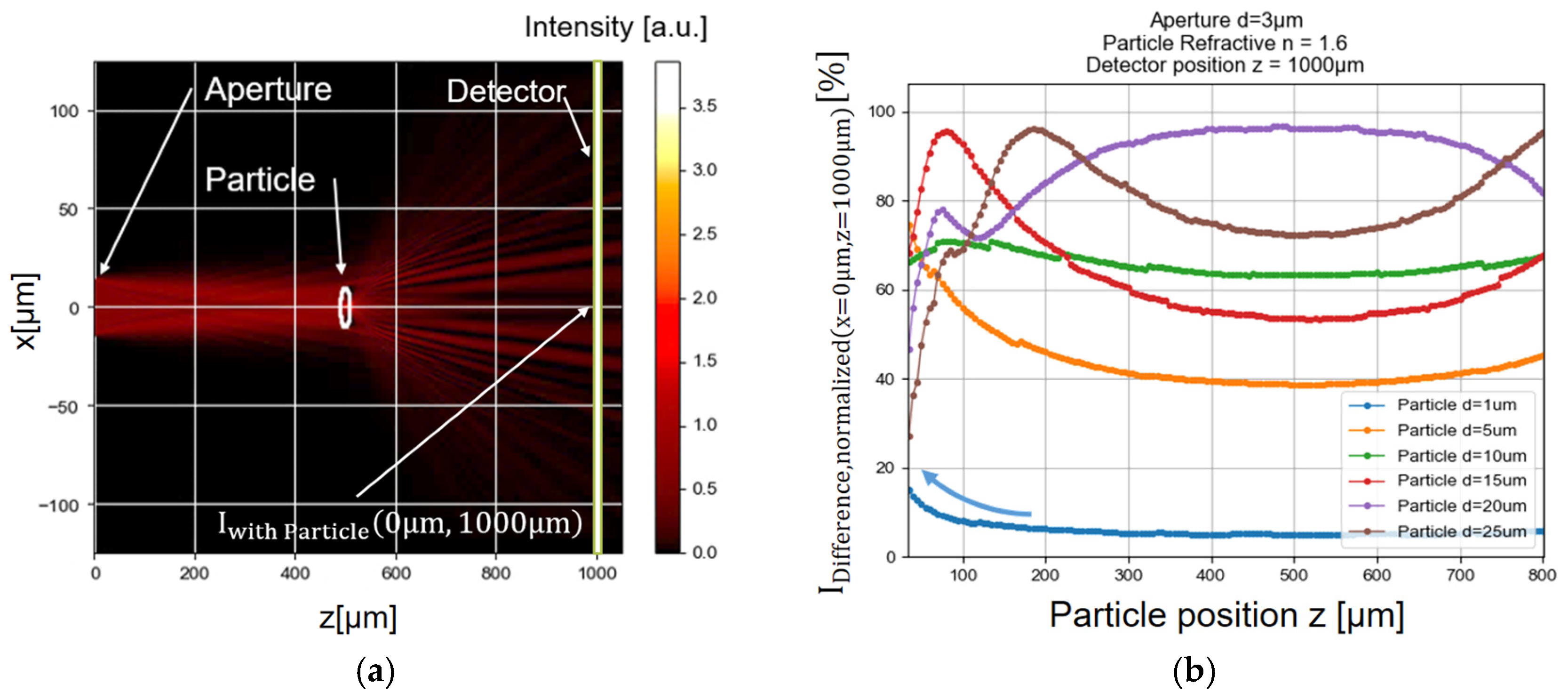
Figure 1. Simulation results of the particle detection setup: (a) the 2D intensity profile with a 30 µm aperture located at z equals zero, and the particle located at x equals zero with a distance of 500 µm between the particle and aperture; (b) normalized difference intensity over the distance between the particle and the 3 µm aperture. An increase in the normalized difference intensity and thus the detectability for the 1 µm particle can be observed for low distances, indicated by the blue arrow.
Proceedings 56 00026 g001
Publisher’s Note: MDPI stays neutral with regard to jurisdictional claims in published maps and institutional affiliations.