Abstract
The aim of this study was to clarify the major differences in the electromyographic (EMG) activities in the hip joint required to achieve a non-rotational (NR) shot as compared with an instep kick from the spatiotemporal data. For this purpose, simulated EMG activities obtained from NR shots and instep kicks were analyzed using principal component analysis (PCA). The PCA was conducted using an input matrix constructed from the time-normalized average and the standard deviation of the EMG activities (101 data x (15 muscles; iliacus, gluteus maximus, rectus femoris, biceos femoris, vastus lateralis, vastus medialis, vastus intermedius, semimembranosus, semitendinosus, sartorius, tensor fasciae latae muscle, adductor magnus muscle, adductor longus muscle, gasctrocnemius, and tibialis anterior)). The PCA revealed that the 3rd, 4th and 8th principal component vectors (PCVs) of the 10 generated PCVs were related to achieving the NR shot (p < 0.05).
1. Introduction
In soccer, ball impact techniques related to the rotation of the ball have been improved; i.e., ball aerodynamics [1,2], the behavior of the ankle joint (while the foot and the ball are in contact, approximately 10 ms [3]), and also the profile of the foot’s trajectories [4,5]. From the viewpoint of the kinematic constraints, the profile of the foot’s trajectory should be a straight line passing through the center of the ball in the impact phase to achieve a non-rotational shot (NR shot), as compared to other complicated curving shots with topspin and sidespin. To achieve the straight-line motion needed to execute an NR shot, the motion of the lower limbs of the kicking and support legs should be coordinated with the motion of the pelvis. We recently demonstrated the coordinated orientation of the hip joint’s angular displacement and the motion of the pelvis to achieve an NR shot [5,6]. Although the electromyographic (EMG) activity patterns in rectus femoris m., vastus medialis m., and adductor longus m. were different depending on the type of kick in soccer [7], the EMG activities of the entire lower limb in achieving the foot’s straight-line trajectory while making an NR shot are still unclear. This is because the lower-limb muscles that contribute to achieving the straight-line-motion to execute an NR shot need both surface and inner muscles.
The principal component analysis (PCA) is attractive because of its usefulness in identifying the movement characteristics of various groups under various conditions using whole data waveforms [6,8]. This PCA statistical method generates principal component vectors (PCVs) and a set of principal component scores (PCSs) for each PCV. Movements with dominant differences arise in lower-numbered PCVs, and the waveforms related to each PCV can be reconstructed by adding and subtracting the PCSs. Thus, it is assumed that the differences in the motion-related simulated-EMG activities in the lower limbs involved in any soccer kick could be detected and evaluated by PCA. Hence, the purpose of this study is to clarify and to detect the differences in the simulated-EMG activities of lower limbs while executing an NR shot from the simulated EMG activities using PCA.
2. Materials and Methods
2.1. Screening and Subjects
Subjects were screened from among 32 players with the Iwate University soccer team. Those selected satisfied the following three criteria. First, they had been youth team members of the Japan Professional Football League and/or a high school soccer club for more than three years. Second, they had no history of major lower limb injuries or neurological disorders. Third, they could make an NR shot with no bounce from outside the penalty area to the goal. Whether they could execute an NR shot was judged by an official Class S leaders’ coach of the Japan Football Association, an incorporated public interest foundation. After screening, three right-leg-dominant male soccer players (age: 21.6 ± 1.3 years; height: 170.0 ± 5.2 cm; weight: 60.7 ± 5.1 kg; competition: more than 10 years, mean ± S.D.) were chosen to participate in this study. All participants gave informed consent, as approved by the ethics committee of Iwate University.
2.2. Experimental Setup
Each subject was asked to execute 10 NR shots toward the front of the goalpost with the target mark (0.75 m high and 1.6 m from the placed ball). To compare the kicking motions, they were also asked to perform 5 straight instep kicks with the right leg. The soccer ball used was the Finale Capitano (Adidas Inc., 0.43 kg weight, 220 mm diameter, size 5). The real-time three-dimensional motion analysis system VENUS3D (Nobby Tech Ltd.) with six cameras and two force platforms was used to detect the kicking motion with twenty reflective markers attached to each subject on both sides of the following anatomical landmarks: the acromia, the anterior superior iliac spine, the posterior superior iliac spine, the greater trochanter, the lateral femoral condyles, the lateral malleoli, the fifth metatarsophalangeal joints, the calcaneus, the thigh’s center of gravity, and the shank’s center of gravity. Three additional markers were placed on the ball to calculate its rotational velocity [5,6,7]. All of the marker coordinates were sampled at 250 Hz for offline analysis. The analyzed section was from t = −200 ms before impact to t = 200 ms after impact through the impact (t = 0). This section was then divided into 101 variables ranging from 0% to 100%. Using the rotational velocity of the ball, the lowest 5 trial datasets of the NR shots were selected. A total of 10 trial (5 NR shots and 5 instep kicks) datasets were selected for further analysis.
2.3. Simulation of the EMG Activities
We simulated the EMG activities in the kicking leg obtained from the 3D coordinates of the landmarks and force data using the musculoskeletal simulation system (AnyBody Modeling System, TERRABYTE Corp., Tokyo, Japan). In this procedure, the 3D coordinates of each landmark were suited to the skeleton model to compensate for differences in the subjects’ physiques. Fifteen EMG activities were calculated: iliac m., gluteus medius m., rectus femoris m., vastus lateralis m., vastus medialis m., vastus intermedius m., biceps femoris m., semitendinosus m., semimembraneous m., sartorius m., tensor fasciae latae m., adductor longus m., adductor magnus m., gastrocnemius m., and tibialis anterior m. The maximum strength of each muscle was depended on the body height and weight. In this study, we defined the maximum strength of each muscle as shown in Table 1. Although the simulated-EMGs might not reflect the actual EMG activities, the validity of simulated EMGs or ligament forces had been described [8,9]. In this study, we denoted each EMG activity as a ratio of own strength, ranging from 0 (no activity) to 1 (maximum activity).

Table 1.
Strength of each muscle (mean ± SD (unit kN)).
2.4. Principal Component Analysis
In the present study, a principal component analysis (PCA) was used to analyze the specific differences in the EMG activities of the kicking leg in the impact phase of an NR shot and those of an instep kick. The PCA generated PCVs and a set of PCSs for each PCV, and the PCV indicated the axis variance, whereas a PCS is the projection of the data input to each PCV. If there are significant differences between the PCSs obtained from the EMG activities of NR shots and instep kicks for a certain PCV, the EMG activities corresponding to that of the PCV could be interpreted as the key EMG activities of kicking related to the differences between NR shots and instep kicks.
The following procedure was used in performing the PCA. First, each trial consisted of a dataset of 1515 variables (i.e., 101 time points and 15 EMG activities). Second, we mean centered each of the 1515 variables using the z-score:
where zt is the standard score of parameter t, Xt is the raw score of parameter t, μt is the mean of parameter t, and σt is the standard deviation of parameter t. The values of parameter t range between 1 and 1515. Third, we constructed 30 by 1515 input matrixes (i.e., 30 trials (3 participants, 10 trial datasets) by 1515 mean-centered parameters). Fourth, we conducted a PCA of the input matrix using a correlation matrix. Fifth, we conducted an independent t-test to compare the significant differences between the PCSs of the data obtained from NR shots and instep kicks. Finally, the EMG activities were recombined [10,11] to interpret the corresponding EMG activities of the PCVs in accordance with the t-test, which showed significant differences between NR shots and instep kicks. Although a recent study [12] demonstrated the applicability of PCA to motion analysis using small sample groups, we repeated the analysis (sub PCA: sPCA) using 30 surrogate input matrices built using the jackknife procedure to validate the results of the main PCA and confirm the stability of the technique. The dimensions of the surrogate input matrices were 29 by 1515. All statistical analyses were executed using commercial statistical analysis software (IBM SPSS Statistics Version 21.0, IBM Japan, Ltd., Tokyo, Japan). The statistical significance was set at p < 0.05.
3. Results
Table 2 shows the PCA results, revealing that the first 10 PCVs explained 71.5% of the EMG activities in the kicking leg. Among these 10 PCVs, the PCSs of PCV3, PCV4, and PCV8 revealed significant differences between NR shots and instep kicks (p < 0.05). Since the cumulative of PCV4 and PCV8 is less than 5%, we would like to focus only on PCV3, and the difference shown by PCV4 and 8 could be ignored.

Table 2.
Results of the principal component analysis (PCA) in EMG activities.
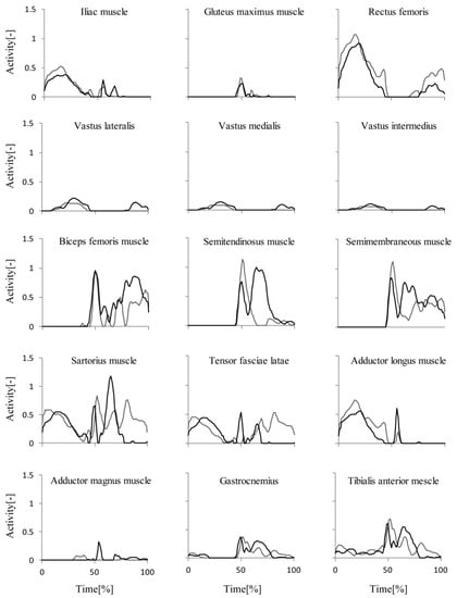
Figure 1 shows the time series of recombined EMG activities in PCV3. From time = 0% to time = 50%, there are remarkable differences in the hip flexion muscle group (rectus femoris m., tensor fasiae latae m. and adductor longus m.). At the impact phase, the simultaneous enhanced EMG activities in tensor fasciae latae m. and adductor longus m. were found while achieving the NR shot as compared to those of the instep kick. On the other hand, immediately before to after the impact phase, the semitendinosus m. and semimembraneous m. show the enhanced EMG activities in an instep kick compared to those of an NR shot.

Figure 1.
Recombined EMG activities in principal component vector 3 (PCV3). Black lines denote a non-rotational (NR) shot, and gray lines are for the instep kick. Remarkable differences were found at the impact phase of the instep kick in semitendinosus m. and semimembraneous m. and of the NR shot in tensor fasciae latae m. and adductor longus m.
4. Discussion
Previously, we demonstrated the coordinated pelvis and thigh motion to achieve a straight-line motion in an NR shot [5,6]. The result that the simultaneous enhancement of EMG activities in tensor fasciae latae m. and adductor longus m. would reflect the reduction in the redundant degree of freedom around the hip joint. In the case of the instep kick, generally, this situation requires higher ball speed than the NR shot. Therefore, the enhancement of semitendinosus m. and semimembraneous m., which are the synergistic muscles for hip extension and knee flexion, would play the role of the driving force against the ball’s moment of inertia at the kicking phase.
5. Conclusions
Achieving the NR shot, the simultaneous enhancement of EMG activities in tensor fasciae latae m. and adductor longus m. would reduce the redundant degree of freedom around the hip joint. On the other hand, enhanced EMG activities in semitendinosus m. and semimembraneous m. would play the role of the driving force against the ball’s moment of inertia at the kicking phase.
Funding
This research received no external funding.
References
- Asai, T.; Kamemoto, K. Flow structure of knuckling effect in footballs. J. Fluid Str. 2011, 27, 727–733. [Google Scholar] [CrossRef][Green Version]
- Kray, T.; Franke, J.; Frank, W. Magnus effect on a rotating soccer ball at high Reynolds numbers. J. Wind Eng. Indust. Aerodyn. 2014, 124, 46–53. [Google Scholar] [CrossRef]
- Nunome, H.; Lake, M.; Georgakis, A.; Stergioulas, L.K. Impact phase kinematics of instep kicking in soccer. J. Sport Sci. 2006, 24, 11–22. [Google Scholar] [CrossRef]
- Hong, S.; Chung, C.; Sakamoto, K.; Asai, T. Analysis of the swing motion on knuckling shot in soccer. Procedia Eng. 2011, 13, 176–181. [Google Scholar] [CrossRef]
- Nakamura, T.; Miyoshi, T.; Takagi, M.; Kamada, Y. Synchronized Lower Limb Kinematics with Pelvis Orientation Achieve the Non-rotational Shot. Sport Eng. 2016, 19, 71–79. [Google Scholar] [CrossRef]
- Nakamura, T.; Miyoshi, T.; Sato, S.; Takagi, M.; Kamada, Y.; Kobayashi, Y. Differences in soccer kicking type identified using principal component analysis. Sport Eng. 2018, 21, 149–159. [Google Scholar] [CrossRef]
- Ozaki, H.; Aoki, K. Kinematic and electromyographic analysis of infront curve soccer kick. Football Sci. 2008, 5, 26–36. [Google Scholar]
- Kang, K.T.; Koh, Y.G.; Nam, J.H.; Jung, M.; Kim, S.J.; Kim, S.H. Biomechanical evaluation of the influence of posterolateral corner structures on cruciate ligaments forces during simulated gait and squatting. PLoS ONE 2019, 14, e0214496. [Google Scholar] [CrossRef] [PubMed]
- Lee, H.; Jung, M.; Lee, K.K.; Lee, S.H. A 3D Human-Machine Integrated Design and Analysis Framework for Squat Exercises with a Smith Machine. Sensors 2017, 17, 299. [Google Scholar] [CrossRef] [PubMed]
- Kobayashi, Y.; Hobara, H.; Matsushita, S.; Mochimaru, M. Key joint kinematic characteristics of the gait of fallers identified by principal component analysis. J. Biomech. 2014, 47, 2424–2429. [Google Scholar] [CrossRef] [PubMed]
- Deluzio, K.J.; Astephen, J.L. Biomechanical features of gait waveform data associated with knee osteoarthritis: An application of principal component analysis. Gait Posture 2007, 25, 86–93. [Google Scholar] [CrossRef] [PubMed]
- Federolf, P.; Reid, R.; Gilgien, M.; Haugen, P.; Smith, G. The application of principal component analysis to quantify technique in sports. Scand. J. Med. Sci. Sport 2014, 24, 491–499. [Google Scholar] [CrossRef] [PubMed]
Publisher’s Note: MDPI stays neutral with regard to jurisdictional claims in published maps and institutional affiliations. |
© 2020 by the authors. Licensee MDPI, Basel, Switzerland. This article is an open access article distributed under the terms and conditions of the Creative Commons Attribution (CC BY) license (https://creativecommons.org/licenses/by/4.0/).