C-28 Esters of Triterpenoid Acids Bearing Tris(hydroxymethyl)aminomethane: Synthesis and Anticancer/Antimicrobial Activity †


Abstract
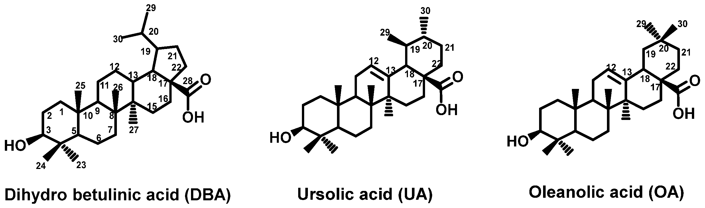
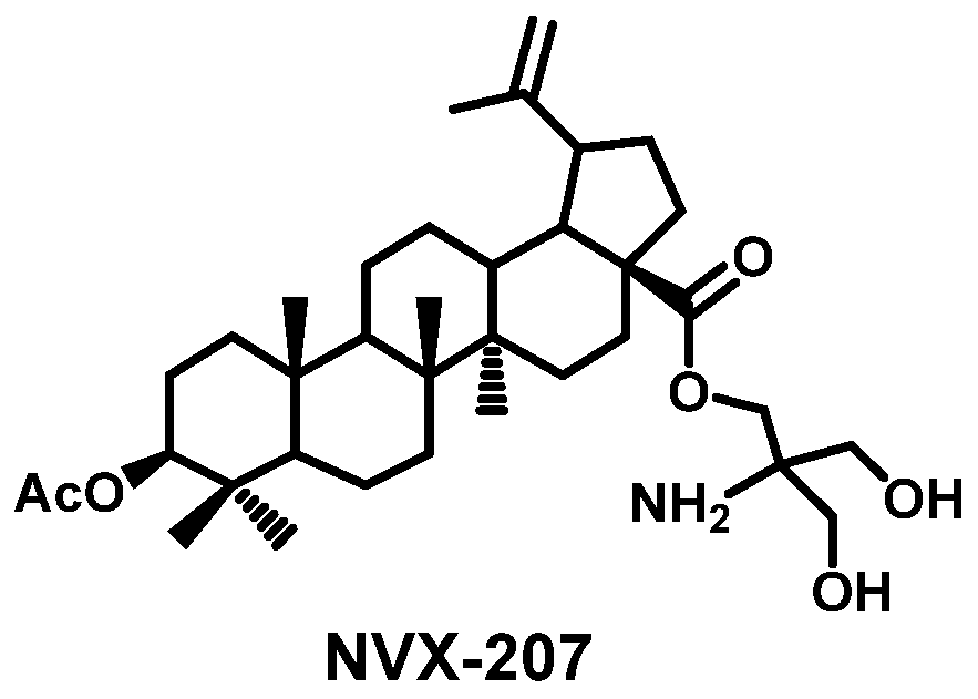
:1. Introduction
2. Experimental
2.1. Chemistry
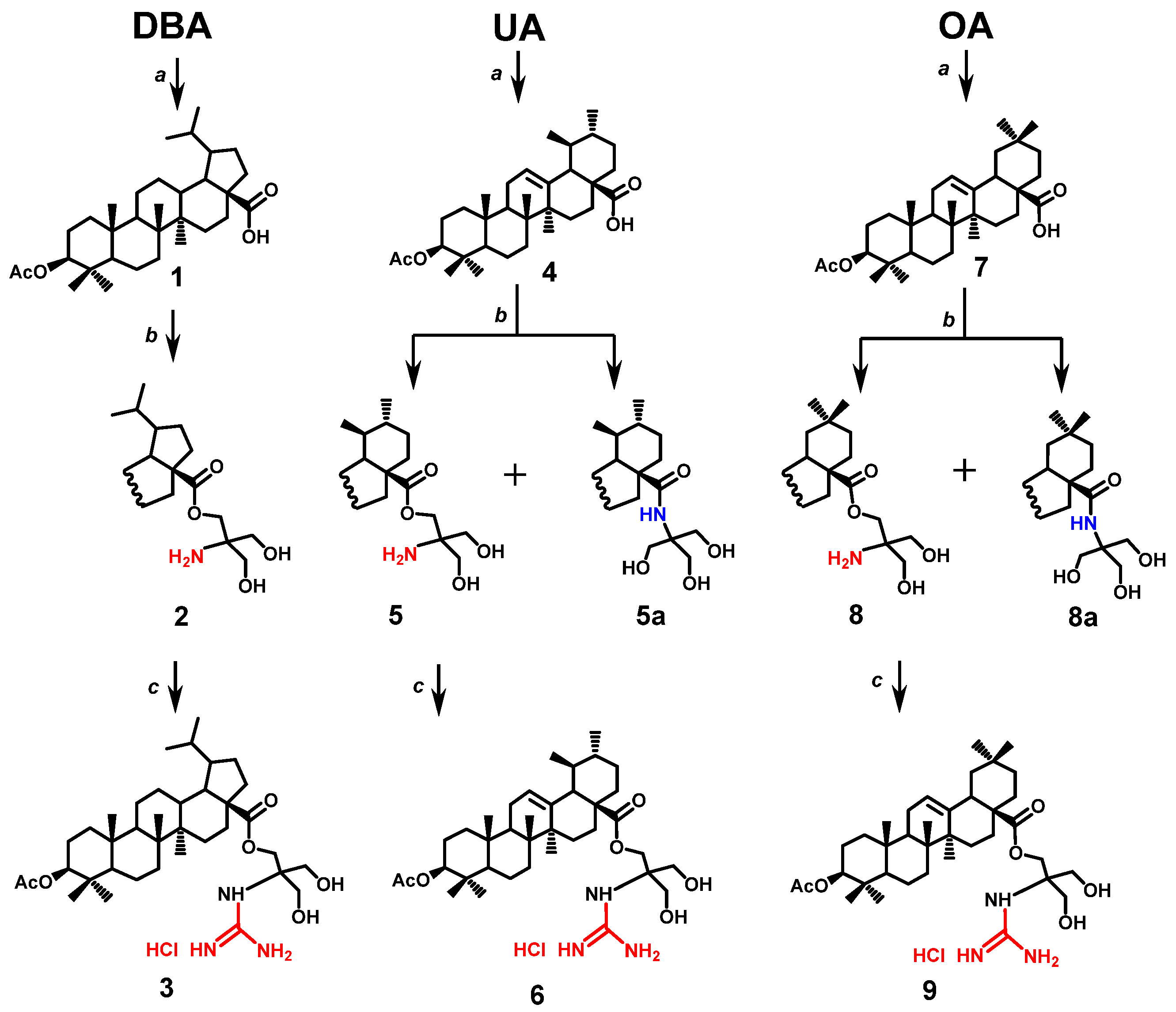
2.1.1. General Procedure for the Synthesis of Amines 2, 5, 8 and Amides 5a, 8a
2.1.2. General Procedure for the Guanilation of Amines 2, 5, and 8
2.2. Biology
2.2.1. Anticancer Activity
Cell Culturing
Cytotoxicity Assay
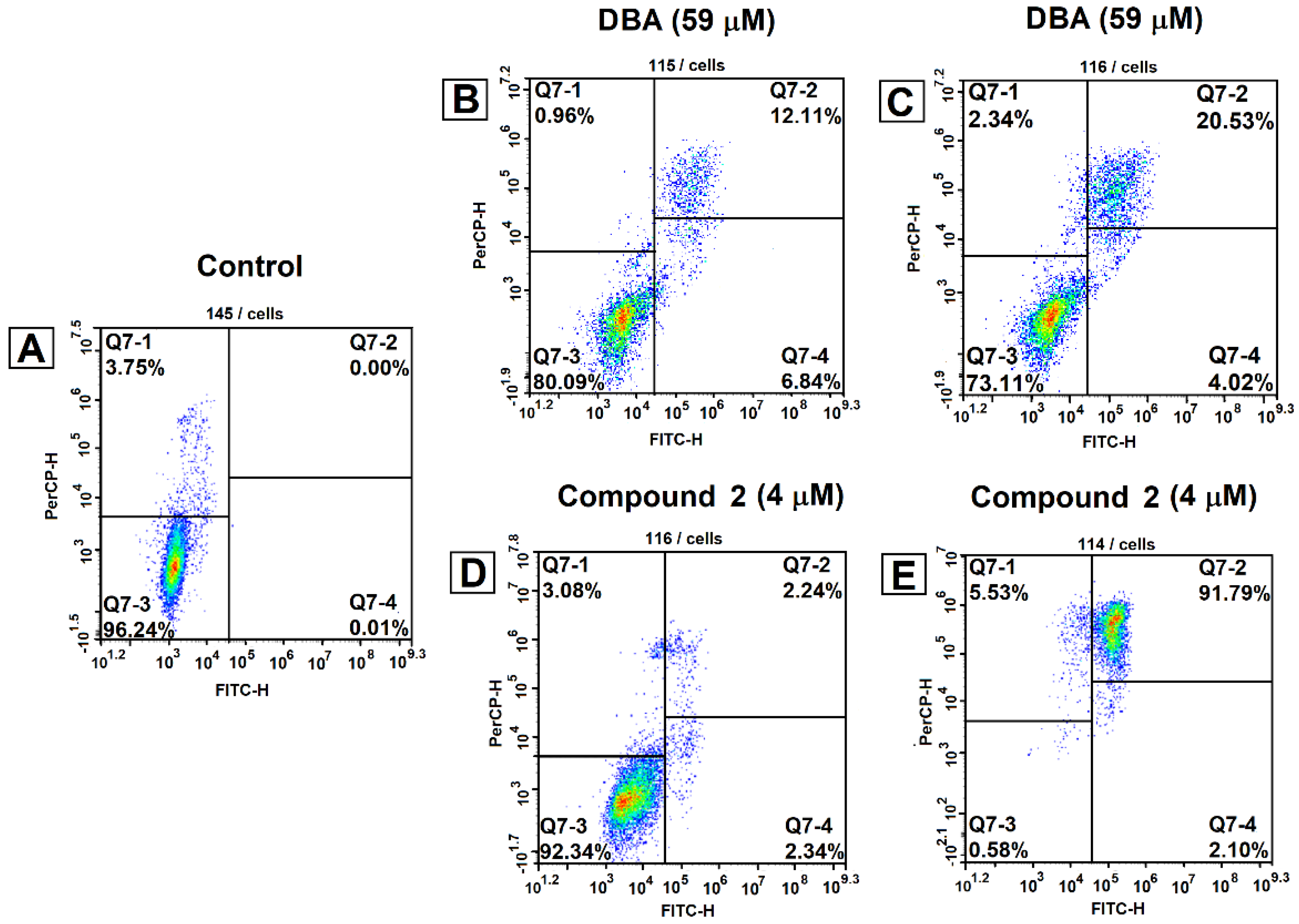
Viability and Apoptosis
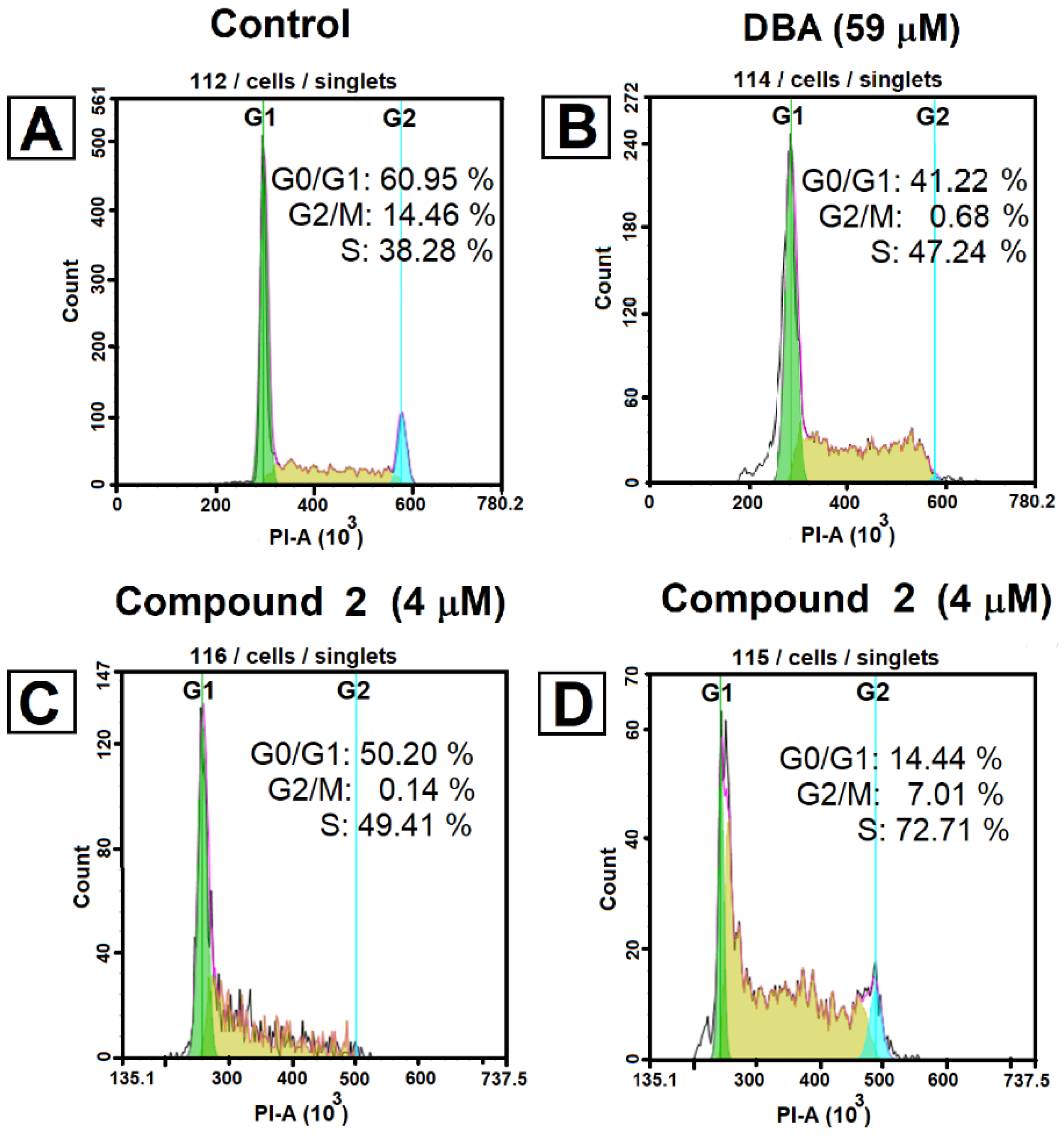
Cell Cycle Analysis
2.2.2. Antimicrobial Activity
Antibacterial Assays
Antifungal Assays
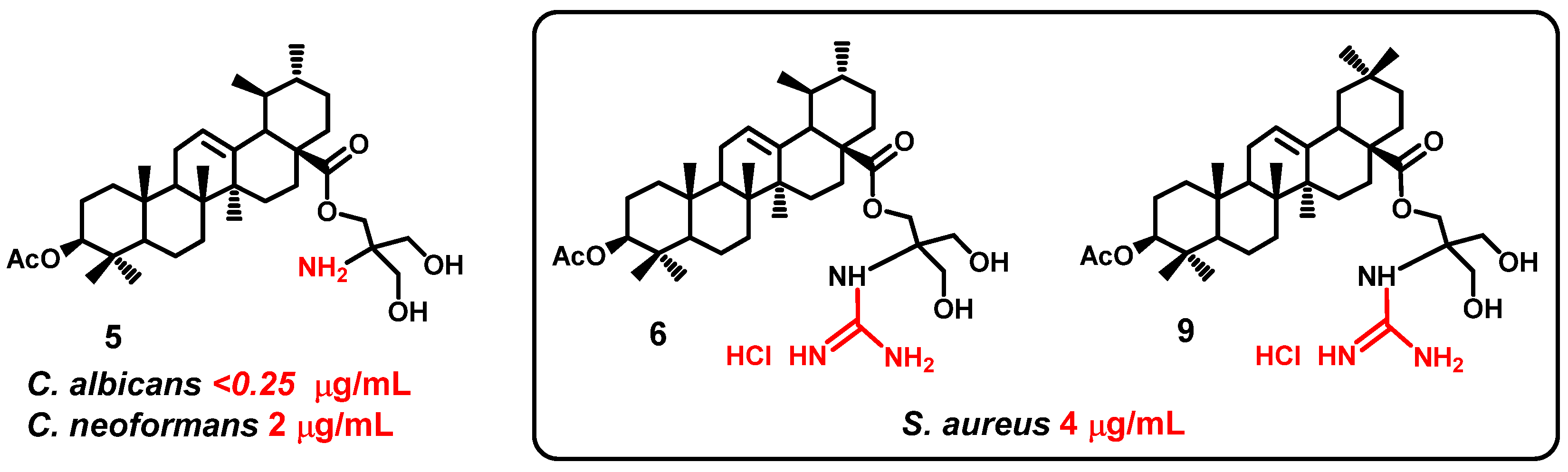
3. Results and Discussion
Chemistry
4. Conclusions
Author Contributions
Funding
Acknowledgments
Conflicts of Interest
References
- Hill, R.A.; Connolly, J.D. Triterpenoids. Nat. Prod. Rep. 2017, 34, 90–122. [Google Scholar] [CrossRef]
- Dzubak, P.; Hajduch, M.; Vydra, D.; Hustova, A.; Kvasnica, M.; Biedermann, D.; Markova, L.; Urban, M.; Sarek, J. Pharmacological activities of natural triterpenoids and their therapeutic implications. Nat. Prod. Rep. 2006, 23, 394–411. [Google Scholar] [CrossRef]
- Sarek, J.; Kvasnica, M.; Vlk, M.; Urban, M.; Dzubak, P.; Hajduch, M. The potential of triterpenoids in the treatment of melanoma. In Research on Melanoma: A Glimpse into Current Directions and Future Trends; Murph, M., Ed.; IntechOpen: Rijeka, Croatia, 2011; pp. 125–158. [Google Scholar]
- Sheng, H.; Sun, H. Synthesis, biology and clinical significance of pentacyclic triterpenes: A multi-target approach to prevention and treatment of metabolic and vascular diseases. Nat. Prod. Rep. 2011, 28, 543–593. [Google Scholar] [CrossRef]
- Salvador, J.A.R.; Moreira, V.M.; Goncalves, B.M.F.; Lealab, A.S.; Jing, Y. Ursane-type pentacyclic triterpenoids as useful platforms to discover anticancer drugs. Nat. Prod. Rep. 2012, 29, 1463–1479. [Google Scholar] [CrossRef] [PubMed]
- Chen, H.; Gao, Y.; Wang, A.; Zhou, X.; Zheng, Y.; Zhou, J. Evolution in medicinal chemistry of ursolic acid derivatives as anticancer agents. Eur. J. Med. Chem. 2015, 92, 648–655. [Google Scholar] [CrossRef] [PubMed]
- Kommera, H.; Kaluderovic, G.N.; Kalbitz, J.; Dräger, B.; Paschke, R. Small structural changes of pentacycliclupane type triterpenoid derivatives lead to significant differences in their anticancer properties. Eur. J. Med. Chem. 2010, 45, 3346–3353. [Google Scholar] [CrossRef] [PubMed]
- Willmann, M.; Wacheck, V.; Buckley, J.; Nagy, K.; Thalhammer, J.; Paschke, R.; Triche, T.; Jansen, B.; Selzer, E. Characterization of NVX-207, a novel betulinic acid-derived anti-cancer compound. Eur. J. Clin. Investig. 2009, 39, 384–394. [Google Scholar] [CrossRef]
- Bache, M.; Bernhardt, S.; Passin, S.; Wichmann, H.; Hein, A.; Zschornak, M.; Kappler, M.; Taubert, H.; Paschke, R.; Vordermark, D. Betulinic acid derivatives NVX-207 and B10 for treatment of glioblastoma—An in vitro study of cytotoxicity and radiosensitization. Int. J. Mol. Sci. 2014, 15, 19777–19790. [Google Scholar] [CrossRef]
- Liebscher, G.; Vanchangiri, K.; Mueller, T.; Feige, K.; Cavalleri, J.M.; Paschke, R. In vitro anticancer activity of betulinic acid and derivatives thereof on equine melanoma cell lines from grey horses and in vivo safety assessment of the compound NVX-207 in two horses. Chem. Biol. Interact. 2016, 246, 20–29. [Google Scholar] [CrossRef]
- Saczewski, F.; Balewski, L. Biological activities of guanidine compounds. Expert Opin. Ther. Pat. 2009, 19, 1417–1448. [Google Scholar] [CrossRef]
- Castagnolo, D.; Schenone, S.; Botta, M. Guanylated diamines, triamines, and polyamines: Chemistry and biological properties. Chem. Rev. 2011, 111, 5247–5300. [Google Scholar] [CrossRef] [PubMed]
- Kim, D.S.; Pezzuto, J.M.; Pisha, E. Synthesis of betulinic acid derivatives with activity against human melanoma. Bioorg. Med. Chem. Lett. 1998, 8, 1707–1712. [Google Scholar] [CrossRef] [PubMed]
- Spivak, A.; Khalitova, R.; Nedopekina, D.; Dzhemileva, L.; Yunusbaeva, M.; Odinokov, V.; D’yakonov, V.; Dzhemilev, U. Synthesis and Evaluation of Anticancer Activities of Novel C-28 Guanidine-Functionalized Triterpene Acid Derivatives. Molecules 2018, 23, 3000. [Google Scholar] [CrossRef] [PubMed]
- Blaskovich, M.A.T.; Zuegg, J.; Elliott, A.G.; Cooper, M.A. Helping Chemists Discover New Antibiotics. ACS Infect. Dis. 2015, 1, 285–287. [Google Scholar] [CrossRef]
- Feichtinger, K.; Zapf, C.; Sings, H.L.; Goodman, M. Diprotected Triflylguanidines: A New Class of Guanidinylation Reagents. J. Org. Chem. 1998, 63, 3804–3805. [Google Scholar] [CrossRef]
- Wolska, K.I.; Grudniak, A.M.; Fiecek, B.; Kraczkiewicz-Dowjat, A.; Kurek, A. Antibacterial activity of oleanolic and ursolic acids and their derivatives. Cent. Eur. J. Biol. 2010, 5, 543–553. [Google Scholar] [CrossRef]
- Jesus, J.A.; Lago, J.H.G.; Laurenti, M.D.; Yamamoto, E.S.; Passero, L.F.D. Antimicrobial Activity of Oleanolic and Ursolic Acids: An Update. Evidence-Based Complement. Altern. Med. 2015, 2015, 620472. [Google Scholar] [CrossRef]
- Haque, S.; Nawrot, D.A.; Alakurtti, S.; Ghemtio, L.; Yli-Kauhaluoma, J.; Tammela, P. Screening and characterisation of antimicrobial properties of semisynthetic betulin derivatives. PLoS ONE 2014, 9, e102696. [Google Scholar] [CrossRef]
- Fontanay, S.; Grare, M.; Mayer, J.; Finance, C.; Duval, R.E. Ursolic, oleanolic and betulinic acids: Antibacterial spectra and selectivity indexes. J. Ethnopharmacol. 2008, 120, 272–276. [Google Scholar] [CrossRef]
- Mallavadhani, U.V.; Mahapatra, A.; Jamil, K.; Reddy, P.S. Antimicrobial Activity of Some Pentacyclic Triterpenes and Their Synthesized 3-O-Lipophilic Chains. Biol. Pharm. Bull. 2004, 27, 1576–1579. [Google Scholar] [CrossRef]
- Innocente, A.; Casanova, B.B.; Klein, F.; Lana, A.D.; Pereira, D.; Muniz, M.N.; Sonnet, P.; Gosmann, G.; Fuentefria, A.M.; Gnoatto, S.C.B. Synthesis of isosteric triterpenoid derivatives and antifungal activity. Chem. Biol. Drug Des. 2014, 83, 344–349. [Google Scholar] [CrossRef] [PubMed]
- Huang, L.; Luo, H.; Li, Q.; Wang, D.; Zhang, J.; Hao, X.; Yang, X. Pentacyclic triterpene derivatives possessing polyhydroxyl ring A inhibit Gram-positive bacteria growth by regulating metabolism and virulence genes expression. Eur. J. Med. Chem. 2015, 95, 64–75. [Google Scholar] [CrossRef] [PubMed]


| Compound | IC50 (µM) a | ||
|---|---|---|---|
| Jurkat | K562 | U937 | |
| NVX-207 | 4.15 ± 0.21 | 4.42 ± 0.26 | 1.87 ± 0.11 |
| 2 | 2.78 ± 0.16 | 3.19 ± 0.17 | 1.14 ± 0.09 |
| 3 | 3.1 ± 0.41 | 2.3 ± 0.34 | 15 ± 0.23 |
| 5 | 6.19 ± 0.27 | 6.59 ± 0.29 | 2.91 ± 0.18 |
| 6 | 3.8 ± 0.23 | 11 ± 0.18 | 5.3 ± 0.29 |
| 8 | 2.96 ± 0.18 | 3.21 ± 0.22 | 2.81 ± 0.15 |
| 9 | 7.6 ± 0.33 | 13 ± 0.45 | 6.8 ± 0.11 |
| DBA | 59 ± 0.31 | 44 ± 0.24 | 39 ± 0.38 |
| UA | 23 ± 0.34 | 68 ± 0.11 | 17 ± 0.12 |
| OA | 271 ± 0.19 | 235 ± 0.24 | 186 ± 0.18 |
| Compound | Gram-Positive Bacteria b, Gram-Negative Bacteria c and Fungal Genetic d Strains | |||||||
|---|---|---|---|---|---|---|---|---|
| Sa b | Ec c | Kp c | Pa c | Ab c | Ca d | Cn d | ||
| 2 | 29.41 | 37.71 | 4.97 | −10.92 | −28.05 | 72.24 | −10.5 | |
| 3 | 63.83 | −19.19 | −12.78 | 2.65 | −42.67 | 72.1 | 69.21 | |
| 5 | 49.64 | 4.43 | 9.19 | 8.96 | 9.71 | 98.8 | 67.64 | |
| 6 | 79.47 | 0.39 | −0.57 | −5.98 | 8.11 | 23.07 | 118.6 | |
| 8 | 14.95 | 5.63 | 13.25 | 13.37 | 3.01 | 40.42 | 1.16 | |
| 9 | 72.3 | 3.74 | 12.35 | −12.6 | −10.66 | 76.58 | 106.1 | |
| DBA | 15.59 | 8.16 | 4.77 | 1.38 | 8.37 | 9.31 | −52.68 | |
| UA | 4.69 | 8.78 | −2.51 | −3.62 | 4.19 | 2.10 | −84.32 | |
| OA | 7.17 | 6.40 | 2.82 | −3.76 | 0.97 | 3.22 | −92.51 | |
© 2019 by the authors. Licensee MDPI, Basel, Switzerland. This article is an open access article distributed under the terms and conditions of the Creative Commons Attribution (CC BY) license (http://creativecommons.org/licenses/by/4.0/).
Share and Cite
Spivak, A.; Nedopekina, D.; Dzhemileva, L.; Khalitova, R. C-28 Esters of Triterpenoid Acids Bearing Tris(hydroxymethyl)aminomethane: Synthesis and Anticancer/Antimicrobial Activity. Proceedings 2019, 41, 45. https://doi.org/10.3390/ecsoc-23-06493
Spivak A, Nedopekina D, Dzhemileva L, Khalitova R. C-28 Esters of Triterpenoid Acids Bearing Tris(hydroxymethyl)aminomethane: Synthesis and Anticancer/Antimicrobial Activity. Proceedings. 2019; 41(1):45. https://doi.org/10.3390/ecsoc-23-06493
Chicago/Turabian StyleSpivak, Anna, Darya Nedopekina, Lilya Dzhemileva, and Rezeda Khalitova. 2019. "C-28 Esters of Triterpenoid Acids Bearing Tris(hydroxymethyl)aminomethane: Synthesis and Anticancer/Antimicrobial Activity" Proceedings 41, no. 1: 45. https://doi.org/10.3390/ecsoc-23-06493
APA StyleSpivak, A., Nedopekina, D., Dzhemileva, L., & Khalitova, R. (2019). C-28 Esters of Triterpenoid Acids Bearing Tris(hydroxymethyl)aminomethane: Synthesis and Anticancer/Antimicrobial Activity. Proceedings, 41(1), 45. https://doi.org/10.3390/ecsoc-23-06493