Proceeding Paper

In-Situ Preparation of Three Types of Noble Metal Nanoparticles-Polyacrylonitrile Nanofibers (NM NPs-PAN NFs, M = Pt, Au, and Ag) Using Electrospinning Technique Assisted with Ultrasound Irradiation †

Research Laboratory of Inorganic Materials Synthesis, Department of Chemistry, Iran University of Science and Technology, Narmak, Tehran 16846, Iran
*
Author to whom correspondence should be addressed.
Presented at the 23rd International Electronic Conference on Synthetic Organic Chemistry, 15 November–15 December 2019; Available online: https://ecsoc-23.sciforum.net/.
Proceedings 2019, 41(1), 19; https://doi.org/10.3390/ecsoc-23-06488
Published: 14 November 2019

Abstract

:
In recent years, the many alluring methods to prepare inorganic-organic hybrid nanomaterials have garnered great interest. The in-situ growth phenomenon is the most straightforward way to form these compounds with multiply dimensions. Here in, we demonstrate the in-situ synthesis of noble metal nanoparticles-polyacrylonitrile nanofibers (M NPs-PAN NFs) using an electrospinning route. Synthesis includes two main paths in the presence of dimethyl formamide (DMF). In the first path, the M NPs were prepared from the precursor solution using an in-situ reduction route in the presence of ultrasound irradiation and DMF (as a solvent and weak reducing agent) at 60 °C for nine min. The mechanism of our second path exhibited that the polymer matrix solution (PAN/DMF) acts as an appropriate host solution for the MNPs, due to possessing a high contents of effectiveness groups. These groups not only anchor NM NPs tightly in PAN fibers via dipole-induced dipole interactions but also they stabilize metal nanoparticles by good bonding interaction with their surface atoms. After preparing all nanofibers, TEM images revealed that both M NPs and their related nanofibers have a particular and unique shape without agglomerated particles with different sizes. Field emission scanning electron microscopy (FESEM), x-ray diffraction (XRD), energy-dispersive x-ray spectroscopy (EDS), and Fourier-transform infrared (FT-IR) were also applied to verify the formation of all samples. In this report, we tried to present a preparative synthesis strategy to the preparation of nanofibers.

1. Introduction

Over the past years, polymers and their related metallic compounds have been considered as promising and suitable materials to modify the enhancement energy and resolve the environmental crisis for many applications such as water treatment, air-batteries, and opto-electronics, due to uniform structures and their surface properties [1,2,3,4]. Several reports have shown that polymers act as stabilizers and hosts for metallic nanoparticles because the functional groups of the polymer chain contain electron-donor functional groups (hydroxyl, amine, etc.) [5,6]. There are many synthetic polymers used to generate a combination between them and metal nanoparticles with specific virtues. Among those are polyacrylamide (PAA), polyaniline (PANI), poly (vinyl alcohol) (PVA), polyurethane (PU), poly (vinylpyrrolidone) (PVA), etc. [7,8,9]. The ability to firm metal particles on the surface of the polymer depends on: the type of functional groups, particle size, abundance of these groups etc. There are a number of earlier studies on the synthesis of metal-polymer such as Ag NPs-PMMA [2], Cu NPs-chitosan [10], Au NPs-polystyrene [11], Pd NPs-poly(3,5-dimethyl aniline) [12], Ag NPs-PVA [13], Cu NPs- poly(o-toluidine) [14], etc. The metal nanoparticles (M NPs)-polymer can be synthesized by various methods such as solvothermal, sonochemical, co-precipitation, electrospinning, and so on.
In this work, we revealed a novel synthetic protocol for the fabrication of noble metal nanoparticles -polyacrylonitrile (NM NPs-PAN, NM NPs = (Pt NPs, Au NPs, and Ag NPs)) nanofibers using mixed method in which NM NPs are highly dispersed in a polymer fiber matrix. Moreover, the ultrasound irradiation, electrospinning route, and DMF were used as the assisted route to form metal nanoparticles and reducing agent for metal ions, respectively. During the reduction reaction, DMF undergoes oxidation and forms its related radicals, which act as reductants for metal precursors, whereas the in-situ reduction of metal ions results in the formation of their corresponding zero-valent metal nanoparticles by ultrasound irradiations, the combining of this way with electrospinning way ultimately forms a “NM NPs-PAN” nanofibers material.

2. Experimental Section

2.1. General

All chemicals used were of analytical grade and used as received.
X-ray diffraction (XRD) patterns were recorded on x-ray diffractometer (X’pert pro analytical company) using Cu-Kα radiation. The morphologies of NM-PAN NFs were determined by field-emission scanning electron microscopy (FESEM-ZISS-Sigma VP-Germany) with an acceleration voltage of 3 KV, and transmission electron microscopy (TEM-ZEISS- EMIOC, operating at 100 KV). A FESEM equipped with an energy-dispersive x-ray analyzer (Oxford Instrument company, England) was used to determine the morphology and composition of the as-prepared samples. FT-IR analysis was carried out on a Shimadzu FT-IR-8400 spectrophotometer using a KBr pellet for sample preparation. A multi wave ultrasonic generator (Sonicator 3000; Bandeline, MS 72, Germany), equipped with a converter/transducer and titanium oscillator (horn), 12.5 mm in diameter, operating at 20 kHz with a maximum power output of 600 W, was used for the ultrasonic irradiation.

2.2. Procedure

Our typical experiment contains two main stages. In the first stage, 1.2 g of polyacrylonitrile was dissolved in 10 mL of DMF under continuous stirring for 3 h. In the second stage, 0.25 g of each raw metallic material was dissolved in 10 mL of DMF under vigorous magnetic stirring for 30 min. Then, this solution (Mn+/DMF) was irradiated with an ultrasonic horn for 9 min under air atmosphere at 25 °C. Then the resulting solution, NMPs/DMF, was slowly dropped into the PAN/DMF solution under continuous magnetic stirring for 25 min at 60 °C. To prepare the electrospun NM NPs precursor PAN nanofibers, the prepared precursor solution was transferred into two syringes with a capacity of 10 mL and an inner diameter of 0.8 mm.

3. Result and Discussion

XRD patterns, FT-IR spectra, FESEM images, EDS with SEM images and TEM micrographs provide the information on the structure of the whole nanofibers.

3.1. XRD Patterns

The XRD patterns of the samples (PAN NFs, and NM NPs-PAN NFs; M = Pt, Au and Ag) were depicted in Figure 1. The PAN NFs a single unique and a broad peak diffraction with hexagonal lattice is observed at 2θ = 17.84°, which are in good agreement with the results reported by Xu et al. [15]. Another two peaks at 17.22 and 27.06° can be related to the amorphous structure of these polymer nanofibers. X-ray diffraction patterns of modified electrospun nanofibers (PAN NFs embedded with three types of noble metals nanoparticles) show other characteristic peaks at different angles, as shown in Figure 1. Table 1 gives a brief of the whole XRD data of all samples. According to Table 1 and Figure 1, XRD data well indicate evidence for the successful rapid in-situ synthesis of PAN nanofibers loaded with noble metal nanoparticles. The intensity diffraction peaks of a polymer is larger than those diffraction peaks of a metal because the corresponding peaks of metals were covered by the polymer peaks [15].

3.2. Morphology Studies

The morphologies of as-synthesized samples were determined by FESEM and TEM micrographs. From Figure 2A1, we can see that the smooth surface of pure PAN NFs was composed of different nano-size fibers in the range (300–320 nm). Figure 2B1,C1,D1 show the FESEM images of the Pt NPs-PAN NFs, Au NPs-PAN NFs, and Ag-PAN NFs, respectively. In all images, after loading NM NPs on the surface of polyacrylonitrile nanofibers, their nanofibers have a relatively rough surface, because the surface of polyacrylonitrile nanofibers was covered by a uniform compact layer of metal particles. TEM results (Figure 2A2,B2,C2,D2) are revealed by both inner and external fiber surface. The metal particles are uniformly embedded in both surfaces of the polymer chain.

3.3. FT-IR Studies

The FT-IR spectra of PAN NFs, and NM NPs-PAN NFs were shown in Figure 3A–C. As seen in Figure 3A, the corresponding vibration characteristic of the PAN nanofibers at 1668 cm−1 with strong intensity belongs to the nitrile group, and the absorption peaks at 3452, 2870–2922, 1452–1460, 1350–1380, 1220–1270, 1093 cm−1 are assigned to vibrations modes of OH, the aliphatic CH in CH2 and CH, respectively. The strong peak at 1988 cm−1 is due to C = O stretching, while the observed peak at 2244 cm−1 related to the stretching vibration of nitrile group. The vibration frequencies of 1065 cm−1 and 1161 cm−1 were associated with the symmetrical bending vibration of hydrogen bonds (OH…OH, CN…HO, etc.). These peaks for all groups are much shifted in the (NM NPs-PAN) NFs than the reported peaks for PAN at 1988, 2242, and 1668 cm−1 [16,17].

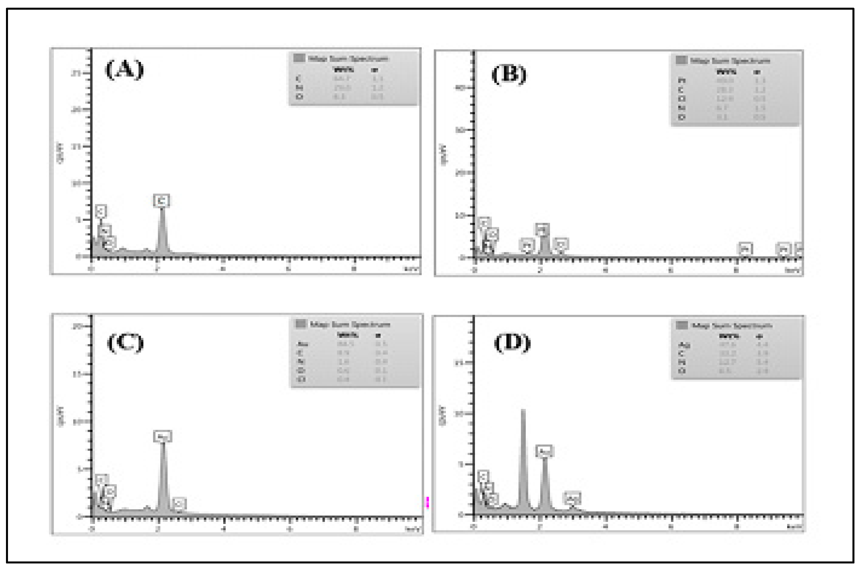
3.4. Elemental Analysis Study

The chemical analysis of pristine PAN NFs and NM NPs-PAN NFs were enabled by EDS Figure 4A–D. With mention to the results in Figure 4A, which were derived from PAN NFs, C, N, and O were only found associated with this sample. The nominal chemical compositions of the NM NPs-PAN NFs estimated using the EDS analysis are positioned in Figure 4B–D, show that all their corresponded metallic elements are in equiatomic proportion.

4. Conclusions

In summary, the synthesis of three types of noble metal nano-particles has been described in polyacrylonitrile nanofibers (NM NPs-PAN NFs) by a simple and easy route (as-called in-situ method). The XRD patterns indicate the well-defined crystallinity and lattice structure type of all as-prepared nanofibers. The reduction of H2PtCl6, HAuCl4, and AgNO3 by DMF in the presence of ultrasound irradiation is considered to be a key step in the formation of such system. The particles diameter of the NM NPs by the current methodology is varied that depended on the type of particles. SEM and TEM images revealed that the particle dimension is in a nanometer regime and the calculations made by the XRD match well with the observations made by TEM. The functional groups such as CN and hydroxyl groups act as capturing agents by electrostatic interactions and stabilizes them by strong bonding interactions between NM NPs and their polar groups of PAN polymers.

References

  1. Bounor-Legare, V.; Cassagnau, P. In situ synthesis of organic–inorganic hybrids or nanocomposites from sol–gel chemistry in molten polymers. Prog. Polym. Sci. 2014, 39, 1473–1497. [Google Scholar] [CrossRef]
  2. He, J.; Kunitake, T.; Nakao, A. Facile in Situ Synthesis of Noble Metal Nanoparticles in Porous Cellulose Fibers. Chem. Mater. 2003, 15, 4401–4406. [Google Scholar] [CrossRef]
  3. Yang, X.; Liu, Q.; Zhang, Y.; Su, X.; Huang, Y.; Zhang, T. In situ synthesis of metal clusters encapsulated within small-pore zeolites via a dry gel conversion method. Nanoscale 2018, 10, 11320–11327. [Google Scholar] [PubMed]
  4. Adnan, M.M.; Dalod, A.R.M.; Balci, M.H.; Glaum, J.; Einarsrud, M.-A. In Situ Synthesis of Hybrid Inorganic⁻Polymer Nanocomposites. Polymers 2018, 10, 1129. [Google Scholar] [CrossRef] [PubMed]
  5. Singh, N.; Khanna, P. In situ synthesis of silver nano-particles in polymethylmethacrylate. Mater. Chem. Phys. 2007, 104, 367–372. [Google Scholar]
  6. Guo, Q.; Ghadiri, R.; Weigel, T.; Aumann, A.; Gurevich, E.L.; Esen, C.; Medenbach, O.; Cheng, W.; Chichkov, B.; Ostendorf, A. Comparison of in Situ and ex Situ Methods for Synthesis of Two-Photon Polymerization Polymer Nanocomposites. Polymers 2014, 6, 2037–2050. [Google Scholar]
  7. Panaitescu, D.M.; Frone, A.N.; Chiulan, I. Nanostructured biocomposites from aliphatic polyesters and bacterial cellulose. Ind. Crop. Prod. 2016, 93, 251–266. [Google Scholar] [CrossRef]
  8. Zeng, F.-R.; Xu, J.; Xiong, Q.; Qin, K.-X.; Xu, W.-J.; Wang, Y.-X.; Liu, Z.-J.; Li, Z.-L.; Li, Z.-C. Aliphatic polyketones via cross-metathesis polymerization: Synthesis and post-polymerization modification. Polymers 2019, 185, 121936. [Google Scholar] [CrossRef]
  9. Carrado, K.A.; Xu, L. In Situ Synthesis of Polymer—Clay Nanocomposites from Silicate Gels. Chem. Mater. 1998, 10, 1440–1445. [Google Scholar] [CrossRef]
  10. Haider, S.; Kamal, T.; Khan, S.B.; Omer, M.; Haider, A.; Khan, F.U.; Asiri, A.M. Natural polymers supported copper nanoparticles for pollutants degradation. Appl. Surf. Sci. 2016, 387, 1154–1161. [Google Scholar] [CrossRef]
  11. Tian, J.; Jin, J.; Zheng, F.; Zhao, H. Self-Assembly of Gold Nanoparticles and Polystyrene: A Highly Versatile Approach to the Preparation of Colloidal Particles with Polystyrene Cores and Gold Nanoparticle Coronae. Langmuir 2010, 26, 8762–8768. [Google Scholar] [CrossRef] [PubMed]
  12. Mallick, K.; Witcomb, M.J.; Dinsmore, A.; Scurrell, M.S. Fabrication of a Metal Nanoparticles and Polymer Nanofibers Composite Material by an in Situ Chemical Synthetic Route. Langmuir 2005, 21, 7964–7967. [Google Scholar] [CrossRef] [PubMed]
  13. Porel, S.; Singh, S.; Harsha, S.S.; Rao, D.N.; Radhakrishnan, T.P. Nanoparticle-Embedded Polymer: In Situ Synthesis, Free-Standing Films with Highly Monodisperse Silver Nanoparticles and Optical Limiting. Chem. Mater. 2005, 17, 9–12. [Google Scholar] [CrossRef]
  14. Mallick, K.; Witcomb, M.J.; Scurrell, M.S. In situ synthesis of copper nanoparticles and poly(o-toluidine): A metal–polymer composite material. Eur. Polym. J. 2006, 42, 670–675. [Google Scholar] [CrossRef]
  15. Xu, Z.; Wei, C.; Jin, J.; Xu, W.; Wu, Q.; Gu, J.; Xu, X. Development of a Novel Mixed Titanium, Silver Oxide Polyacrylonitrile Nanofiber as a Superior Adsorbent and its Application for MB Removal in Wastewater Treatment. J. Braz. Chem. Soc. 2018, 29, 560–571. [Google Scholar] [CrossRef]
  16. Alarifi, I.M.; Alharbi, A.; Khan, W.S.; Swindle, A.; Asmatulu, R. Thermal, Electrical and Surface Hydrophobic Properties of Electrospun Polyacrylonitrile Nanofibers for Structural Health Monitoring. Materials 2015, 8, 7017–7031. [Google Scholar] [CrossRef] [PubMed]
  17. Wang, H.; Zhang, X.; Zhang, Y.; Cheng, N.; Yu, T.; Yang, Y.; Yang, G. Study of carbonization behavior of polyacrylonitrile/tin salt as anode material for lithium-ion batteries. J. Appl. Polym. Sci. 2016, 133. [Google Scholar] [CrossRef]
Figure 1. X-ray diffraction patterns of PAN NFs and NM NPs-PAN NFs (M NPs = Pt NPs, Au NPs, and Ag NPs).
Proceedings 41 00019 g001
Figure 2. Field emission scanning electron microscopy (FESEM) images of (A1) PAN NFs, (B1) Pt NPs-PAN NFs, (C1) Au NPs-PAN NFs, (D1) Ag NPs-PAN NFs, and transmission electron microscope (TEM) images of (A2) PAN NFs, (B2) Pt NPs-PAN NFs, (C2) Au NPs-PAN NFs, (D2) Ag NPs-PAN NFs.
Proceedings 41 00019 g002
Figure 3. FT-IR spectra of (A) PAN NFs, (B) Pt NPs-PAN NFs, (C) Au NPs-PAN NFs, (D) Ag NPs-PAN NFs.
Proceedings 41 00019 g003
Figure 4. EDS spectra of (A) PAN NFs, (B) Pt NPs-PAN NFs, (C) Au NPs-PAN NFs, (D) Ag NPs-PAN NFs.
Proceedings 41 00019 g004
Table 1. A brief of the observed XRD data of all samples (NM NPs loaded on PAN NFs) with JCPDS cards, the lattice type, and crystallite size calculated by the Scherrer formula.
No. SamplePosition [2 Theta (Degree)](h k l)Crystallite Size (D, nm)
1 PAN NFs17.84, 27.06(100), (020)3.42
2 Pt loaded on PAN NFs39.92, 46.39, 67.70, 81.07, 86(111), (002), (022), (113), (222)50.14
3 Au loaded on PAN NFs38.34, 44.44, 64.68, 77.59, 81.93(111), (002), (022), (113), (222)19.52
4 Ag loaded on PAN NFs38.19, 44.37, 64.55, 77.48, 81.34(111), (002), (022), (113), (222)24.93
1 This hexagonal structure can be indexed to its crystalline data (JCPDS card no. 96-432-8472, a = b = 32.49 Å, c = 16.32Å), 2 This FCC structure can be indexed to its crystalline data (JCPDS card no. 96-101-1108, a = b = c = 3.911 Å), 3 This FCC structure can be indexed to its crystalline data (JCPDS card no. 96-901-1613, a = b = c = 4.065 Å), 4 This FCC structure can be indexed to its crystalline data (JCPDS card no. 96-101-1608, a = b = c = 4.079 Å).
Publisher’s Note: MDPI stays neutral with regard to jurisdictional claims in published maps and institutional affiliations.

