Proceeding Paper

Is Oxytocin Proper for Cancer Adjuvant Therapy? †

by
Ali Taghizadehghalehjoughi
1,3,*,
Betul Cicek
2,
Ahmet Hacimuftuoglu
3 and
Mustaf Gul
2
1
Department of Pharmacology and Toxicology, Faculty of Veterinary, Ataturk University Medicine, 25240 Erzurum, Turkey
2
Department of Physiology, Faculty of Medicine, Ataturk University, 25240 Erzurum, Turkey
3
Department of Medical Pharmacology, Ataturk University, Faculty of Medicine, 25240 Erzurum, Turkey
*
Author to whom correspondence should be addressed.
Presented at the 2nd International Cell Death Research Congress, Izmir, Turkey, 1–4 November 2018.
Proceedings 2018, 2(25), 1582; https://doi.org/10.3390/proceedings2251582
Published: 11 December 2018
(This article belongs to the Proceedings of The 2nd International Cell Death Research Congress)

Abstract

:
Neuroblastomas are solid tumors and mostly seen in the adrenal medulla and sympathetic ganglia. It is known that neuroblastoma cell proliferation is inhibited by cisplatin and vincristine. The aim of this study was to investigate the effect of oxytocin on cell viability in human neuroblastoma SH-SY5Y cell line and primary cerebral cortex cell culture exposed to cisplatin and vincristine. In this direction, SH-SY5Y cell line and cortex neurons were obtained from the medical pharmacology department, Ataturk University. Both cells were grown in the appropriate cell culture media. Cisplatin (5, 10, 15 μg), vincristine (0.5, 1 and 2 ng) and oxytocin (1 μM) were applied to SH-SY5Y cell line and primary cortex cell culture for 24 h. MTT and TAC-TOS tests were performed 24 h after the application. As a result of the MTT assay, the combination of cisplatin and vincristine reduced cell viability in both cultures approximately 25% and 22%, respectively, compared to the control group. It appears that oxytocin increases neuroblastoma and cortex neuron viability, 112% and 95%, respectively. In this relation, we need to investigate why oxytocin increases cell viability and what are the possible implications in women in lactation stage.

1. Introduction

Neuroblastoma (NB), derived from neural cells, is a tumor which is seen in the adrenal medulla and sympathetic ganglia. NB is the most common extracranial solid tumor of early childhood neoplasms, which accounts for 15% of all childhood cancer deaths. Despite all current advancements in drug delivery systems and cancer therapy, neuroblastoma displays an aggressive, therapy-resistant phenotype, and prognosis remains poor [1]. Most chemotherapeutic agents used in the clinic for cancer therapy induce cell cycle arrest and apoptosis. Cisplatin (CP) is an inorganic platinum complex, which is mostly effective against many tumors. Cytotoxicity of cisplatin is based on cisplatin-derived DNA inserts, including protein-DNA cross-links, DNA mono adducts, and interstrand or intrastrand cross-links and the formation of reactive oxygen species (ROS) in cells [2]. Vincristine (VCR), a vinca alkaloid isolated from Catharanthus roseus, is used to treat cancers. VCR interacts with tubulin disrupting microtubule polymerization, forces cell cycle arrest at the M phase, which is followed by induction of apoptosis [3]. Although higher doses of CP and VCR are more effective, dramatically high-dose chemotherapy is associated with irreversible neurotoxicity. Oxytocin (OT) is a hypothalamic peptide synthesized in the paraventricular and supraoptical nucleus of the hypothalamus. Different therapeutic roles of OT include anxiety, depression and also cell proliferation and cancer. OT receptors are present in the central nervous system and participates in both basic physiology and cancer pathophysiology. It was shown in brain membranes that OT has antioxidant properties, scavenging free peroxyl radicals, inhibiting LDL oxidation and lipid peroxidation [4]. Current study was designed to investigate the possible protective effect of OT against VCR and CP-induced neurotoxicity.

2. Materials and Methods

Cell culture: Human neuroblastoma SH-SY5Y (ATCC, CRL-2266) cells were cultured in DMEM-F12 (Gibco Sigma, St. Louis, MO, USA) supplemented with a 10% heat-inactivated FBS (FBS; Invitrogen, Carlsbad, CA) and 100 unit’s/mL penicillin and 100 μg/mL streptomycin as described previously. Primary cultures of cortex neurons were obtained from the Pharmacology Department of Medical Faculty of Ataturk University (Erzurum, Turkey). Briefly, after thawing, cryo tubes were centrifuged (5 min, 1200 rpm) and the cells were seeded (2 × 10⁵ cells/well) in the growth neurobasal medium (Gibco Sigma, St. Louis, MO, USA) containing 2% B27, 10% FBS and 0.1% antibiotics (penicillin, streptomycin and amphotericin B) (Gibco Sigma, St. Louis, MO, USA). The cells were maintained at 37 °C in a humidified atmosphere containing 95% air and 5% CO2. Determination of Cell Viability. Cell viability was determined by using 3-[4-5-dimethylthylthiazol-2-yl] -2,5-diphenyltetrazolium bromide (MTT) assay (Gibco Sigma, St. Louis, MO, USA) (1). Different concentrations of cisplatin (5, 10, 15 μg), vincristine (0.5, 1 and 2 ng), and oxytocin (1 μM) were applied to SH-SY5Y cancer cell lines and primary cortex neurons for 24 h [5]. TAC and TOS: In total oxidant status (TOS) and total antioxidant capacity (TAC) analyses, spectrophotometry was used. The cell media were collected when the experiments are completed. According to the manufacturer procedure, for TOS (H2O2 Equiv/mmol L-1), absorbances were measured at 530 nm and for TAC (Trolox Equiv/mmol L-1) absorbances were measured at 660 nm (Rel assay, Antep, Turkey) [6,7]. Statistically Analysis: Mann–Whitney U test was used to compare the difference between groups by using the SPSS 22.0 software. P < 0.05 was considered as statistically significant.

3. Results

MTT test: To determine cell viability, the MTT assay test was used after 24 h exposure to cisplatin, vincristine and oxytocin combination with cisplatin and vincristine (Figure 1).
According to our study generally increased cell viability in both cultures. But oxytocin protected the primary neurons obviously better than neuroblastoma cells. In both cultures, vincristine and cisplatin dose dependently decrease cell viability at high doses. Oxytocin combination with cisplatin and vincristine in neuron cells in all doses did not show any significant difference in comparison with the control group. In neuroblastoma culture, oxytocin did not increase cell viability in high dose of vincristine and cisplatin groups. TAC and TOS: To determine cell antioxidant and oxidant status TAC and TOS tests were done after 24 h exposure to cisplatin, vincristine and oxytocin combination with cisplatin and vincristine (Figure 2).
TAC and TOC results show correlation with MTT results. Oxytocin increases TAC level more in primary cortical neuron culture compared to neuroblastoma cell culture.

4. Discussion

In conclusion, oxytocin shows slight neuroprotective effect by increasing the antioxidant capacity and stabilized oxidant status in comparison with NB cells. Bakos J and colleagues’ [4] studies show 0.01–1 μM oxytocin increases neuroblastoma viability against hydrogen peroxide toxicity. Although they showed an icrease in viability, they did not report increases in neurotrophic factor levels. Increases in cell viability in both neuroblastoma cell line and primary neuron culture due to oxytocin exposure in this study agree with the results of Bakos et al. [4]. Oxytocin shows promise to be used as cancer adjuvant therapy, but it needs to be studied further. Supplementary Materials: none.

Author Contributions

A.T. designed the experiments; A.T., A.H. and B.C. performed the experiments; A.H. and M.G. analyzed the data; A.T., B.C., A.H. and M.G. wrote the paper.

Acknowledgments

All sources of funding of the study were met by the authors.

Conflicts of Interest

The authors declare no conflict of interest.

References

  1. Maris, J.M.; Hogarty, M.D.; Bagatell, R. Neuroblastoma. Lancet 2007, 369, 2106–2120. [Google Scholar] [CrossRef]
  2. Sanie-Jahromi, F.; Saadat, M. Different profiles of the mRNA levels of DNA repair genes in MCF-7 and SH-SY5Y cells after treatment with combination of cisplatin, 50-Hz electromagnetic field and bleomycin. Biomed. Pharmacother. 2017, 94, 564–568. [Google Scholar] [CrossRef] [PubMed]
  3. Tu, Y.; Cheng, S.; Zhang, S.; Sun, H.; Xu, Z. Vincristine induces cell cycle arrest and apoptosis in SH-SY5Y human neuroblastoma cells. Int. J. Mol. Med. 2013, 31, 113–119. [Google Scholar] [CrossRef] [PubMed]
  4. Bakos, J.; Strbak, V.; Ratulovska, N.; Bacova, Z. Effect of oxytocin on neuroblastoma cell viability and growth. Cell. Mol. Neurobiol. 2012, 32, 891–896. [Google Scholar] [CrossRef] [PubMed]
  5. Taghizadehghalehjoughi, A.; Hacimuftuoglu, A.; Cetin, M.; Ugur, A.B.; Galateanu, B.; Mezhuev, Y.; Okkay, U.; Taspinar, N.; Taspinar, M.; Uyanik, A.; et al. Effect of metformin/irinotecan-loaded poly-lactic-co-glycolic acid nanoparticles on glioblastoma: In vitro and in vivo studies. Nanomedicine 2018, 13, 1595–1606. [Google Scholar] [CrossRef] [PubMed]
  6. Erel, O. A novel automated direct measurement method for total antioxidant capacity using a new generation, more stable ABTS radical cation. Clin. Biochem. 2004, 37, 277–285. [Google Scholar] [CrossRef] [PubMed]
  7. Erel, O. A new automated colorimetric method for measuring total oxidant status. Clin. Biochem. 2005, 38, 1103–1111. [Google Scholar] [CrossRef] [PubMed]
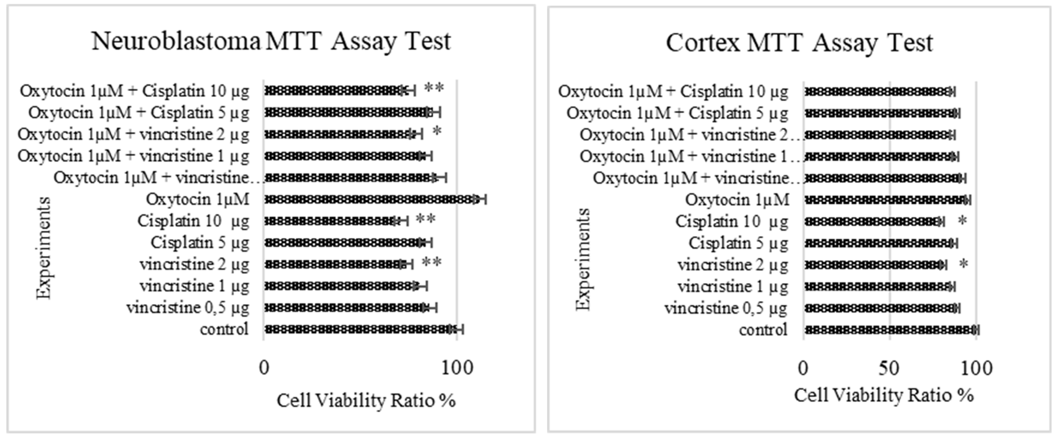
Figure 1. In vitro viability ratio of chemotherapy and oxytocin combination on primary cortex neurons and neuroblastoma cells (n = 6/group) is shown. * Significant differences at p < 0.05 compared to control group; ** Significant differences at the p < 0.001 compared to control group.
Proceedings 02 01582 g001
Figure 2. TAC and TOS results of chemotherapeutics and oxytocin combination on neuroblastoma and primary cortical neuron cells (n = 6/group) were shown. * Significant differences at p < 0.05 compared to control group; ** Significant differences at the p < 0.001 compared to control group.
Proceedings 02 01582 g002
Publisher’s Note: MDPI stays neutral with regard to jurisdictional claims in published maps and institutional affiliations.